The E50K optineurin mutation impacts autophagy-mediated degradation of TDP-43 and leads to RGC apoptosis in vivo and in vitro - Nature
←
→
Page content transcription
If your browser does not render page correctly, please read the page content below
Zhang et al. Cell Death Discovery (2021)7:49
                                    https://doi.org/10.1038/s41420-021-00432-0                                                                                                           Cell Death Discovery

                                     ARTICLE                                                                                                                                                    Open Access

                                    The E50K optineurin mutation impacts autophagy-
                                    mediated degradation of TDP-43 and leads to RGC
                                    apoptosis in vivo and in vitro
                                                           1,2
                                    Shiqi Zhang                , Zhengbo Shao1,3,4, Xinna Liu1,2, Mingying Hou1,2, Fang Cheng1,3, Dawei Lei1 and Huiping Yuan1,3

                                      Abstract
                                      The glaucoma-associated E50K mutation in optineurin (OPTN) is known to affect autophagy and cause the apoptosis
                                      of retinal ganglion cells (RGCs), but the pathogenic mechanism remains unclear. In this study, we investigated whether
                                      the OPTN (E50K) mutation caused TDP-43 aggregation by disrupting autophagy in vivo and in vitro. OPTN (E50K)
                                      mutant mice were generated and analysed for genotype and phenotype. Adeno-associated virus type 2 vectors
                                      containing either GFP only, GFP-tagged wild-type OPTN or GFP-tagged E50K-mutated OPTN were used to transfect
                                      R28 cells. Loss of RGCs decreased retinal thickness and visual impairment were observed in OPTN (E50K) mice
                                      compared with WT mice. Moreover, overexpression of E50K OPTN induced R28 cell apoptosis. Increased p62/SQSTM1
                                      and LC3-II levels indicated that autophagic flux was inhibited and contributed to TDP-43 aggregation in vivo and
                                      in vitro. We found that rapamycin effectively reduced the aggregation of TDP-43 in OPTN (E50K) mice and decreased
                                      the protein levels of p62/SQSTM1 and the autophagic marker LC3-II. Moreover, rapamycin increased the RGC number
                                      and visual function of E50K mice. In addition, we also observed increased cytoplasmic TDP-43 in the spinal cord and
                  1234567890():,;
1234567890():,;
                  1234567890():,;
1234567890():,;

                                      motor dysfunction in 24-month-old OPTN (E50K) mice, indicating that TDP-43 accumulation may be the common
                                      pathological mechanism of glaucoma and amyotrophic lateral sclerosis (ALS). In conclusion, the disruption of
                                      autophagy by OPTN (E50K) affected the degradation of TDP-43 and may play an important role in OPTN (E50K)-
                                      mediated glaucomatous retinal neurodegeneration.

                                    Introduction                                                                                        protein involved in a variety of cellular processes, such as
                                      Glaucoma is an important leading cause of progressive                                             signalling and autophagy5,6.
                                    blindness worldwide. Epidemiologically, normal-tension                                                As an ‘autophagy receptor’, OPTN binds ubiquitin or
                                    glaucoma (NTG) is more prevalent among Asian popu-                                                  ubiquitinated aggregates and directs them to autophago-
                                    lations, including Japanese, Korean and Chinese popula-                                             somes7. It contributes to the maturation of the autopha-
                                    tions, and 21% of patients with NTG have a family history                                           gosome8 and helps to degrade damaged organelles or
                                    of glaucoma, indicating a genetic predisposition to the                                             abnormal proteins to maintain cellular homoeostasis6.
                                    disease1,2. One of the genes associated with NTG is                                                 The E50K mutation of OPTN is the most prevalent
                                    optineurin (OPTN)3,4, which encodes OPTN, an adaptor                                                mutant form that is associated with NTG9; impaired
                                                                                                                                        autophagy has been found in E50K transgenic mice and
                                                                                                                                        E50K OPTN-overexpressing retinal ganglion cell (RGC)-5
                                                                                                                                        cells, and this impairment was linked to apoptosis of
                                    Correspondence: Huiping Yuan (yuanhp2013@126.com)
                                    1
                                     Department of Ophthalmology, The Second Affiliated Hospital of Harbin                               RGCs10–12. Several studies have suggested that OPTN has
                                    Medical University, Harbin, China
                                    2
                                                                                                                                        an important role in mitophagy9,12,13; however, the effect
                                     The Key Laboratory of Myocardial Ischemia, Harbin Medical University,
                                                                                                                                        of E50K on protein metabolism is rarely reported.
                                    Ministry Education, Heilongjiang Province, Harbin, China
                                    Full list of author information is available at the end of the article
                                    Edited by Richard Killick

                                    © The Author(s) 2021
                                                        Open Access This article is licensed under a Creative Commons Attribution 4.0 International License, which permits use, sharing, adaptation, distribution and reproduction
                                                        in any medium or format, as long as you give appropriate credit to the original author(s) and the source, provide a link to the Creative Commons license, and indicate if
                                    changes were made. The images or other third party material in this article are included in the article’s Creative Commons license, unless indicated otherwise in a credit line to the material. If
                                    material is not included in the article’s Creative Commons license and your intended use is not permitted by statutory regulation or exceeds the permitted use, you will need to obtain
                                    permission directly from the copyright holder. To view a copy of this license, visit http://creativecommons.org/licenses/by/4.0/.

                                    Official journal of the Cell Death Differentiation Association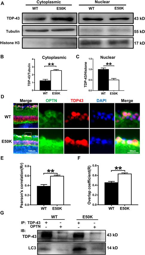
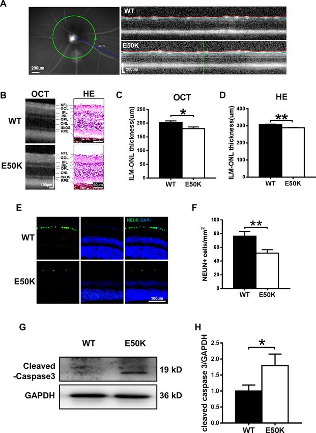
Zhang et al. Cell Death Discovery (2021)7:49                                                                                              Page 2 of 13

  TAR DNA-binding protein 43 (TDP-43 or TARDBP), a                            effect of OPTN (E50K) on the motor system, demon-
known DNA- and RNA-binding protein, has a key role in                         strating that TDP-43 may be the common molecular
mRNA processing and trafficking and microRNA bio-                              mechanism between NTG and ALS. We also examined
genesis14. Both the ubiquitin proteasome system (UPS)                         the efficacy of the autophagic enhancer rapamycin. Our
and autophagy are required for TDP-43 degradation, and                        results revealed that rapamycin reduced TDP-43 accu-
the clearance of cellular TDP-43 macroaggregates                              mulation in the OPTN (E50K) mouse model, and this
depends on the autophagy pathway15. Impairments in                            effect was accompanied by an increase in autophagic flux
degradation pathways implicate failed TDP-43 clearance                        and increased RGC numbers and visual function. These
as a primary disease mechanism in neurodegeneration                           results indicated that TDP-43 accumulation has an
and a variety of neurodegenerative diseases, such as                          important role in neurodegeneration in glaucoma
amyotrophic lateral sclerosis (ALS)16–19. To our knowl-                       and ALS.
edge, the link between OPTN (E50K)-mediated autop-
hagy and TDP-43 is not clear.                                                 Results
  To determine the effect of OPTN (E50K) on TDP-43                            OPTN (E50K) mutant mice exhibit normal IOP and
degradation and whether this effect has a role in glauco-                     abnormal visual function
matous neurodegeneration, a point mutation mouse                                OPTN (E50K) mutant mice were generated by CRISPR/
model was developed by CRISPR/Cas9. In this study, we                         Cas9 technology. The genotypes of the mice were iden-
observed changes in TDP-43 and the level of autophagy in                      tified by gene sequencing, and the OPTN (E50K) mice
the OPTN (E50K) mouse model and OPTN (E50K)-                                  exhibited a homozygous mutation of 148 (G > A) in the
overexpressing R28 cells. In addition, our study shows the                    ORF region (Fig. 1A).

  Fig. 1 Effects of the OPTN-E50K point mutation on visual function and IOP in vivo. A Various domains and the E50K mutation in mouse OPTN.
  NEMO NF-kappa-B essential modulator, LIR LC3-interacting region, UBD ubiquitin-binding domain. B IOP values in WT and E50K mutant mouse eyes.
  n = 17. C Representative recorded waveforms of F-VEP and D decreased amplitude of P2 following visual stimulation in E50K mutant mice compared
  with WT mice. n = 48. E, F The light/dark transition test was used to evaluate visual function, and E50K mutant mice spent more time in the light box
  than WT mice. n = 7. Data are presented as the means ± SEM; *P < 0.05; **P < 0.01.

Official journal of the Cell Death Differentiation Association
Zhang et al. Cell Death Discovery (2021)7:49 Page 3 of 13
The OPTN (E50K) mutation is responsible for NTG4. transfected cells at the same time point. Compared with
We measured the IOP of wild-type and OPTN (E50K) the other three groups, the E50K group had increased
mice. In OPTN (E50K) mice, the mean IOP was 12.06 ± expression of cleaved caspase-3 (Fig. 3C, D), which also
0.74 mmHg (mean ± S.E.M., n = 18), which was similar to suggested that the mutation could lead to increased
the IOP measured in wild-type mice (12.71 ± 0.61 mmHg, apoptosis.
n = 17) (p = 0.39, Fig. 1B).
However, OPTN (E50K) mice exhibit abnormal visual The OPTN (E50K) mutation inhibits autophagic flux in the
function without increased IOP. In the flash visual evoked retina
potential (f-VEP) examination, the P2 amplitude mea- To determine autophagic flux, we analysed the protein
sured for E50K mutant mice was 11.44 ± 0.8074 µV expression of LC3-II and p62/SQSTM1 in retinas. The
(mean ± S.E.M., n = 16), which was significantly lower E50K mutation increased the levels of LC3-II and p62/
than the P2 amplitude measured for wild-type mice SQSTM1 (Fig. 4A–C, p < 0.05), which indicated the
(15.42 ± 0.7107 µV, n = 19) (p < 0.01). This result indi- inhibition of autophagy. In addition, we examined the
cated that axons and the retina were damaged in the E50K pattern of colocalization of OPTN with LC3, which is the
mutant mice (Fig. 1C, D). characteristic signature of autophagic membranes20. The
In addition, the light/dark transition test showed that results of immunofluorescence staining revealed that both
the E50K mutant mice stayed in the light chamber longer wild-type and E50K mutant OPTN colocalized with LC3
than the wild-type mice (375.5 ± 39.36 s, n = 4 and (Rr > 0.5, R > 0.5). Furthermore, a higher degree of colo-
236.3 ± 30.79 s, n = 7, respectively; mean ± S.E.M., p < calization of OPTN with LC3 was found in the OPTN
0.05). The longer time spent in the light box suggested (E50K) mice (Fig. 4D–F, RrWT = 0.560 ± 0.022, RrE50K =
that the preservation of visual behaviours was impaired in 0.712 ± 0.017, RWT = 0.668 ± 0.021, RE50K = 0.779 ±
the E50K mutant mice (Fig. 1E, F). 0.016). The same result was also observed in coimmu-
noprecipitation, in which the interaction between OPTN
The OPTN (E50K) mutation induces histological changes in and LC3 was enhanced in the OPTN (E50K) mice
the mouse retina (Fig. 5F).
For the optical coherence tomography (OCT) exam-
ination, the average thickness of the retina at 3.45 mm The degradation of TDP-43 was affected by the E50K
from the centre of the optic disc was compared between mutation
the two kinds of mice (Fig. 2A). We found that the E50K The effects of the E50K mutation on TDP-43 degrada-
mutant mice showed thinning of the retina compared tion were determined by analysing cytoplasmic and
with wild-type mice (180.0 ± 6.141 µm and 203.0 ± nuclear TDP-43 accumulation in the mouse retina. The
5.143 µm, respectively; n = 10, mean ± S.E.M., p < 0.05, level of cytoplasmic TDP-43 in the retinas of OPTN
Fig. 2B, C). A similar result was observed in histological (E50K) mice was significantly higher than that of wild-
measurements, and it was apparent that the retinal type mice (Fig. 5A, B). Meanwhile, the level of nuclear
thickness at 200 µm from the edge of the optic disc of TDP-43 was lower in OPTN (E50K) mice (Fig. 5A, C).
OPTN (E50K) mice (288.5 ± 2.254 µm, n = 44) was sig- Next, the results of immunofluorescence staining showed
nificantly less than that of wild-type mice (306.5 ± that TDP-43-immunoreactive aggregates tended to colo-
2.972 µm, n = 28, p < 0.01) (Fig. 2B, D). calize with E50K OPTN but not with wild-type OPTN in
As shown in Fig. 2E, F, the number of viable RGCs was the retina (Fig. 5D–F, RrWT = 0.385 ± 0.035, RrE50K =
significantly decreased in the OPTN (E50K) mice. In 0.594 ± 0.021, RWT = 0.452 ± 0.026, RE50K = 0.631 ±
addition, the expression of cleaved caspase-3, a marker of 0.020). Interestingly, we observed a strikingly increased
apoptosis, was also increased in the retinas of the OPTN interaction between TDP-43 and OPTN as well as LC3 in
(E50K) mice (Fig. 2G, H). E50K retinal tissue (Fig. 5G). This result indicated that the
aggregation of TDP-43 may be associated with OPTN-
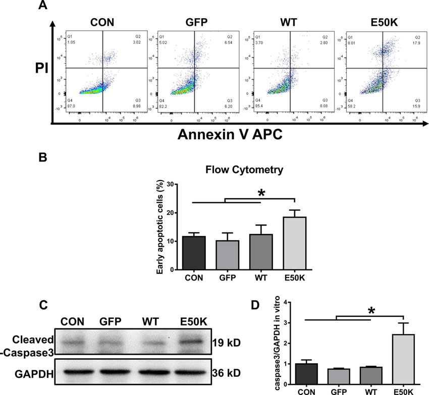
The OPTN (E50K) mutation induces cell apoptosis in the mediated autophagy.
R28 cell line
Apoptosis in the R28 cell line was quantified by flow The E50K mutation inhibits autophagic flux and affects the
cytometry analysis after transfection. Annexin V-positive degradation of TDP-43 in vitro
and PI-negative cells corresponded to early apoptotic In transfected R28 cells, we also detected autophagic
cells. E50K OPTN-overexpressing cells (the E50K group) flux and TDP-43 accumulation. Similar to the results
showed a higher early apoptotic rate than the other three in vivo, analysis of R28 cell protein levels showed an
groups (Fig. 3A, B, 11.66 ± 1.341, 10.19 ± 2.765, 12.39 ± increase in the expression of both LC3-II and p62/
3.31 and 18.47 ± 2.517, p < 0.05). In addition, the protein SQSTM1 with a corresponding increase in TDP-43 in the
expression level of caspase-3 was also measured in E50K group, which indicated the inhibition of autophagy
Official journal of the Cell Death Differentiation Association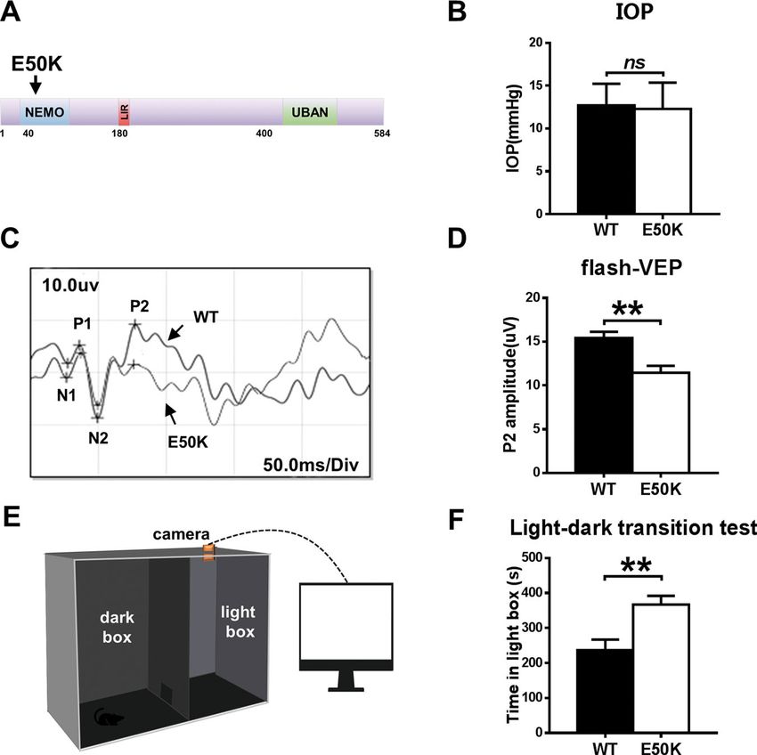
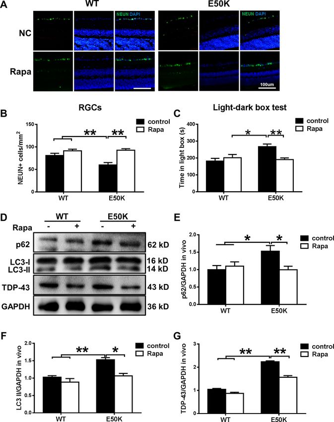
Zhang et al. Cell Death Discovery (2021)7:49                                                                                            Page 4 of 13

  Fig. 2 Comparison of retinal morphology and apoptosis in WT and E50K mutant mice. A Left: example of an SLO image of the central murine
  retina. Right: example of an OCT slice from WT and E50K mutant mice. B OCT scans and histology of retinal layers. C, D Quantification of retinal
  thickness measurements by OCT and histological analysis. For OCT, n = 10; for histology, n = 44/28. E Representative images and F quantification of
  viable RGC immunolabelling by NEUN. WT = 76.33 ± 7.000, n = 10; E50K = 51.67 ± 4.846, n = 12. G Caspase-3 protein expression was evaluated by
  western blot analysis. Representative western blot image and H quantification of caspase-3 protein expression in WT and E50K mutant mice. n = 4.
  Data are presented as the means ± SEM; *P < 0.05; **P < 0.01.

(Fig. 6A, B, D–G). However, the results from the in vivo                     Rapamycin treatment increases autophagy and decreases
and ex vivo experiments in the present study are not                         the aggregation of TDP-43 in OPTN (E50K) mice
completely consistent. TDP-43 did not appear to interact                       To elucidate the effects of rapamycin, WT and OPTN
with OPTN or LC3 in the R28 cells, whereas OPTN                              (E50K) mice were treated as described in the methods.
interacted with LC3, and their interaction was enhanced                      After 5 weeks, the RGC number of the E50K-rapamycin
upon E50K OPTN overexpression (Fig. 6C). These results                       group was significantly increased (Fig. 7A, B). Moreover,
suggest that there are separate pathogenic mechanisms                        the light/dark transition test showed that rapamycin
in vivo and in vitro.                                                        treatment improved the visual function of OPTN (E50K)

Official journal of the Cell Death Differentiation Association
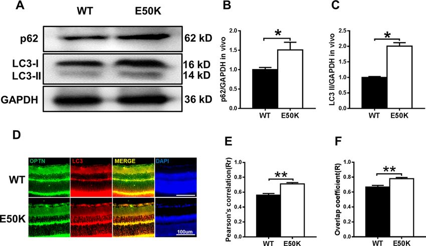
Zhang et al. Cell Death Discovery (2021)7:49                                                                                          Page 5 of 13

  Fig. 3 Effects of OPTN-E50K overexpression on the apoptosis of transgenic R28 cells. A The apoptosis rates were measured via flow cytometry
  and B quantification of early apoptotic cells 48 hours after transfection. GFP-positive cells were counted in the GFP, WT and E50K groups. PI
  propidium iodide. n = 3. C Representative western blot image and D quantification of cleaved caspase-3 protein expression in transfected cells.
  n = 4. Mean ± SEM. *P < 0.05.

  Fig. 4 The OPTN-E50K point mutation affects autophagy and the colocalization of optineurin and LC3. A Western blotting showing
  endogenous p62/SQSTM1 and LC3 levels in retinas from WT and E50K mutant mice. B Quantification of p62/SQSTM1 and C LC3 protein expression
  levels. D Retinal sections were immunostained for optineurin and LC3, and nuclei were stained with DAPI. E, F Quantification of the percentage of
  OPTN-LC3 colocalization. n = 19/14. Mean ± SEM. *P < 0.05; **P < 0.01.

Official journal of the Cell Death Differentiation Association
Zhang et al. Cell Death Discovery (2021)7:49                    Page 6 of 13

  Fig. 5 (See legend on next page.)

Official journal of the Cell Death Differentiation Association
Zhang et al. Cell Death Discovery (2021)7:49                                                                                              Page 7 of 13

  (see figure on previous page)
  Fig. 5 The OPTN-E50K point mutation mediates the degradation of TDP-43 via autophagy. A Western blot analysis of TDP-43 protein
  expression in the retinal cytoplasm and nucleus. β-tubulin and histone h3 were used as loading controls to validate the fractionation of cellular
  extracts. B, C The protein expression of TDP-43 was significantly greater in the cytoplasm and was significantly lower in the nucleus in the retinas of
  E50K mutant mice than in those of WT mice. n = 3. D Fluorescence analysis of the localisation of TDP-43 and optineurin in retinas of WT and E50K
  mutant mice. TDP-43-positive granules (red) were more colocalized with optineurin signals (green) in E50K mutant mice, and E and F both the values
  of Pearson’s correlation and Manders’ overlap coefficient between OPTN and TDP-43 were significantly higher in E50K mutant mice than in WT mice.
  n = 10/14. G Western blot analysis of immunoprecipitation assays of TDP-43 or OPTN in retinas. *P < 0.05; **P < 0.01.

  Fig. 6 Overexpression of E50K optineurin disrupts autophagy and causes the degradation of TDP-43 in vitro. A, B OPTN, p62/SQSTM1 and
  LC3 protein expression and TDP-43 protein expression in the cytoplasm were analysed in blank control, AAV-GFP-, GFP-WT- and GFP-E50K-
  transfected R28 cells by western blotting. C Western blot analysis of immunoprecipitation assays of TDP-43 or OPTN in R28 cells. D–F Quantification
  of OPTN, p62/SQSTM1, LC3 and TDP-43 protein expression levels. n = 3. *P < 0.05; **P < 0.01.

mice. We also analysed endogenous LC3-II and p62/                             Discussion
SQSTM1 modification in mouse retinas. The rapamycin-                             In the present study, we investigated the pathogenic
treated E50K mutant mice demonstrated a decrease in                           mechanisms of E50K-induced RGC apoptosis in a 16-
LC3-II and p62/SQSTM1 expression levels (Fig. 7D–F).                          month-old CRISPR/Cas9-mediated mouse model and an
Furthermore, the aggregation of TDP-43 was significantly                       OPTN (E50K)-overexpressing R28 cell model. We found
decreased in E50K mutant mice after rapamycin treat-                          a strong association between increased autophagy-
ment (Fig. 7D, G). Collectively, these results indicate that                  mediated pathologic TDP-43 levels and increased RGC
the degradation of TDP-43 was associated with autophagy                       apoptosis in the E50K mutant mice. Rapamycin showed a
and RGC viability and function in the mutant mice.                            visual protective effect by reducing RGC apoptosis, which

Official journal of the Cell Death Differentiation Association
Zhang et al. Cell Death Discovery (2021)7:49                                                                                             Page 8 of 13

  Fig. 7 The enhanced autophagy and reduced TDP-43 levels induced by rapamycin treatment decrease E50K-induced apoptosis. A, B After
  treatment with rapamycin, the RGC number in OPTN (E50K) mice significantly increased. Representative images and quantification of viable RGC
  immunolabelling by NEUN are shown. n = 20. C Light/dark transition test. The time spent in the light box was recorded. Rapamycin reduced the time
  spent in the light box by E50K mutant mice. n = 8. D Western blot analysis of p62/SQSTM1, LC3 and TDP-43 protein expression in the retinas of mice
  and E, F quantification of protein expression levels. G The expression of TDP-43 protein was significantly decreased after rapamycin treatment. n = 3.
  *P < 0.05; **P < 0.01.

may be related to upregulation of autophagy and                               it remains unclear whether the mechanisms of this
decreased levels of TDP-43. Our results are helpful for a                     induction are comparable to the situation seen in glau-
better understanding of the underlying mechanism of                           coma. To avoid this problem, we used a point mutation
NTG caused by OPTN (E50K) and targeted treatment                              mouse model to study the glaucoma phenotype of
of NTG.                                                                       humans.
  In the clinic, retinal thickness changes can be observed                      Similar to previous studies, we showed that OPTN
in POAG and NTG patients. In addition, it has previously                      (E50K) mutant mice exhibited thinning of the retinal
been reported that overexpression of E50K mutant OPTN                         layers, loss of RGCs and functional visual impairment
in transgenic mice induces apoptosis of RGCs, leads to                        without higher IOP (Figs. 1 and 2). These findings indi-
progressive retinal degeneration after 16 months3,21,22 and                   cated that the E50K point mutation induced toxic effects
shows a mild glaucoma phenotype23. However, because of                        such as apoptosis so that the mutant mice showed an
endogenous OPTN or the overexpression of E50K OPTN,                           NTG-like phenotype. Based on these results, CRISPR/

Official journal of the Cell Death Differentiation Association
Zhang et al. Cell Death Discovery (2021)7:49                                                                   Page 9 of 13

Cas9 produced an ideal mouse model for evaluating the           a reduction in nuclear expression (Fig. 5A–C). Further-
pathogenic mechanism of NTG.                                    more, both fluorescence microscopy and coimmunopre-
  Without higher IOP, neurodegeneration possibly occurs         cipitation showed that OPTN exhibited a degree of
through a different disease mechanism. Therefore, we            increased interaction with TDP-43 in OPTN (E50K) mice.
next studied the possible underlying mechanism in               In addition, an increased interaction between LC3 and
OPTN-E50K point mutant mice. OPTN belongs to a                  OPTN or TDP-43 was observed in the retinas of mutant
group of ubiquitin-binding autophagy receptors. It is           mice. These results revealed that OPTN (E50K) caused
widely thought to link ubiquitinated cargo through              TDP-43 to accumulate more frequently in the cytoplasm
ubiquitin-binding domains to autophagosomal mem-                and deplete in the nucleus of retinas, and these changes
branes by binding to Atg8 family proteins24; thus,              were related to E50K-mediated autophagy because of the
abnormal function of OPTN is closely related to impaired        altered interactions. To our knowledge, this is the first
autophagy. Autophagy is a crucial mechanism that helps          report of abnormal TDP-43 accumulation in the retinas of
maintain TDP-43 homoeostasis by removing larger                 OPTN (E50K) mice.
aggregates through lysosomal degradation. It has been              The formation of toxic protein aggregates is associated
reported that overexpression of E50K OPTN induces a             with several neurodegenerative diseases33. TDP-43
decrease in autophagosome formation to inhibit autop-           aggregation, which is referred to as TDP-43 proteino-
hagic flux in RGC-5 cells10. Moreover, autophagy                 pathy, is usually identified in ALS34,35. Moreover, some
impairment also occurs in adeno-associated virus type 2         mutations in OPTN, such as the E478G and D477N
(AAV2)-OPTNE50K-transduced eyes of rats11. OPTN-                mutations, have been found to cause ALS, suggesting that
E50K would be expected to lead to an inability to               there is a common pathological mechanism between these
remove aggregates; thus, we hypothesised that impaired          two diseases36. Moreover, it has also been reported that
autophagic flux may result in the formation of TDP-43            retinal neuron loss is preceded by the mislocalization of
aggregates in the OPTN-E50K mutant mouse.                       TDP-43 in Grn-KO mice37 and that increased cyto-
  To test this conjecture, we measured the level of             plasmic TDP-43 induces retinal degeneration, including
autophagic flux, which is usually monitored by detecting         thinning of the retina, in a Drosophila ALS model38,39.
endogenous LC3-II modification and p62/SQSTM1                       However, no OPTN mutation has been reported to
aggregation. Because increased LC3-II may be caused by          cause either glaucoma or ALS, and no E50K-linked case of
autophagy activation or the failure of autophagic lysosome      ALS has been reported. Although the E50K mutation
clearance25,26, p62/SQSTM1 was used to help determine           leads to abnormalities in TDP-43 in the retina, increased
the level of autophagy. The accumulation of p62/                TDP-43 expression does not always result in TDP-43
SQSTM1, a marker of autophagy, is observed in response          pathology40,41, so we cannot determine the effect of E50K
to autophagy inhibition27,28. Therefore, increased levels of    on the motor system. To answer this question, rotarod
p62/SQSTM1 and LC3-II indicate the inhibition of the            tests were used to assess whether the knock-in mice
late stage of degradation, which is usually regarded as the     would develop ALS-like phenotypes. Unlike visual func-
inhibition of autophagic flux. In our results, increased         tion, the knock-in mice appeared indistinguishable from
LC3-II and P62/SQSTM1 were observed, confirming the              age-matched wild-type mice until 18 months of age
impairment of autophagy in E50K mutant mouse retinas.           (Figure S1). Surprisingly, 24-month-old heterozygous
  During the process of autophagy, OPTN recognises              OPTN (E50K) mice exhibited significant motor dysfunc-
protein aggregates to regulate the clearance of abnormal        tion. Moreover, the amount of cytoplasmic TDP-43 was
proteins, helping to maintain cellular homoeostasis29.          increased significantly in the spinal cord of 24-month-old
TDP-43 is aggregation-prone, and its accumulation in the        but not 16-month-old knock-in mice (Figure S2). Intri-
cytoplasm correlates with toxicity14. Clearance of larger       guingly, it seems that E50K-induced TDP-43 abnormal-
TDP-43 aggregates occurs via the autophagy pathway14,30.        ities and dysfunction earlier in retinas than in the spinal
Thus, autophagy-mediated protein clearance has an               cord. The reason for the differences between the two
important role in the balance of TDP-43 synthesis.              systems remains to be discovered. The present results
Moreover, Yamashita et al.31 and Maruyama et al.32 pre-         further indicate that TDP-43 abnormalities may be the
sented evidence that OPTN is associated with TDP-43             common mechanism of NTG and ALS. Therefore, a
and that TDP-43-positive inclusions showed positive             mutation in an ALS-associated mutation knock-in model,
immunolabelling with anti-OPTN antibodies in the                such as E478G, may help clarify the common and differ-
muscles of patients with sporadic inclusion body myositis       ent pathological mechanisms of TDP-43 in these two
and sporadic ALS.                                               diseases.
  In our study, we observed a significantly higher level of         Moreover, recent evidence identified RGC-5 cells as the
TDP-43 in the cytoplasm of retinas from OPTN (E50K)             661 W photoreceptor cell line and indicated that RGC-5
mice than in those from wild-type mice, accompanied by          cells are not of RGC origin, so in the current study, we

Official journal of the Cell Death Differentiation Association
Zhang et al. Cell Death Discovery (2021)7:49                                                                   Page 10 of 13

used the rat retina R28 cell line, which has been char-          reduction and visual impairment in vivo and cell
acterised and used in a variety of in vitro studies of retinal   apoptosis in vitro. The disruption of autophagy by
cell behaviour42 to investigate the effect of E50K. Similar      OPTN-E50K affected the degradation of TDP-43 and
to the in vivo results, overexpression of OPTN-E50K also         may play an important role in E50K-mediated glauco-
induced apoptosis, suppressed autophagy and affected             matous neurodegeneration.
TDP-43 degradation in the R28 cell line. However,
coimmunoprecipitation analysis in transfected R28 cells          Materials and methods
yielded inconsistent results. TDP-43 did not appear to           Development of OPTN (E50K) point mutation mice
interact with OPTN or LC3 in R28 cells, whereas OPTN               The experiments in mice were approved by the Insti-
interacted with LC3, and their interaction was enhanced          tutional Animal Care and Use Committee of Harbin
upon E50K OPTN overexpression. The possible cause                Medical University and performed following the State-
may be associated with the differences between knock-in          ment for the Use of Animals in Ophthalmic and Vision
(in vivo) and overexpression (in vitro). First, in contrast to   Research by the Association for Research in Vision and
the knock-in model, E50K OPTN-overexpressing cells               Ophthalmology. OPTN containing the E50K mutation
express endogenous OPTN, which may affect the results.           was created through homologous recombination-
Furthermore, it is thought that the impairment of pro-           mediated knock-in using the CRISPR/Cas9 system. In
teasomal degradation leads to the initial formation of           brief, the donor vector was constructed containing two
TDP-43 aggregates, and then impairment of autophagy              homologous arms and a point mutation region. Cas9
prevents the removal of aggregated TDP-4315. Ubiquitin           mRNA, gRNA and donor vector were microinjected into
proteasome pathway (UPP) function was also reduced by            fertilised eggs from C57BL/6 mice. To avoid CRISPR off-
overexpression of E50K OPTN11, so we concluded that              target events, F0 lines were outcrossed to F2. Hetero-
the UPP was affected first, leading to the accumulation of        zygous F2 animals were intercrossed to generate mutant
TDP-43 owing to the limited transfection time. Although          and wild-type mice. We examined mice after the F3
the autophagy pathway was also inhibited, it was not the         generation, and the genotypes of the mice were confirmed
main factor contributing to the changes in TDP-43                by sequencing of PCR fragments (896 bp) in the point
in vitro. Moreover, overexpression of OPTN can also              mutation target region amplified from genomic DNA
cause an imbalance in cell homoeostasis. Therefore, it is        isolated from blood using the following primers: forward,
difficult to evaluate the pathological effects of OPTN            AGCCGGGCAGCGTTAACTGGATG; reverse, CTCAC
(E50K) compared with those of wild-type OPTN without             TCTGGGGCCCTGTTCATTC. The mice were main-
appropriate control of their levels of overexpression in         tained on a 12-hour light/dark circle. Sixteen-month-old
transfected cells. This result indicated the importance of       mice were used in our study, and the researchers were not
the knock-in model in investigating the pathogenesis of          blinded to the grouping of the mice during experiments or
the E50K mutation in OPTN. However, validation of this           analysis.
hypothesis will require further investigation.
  To confirm the role of OPTN-mediated autophagy in               Cell culture and transfection
TDP-43 degradation, we adopted the autophagy activator             The R28 retinal precursor cell line was cultured in low-
rapamycin. Rapamycin has been reported to compromise             glucose Dulbecco’s modified Eagle’s medium with 10%
proteasome function and activate the autophagic process          calf serum, 100 U/ml penicillin and 100 mg/ml strepto-
in E50K-transduced rat eyes and was effective in rescuing        mycin in a 37 °C humidified atmosphere containing 5%
adverse OPTN phenotypes11. In our study, we found that           CO2. Cells were transfected with AAV2-EGFP, AAV2-
rapamycin was effective in reducing the aggregation of           OPTNWT-EGFP and AAV2-OPTNE50K-EGFP (Hanbio,
TDP-43 in our mouse model and induced decreases in the           Shanghai, China), which were constructed by inserting the
p62/SQSTM1 protein and the autophagic marker LC3-II              mouse wild-type or E50K OPTN gene according to the
(Fig. 7). These results indicate that the burden of TDP-43       manufacturer’s instructions. Cells were collected through
accumulation will be diminished by enhancing autophagic          trypsin digestion 48 hours after transfection.
flux. Moreover, rapamycin increased the RGC number
and visual function of OPTN (E50K) mice. This result             Intraocular pressure measurement
indicates that the use of rapamycin to reduce TDP-43               A rebound tonometer (Tonolab, iCare Finland Oy,
could suppress E50K-mediated apoptosis. All the above-           Helsinki, Finland) was used to measure the intraocular
mentioned results support our hypothesis that OPTN-              pressure (IOP) of mice according to the manufacturer’s
E50K inhibits autophagy, leading to TDP-43 aggregation           directions. Seven readings were taken per eye immediately
and RGC apoptosis.                                               after the mice were fully anaesthetised. The average values
  In conclusion, our results showed that the glaucoma-           of WT and OPTN (E50K) mice were compared and
associated E50K OPTN mutation induced RGC                        analysed by Student’s t test.

Official journal of the Cell Death Differentiation Association
Zhang et al. Cell Death Discovery (2021)7:49                                                                  Page 11 of 13

Visual function detection                                       (C1005; Beyotime Co., Shanghai, China) at room tem-
  F-VEP and light/dark transition tests were used to            perature for 3 min and placed in anti-fade fluorescence
detect the visual function of mice as previously descri-        mounting medium. All images were acquired by fluores-
bed43. In brief, the active electrode, reference electrode      cence microscopy (Olympus Corporation, Tokyo, Japan),
and ground electrode were inserted under the skin of the        and Pearson’s correlation coefficient (Rr) and Manders’
occiput, mandible and upper limb of mice to perform f-          overlap coefficient (R) were calculated by Image-Pro Plus
VEP. The waveform was recorded three times for each             software to analyse the colocalization. NeuN-positive cells
eye, and the average P2 amplitude was analysed. A light/        were counted by ImageJ from 24 images. Four mice from
dark transition test was conducted as described pre-            each group were collected.
viously44. After 2 hours of dark adaptation, the mouse was
put into the dark box (Fig. 1E), and data were collected for    Western blot
10 min to evaluate visual function by determining the             After removing the anterior portion of the eye and the
time spent in the light box.                                    lens, retinas were lysed in radioimmunoprecipitation lysis
                                                                buffer (P0013B, Beyotime Co.) to extract proteins. For R28
Retinal thickness measurements                                  cells, proteins were extracted in the same way. Cytoplasmic
  We measured the thickness of the inner five layers of          proteins were isolated by a Nuclear and Cytoplasmic
the retina by OCT (Heidelberg, Germany) and histology.          Extraction Kit (CW0199, CWBio) according to the manu-
Mice were placed on a platform after anaesthesia and            facturer’s instructions. Twenty micrograms of pooled ret-
pupil dilation and adjusted to ensure that the incident         inal protein (n = 6 retinas/group) or cellular protein were
beam was perpendicular to the central cornea and passed         resolved by sodium dodecyl sulphate polyacrylamide gel
through the pupil. The retinal thickness of mice at a circle    electrophoresis and transferred to polyvinylidene fluoride
with a diameter of 3.45 mm (system setting) using the           membranes. Western blot analysis was performed as
optic disc as the centre was measured by OCT with a 25D         described44. The primary antibodies included rabbit anti-
lens (50744, Heidelberg, Germany)45. After the measure-         OPTN (1:1000, 10837-1-AP, Proteintech Group), p62/
ment of live animals, the mice were anaesthetised and           SQSTM1 (1:1000, 18420-1-AP, Proteintech Group), LC3A/
cardiac perfused with 4% paraformaldehyde in phosphate          B (1:1000, #4108, CST), caspase-3 (1:1000, 19677-1-AP,
buffer. The eyeballs were removed and fixed in 4% par-           Proteintech Group), TDP-43 (1:1000, 10782-2-AP, Pro-
aformaldehyde overnight at 4 °C. After fixation, the pos-        teintech Group), GAPDH (1:1000, 10494-1-AP, Proteintech
terior segment of the eyeball was dehydrated in 25%             Group), histone H3 (1:1000, 14269 S, CST) and mouse anti-
sucrose overnight and embedded in OCT compound                  β-tubulin (1:2000, bsm-33034M, Bioss) antibodies. The
(Catalogue no. 4583; Sakura Finetek, Tokyo, Japan).             immunoblots were quantitated by using ImageJ software.
Transverse sections (6 µm thick) through the optic disc of
the eye were made for Hematoxylin and Eosin staining.           Measurement of cell apoptosis
All measurements were performed 2 mm away from the                Apoptosis was measured using an APC Annexin V
optic disc edge.                                                Apoptosis Detection Kit with PI (640932, Biolegend)
                                                                according to the manufacturer’s instructions. In brief,
Immunofluorescence                                               after transfection, cells were collected, washed, resus-
  Antigen retrieval was performed using a high-pressure         pended in 100 μl of annexin V binding buffer and then
method in 0.01 M sodium citrate-hydrochloric acid buffer        stained with 5 μl of annexin V and 10 μl of PI in the dark
(pH = 6.0) on tissue sections for 2 min, followed by            for 15 min at room temperature. The percentage of
immersion in antigen retrieval solution (C1035, Solarbio        nonviable apoptotic cells among the transfected cells was
Science & Technology, China) for 5 min and blocking             evaluated by flow cytometry (BD FACSCanto II Flow
with 5% goat serum containing 0.3% Triton X-100 for             Cytometer; BD Bioscience).
1 hour at room temperature. Then, the sections were
incubated with the following antibodies at 4 °C overnight:      Treatment with rapamycin
rabbit anti-OPTN (1:200, 10837-1-AP, Proteintech                  To examine the effects of rapamycin, 15-month-old WT
Group), mouse anti-TDP-43 (1:200, sc-376311, Santa              and OPTN (E50K) mice received 3 mg/kg rapamycin via
Cruz), mouse anti-LC3β (1:200, sc-271625, Santa Cruz)           IP injection three times per week for 5 weeks11,46. The
and rabbit anti-NEUN (1:50, ab177487, Abcam). After the         half-life of rapamycin is ~3 days. After treatment, we
primary antibodies, the sections were incubated with            analysed the visual function of the mice by the light/dark
fluorescein isothiocyanate-AffiniPure goat anti-rabbit IgG        transition test. Later, the mice were killed, and we coun-
(1:250, 111-095-003, Jackson) and Red-X-AffiniPure goat          ted the number of RGCs in the retina. In addition, we
anti-mouse IgG (1:250, 115-295-003, Jackson) antibodies,        tested the protein levels of p62/SQSTM1, LC3 and TDP-
and the cell nuclei were counterstained with DAPI               43 in the retina by western blotting.

Official journal of the Cell Death Differentiation Association
Zhang et al. Cell Death Discovery (2021)7:49 Page 12 of 13
Author details 13. Wong, Y. C. & Holzbaur, E. L. Optineurin is an autophagy receptor for
1
Department of Ophthalmology, The Second Affiliated Hospital of Harbin damaged mitochondria in parkin-mediated mitophagy that is dis-
Medical University, Harbin, China. 2The Key Laboratory of Myocardial Ischemia, rupted by an ALS-linked mutation. Proc. Natl. Acad. Sci. USA 111,
Harbin Medical University, Ministry Education, Heilongjiang Province, Harbin, E4439–E4448 (2014).
China. 3Research Institute, Second Affiliated Hospital of Harbin Medical 14. Chen, H. J. et al. The heat shock response plays an important role in TDP-43
University, Harbin, China. 4Future Medical Laboratory, the Second Affiliated clearance: evidence for dysfunction in amyotrophic lateral sclerosis. Brain 139,
Hospital of Harbin Medical University, Harbin, China 1417–1432 (2016).
15. Scotter, E. L. et al. Differential roles of the ubiquitin proteasome system and
Author contributions autophagy in the clearance of soluble and aggregated TDP-43 species. J. Cell
S.-Q.Z. contributed to the design of the study and the analysis and Sci. 127, 1263–1278 (2014).
interpretation of the data and wrote the manuscript; Z.-B.S. contributed to the 16. Spires-Jones, T. L., Attems, J. & Thal, D. R. Interactions of pathological proteins
design of the study and manuscript writing; X.-N.L. and M.-Y.H. contributed to in neurodegenerative diseases. Acta Neuropathol. 134, 187–205 (2017).
the collection of data and data analysis; F.C. contributed to data analysis; D.-W. 17. Dugger, B. N. & Dickson, D. W. Pathology of neurodegenerative diseases. Cold
L. contributed to conception and design; H.-P.Y. contributed to the design of Spring Harb. Perspect. Biol. 9, a028035 (2017).
the study, manuscript writing and final approval of the manuscript. 18. Neumann, M. et al. Ubiquitinated TDP-43 in frontotemporal lobar degenera-
tion and amyotrophic lateral sclerosis. Science 314, 130–133 (2006).
Funding 19. Tan, R. H., Ke, Y. D., Ittner, L. M. & Halliday, G. M. ALS/FTLD: experimental
This work was supported by grants from the National Natural Science models and reality. Acta Neuropathol. 133, 177–196 (2017).
Foundation of China (81470634 and 81870654), the Supporting Certificate of 20. Dikic, I. & Elazar, Z. Mechanism and medical implications of mammalian
Heilongjiang Postdoctoral Scientific Research Developmental Fund (LBH- autophagy. Nat. Rev. Mol. Cell Biol. 19, 349–364 (2018).
Q18082) and the Postgraduate Research & Practice Innovation Programme of 21. Minegishi, Y. et al. Enhanced optineurin E50K-TBK1 interaction evokes protein
Harbin Medical University (YJSKYCX2018-61HYD). insolubility and initiates familial primary open-angle glaucoma. Hum. Mol.
Genet. 22, 3559–3567 (2013).
22. Chi, Z. L. et al. Overexpression of optineurin E50K disrupts Rab8 interaction and
Conflict of interest
leads to a progressive retinal degeneration in mice. Hum. Mol. Genet. 19,
The authors declare no competing interests.
2606–2615 (2010).
23. Tseng, H. C. et al. Visual impairment in an optineurin mouse model of primary
open-angle glaucoma. Neurobiol. Aging 36, 2201–2212 (2015).
Publisher’s note 24. Padman, B. S. et al. LC3/GABARAPs drive ubiquitin-independent
Springer Nature remains neutral with regard to jurisdictional claims in recruitment of optineurin and NDP52 to amplify mitophagy. Nat.
published maps and institutional affiliations. Commun. 10, 408 (2019).
25. DJ, K. et al. Guidelines for the use and interpretation of assays for monitoring
Supplementary information The online version contains supplementary autophagy. Autophagy 12, 1–222 (2016).
material available at https://doi.org/10.1038/s41420-021-00432-0. 26. Mizushima, N. & Yoshimori, T. How to interpret LC3 immunoblotting. Autop-
hagy 3, 542–545 (2007).
Received: 11 November 2020 Revised: 23 January 2021 Accepted: 13 27. Li, Z. et al. Endothelial-monocyte activating polypeptide II suppresses the
February 2021 in vitro glioblastoma-induced angiogenesis by inducing autophagy. Front.
Mol. Neurosci. 10, 208 (2017).
28. Chen, H. et al. Effect of autophagy on allodynia, hyperalgesia and astrocyte
activation in a rat model of neuropathic pain. Int. J. Mol. Med. 42, 2009–2019
(2018).
References 29. Nakazawa, S. et al. Linear ubiquitination is involved in the pathogenesis of
1. Trivli, A. et al. Normal-tension glaucoma: pathogenesis and genetics. Exp. Ther. optineurin-associated amyotrophic lateral sclerosis. Nat. Commun. 7, 12547
Med. 17, 563–574 (2019). (2016).
2. Zhao, J. et al. Prevalence of normal-tension glaucoma in the Chinese popu- 30. Heyburn, L. et al. Repeated low-level blast overpressure leads to endovascular
lation: a systematic review and meta-analysis. Am. J. Ophthalmol. 199, 101–110 disruption and alterations in TDP-43 and Piezo2 in a rat model of blast TBI.
(2019). Front. Neurol. 10, 766 (2019).
3. Meng, Q. et al. Transgenic mice with overexpression of mutated human 31. Yamashita, S. et al. Optineurin is potentially associated with TDP-43 and
optineurin(E50K) in the retina. Mol. Biol. Rep. 39, 1119–1124 (2012). involved in the pathogenesis of inclusion body myositis. Neuropathol. Appl.
4. Rezaie, T. et al. Adult-onset primary open-angle glaucoma caused by muta- Neurobiol. 39, 406–416 (2013).
tions in optineurin. Science 295, 1077–1079 (2002). 32. Maruyama, H. et al. Mutations of optineurin in amyotrophic lateral sclerosis.
5. Slowicka, K., Vereecke, L. & van Loo, G. Cellular functions of optineurin in health Nature 465, 223–226 (2010).
and disease. Trends Immunol. 37, 621–633 (2016). 33. Pimenta de Castro, I. et al. Genetic analysis of mitochondrial protein misfolding
6. Swarup, G. & Sayyad, Z. Altered functions and interactions of glaucoma- in Drosophila melanogaster. Cell Death Differ. 19, 1308–1316 (2012).
associated mutants of optineurin. Front. Immunol. 9, 1287 (2018). 34. Zhang, T., Baldie, G., Periz, G. & Wang, J. RNA-processing protein TDP-43
7. Wild, P. et al. Phosphorylation of the autophagy receptor optineurin restricts regulates FOXO-dependent protein quality control in stress response. PLoS
Salmonella growth. Science 333, 228–233 (2011). Genet. 10, e1004693 (2014).
8. Bansal, M. et al. Optineurin promotes autophagosome formation by recruiting 35. Flanagan, M. E. et al. TDP-43 neuropathologic associations in the Nun study
the autophagy-related Atg12-5-16L1 complex to phagophores containing the and the Honolulu-Asia aging study. J. Alzheimers Dis. 66, 1549–1558 (2018).
Wipi2 protein. J. Biol. Chem. 293, 132–147 (2018). 36. Minegishi, Y., Nakayama, M., Iejima, D., Kawase, K. & Iwata, T. Significance of
9. Shim, M. S. et al. Optineurin E50K triggers BDNF deficiency-mediated mito- optineurin mutations in glaucoma and other diseases. Prog. Retin Eye Res. 55,
chondrial dysfunction in retinal photoreceptor cell line. Biochem Biophys. Res. 149–181 (2016).
Commun. 503, 2690–2697 (2018). 37. Ward, M. E. et al. Early retinal neurodegeneration and impaired Ran-mediated
10. Chalasani, M. L., Kumari, A., Radha, V. & Swarup, G. E50K-OPTN-induced retinal nuclear import of TDP-43 in progranulin-deficient FTLD. J. Exp. Med. 211,
cell death involves the Rab GTPase-activating protein, TBC1D17 mediated 1937–1945 (2014).
block in autophagy. PLoS ONE 9, e95758 (2014). 38. Matsukawa, K. et al. Familial amyotrophic lateral sclerosis-linked mutations in
11. Ying, H. et al. Induction of autophagy in rats upon overexpression of wild-type profilin 1 exacerbate TDP-43-induced degeneration in the retina of drosophila
and mutant optineurin gene. BMC Cell Biol. 16, 14 (2015). melanogaster through an increase in the cytoplasmic localization of TDP-43. J.
12. Shim, M. S. et al. Mitochondrial pathogenic mechanism and degradation in Biol. Chem. 291, 23464–23476 (2016).
optineurin E50K mutation-mediated retinal ganglion cell degeneration. Sci. 39. Ihara, R. et al. RNA binding mediates neurotoxicity in the transgenic Droso-
Rep. 6, 33830 (2016). phila model of TDP-43 proteinopathy. Hum. Mol. Genet. 22, 4474–4484 (2013).
Official journal of the Cell Death Differentiation AssociationZhang et al. Cell Death Discovery (2021)7:49 Page 13 of 13
40. Huang, S. L. et al. A robust TDP-43 knock-in mouse model of ALS. Acta 44. Lei, D., Shao, Z., Zhou, X. & Yuan, H. Synergistic neuroprotective effect of
Neuropathol. Commun. 8, 3 (2020). rasagiline and idebenone against retinal ischemia-reperfusion injury via the
41. White, M. A. et al. TDP-43 gains function due to perturbed autoregulation in a Lin28-let-7-Dicer pathway. Oncotarget 9, 12137–12153 (2018).
Tardbp knock-in mouse model of ALS-FTD. Nat. Neurosci. 21, 552–563 (2018). 45. Zhang, H. K. et al. Neuroprotective effects of gypenosides in experimental
42. Seigel, G. M. Review: R28 retinal precursor cells: the first 20 years. Mol. Vis. 20, autoimmune optic neuritis. Int J. Ophthalmol. 10, 541–549 (2017).
301–306 (2014). 46. Nalbandian, A., Llewellyn, K. J., Nguyen, C., Yazdi, P. G. & Kimonis, V. E. Rapa-
43. Shao, Z. et al. Young bone marrow Sca-1 cells protect aged retina from mycin and chloroquine: the in vitro and in vivo effects of autophagy-
ischaemia-reperfusion injury through activation of FGF2. J. Cell Mol. Med. 22, modifying drugs show promising results in valosin containing protein mul-
6176–6189 (2018). tisystem proteinopathy. PLoS ONE 10, e0122888 (2015).
Official journal of the Cell Death Differentiation AssociationYou can also read