In Vitro Evaluation of Electrospun Polysaccharide Based Nanofibrous Mats as Surgical Adhesion Barriers - DergiPark
←
→
Page content transcription
If your browser does not render page correctly, please read the page content below
TEKSTİL VE KONFEKSİYON
VOL: 30, NO. 2
DOI: 10.32710/tekstilvekonfeksiyon.548460

In Vitro Evaluation of Electrospun Polysaccharide Based
Nanofibrous Mats as Surgical Adhesion Barriers
Serife Safak1, Ozgur Vatan2, Nilufer Cinkilic2, Esra Karaca3 *

1Namık  Kemal University, Corlu Faculty of Engineering, Department of Textile Engineering, 59860, Corlu, Tekirdag, TURKEY
2Bursa Uludag University, Faculty of Arts and Sciences, Department of Biology, 16059, Gorukle, Bursa, TURKEY
3Bursa Uludag University, Faculty of Engineering, Department of Textile Engineering, 16059, Gorukle, Bursa, TURKEY

Corresponding Author: Şerife Şafak, ssafak@nku.edu.tr

ABSTRACT
                                                                                                         ARTICLE HISTORY
Post-operative adhesions are one of the most important problems faced by patients and surgeons. In
                                                                                                         Received: 02.04.2019
this study, nanofibrous mats were produced as novel surgical adhesion barrier from polysaccharide-
                                                                                                         Accepted: 04.03.2020
based polymers, hyaluronic acid, carboxymethyl cellulose and sodium alginate, via electrospinning.
The produced nanofibrous mats were crosslinked with 1-ethyl-3-(3-dimethylaminopropyl)
carbodiimide hydrochloride and N-hydroxysulfosuccinimide. Furthermore, the morphology, in vitro          KEYWORDS
degradation, cytotoxicity and cell adherence potentials of the nanofibrous mats aimed to be used as
adhesion barriers were evaluated and compared with a commercial adhesion barrier. Results of the in      Nanofiber, adhesion barrier,
vitro experiment showed that the nanofibrous mats have maintained their physical structures during       polysaccharide polymers, in
                                                                                                         vitro
the critical period for adhesion formation, and had non-adherent cell feature and non-cytotoxic nature
required for an ideal adhesion barrier.

     1. INTRODUCTION                                                  and skin care products [3, 4, 11-16].
Nanofibers are defined as fibers with diameters less than             Polysaccharide-based polymers have been widely studied
one micron [1,2]. There are many techniques for the                   as biomaterials for a variety of biomedical applications
production of nanofibers: electrospinning [3,4], self-                including drug delivery and regenerative medicine. Because
assembly [5,6], phase separation [7,8], and template                  of their biochemical similarity with human extracellular
synthesis [9,10]. Among these techniques, only                        matrix (ECM) components, these polymers are readily
electrospinning is versatile, simple, continuous process that         recognized and accepted by the body [17]. Extracellular
can produce large scale nanofibers from wide range of                 matrix (ECM) is a three-dimensional macromolecular
materials for industrial applications.[11-13]. As a                   network that provides structural and biochemical support
production method, electrospinning is utilized to form                for the cell survival, adhesion, proliferation, cellular
nanofibers from melted or dissolved polymer using high                communication and differentiation [18-20]. Native ECM
electric voltage [3,4]. Electrospun nanofibers have many              composed of matrix proteins, glycoproteins, and
good properties which require for biomedical applications             glycosaminoglycans [21]. Glycosaminoglycans (GAGs)
such as high surface-to-volume ratios, small pore sizes,              are highly sulfated, linear polysaccharides and covalently
high porosity and superior mechanical properties [11,14].             linked to a core protein to form proteoglycans [22].
Due to these properties electrospun nanofibers, they are              Proteoglycans provide resitance to compressive forces and
used in biomedical applications such as medical prosthesis            have key roles in regulating cell morphology,
(artificial blood vessels and artificial organ applications),         differentiation, and function [21]. Polysaccharides
wound dressings, drug delivery systems, tissue scaffolds              providing a hydrated space for the diffusion of nutrients and

To cite this article: Şafak S, Vatan Ö, Cinkilic N, Karaca E. 2020. In Vitro evaluation of electrospun polysaccharide based nanofibrous
mats as surgical adhesion barriers. Tekstil ve Konfeksiyon, 30(2), 99-107.

TEKSTİL ve KONFEKSİYON 30(2), 2020                                                                                                      99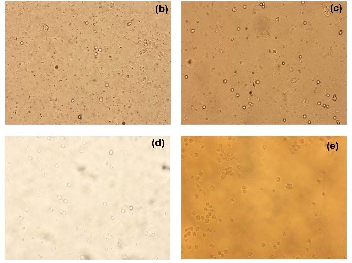
metabolites to and from the cell [23]. The list of There are limited studies in the literature about the use of
polysaccharides used commonly for a variety of biomedical nanofibrous mats produced by the electrospinning method
applications includes cellulose, carboxymethyl cellulose, as adhesion barriers [36-40]. In these studies, poly (lactic-
chitin/chitosan, starch, alginate, hyaluronic acid, pullulan, co-glycolic acid) (PLGA), polycaprolactone (PCL),
guar gum, and glycosaminoglycan [17]. PCL/gelatin, poly-L-lactide acid (PLLA), polyethersulfone
(PES), chitosan and calcium alginate nanofibrous mats
Hyaluronic acid (HA) is one of the major components of
were produced, and their performances were evaluated as
the extracellular matrix between cells in various living
adhesion barrier in vivo experiments. Only Dinanvard et al.
organisms such as cartilage, joint fluid, skin and umbilical
[37] conducted in vitro experiments. In a few studies [41-
cord. Its high viscoelasticity, non-toxic properties, and
44], PLGA, PCL, hyaluronic acid (HA)/PCL nanofiber
ability to absorb water due to high molecular weight and
membranes loaded with drugs, plant extract or silver were
negatively charged nature, make it possible for usage in
produced and proposed as adhesion barriers. Furthermore,
cosmetic, biomedical and food industries. Due to its
biocompatibility and biodegradability, HA polymer has in most studies, the resulting nanofibrous mats were not
been used especially in tissue engineering applications in compared with a commercial adhesion barrier.
the form of gel and/or film [24-27]. Today, complete adhesion prevention problem remains
Anionic and water-soluble carboxymethyl cellulose (CMC) unsolved and the search for an ideal adhesion barrier is still
polymer is produced with carboxylation of the cellulose and ongoing. In this study, electrospun nanofibrous mats were
belongs to the group of cellulose ethers. CMC is used as a first produced from hyaluronic acid (HA), carboxymethyl
binder, blowing, gelling, adhesive and stabilizer agent in cellulose (CMC) and sodium alginate (NaAlg) polymer
textile, paper, pharmaceutical, paint, cosmetic, ceramic and blends to be used as adhesion barriers, and their
food industries. The main reasons for choosing CMC as an performances were evaluated with in vitro experiments by
additive in these fields of use are that it is a physiologically comparing to a commercial adhesion barrier. The produced
inert, water-soluble, non-toxic and biocompatible polymer nanofibrous mats are novel in terms of the polymer blends
having high water retention capacity. Also, CMC is used and have a potential to be alternative to commercial
compatible with colloids and has a bacterial resistance due barriers in film form.
to its high sodium content [28, 29].
2. MATERIAL AND METHOD
Sodium alginate (NaAlg) is a natural polysaccharide
derived from brown seaweed. NaAlg has been used in food, 2.1 Materials
pharmaceutical, medical, textile and paper industries for
HA polymer (Hyaluronic acid sodium salt from
many years. Recently, its utilization has increased,
especially in biomedical and medical fields. The specific Streptococcus equi) with Mw of 1,500,000-2,000,000 g/mol
properties that facilitate wound healing of NaAlg, e.g., high and CMC polymer with Mw of 250,000 g/mol were
moisture absorption and ion exchange abilities, excellent purchased from Sigma Aldrich (USA). NaAlg polymer,
biocompatibility, and bleed inhibitor properties, make it a Cecalgum S1300, with a viscosity of 700-900 cPs was
unique raw material in the production of high absorbent kindly supplied by Cargill (Turkey). Sodium hydroxide
wound dressing [30, 31]. (NaOH, Sigma Aldrich) and dimethyl sulfoxide (DMSO,
Merck) were obtained to be used as the solvents for
Adhesions are described as abnormal connections between electrospinning of HA polymer. Distilled water was used as
organs that are not normally associated with each other and the solvent for CMC and NaAlg polymers.
surrounded by the serous membrane, following injury or
surgical operations. The main reasons of adhesions are For the crosslinking process of the nanofibrous mats, 1-
surgical procedures. Adhesions are common after chest, ethyl-3-(3-dimethylaminopropyl) carbodiimide
heart and intra-abdominal operations. The main approaches hydrochloride (EDC) with Mw of 191.70 g/mol and N-
proposed in the literature to prevent or reduce adhesion are hydroxysulfosuccinimide (NHS) with Mw of 115.09 g/mol
divided into three categories: the development of surgical were obtained from Sigma Aldrich. The crosslinked
techniques, the use of anti-adherence drugs, and the nanofibrous mats were neutralized in ethanol (Merck). All
separation of tissues during the healing process. Adhesion the materials were used without further purification.
barriers allow the surfaces of the injured region to be
separated from each other and freely heal and thus prevent In vitro experiments, a commercially available adhesion
the formation of adhesion. An ideal adhesion barrier should barrier film (Seprafilm, Sanofi-Aventis) containing HA
not affect wound healing but should be non-reactive, and CMC polymers was used to compare with the
effective in the presence of body fluids and blood, easy to nanofibrous adhesion barriers produced in this study.
use, and biodegradable. Furthermore, it should not cause
Human umbilical vein/vascular endothelial cell line
infection and inflammation, and it should be antibacterial
HUVEC (CRL-1730, ATCC) and mouse subcutaneous
and stable in the initial phase of adhesion formation, and
connective tissue fibroblast cell line L-929 (CCL-1, ATCC)
then metabolized [32-35].
100 TEKSTİL ve KONFEKSİYON 30(2), 2020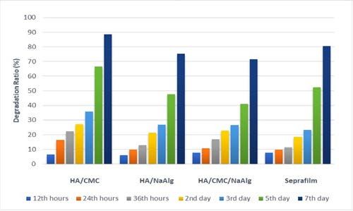
were used to investigate cytotoxicity and cell adherence             barriers [45]. Therefore, to improve their stability in water,
behavior on the nanofibrous mats and Seprafilm. On the               an appropriate crosslinking process was required to apply
cell viability test applied to determine the cytotoxicity, 2,3-      on the electrospun mats.
bis(2-methoxy-4-nitro-5-sulfophenyl)-2H-tetrazolium-5-
                                                                     EDC is a water-soluble, biocompatible and nontoxic
carboxanilide (XTT) cell proliferation kit (Biological
                                                                     crosslinking agent. EDC activates carboxyl groups in the
Industries, Israel) was used. Live cells were determined
                                                                     polysaccharide molecules and forms ester bonds between
with trypan blue solution (Sigma Aldrich).
                                                                     hydroxyl and carboxyl groups. The non-inclusion of EDC
Phosphate-buffered saline (PBS) of pH 7.4 for the                    in the cross-linked structure, i.e., not binding to polymer
degradation and cytotoxicity tests was purchased from PAN            molecules, is particularly recommended for materials used
Biotech (Germany).                                                   in the biomedical field. NHS is a nontoxic, biocompatible
                                                                     and homo-bifunctional crosslinker used to activate
Production of nanofibrous adhesion barriers
                                                                     carboxylic acid groups. When a normal carboxylic acid
In this study, nanofibrous mats aimed as adhesion barriers           forms a salt with amines, the acids which are activated in
were produced from HA/CMC, HA/NaAlg and                              the presence of NHS react with amines to give amides.
HA/CMC/NaAlg polymer blends by electrospinning (Table                EDC productivity increases in NHS presence. The use of
1). CMC and NaAlg solutions were prepared by dissolving              EDC together with NHS causes a formation of hydrolysis-
CMC and NaAlg polymers in distilled water at 80 ºC for 8             resistant and non-rearrangeable intermediates [46-49].
h. The concentration of solutions was 2% w/v. HA polymer
                                                                     The crosslinking medium was prepared by mixing EDC (80
was dissolved in a volume ratio of 4:1 NaOH/DMSO
                                                                     mM) and NHS (100 mM) crosslinking agents of an equal
solvent system at room temperature for 8 h and the
                                                                     ratio in ethanol of 20 ml. The HA/CMC, HA/NaAlg and
prepared HA solution concentration was 12% w/v. For the
                                                                     HA/CMC/NaAlg nanofibrous mats were immersed in
electrospinning, solutions of NaAlg, CMC and HA were
                                                                     crosslinking medium at room conditions for 24 h. After
blended in the volume ratio of 3/1 (HA/CMC), 5/1
                                                                     crosslinking, the mats were washed in ethanol for removing
(HA/NaAlg) and 3/1/1 (HA/CMC/NaAlg). The blended
                                                                     unbound crosslinking agents and then dried in an incubator
solutions were stirred at room temperature for 2 h to
                                                                     at 37 ºC for 12 h.
provide a homogeneous solution.
                                                                     Before in vitro experiments, the crosslinked electrospun
Prepared polymer solutions were fed into a plastic syringe
                                                                     mats were sterilized with ethylene oxide gas at 55 ºC for 4 h
of 20 ml. A spinneret with an inner diameter of 530 μm was
                                                                     and aerated for 8 h.
used as the feeding unit and a cylinder rotating at 200 rpm
was used as the collector. The cylinder collector was                Characterization
covered with aluminum foil. To achieve a smooth and
                                                                     Surface morphologies of the nanofibrous mats and the
beadless nanofiber formation, voltage, flow rate of solution,
                                                                     commercial adhesion barrier (Seprafilm) were characterized
and distance between the spinneret and the collector were
                                                                     by Scanning Electron Microscopy (SEM, Carl Zeiss AG-
adjusted for each polymer solution. All samples were
                                                                     EVO 40 XVP). The samples were coated with a thin layer
produced from 20 ml spinning solution to obtain
                                                                     of gold-palladium before analysis. The nanofiber diameter
uniformity. Electrospinning experiments were carried out at
                                                                     distributions were determined by using ImageJ software
ambient conditions and temperature. The temperature
                                                                     (National Institute of Health, USA) on SEM images. The
range: 20–28 °C, and RH range: 50–80% RH were
                                                                     average fiber diameter and standard deviation were
measured during electrospinning process. The process
                                                                     calculated from 50 random measurements for each sample.
parameters of produced nanofibrous mats are given in
Table 1.                                                             To investigate the degradation behavior of the crosslinked
                                                                     nanofibrous mats and Seprafilm under in vitro conditions,
Crosslinking and sterilization
                                                                     the samples were incubated in 5 ml of PBS at 37°C for 12,
HA, CMC and NaAlg are water-soluble polymers, and the                24, 36 hours, and 2, 3, 5, 7 days. The degradation ratio (D)
resulting nanofibrous mats have low resistance to water and          through the weight changes before and after immersion was
water vapor. This situation would lead to problems in                calculated as [50]:
practical applications of the nanofibrous mats as adhesion

                                  Table 1. The process parameters of the electrospun nanofibrous mats

      Parameters                                     HA/CMC                     HA/NaAlg                HA/CMC/NaAlg
      Voltage                                         20.1 kV                    18.7 kV                    22 kV
      Flow rate of the solution                       0.2 ml/h                   0.6 ml/h                  0.5 ml/h
      Tip-to-collector distance                         9 cm                      7.5 cm                    8.5 cm

TEKSTİL ve KONFEKSİYON 30(2), 2020                                                                                            101
% D = 100 – [Wn / W0] x 100 Cytotoxicity of the crosslinked nanofibrous mats and
Seprafilm were carried out by the XTT cell viability test in
Where W0 is original weight (before incubation), and Wn is
vitro conditions according to ISO 10993-5:2010. Viability
residual dry weight on the assessment day (after incubation).
and proliferation of HUVEC and L-929 cells were
For in vitro cell adherence potential test of the crosslinked determined by the absorbance measurements performed on
nanofibrous mats and Seprafilm, HUVEC and L-929 cells a microplate reader at 450 nm and 630 nm after 24 h. The
were cultured in RPMI-1640 medium supplemented with test was performed three times for each sample, and the
fetal calf serum (10%), penicillin - streptomycin (50 U/ml - percentage cell viability was calculated by quantitative
50 µg/ml), L-glutamine (2 mM) and sodium pyruvate (1%). method.
Fetal Calf Serum (FCS ) is used as a supplement to basal
The test results of cell adherence potential and cytotoxicity
growth medium in cell culture. When used at appropriate
were evaluated statistically with Mann-Whitney U test by
concentrations it supplies many specific metabolic
using SPSS 22.0 software. PBefore the crosslinking process, SEM photographs have (HA/CMC), 5/1 (HA/NaAlg) and 3/1/1 (HA/CMC/NaAlg).
shown that smooth, beadless, uniform and continuous fibers So polymer amount of HA/CMC (1.8 g HA, 0.1 g CMC /
were successfully produced from HA/CMC, HA/NaAlg and 20 ml), HA/NaAlg (2 g HA, 0.067 g NaAlg) and
HA/CMC/NaAlg polymer solutions. The average fiber HA/CMC/NaAlg (1.44 g HA, 0.08 g CMC, 0.08 g NaAlg)
diameters of the uncrosslinked HA/CMC, HA/NaAlg and are not equal. But EDC/NHS crosslinking procedure was
HA/CMC/NaAlg nanofibrous mats were 131±46 nm, conducted in the same amount of crosslinking medium for
199±90 nm and 117±42 nm, respectively. all samples. EDC /NHS activate carboxyl groups in the
polysaccharide molecules and forms ester bonds between
After the crosslinking process, smooth, uniform and the hydroxyl and carboxyl groups. The EDC/NHS is not
continuous nanofiber structure has been deteriorated. binding to polymer molecules [49, 53]. Therefore
Sticking and flattening at the contact points of the fibers, crosslinking medium might be inadequate and crosslinking
and a film-like structure were observed. EDC and NHS efficiency may not be equal for the samples.
activate the carboxyl groups in polymer molecules forming
the nanofibers and ensure bond formation between 3.2 Degradation
hydroxyl (–OH) and carboxyl (–COOH) groups. The Figure 3 shows the weight changes in electrospun
benefit of the EDC and NHS is that they do not become a HA/CMC, HA/NaAlg and HA/CMC/NaAlg crosslinked
part of the crosslinked structure. They just help to facilitate nanofibrous mats and in Seprafilm during the in vitro
bonding within the crosslinked structure. New bonds cause degradation test for a week period.
the squeeze of the structure [49, 53]. Furthermore, the
fibers collapse as flat bands, when a quantity of the solvent
trapped between the fibers evaporates [54]. It is concluded
that the flattening of fibers may occur owing to the rapid
evaporation of the solvent, and the squeezing in the
nanofibrous structure due to the formation of new bonds
between polymer molecules.
In order to compare with the nanofibrous adhesion barriers,
SEM image of the Seprafilm commercial adhesion barrier
is given in Figure 2. When the photograph was examined, it
was observed that the Seprafilm had a rough film structure
and did not contain any pores.
Figure 3. Degradation ratios of the crosslinked nanofibrous mats
and Seprafilm
The critical period for adhesion formation is the first seven
days after trauma. The mechanism of adhesion formation
follows a very rapid course in this period. For this reason,
an ideal surgical adhesion barrier should be able to maintain
its presence by keeping the tissues separated from each
other for the first seven days. However, an adhesion barrier
must biodegrade rapidly in the body after the critical period
for adhesion formation [55]. Consequently, it has been
understood that the produced nanofibrous mats could
protect their structures sufficiently during the critical
healing period and could continue to separate the organs or
Figure 2. SEM photograph of Seprafilm tissues from each other.
No remarkable weight loss differences between the 3.3 Cell adherence potential
nanofibrous mats and Seprafilm were observed within the On a cell adherence potential test, adherent cells take a
first three days. However, the fastest degradation occurred shuttle-like shape when they attached to the surfaces. Due
on HA/CMC nanofibrous mat. Also, on the 5th and the 7th to this feature of adherent cells, shuttle shaped cells on the
days, Seprafilm’s degradation ratio was found to be higher crosslinked nanofibrous mats and Seprafilm were counted
than HA/NaAlg and HA/CMC/NaAlg nanofibrous mats. microscopically after 24 hours from seeding and the
The reason of degradation ratio differences between calculated percentages are presented in Table 2.
nanofibrous mats might be amount of polymer differentiation. Microscopic images of the samples are also shown in
For the electrospinning process, solutions of NaAlg, CMC Figure 4 and Figure 5.
and HA were blended in the volume ratio of 3/1
TEKSTİL ve KONFEKSİYON 30(2), 2020 103Table 2. The percentage of cells adhered to the surfaces after 24 hours from seeding (mean ± standard deviation)
Samples HUVEC cell adherence (%) L929 cell adherence (%)
HA/CMC 0.78 ± 0.19 0.44 ± 0.19
HA/NaAlg 0.56 ± 0.51 0.78 ± 0.19
HA/CMC/NaAlg 0.67 ± 0.33 0.44 ± 0.51
Seprafilm 0.78 ± 0.19 0.89 ± 0.19
Control 98.67 ± 0.33 99.00 ± 0.67
*Control : No adhesion barrier
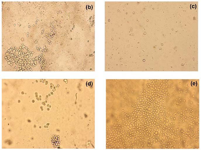
Figure 4. Microscopic images of the samples after 24 hours from seeding with HUVEC cells (X10); a) Control, b) HA/CMC, c)
HA/NaAlg, d) HA/CMC/NaAlg, e) Seprafilm
Figure 5. Microscopic images of the samples after 24 hours from seeding with L929 cells (X10); a) Control, b) HA/CMC, c)
HA/NaAlg, d) HA/CMC/NaAlg, e) Seprafilm
104 TEKSTİL ve KONFEKSİYON 30(2), 2020Cell morphology is an important identifier of adhesion adhesion barrier were evaluated by using the HUVEC and
behavior. If the cells adhere to the surfaces, they take the L-929 cells according to XTT method. The cell viability
shuttle shape and become spread [56]. When the values obtained after 24 hours were given in Table 3.
microscopic images were analyzed, it was observed that the
cells did not adhere onto the nanofibrous mats and Cell viability rates of the nanofibrous mats were determined
Seprafilm for both cell lines. Their cell adherence potentials above 90% for both of the cell lines. Furthermore, the
were lower than 1%. Whereas, cells in the control groups nanofibrous mats did not show statistically any significant
had shuttle shape. They adhered to the petri dishes, and difference in cytotoxicity when compared with the
they were motionless. On the other hand, cells on the Seprafilm and with each other (p ≥ 0.05).
nanofibrous mats and Seprafilm became round in shape. It According to ISO 10993-5:2010 standard, samples are
was noticed that these cells were moving and they could not considered as non-cytotoxic if the cell viability is above
adhere to the surfaces. 70%. To sum up, it was determined that the crosslinked
When the cell adherence capacities of the nanofibrous mats nanofibrous mats had any cytotoxic effect towards to both
and Seprafilm were compared statistically to control group of the cells and they could be used in surgical operations.
for both cell lines; it was found that the percentage of cells 4. CONCLUSIONS
adhered on the sample was significantly low (p < 0.05).
There was no significant statistical difference between the In this study, nanofibrous mats were produced as novel
cell adherence potentials of the nanofibrous mats and surgical adhesion barriers from polysaccharide-based
Seprafilm for both cell lines (p ≥ 0.05). polymers, hyaluronic acid, carboxymethyl cellulose and
sodium alginate, via electrospinning process.
Although the produced nanofibrous mats possess good
As CMC, HA and NaAlg used to produce the nanofibrous
biological compatibility, they were unsuitable for cell
mats were water-soluble polymers, a crosslinking process
adherence. This situation is because of the hydrophilic and
was applied to protect their physical integrity in vitro
polyanionic surfaces do not thermodynamically support cell
environment. This process was performed with EDC/NHS
attachment and tissue formation [48]. Also, it has been
crosslinking medium which do not have toxic effects and
reported that the cell attachment can be observed on the
do not cause damage on morphological properties of the
hydrophobic surfaces more than on hydrophilic surfaces
surfaces. The average fiber diameters of the uncrosslinked
[57, 58]. Therefore, low cell adherence on the nanofibrous
nanofibrous mats were obtained between 108-132 nm.
mats may be explained by their high hydrophilicity and
After the crosslinking, the nanofibers flattened, and the
their polyanionic structure which could prevent the cells to
average fiber diameter values increased almost doubled. On
be attached. For an ideal adhesion barrier, infiltration or
the other hand, it was seen from SEM images that
adherence of blood or cells should be avoided by using
Seprafilm had a rough and nonporous structure.
proper materials and by controlling precisely pore sizes
[59]. It was concluded that the produced nanofibrous mats In the in vitro degradation test, the crosslinked nanofibrous
had non-adherent cell feature and could show the potential mats and Seprafilm have suffered the weight loss in a
to be an ideal adhesion barrier to prevent adhesion of the linearly increasing manner for 7 days. While significant
tissues in the surgical operations. differences between the degradation ratios of produced
nanofibrous adhesion barriers and the commercial adhesion
3.4 Cytotoxicity barrier were not observed on the earlier days, the
nanofibrous barriers, especially HA/NaAlg and HA/CMC/
The ideal adhesion barrier should not have any toxic
NaAlg, kept their mass more than Seprafilm on 5th and 7th
effects, and it should not have an effect on wound healing
days. It was concluded that the produced nanofibrous mats
in the surgical region. Therefore, possible cytotoxic effects
could maintain their presence by keeping the tissues
of the crosslinked nanofibrous mats and the commercial
separate from each other during the healing process.
Table 3. The percentage cell viability (mean ± standard deviation)
Sample HUVEC cell viability (%) L929 cell viability (%)
HA/CMC 92.12 ± 3.90 97.12 ± 6.00
HA/NaAlg 95.45 ± 11.00 95.94 ± 13.00
HA/CMC/NaAlg 91.38 ± 2.80 92.54 ± 11.00
Seprafilm 97.29 ± 2.10 97.45 ± 2.00
Control 0.95 ± 0.001 0.99 ± 0.001
TEKSTİL ve KONFEKSİYON 30(2), 2020 105In vitro cytotoxicity and cell adherence potential of the The cytotoxicity, cell adherence and degradation behaviors
nanofiber mats and Seprafilm were evaluated with HUVEC under in vitro conditions showed that the crosslinked
and L-929 cells. According to the cytotoxicity test results, nanofibrous mats are have potentials to use as adhesion
cell viability rates of the nanofibrous mats were determined barriers.
above 90% for both cell lines. It was concluded that the
produced nanofibrous mats were not cytotoxic. On the other ACKNOWLEDGMENTS
hand, it was found that cell adherence on the nanofibrous The authors thank to TUBİTAK (The Scientific and
mats was very low (below 1%). The cell adherence Technical Research Council of Turkey) for its financial
potential test indicated that the produced nanofibrous mats support in the research project (No. 214M415).
could prevent infiltration of blood or cells.
REFERENCES
1. Deitzel J.M., Kleinmeyer J., Harris DEA., & Tan NB. (2001). The 15. Şafak Ş., Düzyer Ş. & Karaca E. (2016). Evaluation of
effect of processing variables on the morphology of electrospun biocompatibility of fibroin-based electrospun nanofibrous mats for
nanofibers and textiles. Polymer, 42,261–272. medical applications. Industria Textila, 67,3–9.
2. Supaphol P., Suwantong O., Sangsanoh P., Srinivasan S., Jayakumar 16. Rim, N. G., Shin, C. S., & Shin, H. (2013). Current approaches to
R. & Nair SV. (2012). Electrospinning of biocompatible polymers electrospun nanofibers for tissue engineering. Biomedical Materials,
and their potentials in biomedical applications. Advances in Polymer 8(1), 014102.
Science, 246,213–240.
17. Shelke N.B., James R., Laurencin C.T. & Kumbar S.G. (2014).
3. Ramakrishna S., Fujıhara K., Teo WE., Lim TC. & Ma Z. (2005). An Polysaccharide biomaterials for drug delivery and regenerative
introduction to electrospinning and nanofibers. Singapore: World engineering. Polymers for Advanced Technologies, 25,448–460.
Scientific Publishing Co, 396 p.
18. Kusindarta, D. L., & Wihadmadyatami, H. (2018). The Role of
4. Huang C., Chen S., Lai C., Reneker DH., Qiu H., Ye Y. & Hou H. Extracellular Matrix in Tissue Regeneration. Tissue Regeneration,
(2006). Electrospun polymer nanofibres with small diameters. 65.
Nanotechnology, 17,1558–1563.
19. Theocharis, A. D., Skandalis, S. S., Gialeli, C., & Karamanos, N. K.
5. Li, J., Chen, Z., Zhou, M., Jing, J., Li, W., Wang, Y. & Lee, M. (2016). Extracellular matrix structure. Advanced Drug Delivery
(2016). Polyoxometalate‐Driven Self‐Assembly of Short Peptides Reviews, 97, 4-27.
into Multivalent Nanofibers with Enhanced Antibacterial Activity.
20. Abedin, M., & King, N. (2010). Diverse evolutionary paths to cell
Angewandte Chemie International Edition, 55(7), 2592-2595.
adhesion. Trends in Cell Biology. 20(1), 734-742.
6. Tan, C., Qi, X., Liu, Z., Zhao, F., Li, H., Huang, X. & Tang, Z.
21. Varki, A., Cummings, R., Esko, J., Freeze, H., Hart, G., & Marth, J.
(2015). Self-assembled chiral nanofibers from ultrathin low-
(1999). Proteoglycans and glycosaminoglycans. In Essentials of
dimensional nanomaterials. Journal of the American Chemical
glycobiology. Cold Spring Harbor Laboratory Press.
Society, 137(4), 1565-1571.
22. Zhang, F., Zhang, Z., & Linhardt, R. J. (2010). Glycosaminoglycans.
7. Liu, Y., Yan, X., Lan, J., Yu, Y., Yang, X., & Lin, Y. (2017). Phase-
In Handbook of glycomics. Academic Press, pp. 59-80.
separation induced hollow/porous carbon nanofibers containing in
situ generated ultrafine SnO x as anode materials for lithium-ion 23. Rowley, J. A., Madlambayan, G., & Mooney, D. J. (1999). Alginate
batteries. Materials Chemistry Frontiers 1(7), 1331-1337. hydrogels as synthetic extracellular matrix materials. Biomaterials,
20(1), 45-53.
8. Katsogiannis, K. A. G., Vladisavljević, G. T., & Georgiadou, S.
(2015). Porous electrospun polycaprolactone (PCL) fibres by phase 24. Ji Y., Ghosh K., Li B., Sokolov J.C., Clark R.A. & Rafailovich M.H.
separation. European Polymer Journal., 69, 284-295. (2006). Dual-syringe reactive electrospinning of cross-linked
hyaluronic acid hydrogel nanofibers for tissue engineering
9. Yan, C., Chen, G., Zhou, X., Sun, J., & Lv, C. (2016).
applications. Macromolecular Bioscience, 6,811–817.
Template‐Based Engineering of Carbon‐Doped Co3O4 Hollow
Nanofibers as Anode Materials for Lithium‐Ion Batteries. Advanced 25. Schante C.E., Zuber G., Herlin C. & Vandamme T.F. (2011).
Functional Materials 26(9), 1428-1436. Chemical modifications of hyaluronic acid for the synthesis of
derivatives for a broad range of biomedical applications.
10. Mishra, S., & Verma, N. (2016). Carbon bead-supported hollow
Carbohydrate Polymers, 85, 469–489.
carbon nanofibers synthesized via templating method for the removal
of hexavalent chromium. Journal of Industrial and Engineering 26. Aytar P., Buruk Y. & Çabuk A. (2013). Streptecoccus equi ile
Chemistry. 36, 346-354 hyaluronik asit üretiminde optimum koşullarin plackett-burman
yöntemi ile belirlenmesi. Elektronik Mikrobiyoloji Dergisi, 11, 28–
11. Laurencin, C. T., Kumbar, S. G., Nukavarapu, S. P., James, R., &
35.
Hogan, M. V. (2008). Recent patents on electrospun biomedical
nanostructures: an overview. Recent Patents on Biomedical 27. Collins M.N. & Birkinshaw C. (2013). Hyaluronic acid based
Engineering, 68-78. scaffolds for tissue engineering-a review. Carbohydrate Polymers
12. Eatemadi, A., Daraee, H., Zarghami, N., Melat Yar, H., & 28. Qiu X. & Hu S. (2013). Smart materials based on cellulose: a review
Akbarzadeh, A. (2016). Nanofiber: synthesis and biomedical of the preparations, properties and applications. Materials, 6, 738–
applications: Artificial Cells, Nanomedicine, and Biotechnology, 781.
44(1), 111-121.
29. Klemm D., Heublein B., Fink H.P. & Bohn A. (2005). Cellulose:
13. Sharma, J., Lizu, M., Stewart, M., Zygula, K., Lu, Y., Chauhan, R., fascinating biopolymer and sustainable raw material. Angewandte
& Wei, S. (2015). Multifunctional nanofibers towards active Chemie International Edition, 44, 3358–3393.
biomedical therapeutics. Polymers, 7(2), 186-219.
30. Qin Y. (2008). Alginate fibres: an overview of the production
14. Khalil, K. A., Fouad, H., Elsarnagawy, T., & Almajhdi, F. N. (2013). processes and applications in wound management. Polymer
Preparation and characterization of electrospun PLGA/silver International, 57, 171–180.
composite nanofibers for biomedical applications. Reklamlar
International Journal of Electrochemistry, 8(3), 3483-3493.
106 TEKSTİL ve KONFEKSİYON 30(2), 202031. Üstündağ C.G., Karaca E., Özbek S. & Çavuşoğlu İ. (2010). In vivo 46. Fischer R.L., McCoy M.G. & Grant S.A. (2012). Electrospinning
evaluation of electrospun poly (vinyl alcohol)/sodium alginate collagen and hyaluronic acid nanofiber meshes. Journal of Materials
nanofibrous mat as wound dressing. Tekstil Konfeksiyon, 20, 290–298. Science: Materials in Medicine, 23, 1645–1654.
32. Şahiner İ.T. (2011). Simvastatin yüklü polipropilen yama ile 47. Lu P.L., Lai J.Y., Ma D.H.K. & Hsiue G.H. (2008). Carbodiimide
onarilan karın duvarı defektlerinde batın içi yapışıklıkların cross-linked hyaluronic acid hydrogels as cell sheet delivery
karşılaştırılması. Ph.D. Dissertation: Kırıkkale University. vehicles: characterization and interaction with corneal endothelial
cells. Journal of Biomaterials Science Polymer Edition, 19, 1–18.
33. Yeğenoğlu A. (2005). Postoperatif intraperitoneal adezyonların
önlenmesinde değişik dozlardaki heparin ve seprafilm’in 48. Xu S., Li J., He A., Liu W., Jiang X., Zheng J., Han C.C., Hsiao B.S.,
etkinliklerinin karşılaştırılması. Ph.D. Dissertation: Dr. Lütfi Kırdar Chu B. & Fang D. (2009). Chemical crosslinking and biophysical
Education and Research Hospital. properties of electrospun hyaluronic acid based ultra-thin fibrous
membranes. Polymer, 50, 3762–3769.
34. Günaydın M., Güvenç D., Yıldız L., Aksoy A., Tander B., Bıçakcı
Ü., Ayyıldız H.S., Sünter A.T. & Bernay F. (2012). Comparison of 49. Tomihata K, & Ikada Y. (1997). Crosslinking of hyaluronic acid with
substances used for prevention of ıntra-abdominal adhesions: an water-soluble carbodiimide. Journal of Biomedical Material
experimental study in rats. International Journal of Medical Sciences, Research, 37, 243–251.
32, 337–345.
50. Fouad, H., Elsarnagawy, T., Almajhdi, F. N., & Khalil, K. A. (2013).
35. Şafak Ş. (2016). Investigation of usage performance of electrospun Preparation and in vitro thermo-mechanical characterization of
nanofibrous mats produced from biodegradable polymers as surgical electrospun PLGA nanofibers for soft and hard tissue replacement.
adhesion barrier. Ph.D. Dissertation: Uludag University. International Journal of Electrochemical Science, 8(2), 2293-2304.
36. Zong X., Li S., Chen E., Garlick B., Kim K.S., Fang D. & Chu B. 51. Biological Industries. (2019, September, 17), Introduction to Fetal
(2004). Prevention of postsurgery-induced abdominal adhesions by Bovine Serum Class. Retrieved from https://www.bioind.com/
electrospun bioabsorbable nanofibrous poly (lactide-co-glycolide)- worldwide/support/tech-tips-posters/introduction-to-fetal-bovine-
based membranes. Annals of Surgery, 240, 910–915. serum-class/
37. Dinarvand P., Hashemi S.M., Seyedjafari E., Shabani I., 52. Brunner, D., Frank, J., Appl, H., Schöffl, H., Pfaller, W., &
Mohammadi-Sangcheshmeh A., Farhadian S. & Soleimani M. Gstraunthaler, G. (2010). The serum-free media interactive online
(2012). Function of poly (lactic-co-glycolic acid) nanofiber in database. ALTEX-Alternatives to animal experimentation, 27, 53-62.
reduction of adhesion bands. Journal of Surgical Research, 172, 1–9.
53. Sinha M.K., Das B.R., Srivastava A. & Saxena A.K. (2013).
38. Chang J.J., Lee Y.H., Wu M.H., Yang M.C. & Chien C.T. (2012). Needleless electrospinning and coating of poly vinyl alcohol with
Electrospun anti-adhesion barrier made of chitosan alginate for cross-linking agent via in-situ technique. International Journal of
reducing peritoneal adhesions. Carbohydrate Polymers, 88, 1304– Textile and Fashion Technology, 3, 29–38.
1312.
54. Baji A., Mai Y.W., Wong S.C., Abtahi M. & Chen P. (2010).
39. Shi R., Xue J., Wang H., Wang R., Gong M., Chen D. & Tian W. Electrospinning of polymer nanofibers: effects on oriented
(2015). Fabrication and evaluation of a homogeneous electrospun morphology, structures and tensile properties. Composites Science
PCL–gelatin hybrid membrane as an anti-adhesion barrier for and Technology, 70, 703–718.
craniectomy. Journal of Materials Chemistry B, 3, 4063–4073.
55. Hatipoğlu E. (2011). Ameliyat sonrası karın içi yapışıklıkların
40. Lee Y.W., Chu B., Lee Y.G., Kim N.H., Kim J.H., Kim K.I. & Kwon önlenmesinde sodyum hyaluronat karboksimetilselüloz membran,
S.W. (2009). Efficacy and safety of the electrospun nanofibrous polietilen glikol - lysine ve hyaluronik asitin etkinliğinin wistar
adhesion barrier for laparoscopic surgery in a rabbit model. Journal albino tipi sıçanlarda yapılan deneysel çalışma ile araştırılması. Ph.D.
of the Korean Surgical Society, 76, 73–80. Dissertation: Istanbul University.
41. Bölgen N., Vargel I., Korkusuz P., Menceloğlu Y.Z. & Pişkin E. 56. Ma Z., Kotaki M., Yong T., He W. & Ramakrishna S. (2005).
(2007). In vivo performance of antibiotic embedded electrospun Surface engineering of electrospun polyethylene terephthalate (PET)
PCL membranes for prevention of abdominal adhesions. Journal of nanofibers towards development of a new material for blood vessel
Biomedical Materials Research Part B, 81, 530–543. engineering. Biomaterials, 26,2527–2536.
42. Adegani J.F., Seyedjafari E., Gheibi N., Soleimani M. & Sahmani M. 57. Yang D.J., Chen F., Xiong Z.C., Xiong C.D. & Wang Y.Z. (2009).
(2016). Prevention of adhesion bands by ibuprofen-loaded PLGA Tissue anti-adhesion potential of biodegradable PELA electrospun
nanofibers. Biotechnological Programme, 32, 990–997. membranes. Acta Biomaterials, 5, 2467–2474.
43. Shin Y.C., Yang W.J., Lee J.H., Oh J.W., Kim T.W., Park J.C., Hyon 58. Lee J.H., Lee S.J., Khang G. & Lee H.B. (2000). The effect of fluid
S.H. & Han D.W. (2014). PLGA nanofiber membranes loaded with shear stress on endothelial cell adhesiveness to polymer surfaces with
epigallocatechin-3-O-gallate are beneficial to prevention of wettability gradient. Journal of Colloid and Interface Science, 230,
postsurgical adhesions. International Journal of Nanomedicine, 9, 84–90
4067–4078.
59. Mayes S., Schmidth C.E. & Peterson D. (2012). Anti-adhesive
44. Chen C.H., Chen S.H., Shalumon K.T. & Chen J.P. (2015). Dual barrier membrane using alginate and hyaluronic acid for biomedical
functional core–sheath electrospun hyaluronic acid/polycaprolactone aplications. Patent 0088832- A1, USA
nanofibrous membranes embedded with silver nanoparticles for
prevention of peritendinous adhesion. Acta Biomaterialia, 26, 225–
235.
45. Şafak Ş. & Karaca E. (2017). Production and crosslinking of
polysaccharide based nanofibrous mat for biomedical applications.
Uludag University JFE, 22, 127–144.
TEKSTİL ve KONFEKSİYON 30(2), 2020 107You can also read