Metastatic and pathophysiological characteristics of breast cancer with emphasis on hereditary factors
←
→
Page content transcription
If your browser does not render page correctly, please read the page content below

Central Asian Journal of Medical and Pharmaceutical Sciences Innovation 3 (2021) 104-113

REVIEW PAPER

Metastatic and pathophysiological characteristics of
breast cancer with emphasis on hereditary factors
Azita Faramarzi 1, Masoumeh Golestan Jahromi 2, Sareh Ashourzadeh 3, Nasrin Jalilian 4*
1
  Fertility and Infertility Research Center, Health Technology Institute, Kermanshah University of Medical Sciences,
Kermanshah, Iran
2
  Department of Advanced Medical Science & Technologies, School of Medicine, Jahrom University of Medical
Sciences, Jahrom, Iran
3
  Afzalipour clinical center for infertility, Kerman University of Medical Sciences, Kerman, Iran
4
  Department of Obstetrics and Gynecology, School of Medicine, Kermanshah University of Medical Sciences,
Kermanshah, Iran

Highlights                                   Graphical Abstract

• Breast cancer develops from the
breast tissue and is the second
leading cause of cancer deaths.
• Cadherin and integrin are could
be considered in the metastasis
procedure of breast cancer.
• BRCA1 and BRCA2 are the
most important genes involved in
breast cancer pathophysiology.
• Some other genes, including Her
2, p53, p21, bcl-2, etc. play the
central role in breast cancer.

Article Info                                 Abstract
Receive Date: 09 January 2021               Breast cancer is the most common type of cancer among women and the second
Revise Date: 25 May 2021                    leading cause of deaths after lung cancer. Each year, more than 180,000 new cases
Accept Date: 13 June 2021                   of breast cancer are diagnosed in the United States. The risk of breast cancer is low
                                            before the age of 35, but the high prevalence of this type of cancer is diagnosed
Available online: 20 June 2021
                                            after the age of 35. The risk of developing breast cancer in a woman's lifetime is
                                            about 10% and 5-10% with a familiar genetic basis. The accumulation of breast and
Keywords:                                   ovarian cancers in certain families indicates that genetic variations are involved in
Breast cancer                               developing these types of cancers. In 1994, two genes were identified to be linked
Risk-factors                                to familial breast cancers. Gene variations in these two genes (BRCA1 and BRCA2)
Pathophysiology                             are found in most familial breast cancers, and variations in several other genes may
Metastasis                                  also be involved in this cancer. The molecular functions of BRCA1 and BRCA2 are
                                            still unclear, although they could be involved in repairing damaged DNA or
Genetic factors                             regulating the transcription of hormone-responsive genes. Mutations in these genes
                                            are predominantly autosomally transmitted by variable penetration. Women with
                                            BRCA1 variations are up to 50 percent more susceptible to breast cancer pending
                                            their lifetime. However, there are some other genes in which the variation may
                                            result in breast cancer occurrence. In this paper, we assessed some features of breast
                                            cancer to focus on hereditary aspects.
                                                                                                 © 2021 Published by CAS-Press.

   10.22034/CAJMPSI.2021.03.01                                                                              E-ISSN: 2783-0993
*Corresponding author: njalilian@kums.ac.ir (N. Jalilian)
                                                                                                                            104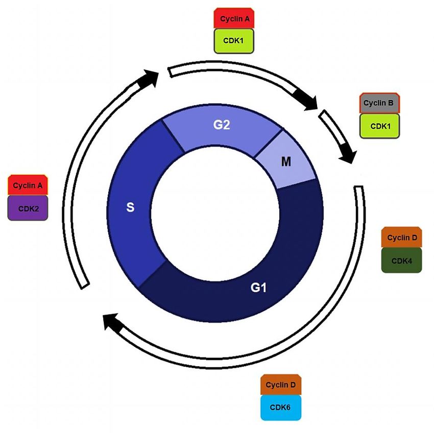
Faramarzi et al.,                                                      Cent Asian J Med Pharm Sci Innov 3 (2021) 104-113

Introduction
   Based on World Health Organization (WHO), breast cancer is the most common cancer type in women and
is the second cause of cancer-related death in women after lung cancer (1). It accounts for 22% of all women
cancers (2). In Iran, breast cancer consists of 16% of all cancers and still ranks first. The fifth leading cause of
death in Iranian women is common cancer in almost all provinces such as Mazandaran and Kermanshah (3, 4).
Breast cancer is a multifactorial disease in which various genetic and environmental factors work in different
ways. Our knowledge of how they work and the fundamental cause of the disease are still incomplete. A risk
factor is any factor that grows the risk of developing a disease such as cancer. Of course, having risk factors
does not mean that a person will get the disease, and not having them will not be a guarantee (5, 6).
    Researches have not shown the definite cause of breast cancer, but it seems that this disease is influenced by
factors such as the genetic structure of hormonal profiles and different life patterns (7). Some predisposing
environmental factors of breast cancer are Geographical status, lifestyle, age of marriage and obesity (8). Genetic
is a significant factor in breast cancer, so the risk of disease for a woman with one or more first-degree relatives
with this cancer is three and ten times higher, respectively (9). According to another source, the risk of breast
cancer for women up to the age of 80 with any first-degree relatives suffering cancer will be 7.8%, and for
women with one or two first-degree relatives is 13 and 21%, respectively (10). Finding the relevant agents of this
disease, including its genetic factors, will provide important information that helps researchers and physicians
find solutions to prevent, treat, or increase life expectancy with the least physical and mental problems for
patients (11).
    BRCA1 and BRCA2 are two primary genes involved in breast cancer. These genes are the dynamic managers
of the integrity of the genome. Mutations in these genes are correlated with cancer development in several
organs, such as the ovary and breast. The BRCA1/2 genes mutations mainly elevate duration risk to the
development of cancers of the ovary and breast, and these mutations are commonly detected in the hereditary
ovary and breast cancers (12). Also, deregulation and changed expressions of BRCA1/2 could result in sporadic
breast cancer. Significantly, both proteins play a role in DNA repair and regulation of gene transcription in
DNA damage events. Thus, defects of BRCA1/2 result in the accumulation of genetic changes and eventually
impact cancer development. Both BRCA1/2 have provided vital signs for their activities in tumor suppression,
definitely for breast cancer (13). In this review, we describe the pathophysiological characteristics and
metastasis of breast cancer, focusing on its genetic aspects.

Breast cancer metastasis
    Breast cancer is the most common cancer, could affect other parts of the body. This migration from the origin
to another is known as metastasis, which is the main cause of cancer death. Cancer can spread in several ways.
Cancer cells, or small packages of cells, can invade healthy tissue near the site of origin. They may also migrate
to other body regions through the lymph or blood vessels and attack healthy tissues. Although most cancers can
spread almost anywhere, bone is the most common site for breast cancer metastasis, so that up to 75% of breast
cancer patients with metastatic cancer have bone involvement (14, 15).
    In cancer metastasis, cells must pass the basement membrane and enter the blood circulation or lymphatic
system. There are specific genes that command the production of adhesive proteins. If these genes are mutated,
proteins are produced that will not be able to function normally. Thus, cells lose their natural adhesion to each
other and the basement membrane. As a result, they go outside their natural limitations (16, 17, 18).
    Researchers have shown that the normal function of these adhesive molecules is gradually reduced in
advanced tumors that cause the onset of metastasis, which is one of the most dangerous stages in the
development of cancer. Removing a malignancy before it enters this dangerous stage will significantly reduce
patient mortality (19). Among the adhesive proteins, cadherin and integrin are the two most well-known types,
which have been the subject of many biological types of research on breast cancer (20). These cells can only raise

105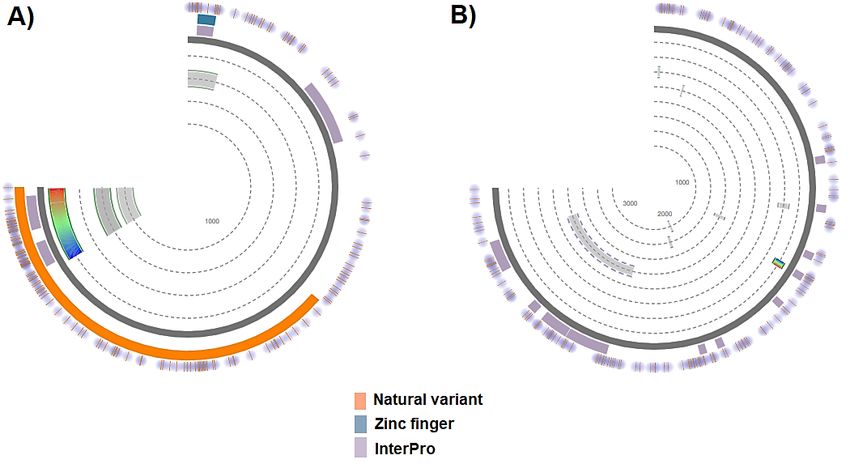
Cent Asian J Med Pharm Sci Innov 3 (2021) 104-113                                                        Faramarzi et al.,

and increase if they have certain abilities like produce new blood vessels. The mechanism of metastasis is
multifaceted, and its ability differs according to the target tissue (21).

Pathophysiology of breast cancer
   Breast cancer, like other cancers, is triggered by contact between an environment and a defective gene. Cell
division in normal cells stops after the desired number; then cells enter their tissues by attaching to other cells
(22). However, in cancer cells, due to the variation, the ability to stop division is lost; as a result, the cell cannot
attach to other cells and be placed in its tissue (23). Normal cells are doomed to cell death when they are no
longer needed, but until then, these cells are protected from apoptosis by several proteins and pathways (24).
One of these pathways is the mTOR/AKT/PI3K pathway (the intracellular signalling pathway that is important
in cell cycle regulation), and the ERK/MEK/Raf/Ras pathway (A group of intracellular proteins that are involved
in leading signals from the receptor on the cell surface into the nucleus) is another one (25). Sometimes some of
the genes involved in the protective pathways mutate into permanent and stable genes that are not capable of
apoptosis. This type of variation, along with other ones, causes cancer (26). Usually, the Phosphatase and tensin
homolog (PTEN) protein blocks the PI3K/AKT/MTOR pathway when the cell is ready for apoptosis.
   In some breast cancers, the gene that makes the PTEN protein is changed. Therefore, the PI3K/AKT/MTOR
pathway remains active, and the cancer cells will not be able to apoptosis and become immortal (27).
Experiments show that variations that can lead to breast cancer are associated with high estrogen levels.
Immune system defects that are naturally responsible for killing malignant cells through a person's life and
anomalous signalling of growth factors in the interaction between stromal cells and epithelial cells can enable
the growth of cancerous cells (28).

Genetic factors of breast cancer
   All breast cancer cases are not inherited, and the hereditary type accounts for just a small percentage (5-10%)
of all cases. In fact, in most cases, the disorder begins in the individual and is not related to higher generations
(29). These are called "unilateral breast cancers." About 70% of breast cancers are non-hereditary, but family
history is a very important risk factor for breast cancer. Women with a family history are at higher risk for
disease (30). Thus, the risk of breast cancer in a woman with an infected sister or mother is 1.5 to 3 times higher,
for instance, in a woman whose mother has breast cancer in comparison with a woman who has no family
history of breast malignancy, the age of onset is lower (31).
   In two-thirds of these cases, inherited variations in the BRCA1 and BRCA2 genes (an autosomal dominant
genetic pattern) increase breast cancer and ovarian cancer (32). In 40% of familial breast cancer cases, a variation
in BRCA1 and 30% of this type of breast cancer, a variation in BRCA2 gene was observed (33). While both
BRCA1 and BRCA2 genes are tumor suppressor genes that control cell growth, death and differentiation, they
are also involved in DNA repair. The protein products of these genes are called caretaker, which maintains
genomic integrity (33).
   In proto-oncogenes, variations in one allele are sufficient to alter gene function in uncontrolled cell growth;
unlike in tumor suppressor genes, both alleles must mutate to disrupt gene function. Therefore, when an
abnormal BRCA1 or BRCA2 gene is inherited, a person gets breast cancer when another allele is mutated too
(34). So far, more than 700 types of BRCAl variations and 300 types of BRCA2 variations have been identified,
which in addition to increasing the risk of breast cancer and ovarian cancer, lead to other cancers, including
breast cancer in men, fallopian tube cancer and prostate cancer. BRCA2 variations also increase the risk of
melanoma and stomach cancer (31).

Somatic variations involved in breast cancer
   Over the past decades, the theory has appeared that somatic variations are the primary reason for cell cancer
in most malignancies. Most cases of breast cancer were reported in women who had no family history of the

                                                                                                                     106
Faramarzi et al., Cent Asian J Med Pharm Sci Innov 3 (2021) 104-113
disease. Also, a review of the history of breast cancer in some families showed that heredity is an essential factor
in breast cancer. Breast cancer is a complex genetic disease whose phenotype is due to connections between
specific genes and environmental factors (35, 36).
That cancer is a genetic disease should not be confused with the fact that cancer is a disease. Although cancer
is caused by the inheritance of a defective gene from parents such as retinoblastoma, this is an exception to the
general rule that cancer is not inherited. However, all cancers, including breast cancer, are genetic, that is, and
they are triggered by the abnormal functioning of genes (37).
Essential genes in cell cancer
BRCA1 and BRCA2
The BRCA1 gene was identified in 1990 on chromosome 17 (17q21) and the BRCA2 gene in 1994 on
chromosome 13 (13q12-13). The structure of this protein was illustrated in Figure 1. There are numerous
variations in the normal BRCA1 and BRCA2 alleles (38). These variations alter the transcriptional structure of
these two genes, leading to the production of abnormal proteins. Variations in the two genes mentioned above
were reported in 5-10% of total cancers of the breast. 40% of breast cancers with a family history had a variation
in the BRCA1 gene, and 30% had a variation in the BRCA2 (39). In male cancers, variations in the BRCA2 gene
are more common than in BRCA1. BRCA1 mutant allele carriers have a worse prognosis than BRCA2 mutant
carriers, even if diagnosed early, which is probably the main reason for the different breast mass structure of
these groups. People with breast cancer with a variation in BRCA1 often have axillary lymph node problems in
the early stages with a negative situation in estrogen receptor (ER-) and progesterone (PR) receptors. BRCA1
and BRCA2 are genes involved in DNA repair (40).
Figure 1. Protein structure homology models. The overview of BRCA1 (A) and BRCA2 (B) gene structures. The
natural variants, zinc finger, and InterPro information for the two mentioned proteins are illustrated in unique
colors. The figure was deduced from the Expasy server and was modified by the authors.
Her 2 and C-erbB-2
These genes, which are part of proto-oncogenes, include erbB1 (B1/EGFR), erbB2 (Her2/neu), erbB3 (Her3),
and erbB4 (Her4), produce protein products that belong to the tyrosine kinase receptor family. In 20 to 30% of
invasive breast tumors, we see an increase in copies of this gene, and there seems to be an association between
107Cent Asian J Med Pharm Sci Innov 3 (2021) 104-113 Faramarzi et al.,
increased copies of this gene and the invasiveness of breast cancer. ErbB2 gene amplification is associated with
lymph node involvement and non-recurrent survival in breast and ovarian cancers (41). Increased erbB2
expression is associated with a growth in the degree of tumor in the negative state of estrogen and progesterone
receptors and causes bone marrow metastasis (42, 43).
c-Myc
The c-Myc is a nuclear protein and a component of proto-oncogenes that plays a vital role in regulating cell
growth and proliferation. C-Myc mRNA overexpression is associated with inflammatory carcinoma (44).
Deregulation of MYC could play a role in the progression and development of breast cancer. Several
mechanisms contributed to the deregulation of MYC in breast cancer, consisting of transcriptional regulation,
gene amplification, and mRNA and protein stabilization, associated with loss of tumor inhibitors and
stimulation of oncogene paths. Breast cancer is categorized into at least 5 subgroups according to gene
expression profiles, and each subgroup has different biological properties and clinical consequences. The
BRCA1 prevents MYC’s transforming and transcriptional action. Lack of BRCA1, along with overexpression of
MYC results in breast cancer development (45).
Cyclin
Cyclins are a family of genes that could regulate the cell progression via the cell cycle by triggering cyclin-
dependent kinase (CDK) proteins or a group of proteins essential for cell cycle synthesis (46). If the cyclins bind
to the related kinases, including the p34/cdc2/cdk1 complex, they create the maturation-promoting factor
(MPFs). MPFs trigger other proteins via phosphorylation procedure. The phosphorylated molecules are then
responsible for definite actions throughout cycle division, including remodeling chromatin and microtubule
formation (47). Many types of cyclins play a different role in cell division and also in cancer development.
Increased expression of B1 cyclin, cyclin E and overexpression of D cyclin was detected in studies on breast
cancer cell line and breast biopsies. The cyclin family, including D1 and E, control the cell cycle and activate in
the G1 stage of the cell cycle (48). The role and schematic representation of the cell cycle and its regulatory
proteins are shown in Figure 2.
EGFR, IGF-I and IGF-II
Epidermal growth factor receptor (EGFR) gene product is a glycoprotein with a molecular weight of 170KD,
which is found in many cell types, including breast cancer cells. In addition, some growth factors bind to and
are activated by EGFR, which include altered epidermal growth factor receptor (EGFR-α), epidermal growth
factor, and amphiregulin (49).
The insulin-like growth factor (IGF) family and the normal growth and development of the breast also play a
role in the development of cancer. The IGF family, especially IGF-I, plays an important role in cell division,
metastasis, and inhibiting apoptosis in breast cancer cells (50, 51).
Waf1/cip1, Tenascin and CD44
The Cip / Kip gene family includes p21 / WAF1 / CIP1, p27 Kip1, p57 Kip2 proteins, which negatively
regulate the activity of cyclin-CDK complexes in the G1 phase and, to a lesser extent Cycline B/CDK1 (52,
53).P21 (a product of the CDKNA1 gene), located on chromosome 6p21.2, inhibits cell cycle progress in two
ways: stress-induced p53-dependent ending and p53-independent ending (54).
Tenascin is an extracellular matrix glycoprotein that has been considered a regulator of cell migration and
organogenesis. Tenasine is strongly associated with ductal and lobular breast cancers, and its other expression
is associated with the degree of tumor differentiation (55). The product of CD44 is a transmembrane
glycoprotein that has several isoforms. In one study, v6 isoform was observed in 84% of primary breast tumors
and 100% of cases of lymph node metastasis (55).
108Faramarzi et al., Cent Asian J Med Pharm Sci Innov 3 (2021) 104-113
Figure 2. Cell cycle and its regulator. The cell cycle consists of four phases, including G1, S, G2, and M. The
special cyclins and CDKs for each step are different. For example, cyclin D and CDK4 and cyclin D and CDK6
are involved in the G1 phase. This cycle is disrupted in the cancerous process.
Rb, p53, and p21, and bcl-2
Rb gene is located on the extensive arm of chromosome 13. In approximately 25% of breast cancers, the
absence of alleles on chromosome 13 is found in the Rb gene region (56). Retinoblastoma is a tumor-inhibitor
that, in the non-phosphorylated state, binds to E2F transcription factors and stops the cell cycle in the G1 stage.
Rb variations have been observed in advanced breast cancer cases or in breast aneuploid cancers, which
demonstrates that Rb variation is not the trigger for breast cancer but is a phenomenon that occurs during
genetic instability (57).
The p53 is a tumor protein (TP) and a regulatory transcription factor in the cell cycle, so it acts as a tumor
inhibitor.P53 is considered an expert genomic protector because of its role in sustaining cell balance by
preventing gene variation (58).P53 is located on the short arm of chromosome 17, and its deletion is common in
primary breast cancer (59).
P53 plays a vital role in cell cycle control. The normal function of this gene not only detects DNA damage
but also allows the cell to enter a stop phase to repair the damage. If the cell fails to repair the damage, P53
inhibits the transfer of mutated genes to daughter cells by inducing programmed death (60). P53 variations are
mainly acquired, but the mutated gene is inherited from parents in rare cases causes-Fraumeni syndrome.
People with this syndrome are at risk for various cancers, including breast cancer, at an early age.
The p21 is the primary mediator which through it, P53 induces cell growth arrest. In addition, p21 induces
cell death. In addition, the bcl-2 gene is a proto-oncogene, which regulates the mitochondrial pathway of
apoptosis. It can play a stimulatory or inhibitory role in apoptosis. There is a weak association between bcl-2
gene expression and breast cancer (61-63). However, a detailed study of this dilemma requires a more detailed
and comprehensive study of the genome in this regard, as has been done for some disorders and inadequacies.
109Cent Asian J Med Pharm Sci Innov 3 (2021) 104-113 Faramarzi et al.,
Concluion
Breast cancer is a type of cancer that starts in the breast tissue. There are many known risk factors for breast
cancer. Lifestyle risk factors including alcohol consumption, smoking, eating unhealthy foods, being
overweight, inactivity and exercise are a group of risk factors for this disease. Environmental risk factors such
as UV rays, pollution, pesticides and toxins are other risk factors. The disease also depends on other factors
such as gender, race, age and skin color. Hereditary risk factors, including specific mutated genes, can also
increase the risk of breast cancer in carriers of these variations. The BRCA1 and BRCA2 genes, Her 2 and C-
erbB-2, c-Myc, cyclin, EGFR, IGF-I and IGF-II, Waf1 / cip1, Tenascin, CD44, bcl-2, Rb, p53, p21 are the most
important genes involved in this disease. Meanwhile, BRCA1 and BRCA2 genes are more important than other
genes and play a key role in breast cancer. These genes are responsible for 20% of inherited breast cancers.
References
1. Mokhtari L, baradaran RM, Mohammadpour AA, Mousavi S. Health beliefs about mammography and
clinical breast examination among female healthcare providers in Tabriz health centers. Iran J Nurs 2011;
24(71): 63-73.
2. Saadat S. Can we prevent breast cancer? Int J Health Sci 2008; 2(2): 167-170.
3. Naghibi A, Shojaeezade D, Montazeri A. Early detection of breast cancer among women in Mazandaran,
Iran. Iran J Health Sci 2013; 1(1): 44-49. https://doi.org/10.18869/acadpub.jhs.1.1.44
4. Oldenburg RA, Meijers-Heijboer H, Cornelisse CJ, Devilee P. Genetic susceptibility for breast cancer: how
many more genes to be found? Crit Rev Oncol Hematol 2007; 63(2): 125-149.
https://doi.org/10.1016/j.critrevonc.2006.12.004
5. Byers T, Nestle M, McTiernan A, Doyle C, Currie‐Williams A, Gansler T, Thun M. American Cancer
Society 2001 Nutrition and Physical Activity Guidelines Advisory Committee. American Cancer Society
guidelines on nutrition and physical activity for cancer prevention: reducing the risk of cancer with healthy
food choices and physical activity. Cancer J Clinic 2002; 52(2): 92-119.
https://doi.org/10.3322/canjclin.52.2.92
6. Osborne J, Hutchinson P. Vitamin D and systemic cancer: is this relevant to malignant melanoma? Br J
Dermatol 2002; 147(2): 197-213. https://doi.org/10.1046/j.1365-2133.2002.04960.x
7. Aceves C, Anguiano B, Delgado G. Is iodine a gatekeeper of the integrity of the mammary gland? J
Mammary Gland Biol Neoplasia 2005; 10(2): 189-196. https://doi.org/10.1007/s10911-005-5401-5
8. Bevier M, Sundquist K, Hemminki K. Risk of breast cancer in families of multiple affected women and
men. Breast Cancer Res Treat 2012; 132(2): 723-728. https://doi.org/10.1007/s10549-011-1915-2
9. Byrne C, Schairer C, Wolfe J, Parekh N, Salane M, Brinton LA, Hoover R, Haile R. Mammographic features
and breast cancer risk: effects with time, age, and menopause status. J Nat Cancer Inst 1995; 87(21): 1622-
1629. https://doi.org/10.1093/jnci/87.21.1622
10. Volkow ND, Li TK. Drugs and alcohol: treating and preventing abuse, addiction and their medical
consequences. Pharmacol Ther 2005; 108(1): 3-17. https://doi.org/10.1016/j.pharmthera.2005.06.021
11. Paul A, Paul S. The breast cancer susceptibility genes (BRCA) in breast and ovarian cancers. Front Biosci
2014; 19: 605. https://doi.org/10.2741/4230
12. Davis JD, Lin SY. DNA damage and breast cancer. World J Clin Oncol 2011; 2(9): 329.
https://doi.org/10.5306/wjco.v2.i9.329
13. Suva LJ, Griffin RJ, Makhoul I. Mechanisms of bone metastases of breast cancer. Endocr Relat Cancer 2009;
16(3): 703-713. https://doi.org/10.1677/ERC-09-0012
14. Hermann PC, Huber SL, Herrler T, Aicher A, Ellwart JW, Guba M, Bruns CJ, Heeschen C. Distinct
populations of cancer stem cells determine tumor growth and metastatic activity in human pancreatic
cancer. Cell Stem Cell 2007; 1(3): 313-323. https://doi.org/10.1016/j.stem.2007.06.002
110Faramarzi et al., Cent Asian J Med Pharm Sci Innov 3 (2021) 104-113
15. Chang J, Chaudhuri O. Beyond proteases: Basement membrane mechanics and cancer invasion. J Cell Biol
2019; 218(8): 2456-2469. https://doi.org/10.1016/j.pharmthera.2005.06.021
16. Parsa Y, Mirmalek SA, Kani FE, Aidun A, Salimi-Tabatabaee SA, Yadollah-Damavandi S, Jangholi E, Parsa
T, Shahverdi E. A review of the clinical implications of breast cancer biology. Electron Physician 2016; 8(5):
2416. https://doi.org/10.19082/2416
17. Lu J, Doyle AD, Shinsato Y, Wang S, Bodendorfer MA, Zheng M, Yamada KM. Basement membrane
regulates fibronectin organization using sliding focal adhesions driven by a contractile winch. Dev Cell
2020; 52(5): 631-646. https://doi.org/10.1016/j.devcel.2020.01.007
18. Gubbels JA, Claussen N, Kapur AK, Connor JP, Patankar MS. The detection, treatment, and biology of
epithelial ovarian cancer. J Ovarian Res 2010; 3(1): 8-18. https://doi.org/10.1186/1757-2215-3-8
19. Subbaram S, DiPersio CM. Integrin α3β1 as a breast cancer target. Expert Opin Ther Targets 2011; 15(10):
1197-1210. https://doi.org/10.1517/14728222.2011.609557
20. Scully OJ, Bay BH, Yip G, Yu Y. Breast cancer metastasis. Cancer Genom Proteom 2012; 9(5): 311-320.
https://doi.org/10.1016/j.ajpath.2013.06.012
21. Hejmadi M. Introduction to cancer biology. Bookboon; 2014.
22. Jakóbisiak M, Lasek W, Gołab J. Natural mechanisms protecting against cancer. Immunol Lett 2003; 90(2-3):
103-122. https://doi.org/10.1016/j.imlet.2003.08.005
23. Fiers W, Beyaert R, Declercq W, Vandenabeele P. More than one way to die: apoptosis, necrosis and
reactive oxygen damage. Oncogene 1999; 18(54): 7719-7730. https://doi.org/10.1038/sj.onc.1203249
24. Steelman LS, Chappell WH, Abrams SL, Kempf CR, Long J, Laidler P, Mijatovic S, Maksimovic-Ivanic D,
Stivala F, Mazzarino MC, Donia M. Roles of the Raf/MEK/ERK and PI3K/PTEN/Akt/mTOR pathways in
controlling growth and sensitivity to therapy-implications for cancer and aging. Aging 2011; 3(3): 192-222.
https://doi.org/10.18632/aging.100296
25. Hayes JD, McMahon M. NRF2 and KEAP1 mutations: permanent activation of an adaptive response in
cancer. Trends Biochem Sci 2009; 34(4): 176-188.
26. Bononi A, Agnoletto C, De Marchi E, Marchi S, Patergnani S, Bonora M, Giorgi C, Missiroli S, Poletti F,
Rimessi A, Pinton P. Protein kinases and phosphatases in the control of cell fate. Enzyme Res 2011; 2011:
329098. https://doi.org/10.4061/2011/329098
27. Rich TA, Woodson AH, Litton J, Arun B. Hereditary breast cancer syndromes and genetic testing. J Surg
Oncol 2015; 111(1): 66-80. https://doi.org/10.1002/jso.23791
28. Vaittinen P, Hemminki K. Risk factors and age-incidence relationships for contralateral breast cancer. Int J
Cancer 2000; 88(6): 998-1002. https://doi.org/10.1002/1097-0215(20001215)88:63.0.CO;2-0
29. Kobayashi H, Ohno S, Sasaki Y, Matsuura M. Hereditary breast and ovarian cancer susceptibility genes.
Oncol Rep 2013; 30(3): 1019-1029. https://doi.org/10.3892/or.2013.2541
30. Lux MP, Fasching PA, Beckmann MW. Hereditary breast and ovarian cancer: review and future
perspectives. J Mol Med 2006; 84(1): 16-28. https://doi.org/10.1007/s00109-005-0696-7
31. Lundberg C, Skoog L, Cavenee WK, Nordenskjöld M. Loss of heterozygosity in human ductal breast
tumors indicates a recessive mutation on chromosome 13. Proc Natl Acad Sci 1987; 84(8): 2372-2376.
https://doi.org/10.1073/pnas.84.8.2372
32. Armstrong K, Eisen A, Weber B. Assessing the risk of breast cancer. N Engl J Med 2000; 342(8): 564-571.
https://doi.org/10.1056/nejm200002243420807
33. Steel C, Smyth E. Molecular pathology of breast cancer and its impact on clinical practice. Schweiz Med
Wochenschr 1999; 129(46): 1749-1757.
34. Narod SA, Foulkes WD. BRCA1 and BRCA2: 1994 and beyond. Nat Rev Cancer 2004; 4(9): 665-676.
https://doi.org/10.1038/nrc1431
35. Rosen EM, Fan S, Pestell RG, Goldberg ID. BRCA1 gene in breast cancer. J Cell Physiol 2003; 196(1): 19-41.
https://doi.org/10.1002/jcp.10257
111Cent Asian J Med Pharm Sci Innov 3 (2021) 104-113 Faramarzi et al.,
36. Brown AJ, Slatopolsky E. Vitamin D analogs: therapeutic applications and mechanisms for selectivity. Mol
Asp Med 2008; 29(6): 433-452. https://doi.org/10.1016/j.mam.2008.04.001
37. Carraway III KL, Cantley LC. A neu acquaintance for erbB3 and erbB4: a role for receptor
heterodimerization in growth signaling. Cell 1994; 78(1): 5-8. https://doi.org/10.1016/0092-8674(94)90564-9
38. Eissa S, Khalifa A, El-Gharib A, Salah N, Mohamed M. Multivariate analysis of DNA ploidy, p53, c-erbB-2
proteins, EGFR, and steroid hormone receptors for short-term prognosis in breast cancer. Anticancer Res
1997; 17(4B): 3091-3097.
39. Tsongalis GJ, Ricci Jr A. HER2: the neu prognostic marker for breast cancer. Crit Rev Clin Lab Sci 2001;
38(2): 167-182. https://doi.org/10.1080/20014091084191
40. Dang CV. c-Myc target genes involved in cell growth, apoptosis, and metabolism. Mol Cell Biol 1999; 19(1):
1-11. https://doi.org/10.1128/mcb.19.1.1
41. Xu J, Chen Y, Olopade OI. MYC and Breast Cancer. Genes Cancer 2010; 1(6): 629-640.
https://doi.org/10.1177/1947601910378691
42. Galderisi U, Jori FP, Giordano A. Cell cycle regulation and neural differentiation. Oncogene 2003; 22(33):
5208-5219. https://doi.org/10.1038/sj.onc.1206558
43. Grant S, Roberts JD. The use of cyclin-dependent kinase inhibitors alone or in combination with established
cytotoxic drugs in cancer chemotherapy. Drug Resist Updat 2003; 6(1): 15-26. https://doi.org/10.1016/S1368-
7646(02)00141-3
44. Xu X, Qiao W, Linke SP, Cao L, Li WM, Furth PA, Harris CC, Deng CX. Genetic interactions between
tumor suppressors Brca1 and p53 in apoptosis, cell cycle and tumorigenesis. Nat Genet 2001; 28(3): 266-271.
https://doi.org/10.1038/90108
45. Yarden RI, Pardo-Reoyo S, Sgagias M, Cowan KH, Brody LC. BRCA1 regulates the G2/M checkpoint by
activating Chk1 kinase upon DNA damage. Nat Genet 2002; 30(3): 285-289. https://doi.org/10.1038/ng837
46. Joe AK, Memeo L, Mckoy J, Mansukhani M, Liu H, Avila-Bront A, Romero J, Li H, Troxel A, Hibshoosh H.
Cyclin D1 overexpression is associated with estrogen receptor expression in Caucasian but not African-
American breast cancer. Anticancer Res 2005; 25(1A): 273-281.
47. Zeng X, Yee D. Insulin-like growth factors and breast cancer therapy. Breast Cancer Chemosens 2007; 101-
112. https://doi.org/10.1007/978-0-387-74039-3_7
48. Vermeulen K, Van Bockstaele DR, Berneman ZN. The cell cycle: a review of regulation, deregulation and
therapeutic targets in cancer. Cell Prolif 2003; 36(3): 131-149. https://doi.org/10.1046/j.1365-
2184.2003.00266.x
49. Meeran SM, Katiyar SK. Cell cycle control as a basis for cancer chemoprevention through dietary agents.
Front Biosci 2008; 13: 2191. https://doi.org/10.2741/2834
50. Parry D, Bates S, Mann DJ, Peters G. Lack of cyclin D‐Cdk complexes in Rb‐negative cells correlates with
high levels of p16INK4/MTS1 tumour suppressor gene product. EMBO J. 1995; 14(3): 503-511.
https://doi.org/10.1002/j.1460-2075.1995.tb07026.x
51. Williams C, Brunskill S, Altman D, Briggs A, Campbell H, Clarke M, Glanville J, Gray A, Harris A,
Johnston K, Lodge M. Cost-effectiveness of using prognostic information to select women with breast
cancer for adjuvant systemic therapy. Health Technol Assess 2006; 10(34): 1-204.
https://doi.org/10.3310/hta10340
52. Frasca F, Pandini G, Sciacca L, Pezzino V, Squatrito S, Belfiore A, Vigneri R. The role of insulin receptors
and IGF-I receptors in cancer and other diseases. Arch Physiol Biochem 2008; 114(1): 23-37.
https://doi.org/10.1080/13813450801969715
53. Uddin MB. Regulating the Mutant Protein Expression of Missense Mutation TP53 and the Oncogenic Gain-
of-Function. University of Louisiana at Monroe 2018.
112Faramarzi et al., Cent Asian J Med Pharm Sci Innov 3 (2021) 104-113
54. Baker SJ, Fearon ER, Nigro JM, Preisinger AC, Jessup JM, Ledbetter DH, Barker DF, Nakamura Y, White R,
Vogelstein B. Chromosome 17 deletions and p53 gene mutations in colorectal carcinomas. Science 1989;
244(4901): 217-221. https://doi.org/10.1126/science.2649981
55. Di Leonardo A, Linke SP, Clarkin K, Wahl GM. DNA damage triggers a prolonged p53-dependent G1
arrest and long-term induction of Cip1 in normal human fibroblasts. Genes Dev 1994; 8(21): 2540-2551.
https://doi.org/10.1101/gad.8.21.2540
56. Børresen AL, Andersen TI, Garber J, Barbier-Piraux N, Thorlacius S, Eyfjörd J, Ottestad L, Smith-Sørensen
B, Hovig E, Malkin D, Friend SH. Screening for germ line TP53 mutations in breast cancer patients. Cancer
Res 1992; 52(11): 3234-3236.
57. Bell DW, Varley JM, Szydlo TE, Kang DH, Wahrer DC, Shannon KE, Lubratovich M, Verselis SJ,
Isselbacher KJ, Fraumeni JF, Birch JM. Heterozygous germ line hCHK2 mutations in Li-Fraumeni
syndrome. Science 1999; 286(5449): 2528-2531. https://doi.org/10.1126/science.286.5449.2528
58. Staalesen V, Knappskog S, Chrisanthar R, Nordgard SH, Løkkevik E, Anker G, Østenstad B, Lundgren S,
Risberg T, Mjaaland I, Gram IT. The novel p21 polymorphism p21G251A is associated with locally
advanced breast cancer. Clin Cancer Res 2006; 12(20): 6000-6004.
59. Haldar S, Negrini M, Monne M, Sabbioni S, Croce CM. Down-regulation of bcl-2 by p53 in breast cancer
cells. Cancer Res 1994; 54(8): 2095-2097.
60. Joensuu H, Pylkkänen L, Toikkanen S. Bcl-2 protein expression and long-term survival in breast cancer.
Am J Pathol 1994; 145(5): 1191.
61. Yang M, Abdalrahman H, Sonia U, Mohammed AI, Vestine U, Wang M, Ebadi AG, Toughani M. The
application of DNA molecular markers in the study of Codonopsis species genetic variation, a review. Cell
Mol Biol 2020; 66(2): 23-30. https://doi.org/10.14715/cmb/2020.66.2.3
62. Yang M, Shi D, Wang Y, Ebadi AG, Toughani M. Study on Interaction of Coomassie Brilliant Blue G-250
with Bovine Serum Albumin by Multispectroscopic. Int J Pept Res Ther 2021; 27(1): 421-431.
https://doi.org/10.1007/s10989-020-10096-6
63. Wen L, Zhang Y, Yang B, Han F, Ebadi AG, Toughani M. Knockdown of Angiopoietin-like protein 4
suppresses the development of colorectal cancer. Cell Mol Biol 2020; 66(5): 117-124.
https://doi.org/10.14715/cmb/2020.66.5.21
© 2021 by the authors. Submitted for possible open access publication under the terms
and conditions of the Creative Commons Attribution (CC BY) license
(https://creativecommons.org/licenses/by/4.0/).
How to cite this paper:
Faramarzi A, Golestan Jahromi M, Ashourzadeh S, Jalilian N. Metastatic and pathophysiological
characteristics of breast cancer with emphasis on hereditary factors. Cent Asian J Med Pharm Sci Innov 2021;
1(3): 104-113.
113You can also read