Duodenal Metastasis in Triple-Negative Invasive Ductal Breast Carcinoma With Negative Mammography: A Case Report and Review of the Literature
←
→
Page content transcription
If your browser does not render page correctly, please read the page content below
䊏 CASE REPORT
          Duodenal Metastasis in Triple-Negative Invasive Ductal Breast
          Carcinoma With Negative Mammography: A Case Report and Review
          of the Literature
          Naila A Khan, DO1; Sonha T Nguyen, MD1; Phildrich G Teh, MD1; Vishal N Ranpura, MD2; Taruna Bhatia, MD3                                                        Perm J 2021;00:20.244
          E-pub: 07/28/2021                                                                                                                                https://doi.org/10.7812/TPP/20.244

              ABSTRACT                                                                              melena, hematochezia, or hematuria. She reported a non-
              Breast cancer metastasis to the gastrointestinal tract is uncom-                      tender lump on the right side of the neck for several weeks.
              mon, and duodenal involvement is exceptionally rare. Those cases                      The patient had an unremarkable screening colonoscopy 7
              that do metastasize are reported to be lobular, with ductal carci-                    years earlier. In addition, routine mammogram was negative
              nomas comprising only a small percentage of reported cases. Fur-                      6 months earlier. She had a history of remote tobacco use.
              thermore, these invasive carcinomas are typically estrogen                            Family history was negative for breast or ovarian cancer. Lab-
              receptor–, progesterone receptor–positive 6 human epidermal                           oratory results revealed new onset of severe anemia, with a
              growth factor receptor 2 malignancies. We present a unique                            hemoglobin level of 7.0 g/dL (normal, 12.0-15.5 g/dL), a
              case of a patient with duodenal metastasis as the first sign of met-                   notable decline from her baseline of 14.5 g/dL 6 months prior.
              astatic breast cancer. The rarity of this case is highlighted by the
                                                                                                    The patient was admitted to the hospital for further workup.
              fact that the patient had no known breast malignancy, and patho-
                                                                                                      On arrival, the patient was afebrile with mild sinus tachycar-
              logical findings revealed triple-negative invasive ductal carcinoma
              consistent with primary breast cancer. Diagnostic mammogram
                                                                                                    dia. She had conjunctival pallor and right supraclavicular
              and ultrasound were negative for any lesions.                                         lymphadenopathy (2.0 3 2.0 cm). Abdominal examination
                                                                                                    revealed diffuse tenderness without guarding or rebound.
                                                                                                    No palpable breast or axillary masses were noted. Fecal occult
                                                                                                    blood test was positive. Laboratory studies confirmed micro-
          INTRODUCTION
                                                                                                    cytic anemia (hemoglobin, 7.1 g/dL). Contrast-enhanced
             Breast cancer is the most common neoplasm in women and
                                                                                                    computed tomography (CT) scan of the abdomen and pelvis
          the second leading cause of cancer-related death in women
                                                                                                    revealed bulky mesenteric and retroperitoneal adenopathy
          worldwide.1 Although atypical, gastrointestinal (GI) tract
                                                                                                    and several segments of small bowel wall thickening (Figure
          metastasis of breast cancer can occur but is exceedingly rare
                                                                                                    1). CT scan of the thorax with contrast showed bilateral
          in the small intestine. GI disturbances may be the first present-                          supraclavicular and extensive mediastinal and right hilar
          ing sign of metastatic breast cancer. If diagnosis and treatment                          lymphadenopathy. The patient underwent urgent esophago-
          are delayed, this can result in a grim overall prognosis. GI                              gastroduodenoscopy, which revealed a semicircumferential,
          involvement of breast malignancy is not only limited to inva-                             necrotic, and fragile mass in the second portion of the duode-
          sive lobular carcinoma (ILC) but can also occur in invasive                               num (Figure 2). Colonoscopy revealed diverticulosis but was
          ductal carcinoma (IDC), which is a rare entity.2,3 Screening                              otherwise unremarkable.
          mammography improves mortality by facilitating early detec-
          tion and treatment.4 However, its sensitivity and specificity
          can depend on patient age and density of breast tissue, further
                                                                                                    Author Affiliations
          complicated by the possibility of imaging-negative malig-                                 1
                                                                                                      Department of Internal Medicine, University of California Riverside, Riverside, CA
          nancy. We report a case of a 57-year-old woman with GI                                    2
                                                                                                      Department of Hematology and Oncology, Kaiser Permanente, Riverside, CA
          metastasis as the presenting finding of triple-negative breast                             3
                                                                                                      Department of Gastroenterology, Kaiser Permanente, Riverside, CA
          cancer with negative mammography.
                                                                                                    Corresponding Author:
                                                                                                    Naila A Khan, DO (naila.ahmad.khan@gmail.com)
          CASE PRESENTATION
            A 57-year-old Caucasian woman presented to clinic with a                                Keywords: breast cancer metastasis, duodenal metastasis, invasive ductal carcinoma, negative
                                                                                                    mammogram, triple negative
          2-month history of recurrent postprandial epigastric pain with
          associated nausea, decreased appetite, and bloating. The                                  Abbreviations: CA 15-3, cancer antigen 15-3; CA 19-9, cancer antigen 19-9; CDX2, caudal type homeobox
          patient visited the clinic multiple times and was prescribed                              transcription factor 2; CK7, cytokeratin 7; CK20, cytokeratin 20; CT, computed tomography; EGD,
                                                                                                    esophagogastroduodenoscopy; ER, estrogen receptor; GATA3, GATA binding protein 3; GI, gastrointestinal;
          proton pump inhibitors (pantoprazole) and antacid (sucral-                                HER2, human epidermal growth factor receptor 2; IDC, invasive ductal carcinoma; IHC,
          fate) for presumed dyspepsia, but her symptoms persisted.                                 immunohistochemistry; ILC, invasive lobular carcinoma; MRI, magnetic resonance imaging; PR,
          She denied fevers, night sweats, unexplained weight loss,                                 progesterone receptor

The Permanente Journal • https://doi.org/10.7812/TPP/20.244   The Permanente Journal • For personal use only. No other uses without permission. Copyright © 2021 The Permanente Press. All rights reserved.   1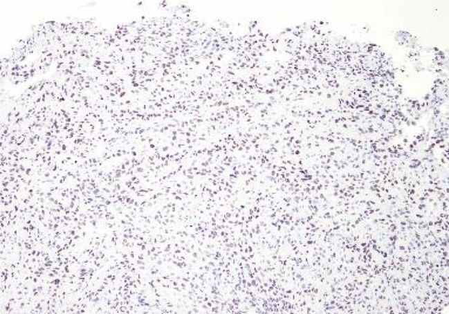
CASE REPORT
Duodenal Metastasis in Triple-Negative Invasive Ductal Breast Carcinoma With Negative Mammography
A
B
Figure 1. Contrast-enhanced computed tomography scan of the abdomen and pelvis with (a) coronal view and (b) axial view revealing bulky mesenteric and retro-
peritoneal adenopathy with segments of small bowel wall thickening
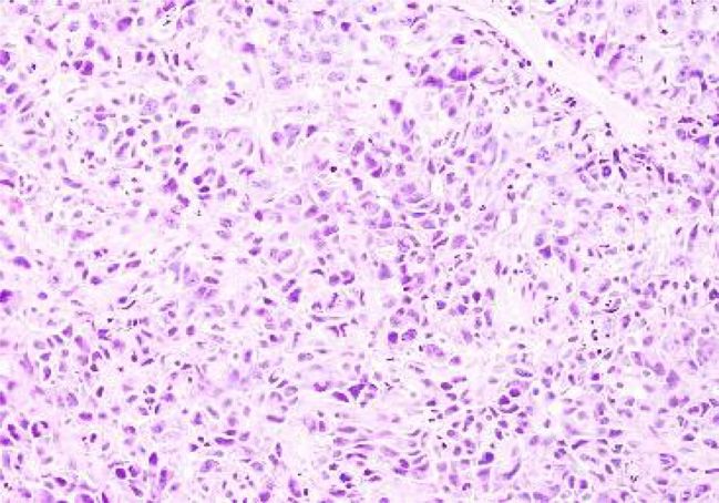
Histopathologic findings from the duodenal biopsies breast ultrasound were negative for any breast lesion (Figure 5).
revealed poorly differentiated, primary, triple-negative ductal The cancer antigen 15-3 (CA 15-3) level was normal, at
breast carcinoma (Figure 3). Specifically, immunohistochem- 5.0 U/mL (normal , 31.3 U/mL). During her hospital course,
istry (IHC) staining showed positive cytokeratin 7 (CK7), the patient was treated with blood transfusions and discharged
GATA binding protein 3, and vimentin (Figure 4a, 4c) with home with medical and radiation oncology follow-up.
negative caudal type homeobox transcription factor 2 The patient received palliative radiation therapy to the distal
(CDX2) and cytokeratin 20 (CK20; Figure 4b), suggestive stomach and duodenum at a dose of 2000 cGy in 250 cGy frac-
of metastatic breast carcinoma. Furthermore, estrogen recep- tions for 10 treatment sessions. She was then started on palli-
tor (ER), progesterone receptor (PR), and human epidermal ative chemotherapy with weekly paclitaxel. However, after 1
growth factor receptor 2 (HER2) results were all negative dose of chemotherapy, the patient returned to the emergency
(Figure 4d). Diagnostic bilateral mammogram and bilateral department with orthopnea, abdominal pain, distention, and
A B
Figure 2. Esophagogastroduodenoscopy revealing a semicircumferential necrotic and fragile mass in the second portion of the duodenum (a and b).
2 The Permanente Journal • For personal use only. No other uses without permission. Copyright © 2021 The Permanente Press. All rights reserved. The Permanente Journal • https://doi.org/10.7812/TPP/20.244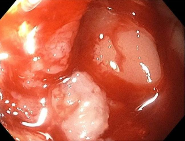
CASE REPORT
Duodenal Metastasis in Triple-Negative Invasive Ductal Breast Carcinoma With Negative Mammography

               A                                                                                      B

          Figure 3. Histologic staining with hematoxylin and eosin from the duodenal biopsy shows (a) invasive ductal carcinoma with mitosis (320) and (b) malignant cells
          surrounded by tumor necrosis (310).

          bilateral peripheral edema. Laboratory studies revealed a                                   (normal, 3.5-4.5 mEq/L), and acute renal failure with a creat-
          hemoglobin level of 7.1 g/dL, hyponatremia of 124 mEq/L                                     inine level of 1.69 mg/dL (normal, 0.5-1.0 mg/dL).
          (normal, 135-145 mEq/L), hyperkalemia of 6.2 mEq/L                                           A repeat CT scan of the abdomen and pelvis showed

               A                                                                                      B

              C                                                                                      D

          Figure 4. Immunohistochemical staining from the duodenal biopsy was (a) positive for cytokeratin 7 (310), (b) negative for cytokeratin 20 and caudal type homeobox tran-
          scription factor 2 (310), (c) positive for GATA binding protein 3 (310), and (d) negative for estrogen, progesterone, and human epidermal growth factor receptor 2 (310).

The Permanente Journal • https://doi.org/10.7812/TPP/20.244     The Permanente Journal • For personal use only. No other uses without permission. Copyright © 2021 The Permanente Press. All rights reserved.   3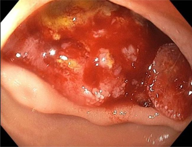
CASE REPORT
                                                                                          Duodenal Metastasis in Triple-Negative Invasive Ductal Breast Carcinoma With Negative Mammography

                                                                                                               metastasis – an eventual 20% to 50% will develop metastasis
           A                                               B                                                   during their disease course.10 Furthermore, the interval
                                                                                                               between breast cancer diagnosis and GI metastasis can be var-
                                                                                                               iable, with a latency period of several months to decades.11,12 In
                                                                                                               our case, the patient had recurrent visits to the clinic for gen-
                                                                                                               eralized abdominal pain, which was misdiagnosed and treated
                                                                                                               as dyspepsia. Unlike most reported cases, she had no prior
                                                                                                               diagnosis of breast malignancy and, therefore, metastatic dis-
                                                                                                               ease had a lower index of suspicion. This cautions clinicians on
                                                                                                               the importance of obtaining a detailed medical history and for-
                                                                                                               mulating a broad differential. Our patient had also presented
                                                                                                               with a neck lump during her first clinic visit for which a CT
                                                                                                               scan of the neck was ordered on a routine basis; however,
                                                                                                               the patient was admitted before the scheduled outpatient
                                                                                                               scan. Expediting imaging orders in many cases may prevent
                                                                                                               an additional delay in diagnosis.
        Figure 5. Diagnostic mammogram of (a) right and (b) left breast revealing no evi-                         Metastatic patterns vary depending on the 2 histologic sub-
        dence of malignancy.                                                                                   types of breast cancer. ILC accounts for only 10% to 14% of all
                                                                                                               invasive breast carcinomas, whereas IDC constitutes 80% of
                                                                                                               cases.11,13,14 Interestingly, ILC more frequently metastasizes
        multiple dilated loops of small bowel with air-fluid levels                                             to the GI tract compared with ductal cancer and is reported
        suggestive of small bowel obstruction. A chest radiograph                                              in up to 64% of all lobular metastatic cases.11,13,15,16 The exact
        demonstrated further findings of left pleural effusion. The                                             mechanism of lobular versus ductal spread remains unknown.
        general surgery team on consult determined no intervention                                             It has been hypothesized that ILC metastasis is a result of tro-
        was necessary, given her overall poor prognosis. The patient                                           pism of lobular cells16,17 and loss of the cell adhesion molecule
        and her family opted for comfort measures; she went into                                               E-cadherin, which may contribute to its predilection for the
        cardiorespiratory arrest and died 2 days later (Table 1).                                              GI system.18,19 Lobular metastasis may also be caused by
                                                                                                               hematogenous dissemination of malignant cells to the GI
        DISCUSSION                                                                                             tract, given its rich blood supply or via peritoneal and lym-
           Two million new cases of breast cancer occur globally,                                              phatic spread.20,21 In contrast, the GI metastasis of IDC is
        with a lifetime risk of 1 in 8 women in the United States.5                                            uncommon, with duodenal involvement being exceptionally
        The disease metastasizes to the brain, lymph nodes, skin,                                              rare.3,22 In a landmark article, Harris et al23 studied 76 post-
        lung, liver, and bone.2 GI tract metastasis is infrequently                                            mortem cases of metastatic ductal carcinoma, in which only
        reported, and when present, is an indication of a poor                                                 3 had metastasized to the intestines. They also noted that lob-
        prognosis.6 In a study of approximately 2600 patients                                                  ular metastatic patterns involved tiny nodules leading to a
        with breast malignancy, , 1% were noted to have GI                                                     more confluent spread, whereas ductal metastasis formed dis-
        metastatic involvement.3 Furthermore, Ambroggi et al7                                                  tinct nodules.
        studied the specific metastatic sites of breast malignancy                                                 Although duodenal metastasis of primary ductal breast car-
        within the GI tract and found that the most common                                                     cinoma is rare, it has been reported in previous studies.24–30
        involvement is the stomach (60%), followed by the esoph-                                               When small bowel involvement is present in metastatic breast
        agus (12%), colon (11%), and rectum (7%). Breast cancer                                                cancer, the terminal ileum is a more common site of metastasis
        metastasis to the small intestine (8%), especially the duo-                                            compared with the duodenum.2 The presentation in our case –
        denum, is very rare and usually discovered on autopsy.7–9                                              small bowel obstruction as the first manifestation of metastasis
           It can be challenging to diagnose GI metastasis secondary to                                        to the GI tract – is documented in previous patient
        primary breast carcinoma because it is uncommon and may be                                             reports.13,26,31–33 However, each of these cases involves
        overlooked in the initial presentation. The clinical diagnosis                                         patients with a known diagnosis and prior treatment of breast
        can be delayed because of symptoms of nausea, vomiting,                                                cancer who eventually developed GI metastasis after some
        anorexia, abdominal pain, changes in stool, hemorrhage, or,                                            interval. Our case is unique in that the patient had no prior his-
        less commonly, perforation; these nonspecific findings can                                               tory of breast cancer, including no breast complaints or focal
        mimic primary GI disorders and lead to underdiagnosis of                                               findings. In 1 reported case by Woo et al,34 a patient with
        breast cancer metastasis.2,6 At the time of initial diagnosis,                                         hormone-positive ILC presented with an initial manifestation
        5% to 10% of patients with breast cancer present with                                                  of malignancy as gastric metastasis; this patient’s screening

4   The Permanente Journal • For personal use only. No other uses without permission. Copyright © 2021 The Permanente Press. All rights reserved.   The Permanente Journal • https://doi.org/10.7812/TPP/20.244
CASE REPORT
Duodenal Metastasis in Triple-Negative Invasive Ductal Breast Carcinoma With Negative Mammography
Table 1. Case report timeline and relevant history
Date: Summaries from initial and follow-up visits Diagnostic testing Interventions
04/18/2019 Patient presented to primary care for No imaging ordered at this time. Dietary and lifestyle modification
epigastric pain and nausea for recommended along with trial of proton
past month. pump inhibitor (omeprazole).
04/30/2019 Patient returns for continued and CT of the abdomen and pelvis ordered Dietary and lifestyle modification along with
worsening abdominal pain now radiating along with laboratory tests, including addition of sucralfate. Omeprazole was
to back with no relief from proton pump CBC, CMP, lipase, and Helicobacter switched to pantoprazole.
inhibitor therapy. pylori stool testing.
04/30/2019 Laboratory testing performed as an Hemoglobin drop to 7.0 g/dL from baseline Patient was admitted to inpatient service
outpatient revealed acute drop in of 14 g/dL and positive fecal occult for further workup of anemia,
hemoglobin. Patient was called and blood test. CT of the abdomen and gastrointestinal bleeding, and abdominal
advised to go to the ED immediately. pelvis revealed adenopathy and several pain.
segments of wall thickening.
05/01/2019 Gastroenterology consulted for epigastric EGD and colonoscopy planned. EGD revealed necrotic and fragile
pain and anemia. semicircumferential mass found in
second portion of duodenum; multiple
biopsies taken. Colonoscopy was
unremarkable.
05/02/2019 Patient was treated with 2 units of packed Patient was discharged home with
red blood cell transfusion and outpatient follow-up.
improvements noted in her hemoglobin.
05/07/2019 Final pathological results from duodenal Patient referred for urgent oncologic
biopsies revealed poorly differentiated evaluation.
triple-negative primary breast cancer.
05/08/2019 Patient seen by oncology for newly Diagnostic mammogram and ultrasound Overall poor prognosis and plan for
diagnosed metastatic breast cancer. of the breasts ordered; both returned palliative chemotherapy. Referral to
Previous screening mammograms, last negative and without any suggestive radiation oncology also placed.
one in 10/2018, were negative for breast findings. Tumor marker CA 15-3 levels
lesions. were normal at 5.0 U/mL.
05/10/2019 Patient started on course of radiation
therapy to duodenum for 10 planned
treatment sessions.
05/16/2019 Patient started on chemotherapy with
paclitaxel weekly planned for 6 cycles.
05/21/2019 Patient presents to the ED with fatigue, CT of the abdomen and pelvis revealed Patient was admitted for further evaluation
nausea, vomiting, and worsening multiple dilated loops of small bowel and surgical consultation for abdominal
abdominal pain. suggestive of obstruction. Chest x-ray findings.
revealed bilateral pleural effusions.
Significant laboratory findings included
hyponatremia, hyperkalemia, and acute
renal injury.
05/22/2019 Patient deemed not a surgical candidate for Palliative care team consulted given overall
small bowel obstruction. Patient poor prognosis. Patient opted for comfort
symptomatically treated for pain and measures and died on 5/24/2019.
nausea. Hydration therapy initiated and
electrolyte impairments monitored.
Relevant medical history and interventions
Medical history Hyperlipidemia, attention deficit hyperactivity disorder.
Past medical testing Screening colonoscopy in 2017 was normal. Screening mammograms biennially from
2006 to 2018 were negative.
Family history Negative for breast or ovarian cancer. Paternal grandfather with colon cancer at
unknown age.
Psychosocial history Former tobacco use (21 pack-years and quit 10 y prior). Recently under a lot of stress,
working 3 jobs and over 80 h per week. Divorced with 2 adult children.
CA 15-3 5 cancer antigen 15-3; CBC5 complete blood count; CMP 5 comprehensive metabolic panel; CT 5 computed tomography; ED 5 emergency department; EDG 5
esophagogastroduodenoscopy.
The Permanente Journal • https://doi.org/10.7812/TPP/20.244 The Permanente Journal • For personal use only. No other uses without permission. Copyright © 2021 The Permanente Press. All rights reserved. 5CASE REPORT
Duodenal Metastasis in Triple-Negative Invasive Ductal Breast Carcinoma With Negative Mammography
mammography was also negative at the time of diagnosis. Fur- specificity in patients with signs of breast cancer compared
ther literature review demonstrated another case by Khairy with screening mammography. Perhaps using a more accurate
et al26 of a patient with no prior history of breast cancer who diagnostic modality like magnetic resonance imaging (MRI)
first presented with small bowel obstruction, and biopsy in our patient scenario could have detected a breast lesion.
revealed triple-positive IDC, also with negative mammogra- MRIs are far more sensitive and specific than mammograms
phy. This patient did have axillary lymphadenopathy, and a or ultrasounds in detecting small breast malignancies.37 In
biopsy helped confirm the diagnosis. Similarly, our patient our patient, MRI was ordered but she clinically deteriorated
had supraclavicular lymph nodes as a late presentation of before the test could be performed. Despite any evidence of
advanced disease for which biopsy was planned; however, breast pain, mass, or axillary adenopathy, a breast biopsy
this biopsy was not completed, given her rapid decline. may have been another option to potentially diagnose primary
Our patient received annual screening mammograms start- carcinoma in the breast tissue. For instance, a case presentation
ing at 44 years of age. She had a routine mammogram just 6 written by Zuhair and Maron38 reports a patient with no
months prior, which was negative, with a BI-RADS (Breast known diagnosis of breast cancer who presented with abdom-
Imaging, Reporting and Data System) score of 1. Notably, inal pain and was found to have ER-positive lobular carcinoma
diagnostic mammogram and ultrasonography revealed no evi- in the GI tract. Previous mammography in this patient was
dence of malignancy after her biopsy-proven IDC in the duo- notable for nonspecific findings of dense nodular parenchyma,
denum. This illustrates an important concept of diagnostic but ILC was identified only after core biopsy of breast tissue
accuracy. Although regular screening mammography is the was completed.
most effective tool to reduce breast cancer-related mortality, Differentiating metastasis from an unknown primary can be
it has its limitations. The sensitivity of screening mammo- challenging, especially in a patient without a history of malig-
grams is 85%, and specificity is approximately 96%.35 How- nancy. Distinction can be made with analysis of morphologic
ever, sensitivity is reduced to 47.8% to 64.4% in females patterns through histopathologic studies and IHC staining.
with denser breast tissue. In a large prospective study by Bar- Determining CK7 and CK20 immunophenotypes is valuable
low et al36 involving 41,427 diagnostic mammograms, it was to distinguish colorectal from extraintestinal malignancies.39
concluded that these have higher sensitivity but lower The CK71/CK20 pattern, as in our case, is highly specific
Table 2. Summary of select cases of metastatic breast cancer to the gastrointestinal tract, highlighting the rarity of triple-negative IDC
metastasis to the duodenum
Gastrointestinal Characteristic
metastasis of breast Primary breast diagnosed Primary breast Subtype Receptor status Reference
cancer undiagnosed
Duodenum x IDC HR1/HER22 24
Duodenum x IDC HR1/HER22 25
Duodenum x IDC HR1/HER22 27
Duodenum x IDC HR1/HER22 29
Duodenum x IDC HR1/HER22 16
Duodenum x IDC HR1/HER21 26
Duodenum x ILC HR1/HER22 52
Duodenum x ILC HR1/HER22 32
Duodenum x ILC HR1/HER22 38
Jejunum x ILC HR2/HER22 31
Jejunum x IDC HR2/HER22 51
Colon x IDC HR1/HER22 17
Colon x ILC HR1/HER22 53
Stomach x ILC HR1/HER22 11
Stomach x ILC HR2/HER22 50
Stomach x IDC HR2/HER22 48
Stomach x ILC HR1/HER22 38
Stomach x ILC HR1/HER22 34
HR 5 hormone receptor; HER2 5 human epidermal growth factor receptor 2; IDC 5 invasive ductal carcinoma; ILC 5 invasive lobular carcinoma
6 The Permanente Journal • For personal use only. No other uses without permission. Copyright © 2021 The Permanente Press. All rights reserved. The Permanente Journal • https://doi.org/10.7812/TPP/20.244CASE REPORT
Duodenal Metastasis in Triple-Negative Invasive Ductal Breast Carcinoma With Negative Mammography
for breast carcinomas.40 Caudal type homeobox transcription involvement from breast carcinoma, only 6 were treated surgi-
factor 2, a transcription factor, is a strong marker for carcino- cally, all of them for palliative reasons and relief of obstructive
mas of intestinal origin and was negative in our patient.41 symptoms. Mourra et al6 found that no patient survived
Vimentin, a structural protein, was positively expressed on beyond 5 years, even with surgery, and there was no significant
our patient’s biopsy and has been associated with invasive improvement in survival compared with those without surgical
and chemoresistant ductal carcinomas.42 Additionally, the management. Our patient died within 3 months of diagnosis.
presence of the GATA binding protein 3 is a hallmark of Duodenal metastasis of primary breast carcinoma has a dismal
metastasis due to breast origin, especially triple-negative prognosis, and the overall median survival in patients with GI
malignancies.43 To date, there are no reliable serum tests to metastasis from breast malignancy is 28 months.16 Although
serve as screening and diagnostic markers for breast cancer. GI involvement is not common in metastatic triple-negative
The CA 15-3 is a common tumor marker used for prognostic breast cancer, it must be considered to prevent delay in diagno-
purposes and to detect breast cancer recurrence, but its use is sis and improve patient outcomes.
controversial.44 CA 15-3 levels may be elevated in metastatic
disease and its presence is associated with poor prognosis in CONCLUSION
nonmetastatic cases.45 However, the CA 15-3 level was nor- Breast cancer rarely metastasizes to the GI tract, especially
mal in our patient. A study conducted by Dede et al46 found the small intestine. This pattern of metastatic spread is not
that CA 15-3 levels in triple-negative breast cancer cases limited to ILC and is possible with IDCs. In these rare cases,
were significantly lower at the time of diagnosis and during the indolent symptoms can cause a delay in diagnosis and
metastasis compared with other forms of breast cancer. We treatment of the aggressive cancer. Differentiating metastasis
did not analyze cancer antigen 19-9 (CA 19-9) levels, but 1 from an unknown primary can be challenging; we rely on
study reports elevated levels in metastatic lobular breast carci- detailed pathological analysis including immunohistochemi-
noma to the GI tract.47 cal staining as important diagnostic tools. These collective
Triple-negative breast carcinomas do not express ER, PR, findings belie the shortcomings of screening mammography,
and HER2 receptors. They are high-grade with increased because imaging-negative breast malignancy may exist. Our
mitotic activity and poor overall prognosis. Triple-negative case illustrates unusual findings of small bowel metastasis as
findings are present in 20% to 25% of all breast malignancies31; the initial manifestation of triple-negative ductal carcinoma
they are more frequently seen in ductal carcinomas and, thus, in a patient with no prior history of breast malignancy. v
usually metastasize to the brain, lung, and liver.48,49 As
discussed previously, GI metastasis of IDC is an overall rare
Disclosure Statement
phenomenon and predominantly hormonal (ER/PR) positive The author(s) have no conflicts of interest to disclose.
6 HER2.13,26,31–33 There are only a handful of case reports
describing metastasis of triple-negative carcinoma to the GI
Funding and Sponsor Statement
tract, and these include both ILC and IDC subtypes.5,31,38,50,51 No funding or sponsor contributions.
Geredeli et al50 report a case of lobular triple-negative carci-
noma to the stomach. Another article by Baa et al48 reports
Authors’ Contributions
a patient who presented with a breast lump and dyspepsia Naila A Khan, DO, participated in the critical review, drafting, literature
who was diagnosed with triple-negative IDC with stomach research, and submission of the final manuscript. Sonha T Nguyen, MD, and
involvement. Only 1 case of triple-negative IDC to the small Phildrich G Teh, MD, participated in the literature research and drafting of the
bowel, specifically the jejunum, was reported.51 Of note, all final manuscript. Vishal N Ranpura, MD, and Taruna Bhatia, MD, participated
in direct patient care and editing of manuscript. All authors have given final
these patients had either a known previous breast cancer diag- approval to the manuscript.
nosis or presented with focal breast findings. To our knowl-
edge, there have been no case reports on triple-negative References
IDC to the duodenum, which is unique to our patient scenario 1. Siegel RL, Miller KD, Jemal A. Cancer statistics, 2020. CA Cancer J Clin 2020 Jan;70(1):7-
30. DOI: https://doi.org/10.3322/caac.21590
(Table 2). 2. Nazareno J, Taves D, Preiksaitis H-G. Metastatic breast cancer to the gastrointestinal tract:
Treatment of triple-negative metastatic breast carcinoma A case series and review of the literature. World J Gastroenterol 2006 Oct;12(38):6219-24.
DOI: https://doi.org/10.3748/wjg.v12.i38.6219
usually involves palliative systemic chemotherapy, immuno- 3. Borst MJ, Ingold JA. Metastatic patterns of invasive lobular versus invasive ductal
therapy combined with chemotherapy, or targeted treatment carcinoma of the breast. Surgery 1993 Oct;114(4):637-41; discussion 641-2 PubMed
with poly (ADP-ribose) polymerase inhibitors. Radiotherapy PMID: 8211676
4. Lehman CD, Arao RF, Sprague BL, et al. National performance benchmarks for modern
is reserved for palliation to provide symptomatic relief. In screening digital mammography: Update from the breast cancer surveillance consortium.
select cases, surgery can be considered for refractory bleeding, Radiology 2017 Apr;283(1):49-58. DOI: https://doi.org/10.1148/radiol.2016161174
5. Ghoncheh M, Pournamdar Z, Salehiniya H. Incidence and mortality and epidemiology of
obstruction, or intestinal perforation.11,16 In a retrospective breast cancer in the world. Asian Pac J Cancer Prev 2016;17(S3):43-6. DOI: https://doi.org/
study conducted by Taal et al,11 of 51 patients with GI 10.7314/apjcp.2016.17.s3.43 June
The Permanente Journal • https://doi.org/10.7812/TPP/20.244 The Permanente Journal • For personal use only. No other uses without permission. Copyright © 2021 The Permanente Press. All rights reserved. 7CASE REPORT
Duodenal Metastasis in Triple-Negative Invasive Ductal Breast Carcinoma With Negative Mammography
6. Mourra N, Jouret-mourin A, Lazure T, et al. Metastatic tumors to the colon and rectum: A 31. Khokhlova M, Roppelt H, Gluck B, et al. Triple negative invasive lobular carcinoma of the
multi-institutional study. Arch Pathol Lab Med 2012 Nov;136(11):1397-401. DOI: https://doi. breast presents as small bowel obstruction. Int J Surg Case Rep 2017 Jun;37:79-82. DOI:
org/10.5858/arpa.2011-0432-OA https://doi.org/10.1016/j.ijscr.2017.06.002
7. Ambroggi M, Stroppa EM, Mordenti P, et al. Metastatic breast cancer to the gastrointestinal 32. Shrestha S, Shah BK, Tandukar S. Duodenal obstruction as the presenting manifestation of
tract: Report of five cases and review of the literature. Int J Breast Cancer 2012;2012: recurrent breast cancer. J Cancer Res Ther 2014 Jul-Sep;10(3):761-2. DOI: https://doi.org/
439023. DOI: https://doi.org/10.1155/2012/439023 July 10.4103/0973-1482.136031
8. Asch MJ, Wiedel PD, Habif DV. Gastrointestinal metastases from crcinoma of the 33. Hussain T, Elahi B, Mcmanus P, Mahapatra T, Kneeshaw PJ. Gastric obstruction secondary
breast. Autopsy study and 18 cases requiring operative intervention. Arch Surg 1968 May; to metastatic breast cancer: A case report and literature review. J Med Case Rep 2012 Aug;
96(5):840-3. DOI: https://doi.org/10.1001/archsurg.1968.01330230148023 6:232. DOI: https://doi.org/10.1186/1752-1947-6-232
9. Cifuentes N, Pickren JW. Metastases from carcinoma of mammary gland: An autopsy 34. Woo J, Lee JH, Lee KE, Sung SH, Lim W. Gastric metastasis as the first presentation one
study. J Surg Oncol 1979 Jan;11(3):193-205. DOI: https://doi.org/10.1002/jso.2930110303 year before diagnosis of primary breast cancer. Am J Case Rep 2018 Mar;19:354-9. DOI:
10. Cardoso F, Harbeck N, Fallowfield L, Kyriakides S, Senkus E. Locally recurrent or
https://doi.org/10.12659/ajcr.908039
metastatic breast cancer: ESMO Clinical Practice Guidelines for diagnosis, treatment and
35. Thigpen D, Kappler A, Brem R. The role of ultrasound in screening dense breasts-A review
follow-up. Ann Oncol 2012 Oct;23(Suppl 7):vii11-9. DOI: https://doi.org/10.1093/annonc/
of the literature and practical solutions for implementation. Diagnostics (Basel) 2018 Mar;8(
mds232
11. Taal BG, Peterse H, Boot H. Clinical presentation, endoscopic features, and treatment of 1):20. DOI: https://doi.org/10.3390/diagnostics8010020
gastric metastases from breast carcinoma. Cancer 2000 Dec;89(11):2214-21. DOI: https:// 36. Barlow WE, Lehman CD, Zheng Y, et al. Performance of diagnostic mammography for
doi.org/10.1002/1097-0142(20001201)89:11,2214::aid-cncr9.3.0.co;2-d women with signs or symptoms of breast cancer. J Natl Cancer Inst 2002 Aug;94(15):
12. Ayantunde AA, Agrawal A, Parsons SL, Welch NT. Esophagogastric cancers secondary to 1151–9. DOI: https://doi.org/10.1093/jnci/94.15.1151
a breast primary tumor do not require resection. World J Surg 2007 Aug;31(8):1597-601. 37. Warner E, Plewes DB, Hill KA, et al. Surveillance of BRCA1 and BRCA2 mutation carriers
DOI: https://doi.org/10.1007/s00268-007-9099-y with magnetic resonance imaging, ultrasound, mammography, and clinical breast
13. Schwarz RE, Klimstra DS, Turnbull AD. Metastatic breast cancer masquerading as examination. J Am Med Assoc 2004 Sep;292(11):1317-25. DOI: https://doi.org/10.1001/
gastrointestinal primary. Am J Gastroenterol 1998 Jan;93(1):111-4. DOI: https://doi.org/10. jama.292.11.1317
1111/j.1572-0241.1998.111_c.x 38. Zuhair AR, Maron AR. Occult bilateral invasive lobular carcinoma of the breast presenting
14. Dixon AR, Ellis IO, Elston CW, Blamey RW. A comparison of the clinical metastatic patterns as gastroduodenal metastases: A case report. Breast Dis 2015 Jan;35(1):63-5. DOI: https://
of invasive lobular and ductal carcinomas of the breast. Br J Cancer 1991 Apr;63(4):634-5. doi.org/10.3233/BD-140376
DOI: https://doi.org/10.1038/bjc.1991.145 39. Bayrak R, Haltas H, Yenidunya S. The value of CDX2 and cytokeratins 7 and 20 expression
15. Rosen PP. Invasive duct carcinoma and morphological prognostic markers. In: Rosen’s in differentiating colorectal adenocarcinomas from extraintestinal gastrointestinal
breast pathology. Rosen PP, editor. Philadelphia: Lippincott-Raven: 1997; p 545-65. adenocarcinomas: Cytokeratin 7-/201 phenotype is more specific than CDX2 antibody.
[Doi_inserted_from_Crossref] Diagn Pathol 2012 Jan;7:9. DOI: https://doi.org/10.1186/1746-1596-7-9
16. Mclemore EC, Pockaj BA, Reynolds C, et al. Breast cancer: Presentation and intervention 40. Oien KA, Dennis JL. Diagnostic work-up of carcinoma of unknown primary: From
in women with gastrointestinal metastasis and carcinomatosis. Ann Surg Oncol 2005 Nov; immunohistochemistry to molecular profiling. Ann Oncol 2012 Sep;23(Suppl 10):x271-7.
12(11):886-94. DOI: https://doi.org/10.1245/ASO.2005.03.030 DOI: https://doi.org/10.1093/annonc/mds357
17. Samo S, Sherid M, Husein H, et al. Metastatic infiltrating ductal carcinoma of the breast to 41. Werling RW, Yaziji H, Bacchi CE, Gown AM. CDX2, a highly sensitive and specific marker
the colon: A case report and literature review. Case Rep Gastrointest Med 2013;2013: of adenocarcinomas of intestinal origin: An immunohistochemical survey of 476 primary
603683. DOI: https://doi.org/10.1155/2013/603683 October and metastatic carcinomas. Am J Surg Pathol 2003 Mar;27(3):303-10. DOI: https://doi.org/
18. Sastre-garau X, Jouve M, Asselain B, et al. Infiltrating lobular carcinoma of the breast.
10.1097/00000478-200303000-00003
Clinicopathologic analysis of 975 cases with reference to data on conservative therapy and
42. Korsching E, Packeisen J, Liedtke C, et al. The origin of vimentin expression in invasive
metastatic patterns. Cancer 1996 Jan;77(1):113-20. DOI: https://doi.org/10.1002/
breast cancer: Epithelial-mesenchymal transition, myoepithelial histogenesis or
(SICI)1097-0142(19960101)77:1,113::AID-CNCR19.3.0.CO;2-8
19. Critchley AC, Harvey J, Carr M, Iwuchukwu O. Synchronous gastric and colonic histogenesis from progenitor cells with bilinear differentiation potential? J Pathol 2005 Aug;
metastases of invasive lobular breast carcinoma: Case report and review of the literature. 206(4):451-7. DOI: https://doi.org/10.1002/path.1797
Ann R Coll Surg Engl 2011 Jul;93(5):e49-50. DOI: https://doi.org/10.1308/ 43. Cimino-mathews A, Subhawong AP, Illei PB, et al. GATA3 expression in breast carcinoma:
147870811X582800 Utility in triple-negative, sarcomatoid, and metastatic carcinomas. Hum Pathol 2013 Jul;44(
20. Kim SY, Kim KW, Kim AY, et al. Bloodborne metastatic tumors to the gastrointestinal tract: 7):1341-9. DOI: https://doi.org/10.1016/j.humpath.2012.11.003
CT findings with clinicopathologic correlation. AJR Am J Roentgenol 2006 Jun;186(6):1618- 44. Harris L, Fritsche H, Mennel R, et al. American Society of Clinical Oncology 2007 update of
26. DOI: https://doi.org/10.2214/AJR.05.0095 recommendations for the use of tumor markers in breast cancer. J Clin Oncol 2007 Nov;25(
21. Chang SF, Burrell MI, Brand MH, Garsten JJ. The protean gastrointestinal manifestations of 33):5287-312. DOI: https://doi.org/10.1200/JCO.2007.14.2364
metastatic breast carcinoma. Radiology 1978 Mar;126(3):611-7. DOI: https://doi.org/10. 45. Rafey M, Akhtar K, Rab AZ, Siddiqui SA. Serum Ca 15-3: A useful tumor marker in the
1148/126.3.611 prognostication of locally advanced breast cancer. Annals of Woman and Child Health
22. Fondrinier E, Guerin O, Lorimier G. [A comparative study of metastatic patterns of ductal 2017;4(4):45-9. December. https://doi.org/10.21276/AWCH.1782
and lobular carcinoma of the breast from two matched series (376 patients)]. Bull Cancer 46. Dede DS, Arslan C, Altundag K. Serum levels of CEA and CA 15-3 in triple-negative breast
1997 Dec;84(12):1101–7 PubMed PMID: 9587361 cancer at the time of diagnosis. Med Oncol 2009 Dec;27(15):1429. DOI: https://doi.org/10.
23. Harris M, Howell A, Chrissohou M, Swindell RI, Hudson M, Sellwood RA. A comparison of 1007/s12032-009-9310-0
the metastatic pattern of infiltrating lobular carcinoma and infiltrating duct carcinoma of the 47. Santini D, Altomare A, Vincenzi B, et al. An increase of CA 19.9 as the first clinical sign of
breast. Br J Cancer 1984 Jul;50(1):23-30. DOI: https://doi.org/10.1038/bjc.1984.135 ileocecal valve metastasis from breast cancer. In Vivo 2006 Jan-Feb;20(1):165-8. PubMed
24. Wang X, Jin M, Ye Q, et al. Solitary duodenum metastasis from breast cancer with 8 years' PMID: 16433047.
latency: A case report. Medicine (Baltimore) 2018 Jan;97(2):e9550. DOI: https://doi.org/10. 48. Baa AK, Naik RD, Vanidassane I, et al. Unusual gastric metastasis in triple-negative
1097/MD.0000000000009550 (estrogen receptor/progesterone receptor/HER2neu negative) GATA-binding protein
25. Kumano H, Hozumi Y, Shiozawa M, et al. Recurrent invasive ductal carcinoma of the breast 3-positive breast cancer. Indian J Nucl Med 2020 Jan-Mar;35(1):82-3. DOI: https://doi.org/
presenting as a metastasis to the duodenum with long-term survival. Am Surg 2011 Jun;77 10.4103/ijnm.IJNM_156_19
(6):e107-8. DOI: https://doi.org/10.1177/000313481107700606 49. Ishikawa Y, Horiguchi J, Toya H, et al. Triple-negative breast cancer: Histological subtypes
26. Khairy S, Azzam A, Mohammed S, Suleman K, Khawaji A, Amin T. Duodenal obstruction
and immunohistochemical and clinicopathological features. Cancer Sci 2011 Mar;102(3):
as first presentation of metastatic breast cancer. Case Rep Surg 2015 Jul;2015:605719.
656-62. DOI: https://doi.org/10.1111/j.1349-7006.2011.01858.x
DOI: https://doi.org/10.1155/2015/605719
50. Geredeli C, Dogru O, Omeroglu E, Yilmaz F, Cicekci F. Gastric metastasis of triple negative
27. Wang XW, Meng XJ, Zhang TG, et al. A case of breast cancer found as metastasis to the
duodenum. J Canc Res Exp Oncol 2009;1(2):12–4. Journal of Cancer Research and invasive lobular carcinoma. Rare Tumors 2015 May;7(2):5764. DOI: https://doi.org/10.
Experimental Oncology Vol. 1(2) pp. 012-014, December, 2009. https://doi.org/10.5897/ 4081/rt.2015.5764
JCREO.9000001 51. Paiva C, Garcia J, Silva C, Araujo A, Araujo A, Santos MD. Single jejunum metastasis from
28. Qishan Z, Jinping W, Zhengsheng Y, Leji LZ. Breast cancer found as metastasis to the breast cancer arising twelve years after the initial treatment. Case Rep Oncol Med 2016;
duodenum: A case report. African J Breast Cancer 2015;2(11):133–4. November. 2016:8594652. DOI: https://doi.org/10.1155/2016/8594652 October
29. Ferrari AB, Pulcini G, Gheza F, et al. Duodenal metastasis from male breast cancer: A case 52. Gangireddy M, Shrimanker I, Saintelia S, Gomez J, Peroutka KA. From the breast to the
report and review of the literature. J Med Case Rep 2009 Jul;3:8331. DOI: https://doi.org/ bowel: An unconventional metastatic presentation. Cureus 2019 Nov;11(11):e6199. DOI:
10.4076/1752-1947-3-8331 https://doi.org/10.7759/cureus.6199
30. Liu M, Zhang L, Guo L, Lv J, Shi W, Liu B. Intestinal metastasis from breast invasive ductal 53. Bispo M, Rio-Tinto R, Fidalgo P, Castillo-Martin M, Deviere J. Late colon involvement by
carcinoma after a long latency: Case report and literature review. Onco Targets Ther 2018 lobular carcinoma of the breast: A diagnosis to keep in mind!. GE Port J Gastroenterol 2020
Dec;11:8599-603. DOI: https://doi.org/10.2147/OTT.S180949 Jan;27:47-9. DOI: https://doi.org/10.1159/000497390
8 The Permanente Journal • For personal use only. No other uses without permission. Copyright © 2021 The Permanente Press. All rights reserved. The Permanente Journal • https://doi.org/10.7812/TPP/20.244You can also read


