Altered Molecular Pathways in the Proteome of Cryopreserved Sperm in Testicular Cancer Patients before Treatment
←
→
Page content transcription
If your browser does not render page correctly, please read the page content below
International Journal of
           Molecular Sciences

Article
Altered Molecular Pathways in the Proteome of
Cryopreserved Sperm in Testicular Cancer Patients
before Treatment
Manesh Kumar Panner Selvam 1 , Ashok Agarwal 1, *           and Peter N. Pushparaj 2
 1    American Center for Reproductive Medicine, Cleveland Clinic, Cleveland, OH 44195, USA;
      manesh.balu@gmail.com
 2    Center of Excellence in Genomic Medicine Research, Jeddah 21589, Saudi Arabia;
      peter.n.pushparaj@gmail.com
 *    Correspondence: agarwaa@ccf.org

 Received: 30 December 2018; Accepted: 1 February 2019; Published: 5 February 2019

 Abstract: Testicular cancer (TC) represents the most common cancer affecting men within
 the reproductive age and is often accompanied by major disturbances in semen parameters.
 Cryopreservation is recommended in these patients before initiating cancer treatment. Currently,
 there are no studies reporting the molecular mechanisms associated with altered semen quality in
 these men. The main objective of this study was to compare the sperm proteome of normozoospermic
 (motility >40%) and asthenozoospermic (motility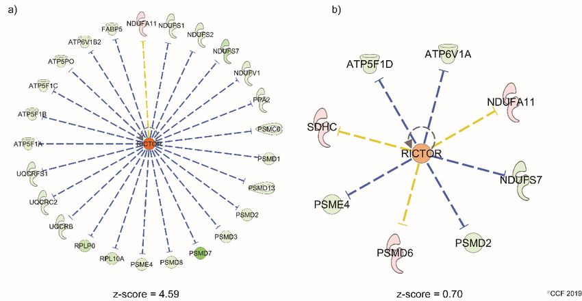
Int. J. Mol. Sci. 2019, 20, 677                                                                      2 of 15

     The relationship between TC and impaired semen parameters before the onset of treatment is well
established [13,14]. In general, a reduction in sperm concentration is seen in TC patients compared to
other types of cancer [15,16]. In particular, asthenozoospermia (sperm motility < 40%) is prevalent in
30% to 45% of TC cases [15,17,18]. Several studies have reported sub-normal semen parameters and
poor fertilization rates in men with TC [11,19–23]. However, 50% of TC patients are found to have
normal semen parameters (normozoospermia) according to WHO 2010 reference values [6,13]. The
reason for infertility in these men with normal semen parameters is unclear.
     Unexplained male infertility is a multifactorial disorder and conventional semen analysis fails to
explain the etiology of infertility in such cases [24]. Furthermore, asthenozoospermia is a common
cause of male infertility and it is observed in 18% of infertile men [25] and accounts for up to 81% of all
abnormal semen analyses [26]. Over the past decade, the proteomic platform has been used to study
the changes in the protein profiles of spermatozoa [27–32]. Advancement in sperm proteomic research
has provided the molecular basis of unexplained infertility [31]. Several sperm proteomic studies on
normozoospermic infertile men have reported changes at the subcellular level of spermatozoa [32–35].
These changes are associated with proteins related to sperm capacitation and acrosome reaction that
are essential for the fertilization of oocyte [26,36].
     The majority of TC patients are referred for sperm banking by their oncologists before the start of
their cancer treatment. Fertility history in the majority of these patients is not established. In addition,
they are not referred to a fertility specialist because of the urgency of oncological treatment. Therefore,
to understand the possible underlying cause(s) of infertility in TC patients, we compared the sperm
proteome of normozoospermic and asthenozoospermic TC patients with that of normozoospermic
infertile men without cancer (control group).

2. Results

2.1. Semen Parameters
     Semen parameters of the normozoospermic and asthenozoospermic TC patients, and control
group are presented in Supplementary Materials Table S1. No difference was seen in the sperm
concentration between normozoospermic TC patients (n = 20) and normozoospermic infertile men
without cancer (control group) (n = 20), and between asthenozoospermic TC patients (n = 20) and
the control group. Sperm motility was significantly (p < 0.0001) decreased in asthenozoospermic TC
patients (Supplementary Materials Table S1).

2.2. Sperm Proteome of TC Patients and Normozoospermic Infertile Men
     Liquid chromatography-tandem mass spectrometry (LC-MS/MS) detected a total of 1085, 846, and
982 proteins in normozoospermic TC, asthenozoospermic TC and control groups, respectively. Based
on the normalized spectral abundance factor (NSAF) ratio and protein abundance, 168 differentially
expressed proteins (DEPs) were identified in normozoospermic TC and 347 in asthenozoospermic TC
patients compared with the control group. The overexpressed and under-expressed DEPs, and unique
proteins are shown in Figure 1.

2.3. Biological Pathways Dysregulated in Spermatozoa of Normozoospermic and Asthenozoospermic
TC Patients
     Ingenuity pathway analysis (IPA) revealed phagosome maturation, sirtuin signaling pathway,
mitochondrial dysfunction, atherosclerosis signaling, and remodeling of epithelial adherens junctions
as the top five canonical pathways in normozoospermic TC patients (Table 1). Mitochondrial
dysfunction, oxidative phosphorylation, sirtuin signaling pathway, protein ubiquitination pathway,
and phagosome maturation were identified as top canonical pathways in asthenozoosp ermic TC
patients (Table 1).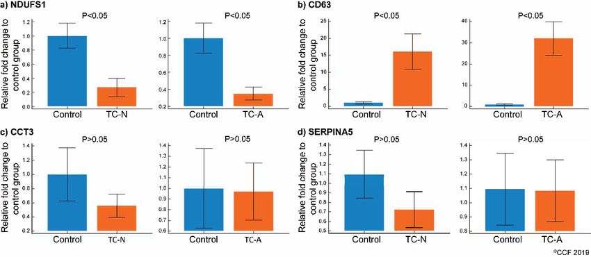
Int. J. Mol. Sci. 2019, 20, 677                                                                                    3 of 15
   Int. J. Mol. Sci. 2018, 19, x FOR PEER REVIEW                                                                3 of 15

      Figure  1. Differentially
         Figure                 expressed
                  1. Differentially        sperm proteins
                                        expressed    sperm (DEPs)   in normozoospermic
                                                              proteins                      and asthenozoospermic
                                                                         (DEPs) in normozoospermic           and
         asthenozoospermic
      testicular patients andtesticular  patients and
                               normozoospermic         normozoospermic
                                                   infertile              infertile(control
                                                             men without cancer      men without
                                                                                            group).cancer
                                                                                                    TC-N:(control
                                                                                                          testicular
         group).
      cancer     TC-N: testicular cancer
              normozoospermic,     TC-A: normozoospermic,     TC-A: testicular cancer asthenozoospermic.
                                          testicular cancer asthenozoospermic.

   2.3. Biological Pathways Dysregulated in Spermatozoa of Normozoospermic and Asthenozoospermic TC
      Table 1. List of differentially expressed proteins involved in top 5 canonical pathways associated with
   Patients
      normozoospermic and asthenozoospermic testicular cancer patients.
        Ingenuity pathway analysis (IPA) revealed phagosome maturation, sirtuin signaling pathway,
   mitochondrial    dysfunction,  atherosclerosis     −log
                                                     signaling, and remodeling of      epithelial adherens
          Groups           Canonical Pathways                                       DEPs
                                                    (p-Value)
   junctions as the top five canonical pathways in normozoospermic TC patients (Table 1).
                                                                  DYNC1H1,M6PR,TUBB3,MPO,PRDX1,TUBB4B,
                          Phagosomeoxidative
   Mitochondrial dysfunction,        Maturation phosphorylation,
                                                       7.19         sirtuin signaling pathway, protein
                                                                   TUBA3C/TUBA3D,CANX,ATP6V1A,NAPA
   ubiquitination pathway, Sirtuin
                             and phagosome
                                   Signaling     maturation were identified as top canonical pathways in
                                                                   SLC25A6,PPIF,ATP5F1D,NDUFS7,NDUFA11,
     Normozoospermic                                   6.15           CPT1B,TUBA3C/TUBA3D,HIST1H1D,
   asthenozoosp   ermic
        TC patients
                        TC patients  (Table
                                Pathway      1).                         SDHC,SLC25A5,VDAC3,VDAC1
                                       Mitochondrial                    ATP5F1D,NDUFA11,NDUFS7,CPT1B,SDHC,
                                                              4.67
                                        Dysfunction                             OGDH,VDAC1,VDAC3
                                  Atherosclerosis Signaling   4.61    ALB,APOB,APOA4,LPL,SERPINA1,COL18A1,CLU
                                  Remodeling of Epithelial
                                                              3.99    TUBB3,TUBB4B,TUBA3C/TUBA3D,ACTN4,ACTN1
                                    Adherens Junctions
                                                                      HSD17B10,NDUFV1,NDUFS7,ATP5F1A,ATP5PO,
                                       Mitochondrial                  ATP5S,VDAC3,UQCRB,VDAC2,PDHA1,ATP5F1C,
                                                              15.7
                                       Dysfunction                     MTND5,NDUFS1,ATP5F1B,NDUFA11,UQCRC2,
                                                                         NDUFS2,UQCRFS1,GPX4,VDAC1,OGDH
                                                                       NDUFV1,ATP5F1C,MTND5,NDUFS1,ATP5F1B,
                                         Oxidative
                                                              10.9    NDUFS7,NDUFA11,ATP5F1A,UQCRC2,ATP5PO,
                                      Phosphorylation
                                                                            NDUFS2,UQCRFS1,ATP5S,UQCRB
                                                                      NDUFV1,PPIF,NDUFS7,ATP5F1A,VDAC3,VDAC2,
     Asthenozoospermic
                                      Sirtuin Signaling                 PDHA1,ATP5F1C,NDUFS1,MTND5,ATP5F1B,
         TC patients                                          9.31
                                          Pathway                     NDUFA11,UQCRC2,TUBA3C/TUBA3D,NDUFS2,
                                                                            UQCRFS1,VDAC1,SLC25A5,LDHA
                                                                       PSMD7,PSMD13,HSPH1,HSPA9,TRAP1,PSMD3,
                                   Protein Ubiquitination
                                                              6.67         PSMD8,UCHL3,USP7,PSMC6,PSMD2,
                                          Pathway
                                                                            DNAJB11,PSMD1,DNAJB1,HSPA4L
                                                                           DYNC1H1,CTSD,MPO,TUBB4B,CTSB,
                                  Phagosome Maturation        5.3                 TUBA3C/TUBA3D,
                                                                           CANX,PRDX6,ATP6V1B2,HLA-DRB5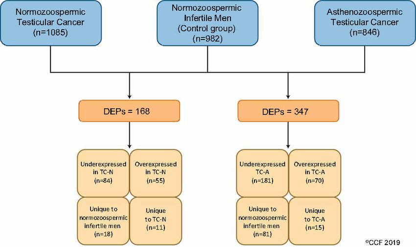
Int. J. Mol. Sci. 2019, 20, 677                                                                        4 of 15

        Canonical pathways-related reproductive function and their significance in both
normozoospermic and asthenozoospermic TC patients are shown in Figure 2. Upstream regulator
analysis revealed rapamycin-insensitive companion of mammalian target of rapamycin (RICTOR)
function was significantly activated in asthenozoospermic TC patients with a z-score of 4.59.
No significant activation of RICTOR was seen in normozoospermic TC patients. Differentially
expressed
Int. J. Mol. Sci.sperm   proteins
                  2018, 19,         associated
                            x FOR PEER REVIEW with RICTOR are shown in Figure 3.       5 of 15

             2. AA comparison
      Figure 2.     comparison pathway
                                  pathway analysis
                                           analysis depicting
                                                    depicting the
                                                               the canonical
                                                                   canonical pathways
                                                                              pathways associated
                                                                                        associated with
      reproductive process function between the TC patients with normal and abnormal semen parameters
      when compared with
                       with normozoospermic
                             normozoospermic infertile
                                                infertile men
                                                          men (control
                                                               (control group).  TC-N: testicular cancer
                                                                         group). TC-N:
      normozoospermic, TC-A: testicular cancer asthenozoospermic.

     Functional analysis of DEPs identified the top pathways associated with disease and disorders,
molecular and cellular functions in both normozoospermic and asthenozoospermic TC groups.
Inflammatory response was identified as the top pathway in disease and disorders, and cellular
compromise in molecular and cellular functions. However, the pathways associated with physiological
system development and function were identified only in the asthenozoospermic TC patients (Table 2).
Forty DEPs were involved in reproductive system development pathway (Table 2).
Figure 2. A comparison pathway analysis depicting the canonical pathways associated with
reproductive process function between the TC patients with normal and abnormal semen parameters
Int. when compared
J. Mol. Sci. with normozoospermic infertile men (control group). TC-N: testicular cancer
2019, 20, 677 5 of 15
normozoospermic, TC-A: testicular cancer asthenozoospermic.
Figure
Figure Differentiallyexpressed
3.3.Differentially expressedproteins
proteinsunder
under the
the regulation
regulation of RICTOR
RICTOR in in (a)
(a) asthenozoospermic
asthenozoospermic
testicularcancer
testicular cancergroup,
group, (b)
(b) normozoospermic
normozoospermic testicular cancercancer group.
group. A A z-score
z-score>2 and2and 2 isisInt. J. Mol. Sci. 2019, 20, 677 6 of 15
Table 2. List of pathways associated with diseases and disorders, molecular and cellular functions, physiological system development and functions in
normozoospermic and asthenozoospermic testicular cancer patients.
Groups Normozoospermic TC Asthenozoospermic TC
Category Pathways p-Value Number of DEPs Pathways p-Value Number of DEPs
Inflammatory response 2.36 × 10⁻¹⁴ –6.15 × 10⁻⁷ 46 Inflammatory Response 1.96 × 10⁻²² –2.37 × 10⁻³ 84
Cancer 4.65 × 10⁻¹¹ –7.06 × 10⁻⁶ 142 Cancer 3.9 × 10⁻¹¹ –2.19 × 10⁻³ 228
Disease and Organism injury and Organism injury and
4.65 × 10⁻¹¹ –7.06 × 10⁻⁶ 142 3.9 × 10⁻¹¹ –2.39 × 10⁻³ 232
disorders abnormalities abnormalities
Reproductive system disease 1.3 ×10⁻¹⁰ –7.06 × 10⁻⁶ 104 Metabolic disease 1.2 × 10⁻¹⁰ –2.36 × 10⁻³ 90
Neurological disease 3.21 × 10⁻¹⁰ –7.06 × 10⁻⁶ 47 Gastrointestinal disease 2.02 × 10⁻⁹ –2.13 × 10⁻³ 214
Cellular compromise 2.36 × 10⁻¹⁴ –2.45 × 10⁻⁷ 30 Cellular compromise 1.96 × 10⁻²² –1.88 × 10⁻³ 59
Protein synthesis 1.06 × 10⁻¹¹ –2.54 × 10⁻⁶ 39 Protein synthesis 3.16 × 10⁻¹⁴ –2.36 × 10⁻³ 74
Post-translational modification 2.17 × 10⁻¹¹ –6.41 × 10⁻¹⁰ 18 Protein degradation 3.93 × 10⁻¹⁴ –2.36 × 10⁻³ 39
Molecular and
Cellular assembly and
cellular functions Protein degradation 2.17 × 10⁻¹¹ –6.41 × 10⁻¹⁰ 26 8.2 × 10⁻¹¹ –2.36 × 10⁻³ 46
organization
Post-translational
Lipid metabolism 4.3 × 10⁻¹¹ –6.79 × 10⁻⁶ 26 2.08 × 10⁻¹⁰ –2.36 × 10⁻³ 52
modification
Reproductive system
NA NA NA 4.14 × 10⁻¹⁰ –2.05 × 10⁻³ 40
development and function
Physiological system Hematological system
NA NA NA 3.85 × 10⁻⁷ –2.37 × 10⁻³ 38
development and development and function
functions NA NA NA Immune cell trafficking 3.85 × 10⁻⁷ –2.37 × 10⁻³ 39
NA NA NA Organ development 9.75 × 10⁻⁶ –2.4 × 10⁻³ 36
Cardiovascular system
NA NA NA 2.62 × 10⁻⁵ –2.23 × 10⁻³ 40
development and functionInt. J. Mol. Sci. 2018, 19, x FOR PEER REVIEW 7 of 15
Cardiovascular
system 2.62E-05-
NA NA NA 40
Int. J. Mol. Sci. 2019, 20, 677 development and 2.23E-03 7 of 15
function
2.4.2.4.
Protein Networks
Protein andand
Networks Biofunctions
BiofunctionsAffected
AffectedininAsthenozoospermic
Asthenozoospermic TC
TC Group
Group
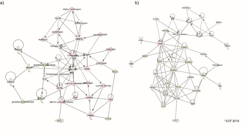
Bioinformatic analysis identified the DEPs associated with sperm function and fertilization
process Bioinformatic analysis
that were altered in theidentified the DEPs associated
asthenozoospermic with
TC patients sperm
(Table function and
3). Network fertilization
analysis revealed
process that were altered in the asthenozoospermic TC patients (Table 3). Network analysis revealed
the involvement of sperm proteins in cellular assembly and organization, cell-to-cell signaling and
the involvement of sperm proteins in cellular assembly and organization, cell-to-cell signaling and
interaction, reproductive system development and function (Figure 4a), and cellular compromise,
interaction, reproductive system development and function (Figure 4a), and cellular compromise,
inflammatory response, and infectious diseases (Figure 4b).
inflammatory response, and infectious diseases (Figure 4b).
Table 3. Reproductive system development and functions affected due to altered expression of DEPs
Table 3. Reproductive system development and functions affected due to altered expression of DEPs
in asthenozoospermic testicular cancer patients.
in asthenozoospermic testicular cancer patients.
Function
Function p-Valuep-Value DEPs
DEPs
CCT2,CCT3,CCT5,CCT6A,CCT7,CCT8,
CCT2,CCT3,CCT5,CCT6A,CCT7,CCT8,PRSS37,SPAM1
−10
Binding
Binding of sperm
of sperm 4.14 × 10
4.14E-10 PRSS37,SPAM1,TCP1,VDAC2
,TCP1,VDAC2
APOB,ATP1A4,GPX4,H2AFX,HSD17B4,
Spermatogenesis APOB,ATP1A4,GPX4,H2AFX,HSD17B4,HSPA4L,KIA
Spermatogenesis 0.0004290.000429 HSPA4L,KIAA1324,
A1324,NPEPPS,NUP210L,PRSS37
NPEPPS,NUP210L,PRSS37
Maturation
Maturation of sperm
of sperm 0.001140.00114 CLU,TPP2
CLU,TPP2
AKAP4,ATP1A4,ATP2B4,PRSS37,
Function of sperm
Function of sperm 9.75 × 10−AKAP4,ATP1A4,ATP2B4,PRSS37,SPESP1,VDAC1
9.75E-06 6
SPESP1,VDAC1
AKAP4,APOB,ATP1A4,ATP2B4,CHDH,GAPDHS,SPA
AKAP4,APOB,ATP1A4,ATP2B4,CHDH,
Cell movement of sperm
Cell movement of sperm 0.0000620.000062
G6,VDAC3
GAPDHS,SPAG6,VDAC3
ALB,APOB,ATP1A4,GPX4,
ALB,APOB,ATP1A4,GPX4,H2AFX,HSD17B4,HSPA4L,
Development of genital
Development organ
of genital organ 0.002050.00205 H2AFX,HSD17B4,HSPA4L,KIAA1324,
KIAA1324,NPEPPS,NUP210L
NPEPPS,NUP210L
AKAP3,AKAP4,APOB,ATP1A4,PRSS37,SERPINA5,SP
AKAP3,AKAP4,APOB,ATP1A4,PRSS37,
Fertilization
Fertilization 0.0001370.000137
AM1,SPESP1,VDAC1
SERPINA5,SPAM1,SPESP1,VDAC1
APOB,ATP2B4,CHDH,GPX4,H2AFX,
APOB,ATP2B4,CHDH,GPX4,H2AFX,HSD17B4,HSPA4
Fertility
Fertility 0.0002630.000263 HSD17B4,HSPA4L,LAMB2,LCN2,MMP9
L,LAMB2,LCN2,MMP9
Figure 4. Network showing differentially expressed proteins associated with (a) cellular assembly and
organization, cell-to-cell
Figure 4. Network signaling
showing and interaction,
differentially andproteins
expressed reproductive system
associated development
with pathways,
(a) cellular assembly
(b) cellular compromise, inflammatory response, infectious diseases in asthenozoospermic
and organization, cell-to-cell signaling and interaction, and reproductive system development testicular
cancer group. Dashed lines: indirect interaction, continuous lines: direct interaction.
pathways, (b) cellular compromise, inflammatory response, infectious diseases in asthenozoospermic
2.5. Western Blot Analysis of Validated DEPs
Of the four validated proteins CD63 antigen (CD63) was overexpressed and NADH:ubiquinone
oxidoreductase core subunit S1 (NDUFS1) was under-expressed (p < 0.05) in both normozoospermic
and asthenozoospermic TC groups (Figure 5a,b). Chaperonin containing TCP1 subunit 3 (CCT3)Int. J. Mol. Sci. 2018, 19, x FOR PEER REVIEW 8 of 15
testicular cancer group. Dashed lines: indirect interaction, continuous lines: direct interaction.2.5.
Western Blot Analysis of Validated DEPs.
OfSci.
Int. J. Mol. the four
2019, 20,validated
677 proteins CD63 antigen (CD63) was overexpressed and NADH:ubiquinone
8 of 15
oxidoreductase core subunit S1 (NDUFS1) was under-expressed (p < 0.05) in both normozoospermic
and asthenozoospermic TC groups (Figure 5a,b). Chaperonin containing TCP1 subunit 3 (CCT3) and
and plasma
plasma serine
serine protease
protease inhibitor
inhibitor (SERPINA5)
(SERPINA5) expression
expression was comparable
was comparable in the
in the three three
groups groups
(Figure
(Figure
5c,d).5c,d).
Figure
Figure 5. Protein
5. Protein expression
expression levels
levels of theofdifferentially
the differentially expressed
expressed proteins
proteins (DEPs) (DEPs)
selectedselected for
for validation
by validation
Western blotby Western blot in normozoospermic
in normozoospermic (n = 10), asthenozoospermic
(n = 10), asthenozoospermic (n = 10)(n
TC= 10) TC patients
patients with
with control
control
group (n =group
7). (a) (n = 7). (a)(b)
NDUFS1, NDUFS1, (b)CCT3,
CD63, (c) CD63,(d)
(c)SERPINA5.
CCT3, (d) SERPINA5. Control: normozoospermic
Control: normozoospermic infertile men
infertile men without cancer, TC-N: testicular cancer normozoospermic,
without cancer, TC-N: testicular cancer normozoospermic, TC-A: testicular cancer TC-A:asthenozoospermic.
testicular cancer
asthenozoospermic.
3. Discussion
3. Discussion
Testicular cancer-associated male infertility is due to the side effect of aggressive oncology
treatment [37]. Treatment
Testicular options for
cancer-associated maleTCinfertility
such as radiation-
is due to and chemotherapy
the side damages oncology
effect of aggressive the gonads
andtreatment
results in[37]. Treatment
impaired options for TC such
spermatogenesis as radiation-
process [12,38,39].andTochemotherapy
improve the qualitydamages ofthe gonads
life, fertility
and results in impaired spermatogenesis process [12,38,39]. To improve
preservation is recommended in TC patients [40]. Sperm cryopreservation before treatment is a the quality of life, fertility
preservationstrategy
cost-effective is recommended
to establishin TC a patients
successful[40].pregnancy
Sperm cryopreservation
[41]. Therefore, before treatment
it is crucial to is aanalyze
cost-
effective strategy to establish a successful pregnancy [41]. Therefore, it is crucial
and define the patient’s pretreatment fertility and improve our understanding of the impact of TC to analyze and define
andthefuture
patient’s pretreatment
fertilization fertilityinand
potential improve
these men. our understanding
Several studies haveof the impact of
reported theTC and futureuse
successful
fertilization potential in these men. Several studies have reported the successful
of cryopreserved sperm of TC patients for fathering a child [42–44]. Záková et al. [17] reported a use of cryopreserved
sperm of TC patients for fathering a child [42–44]. Záková et al. [17] reported a pregnancy rate of
pregnancy rate of 34.8% after using the cryopreserved sperm from TC patients. Similarly, conception
34.8% after using the cryopreserved sperm from TC patients. Similarly, conception rate was 30.4% in
rate was 30.4% in the men before diagnosis of TC [22]. Poor semen quality may be the possible reason
the men before diagnosis of TC [22]. Poor semen quality may be the possible reason for low
for low pregnancy rate in these men.
pregnancy rate in these men.
In TC patients, asthenozoospermia is well documented [11,45]. However, certain populations of
In TC patients, asthenozoospermia is well documented [11,45]. However, certain populations of
TC patients also have normal semen parameters before treatment [46] and their fertility status remains
TC patients also have normal semen parameters before treatment [46] and their fertility status
questionable. In the current
remains questionable. study,
In the semen
current analysis
study, semen results showed
analysis noshowed
results significant difference in
no significant the sperm
difference
concentration and motility of normozoospermic TC patients prior to cancer treatment
in the sperm concentration and motility of normozoospermic TC patients prior to cancer treatment compared with
thecompared
control group. Hence, it is important to understand the changes in the molecular
with the control group. Hence, it is important to understand the changes in the molecular mechanisms
associated
mechanisms withassociated
sperm function in normozoospermic
with sperm TC men utilizing
function in normozoospermic TC themenproteomic
utilizing theapproach.
proteomic
The
approach.sperm proteome is highly complex and requires high throughput instruments such as
LC-MS/MS to detect the maximum number of peptides and proteins [47–51].
The sperm proteome is highly complex and requires high throughput instruments such as LC-In the current experiment,
weMS/MS
also used LC-MS/MS
to detect to profile
the maximum sperm
number ofproteins
peptidesin TCproteins
and patients[47–51].
and the Incontrol group.
the current Until now,
experiment,
thewe also used
majority of LC-MS/MS
the sperm to profile sperm
proteomic proteins
studies have in TC patients
been and on
carried out theasthenozoospermic
control group. Until [52–56]now,
the majority of the sperm proteomic studies have been carried out on asthenozoospermic
or normozoospermic infertile men [32–35]. However, no reports are available on the proteomic [52–56] or
changes associated with spermatozoa in normozoospermic and asthenozoospermic TC patients.
Hence, comparing the proteome profiles of normozoospermic and asthenozoospermic TC patients with
normozoospermic infertile men (control group) may provide an insight into the subcellular changes
responsible for male infertility in these patients before initiating cancer treatment. Our proteomic
results revealed fewer number of DEPs in spermatozoa of asthenozoospermic TC patients and the
majority of the identified DEPs were either under-expressed or absent. This finding suggests thatInt. J. Mol. Sci. 2019, 20, 677 9 of 15
certain biological pathways are dysregulated in the spermatozoa, thus affecting sperm cell homeostasis
in asthenozoospermic TC patients.
Progression of TC impairs the spermatogenesis process [12]. Any defects during the sperm
formation may also contribute to mitochondrial dysfunction. Mitochondrion is the power house
of sperm and its proper functioning is crucial for motility, hyperactivation, capacitation, acrosome
reaction, and fertilization of spermatozoa [57]. Bracke et al. [26] and Cao et al. [58] reviewed sperm
proteomic studies and reported that energy metabolism was dysfunctional in asthenozoospermic men.
In asthenozoospermic TC patients in the current study, we noted that mitochondrial dysfunction was
the top canonical pathway affected followed by oxidative phosphorylation (Table 1). Even though
the dysfunctional mitochondrial pathway was also noted to be affected in the normozoospermic
TC patients, the dysregulation was comparatively more pronounced in the asthenozoospermic TC
patients (Figure 2). Validation of the under-expressed mitochondrial protein NDUFS1 using Western
blot supports our proteomic findings. This inner mitochondrial membrane protein is involved in
the transfer of electrons in the oxidative phosphorylation process. Under-expression of NDUFS1
suggests of mitochondrial dysfunction. In addition, NDUFS1 is under the regulation of the RICTOR
signaling pathway which regulates the spermatogenesis process and helps in the maintenance of the
blood–testis barrier [59,60]. Upstream regulator analysis predicted the activation state of RICTOR
in asthenozoospermic TC patients (Figure 3). Hence, activation of RICTOR and under-expression
of NDUFS1 implicates spermatogenic failure and mitochondrial dysfunction in asthenozoospermic
TC patients. However, under-expression of NDUFS1 without activation of RICTOR is suggestive of
mitochondrial dysfunction without compromising sperm motility in normozoospermic TC patients.
The functionality of spermatozoa is regulated by the molecular pathways associated with cellular
functions. Our bioinformatic analysis revealed that the cellular compromised pathway was affected
in both normozoospermic and asthenozoospermic TC patients (Table 2). Siva et al. reported [61]
the pathways compromising sperm functions such as stress response and sperm maturity were
dysregulated in asthenozoospermic males. Proteins involved in vesicular trafficking were also found
to be deregulated in asthenozoospermic men [52]. Our proteomic results identified proteins interacting
with each other in the cellular compromise network (Figure 4b) were altered in asthenozoospermic TC
patients. Validation of CD63 protein identified in the network further strengthens our bioinformatic
results. Furthermore, CD63 is an exosomal marker protein [62] and is involved in the cellular molecule
trafficking [63]. In general, spermatozoa fuse with the epididymosomes (exosomes) during its
epididymal transit and undergoes maturation [64]. Aberrant expression of CD63 may affect the
vesicle fusion and result in the production of immature spermatozoa. In the present study, using the
Western blot technique we demonstrated the overexpression of CD63 in normozoospermic as well as
asthenozoospermic TC patients (Figure 5). This finding suggests that the sperm maturation process is
defective in TC patients.
A deep insight into the proteins related to the fertilization process provides a better understanding
about the fertilization potential of spermatozoa. We identified the proteins involved in the reproductive
system and developmental functions that were dysregulated in asthenozoospermic TC patients.
Chaperonin protein CCT3 involved in sperm–egg and sperm–zona pellucida binding was detected in
the network (Figure 4b) [65]. Under-expression of CCT3 in asthenozoospermic TC patients indicates a
compromised fertilization process. Serpin family protein SERPINA5 is implicated in the fertilization
process, as it inhibits the binding and penetration of sperm [66] and plays a prominent role in male
infertility [67]. Our proteomic results showed overexpression of SERPINA5 in asthenozoospermic TC
patients (Table 3). However, Western blot validation of CCT3 and SERPINA5 did not show significant
difference in the expression pattern among TC patients and control group. These discrepancies may be
due to the reduced specificity and sensitivity of the conventional Western blot technique compared to
robust LC-MS/MS technique. Our proteomic findings suggest that even though normozoospermic TC
patients exhibit normal semen parameters, sperm proteins associated with the fertilization process are
dysregulated in these men.Int. J. Mol. Sci. 2019, 20, 677 10 of 15
To our knowledge, this is the first proteomic study to investigate the molecular pathways
associated with altered reproductive functions in normozoospermic and asthenozoospermic TC
patients. A limitation of the current study was that we did not have a follow-up of the fertility status
of TC patients. We have demonstrated by use of proteomic analysis that mitochondrial dysfunction
is the main cause of infertility in TC patients. Our data suggests that NDUFS1 and CD63 may serve
as potential protein biomarkers for mitochondrial dysfunction and sperm maturation in TC patients.
Further research on fertility associated proteins CCT3 and SERPINA5 is warranted to establish their
utility as clinical biomarkers in these men.
4. Materials and Methods
4.1. Study Participants
This study was approved by the Institutional Review Board (IRB) of Cleveland Clinic. All the
participants signed an informed written consent at the time of sperm banking at the Andrology
Center, Cleveland Clinic. The cryopreserved semen samples from TC patients before starting cancer
therapy were used for proteomic analysis in compliance with the Minimum Information about a
Proteomics Experiment (MIAPE) guidelines of the Human Proteome Organization’s Proteomics
Standards Initiative (HUPO-PSI) for reporting proteomics studies [68]. Patients also consented to the
use of discarded samples in research.
The inclusion criteria was the use of cryopreserved samples from all patients before the start of
any cancer therapy. This was regardless of the stage or specific disease subtype. Based on the WHO
2010 guidelines, semen samples were divided into normozoospermic (motility > 40%), n = 20; and
asthenozoospermic (motility < 40%), n = 20. We also included a control group (n = 20) comprising of
normozoospermic infertile men without cancer. These infertile men had not fathered a child in the
past 2 years before their enrollment in the study. Female partners of these infertile men were reported
to have normal reproductive health following general gynecological evaluation.
4.2. Semen Analysis and Cryopreservation
Semen samples were collected after 2–3 days of sexual abstinence and allowed to liquefy
completely for 20–30 min at 37 ◦ C. Semen volume, and sperm motility and concentration were
evaluated according to the WHO 2010 guidelines [69]. Semen samples from TC patients and control
group were cryopreserved in TEST-yolk buffer (TYB; Irvine Scientific, Santa Ana, CA, USA) using the
slow-freezing protocol [70].
4.3. Sperm Protein Extraction and Quantification
Cryopreserved samples were thawed at 37 ◦ C for 20 min and centrifuged at 4000× g for
10 min to isolate spermatozoa. The sperm pellet was washed four times with phosphate buffered
saline (PBS; Irvine Scientific, Santa Ana, CA, USA) and centrifuged at 4000× g for 10 min at 4 ◦ C.
Radio-immunoprecipitation assay (RIPA; Sigma–Aldrich, St. Louis, MO, USA) buffer supplemented
with Protease Inhibitor Cocktail, cOmpleteTM ULTRA Tablets, EDTA-free (Roche, Mannheim,
Germany) was added to sperm pellet (100 µL RIPA/106 sperm) and left overnight at 4 ◦ C for cell lysis.
Samples were centrifuged at 10,000× g for 30 min at 4 ◦ C and the supernatant was transferred to a new
centrifuge tube. Protein quantification in the fractions was performed using the Pierce BCA Protein
Assay kit (Thermo Fisher Scientific, Waltham, MA, USA) according to the manufacturer’s instructions.
4.4. Liquid Chromatography-Tandem Mass Spectrometry
Pooled samples from 20 normozoospermic men with TC, 11 from asthenozoospermia men with
TC and 9 from control group were used for global proteomic analysis by LC-MS/MS. The samples
in each pool were mixed with SDS Page buffer and separated on a 1D gel and run in triplicates. For
the protein digestion step, the bands were cut to minimize excess polyacrylamide, and divided intoInt. J. Mol. Sci. 2019, 20, 677 11 of 15 a number of smaller pieces. The gel pieces were washed with water and dehydrated in acetonitrile. The bands were then reduced with dithiothreitol and alkylated with iodoacetamide. Subsequently all bands were digested in-gel using trypsin, by adding five µL of 10 ng/µL trypsin in 50 mM ammonium bicarbonate and incubating overnight at room temperature to achieve complete digestion. The peptides formed were extracted from the polyacrylamide in two aliquots of 30 µL 50% acetonitrile with 5% formic acid. These extracts were combined and evaporated to
Int. J. Mol. Sci. 2019, 20, 677                                                                                12 of 15

asthenozoospermia TC patients with that of control group, and a p < 0.05 was considered as significant.
The same test was used to compare the expression levels of the proteins validated using western blot
technique in both the groups.

Supplementary Materials: Supplementary materials can be found at http://www.mdpi.com/1422-0067/20/3/
677/s1. Supplementary Table S1. Sperm concentration and motility in normozoospermic and asthenozoospermia
testicular cancer patients, and normozoospermic infertile men without cancer (control group). Supplementary
Table S2. List of primary and secondary antibodies
Author Contributions: Project administration and study design, A.A.; methodology, data curation, software,
writing—original draft preparation, validation, review and editing, M.K.P.S.; data curation, software, P.N.P.;
writing—review and editing, A.A. and P.N.P. All authors read and approved the final manuscript.
Funding: Financial support for this study was provided by the American Center for Reproductive Medicine,
Cleveland Clinic, Ohio, USA.
Acknowledgments: Belinda Willard, Ph.D., Director of Proteomic Core Laboratory, Lerner Research Institute
assisted with the proteomic analysis. The authors thank Eva Tvrdá, Ph.D., for helping with sample collection, and
Rakesh Sharma, Ph.D., and Damayanthi Durairajanayagam, Ph.D., for reviewing the manuscript. This study was
supported by research funds from the American Center for Reproductive Medicine.
Conflicts of Interest: The authors declare no conflict of interest.

References
1.    Siegel, R.L.; Miller, K.D.; Jemal, A. Cancer statistics, 2018. CA Cancer J. Clin. 2018, 68, 7–30. [CrossRef]
      [PubMed]
2.    Jacobsen, R.; Bostofte, E.; Engholm, G.; Hansen, J.; Olsen, J.H.; Skakkebæk, N.E.; Møller, H. Risk of testicular
      cancer in men with abnormal semen characteristics: Cohort study. BMJ 2000, 321, 789–792. [CrossRef]
      [PubMed]
3.    Walsh, T.J.; Croughan, M.S.; Schembri, M.; Chan, J.M.; Turek, P.J. Increased risk of testicular germ cell cancer
      among infertile men. Arch. Intern. Med. 2009, 169, 351–356. [CrossRef] [PubMed]
4.    Hanson, H.A.; Anderson, R.E.; Aston, K.I.; Carrell, D.T.; Smith, K.R.; Hotaling, J.M. Subfertility increases
      risk of testicular cancer: Evidence from population-based semen samples. Fertil. Steril. 2016, 105, 322–328.
      [CrossRef] [PubMed]
5.    Hanson, B.M.; Eisenberg, M.L.; Hotaling, J.M. Male infertility: A biomarker of individual and familial cancer
      risk. Fertil. Steril. 2018, 109, 6–19. [CrossRef] [PubMed]
6.    Ostrowski, K.A.; Walsh, T.J. Infertility with Testicular Cancer. Urol. Clin. N. Am. 2015, 42, 409–420. [CrossRef]
      [PubMed]
7.    Paoli, D.; Pallotti, F.; Lenzi, A.; Lombardo, F. Fatherhood and Sperm DNA Damage in Testicular Cancer
      Patients. Front. Endocrinol. 2018, 9, 506. [CrossRef]
8.    Magelssen, H.; Melve, K.K.; Skjærven, R.; Fosså, S.D. Parenthood probability and pregnancy outcome in
      patients with a cancer diagnosis during adolescence and young adulthood. Hum. Reprod. 2008, 23, 178–186.
      [CrossRef]
9.    Gunnes, M.W.; Lie, R.T.; Bjørge, T.; Ghaderi, S.; Ruud, E.; Syse, A.; Moster, D. Reproduction and marriage
      among male survivors of cancer in childhood, adolescence and young adulthood: A national cohort study.
      Br. J. Cancer 2016, 114, 348. [CrossRef]
10.   Girasole, C.R.; Cookson, M.S.; Smith, J.A., Jr.; Ivey, B.S.; Roth, B.J.; Chang, S.S. Sperm banking: Use and
      outcomes in patients treated for testicular cancer. BJU Int. 2007, 99, 33–36. [CrossRef]
11.   Djaladat, H.; Burner, E.; Parikh, P.M.; Beroukhim Kay, D.; Hays, K. The Association Between Testis Cancer
      and Semen Abnormalities Before Orchiectomy: A Systematic Review. J. Adolesc. Young Adult Oncol. 2014, 3,
      153–159. [CrossRef] [PubMed]
12.   Agarwal, A.; Allamaneni, S.S.R. Disruption of Spermatogenesis by the Cancer Disease Process. JNCI Monogr.
      2005, 2005, 9–12. [CrossRef] [PubMed]
13.   Auger, J.; Sermondade, N.; Eustache, F. Semen quality of 4480 young cancer and systemic disease patients:
      Baseline data and clinical considerations. Basic Clin. Androl. 2016, 26, 3. [PubMed]
Int. J. Mol. Sci. 2019, 20, 677 13 of 15
14. Rives, N.; Perdrix, A.; Hennebicq, S.; Saïas-Magnan, J.; Melin, M.-C.; Berthaut, I.; Barthélémy, C.; Daudin, M.;
Szerman, E.; Bresson, J.-L.; et al. The Semen Quality of 1158 Men with Testicular Cancer at the Time of
Cryopreservation: Results of the French National CECOS Network. J. Androl. 2012, 33, 1394–1401. [CrossRef]
[PubMed]
15. Hamano, I.; Hatakeyama, S.; Nakamura, R.; Fukuhara, R.; Noro, D.; Tanaka, T.; Yoneyama, T.; Yamamoto, H.;
Yoneyama, T.; Hashimoto, Y.; et al. Differences in semen characteristics between patients with testicular
cancer and other malignancies using various cut-off values. Int. J. Urol. 2018, 25, 817–824. [CrossRef]
[PubMed]
16. Bussen, S.; Sütterlin, M.; Steck, T.; Dietl, J. Semen parameters in patients with unilateral testicular cancer
compared to patients with other malignancies. Arch. Gynecol. Obstet. 2004, 269, 196–198. [CrossRef]
[PubMed]
17. Záková, J.; Lousová, E.; Ventruba, P.; Crha, I.; Pochopová, H.; Vinklárková, J.; Tesařová, E.; Nussir, M.
Sperm cryopreservation before testicular cancer treatment and its subsequent utilization for the treatment of
infertility. Sci. World J. 2014, 2014, 575978. [CrossRef]
18. MacKenna, A.; Crosby, J.; Huidobro, C.; Correa, E.; Duque, G. Semen quality before cryopreservation and
after thawing in 543 patients with testicular cancer. JBRA Assist. Reprod. 2017, 21, 31–34. [CrossRef]
19. Ragni, G.; Somigliana, E.; Restelli, L.; Salvi, R.; Arnoldi, M.; Paffoni, A. Sperm banking and rate of assisted
reproduction treatment. Cancer 2003, 97, 1624–1629. [CrossRef]
20. Bahadur, G.; Ozturk, O.; Muneer, A.; Wafa, R.; Ashraf, A.; Jaman, N.; Patel, S.; Oyede, A.W.; Ralph, D.J.
Semen quality before and after gonadotoxic treatment. Hum. Reprod. 2005, 20, 774–781. [CrossRef]
21. Hallak, J.; Mahran, A.; Chae, J.; Agarwal, A. Poor semen quality from patients with malignancies does not
rule out sperm banking. Urol. Res. 2000, 28, 281–284. [CrossRef] [PubMed]
22. Ping, P.; Gu, B.-H.; Li, P.; Huang, Y.-R.; Li, Z. Fertility outcome of patients with testicular tumor: Before and
after treatment. Asian J. Androl. 2014, 16, 107.
23. Gandini, L.; Lombardo, F.; Salacone, P.; Paoli, D.; Anselmo, A.P.; Culasso, F.; Dondero, F.; Lenzi, A. Testicular
cancer and Hodgkin’s disease: Evaluation of semen quality. Hum. Reprod. 2003, 18, 796–801. [CrossRef]
[PubMed]
24. Hamada, A.; Esteves, S.C.; Agarwal, A. Unexplained male infertility: Potential causes and management.
Hum. Androl. 2011, 1, 2–16. [CrossRef]
25. Curi, S.; Ariagno, J.; Chenlo, P.; Mendeluk, G.; Pugliese, M.; Sardi Segovia, L.; Repetto, H.; Blanco, A.
Asthenozoospermia: Analysis of a large population. Arch. Androl. 2003, 49, 343–349. [CrossRef] [PubMed]
26. Bracke, A.; Peeters, K.; Punjabi, U.; Hoogewijs, D.; Dewilde, S. A search for molecular mechanisms underlying
male idiopathic infertility. Reprod. Biomed. Online 2018, 36, 327–339. [CrossRef] [PubMed]
27. Oliva, R.; De Mateo, S.; Castillo, J.; Azpiazu, R.; Oriola, J.; Ballescà, J.L. Methodological advances in sperm
proteomics. Hum. Fertil. 2010, 13, 263–267. [CrossRef] [PubMed]
28. Baker, M.A.; Witherdin, R.; Hetherington, L.; Cunningham-Smith, K.; Aitken, R.J. Identification of
post-translational modifications that occur during sperm maturation using difference in two-dimensional
gel electrophoresis. Proteomics 2005, 5, 1003–1012. [CrossRef]
29. Ayaz, A.; Agarwal, A.; Sharma, R.; Arafa, M.; Elbardisi, H.; Cui, Z. Impact of precise modulation of reactive
oxygen species levels on spermatozoa proteins in infertile men. Clin. Proteom. 2015, 12, 4. [CrossRef]
30. Sharma, R.; Agarwal, A.; Mohanty, G.; Hamada, A.J.; Gopalan, B.; Willard, B.; Yadav, S.; du Plessis, S.
Proteomic analysis of human spermatozoa proteins with oxidative stress. Reprod. Biol. Endocrinol. 2013, 11,
48. [CrossRef]
31. Agarwal, A.; Durairajanayagam, D.; Halabi, J.; Peng, J.; Vazquez-Levin, M. Proteomics, oxidative stress and
male infertility. Reprod. Biomed. Online 2014, 29, 32–58. [CrossRef] [PubMed]
32. Légaré, C.; Droit, A.; Fournier, F.; Bourassa, S.; Force, A.; Cloutier, F.; Tremblay, R.; Sullivan, R. Investigation of
Male Infertility Using Quantitative Comparative Proteomics. J. Proteome Res. 2014, 13, 5403–5414. [CrossRef]
[PubMed]
33. Xu, W.; Hu, H.; Wang, Z.; Chen, X.; Yang, F.; Zhu, Z.; Fang, P.; Dai, J.; Wang, L.; Shi, H.; et al. Proteomic
characteristics of spermatozoa in normozoospermic patients with infertility. J. Proteom. 2012, 75, 5426–5436.
[CrossRef] [PubMed]Int. J. Mol. Sci. 2019, 20, 677 14 of 15
34. Pixton, K.L.; Deeks, E.D.; Flesch, F.M.; Moseley, F.L.C.; Björndahl, L.; Ashton, P.R.; Barratt, C.L.R.; Brewis, I.A.
Sperm proteome mapping of a patient who experienced failed fertilization at IVF reveals altered expression
of at least 20 proteins compared with fertile donors: Case report. Hum. Reprod. 2004, 19, 1438–1447.
[CrossRef] [PubMed]
35. Azpiazu, R.; Amaral, A.; Castillo, J.; Estanyol, J.M.; Guimerà, M.; Ballescà, J.L.; Balasch, J.; Oliva, R.
High-throughput sperm differential proteomics suggests that epigenetic alterations contribute to failed
assisted reproduction. Hum. Reprod. 2014, 29, 1225–1237. [CrossRef] [PubMed]
36. Stival, C.; Puga Molina, L.d.C.; Paudel, B.; Buffone, M.G.; Visconti, P.E.; Krapf, D. Sperm Capacitation and
Acrosome Reaction in Mammalian Sperm. In Sperm Acrosome Biogenesis and Function during Fertilization;
Buffone, M.G., Ed.; Springer: Cham, Switzerland, 2016; pp. 93–106.
37. Agarwal, A.; Said, T.M. Implications of systemic malignancies on human fertility. Reprod. Biomed. Online
2004, 9, 673–679. [CrossRef]
38. Albers, P.; Albrecht, W.; Algaba, F.; Bokemeyer, C.; Cohn-Cedermark, G.; Horwich, A.; Klepp, O.;
Laguna, M.P.; Pizzocaro, G. Guidelines on testicular cancer. Eur. Urol. 2005, 48, 885–894. [CrossRef]
39. Howell, S.J.; Shalet, S.M. Spermatogenesis after cancer treatment: Damage and recovery. JNCI Monogr. 2005,
2005, 12–17. [CrossRef]
40. Mehta, A.; Sigman, M. The effect of cancer therapies on sperm: Current guidelines. In Biennial Review of
Infertility; Springer: Berlin, Germany, 2011; pp. 117–131.
41. Gilbert, K.; Nangia, A.K.; Dupree, J.M.; Smith, J.F.; Mehta, A. Fertility preservation for men with testicular
cancer: Is sperm cryopreservation cost effective in the era of assisted reproductive technology? Urol. Oncol.
Semin. Orig. Investig. 2018, 36, e1–e92. [CrossRef]
42. Huyghe, E.; Matsuda, T.; Daudin, M.; Chevreau, C.; Bachaud, J.-M.; Plante, P.; Bujan, L.; Thonneau, P. Fertility
after testicular cancer treatments. Cancer 2004, 100, 732–737. [CrossRef]
43. Molnár, Z.; Berta, E.; Benyó, M.; Póka, R.; Kassai, Z.; Flaskó, T.; Jakab, A.; Bodor, M. Fertility of testicular
cancer patients after anticancer treatment–experience of 11 years. Die Pharm. Int. J. Pharm. Sci. 2014, 69,
437–441.
44. García, A.; Herrero, M.B.; Holzer, H.; Tulandi, T.; Chan, P. Assisted reproductive outcomes of male cancer
survivors. J. Cancer Surviv. 2015, 9, 208–214. [CrossRef] [PubMed]
45. Amirjannati, N.; Sadeghi, M.; Hosseini Jadda, S.; Ranjbar, F.; Kamali, K.; Akhondi, M. Evaluation of semen
quality in patients with malignancies referred for sperm banking before cancer treatment. Andrologia 2011,
43, 317–320. [CrossRef] [PubMed]
46. Rofeim, O.; Gilbert, B.R. Normal semen parameters in cancer patients presenting for cryopreservation before
gonadotoxic therapy. Fertil. Steril. 2004, 82, 505–506. [CrossRef] [PubMed]
47. Agarwal, A.; Sharma, R.; Durairajanayagam, D.; Ayaz, A.; Cui, Z.; Willard, B.; Gopalan, B.; Sabanegh, E. Major
protein alterations in spermatozoa from infertile men with unilateral varicocele. Reprod. Biol. Endocrinol. RBE
2015, 13, 8. [CrossRef] [PubMed]
48. Samanta, L.; Agarwal, A.; Swain, N.; Sharma, R.; Gopalan, B.; Esteves, S.C.; Durairajanayagam, D.;
Sabanegh, E. Proteomic Signatures of Sperm Mitochondria in Varicocele: Clinical Use as Biomarkers of
Varicocele Associated Infertility. J. Urol. 2018, 200, 414–422. [CrossRef] [PubMed]
49. Baker, M.A.; Reeves, G.; Hetherington, L.; Aitken, R.J. Analysis of proteomic changes associated with sperm
capacitation through the combined use of IPG-strip pre-fractionation followed by RP chromatography
LC-MS/MS analysis. Proteomics 2010, 10, 482–495. [CrossRef]
50. Cui, Z.; Sharma, R.; Agarwal, A. Proteomic analysis of mature and immature ejaculated spermatozoa from
fertile men. Asian J. Androl. 2016, 18, 735–746.
51. Oliva, R.; Martinez-Heredia, J.; Estanyol, J.M. Proteomics in the study of the sperm cell composition,
differentiation and function. Syst. Biol. Reprod. Med. 2008, 54, 23–36. [CrossRef]
52. Amaral, A.; Paiva, C.; Attardo Parrinello, C.; Estanyol, J.M.; Ballescà, J.L.; Ramalho-Santos, J.; Oliva, R.
Identification of Proteins Involved in Human Sperm Motility Using High-Throughput Differential
Proteomics. J. Proteome Res. 2014, 13, 5670–5684. [CrossRef]
53. Hashemitabar, M.; Sabbagh, S.; Orazizadeh, M.; Ghadiri, A.; Bahmanzadeh, M. A proteomic analysis on
human sperm tail: Comparison between normozoospermia and asthenozoospermia. J. Assist. Reprod. Genet.
2015, 32, 853–863. [CrossRef] [PubMed]Int. J. Mol. Sci. 2019, 20, 677 15 of 15
54. Martínez-Heredia, J.; de Mateo, S.; Vidal-Taboada, J.M.; Ballescà, J.L.; Oliva, R. Identification of proteomic
differences in asthenozoospermic sperm samples. Hum. Reprod. 2008, 23, 783–791. [CrossRef] [PubMed]
55. Shen, S.; Wang, J.; Liang, J.; He, D. Comparative proteomic study between human normal motility sperm
and idiopathic asthenozoospermia. World J. Urol. 2013, 31, 1395–1401. [CrossRef] [PubMed]
56. Zhao, C.; Huo, R.; Wang, F.-Q.; Lin, M.; Zhou, Z.-M.; Sha, J.-H. Identification of several proteins involved in
regulation of sperm motility by proteomic analysis. Fertil. Steril. 2007, 87, 436–438. [CrossRef] [PubMed]
57. Moraes, C.R.; Meyers, S. The sperm mitochondrion: Organelle of many functions. Anim. Reprod. Sci. 2018,
194, 71–80. [CrossRef]
58. Cao, X.; Cui, Y.; Zhang, X.; Lou, J.; Zhou, J.; Bei, H.; Wei, R. Proteomic profile of human spermatozoa in
healthy and asthenozoospermic individuals. Reprod. Biol. Endocrinol. RBE 2018, 16, 16. [CrossRef] [PubMed]
59. Oliveira, P.F.; Cheng, C.Y.; Alves, M.G. Emerging Role for Mammalian Target of Rapamycin in Male Fertility.
Trends Endocrinol. Metab. TEM 2017, 28, 165–167. [CrossRef] [PubMed]
60. Dong, H.; Chen, Z.; Wang, C.; Xiong, Z.; Zhao, W.; Jia, C.; Lin, J.; Lin, Y.; Yuan, W.; Zhao, A.Z.; et al. Rictor
Regulates Spermatogenesis by Controlling Sertoli Cell Cytoskeletal Organization and Cell Polarity in the
Mouse Testis. Endocrinology 2015, 156, 4244–4256. [CrossRef]
61. Siva, A.B.; Kameshwari, D.B.; Singh, V.; Pavani, K.; Sundaram, C.S.; Rangaraj, N.; Deenadayal, M.; Shivaji, S.
Proteomics-based study on asthenozoospermia: Differential expression of proteasome alpha complex.
MHR Basic Sci. Reprod. Med. 2010, 16, 452–462. [CrossRef]
62. Vojtech, L.; Woo, S.; Hughes, S.; Levy, C.; Ballweber, L.; Sauteraud, R.P.; Strobl, J.; Westerberg, K.; Gottardo, R.;
Tewari, M. Exosomes in human semen carry a distinctive repertoire of small non-coding RNAs with potential
regulatory functions. Nucleic Acids Res. 2014, 42, 7290–7304. [CrossRef]
63. Pols, M.S.; Klumperman, J. Trafficking and function of the tetraspanin CD63. Exp. Cell Res. 2009, 315,
1584–1592. [CrossRef] [PubMed]
64. Sullivan, R.; Saez, F.; Girouard, J.; Frenette, G. Role of exosomes in sperm maturation during the transit along
the male reproductive tract. Blood Cells Mol. Dis. 2005, 35, 1–10. [CrossRef] [PubMed]
65. Skerget, S.; Rosenow, M.A.; Petritis, K.; Karr, T.L. Sperm proteome maturation in the mouse epididymis.
PLoS ONE 2015, 10, e0140650. [CrossRef] [PubMed]
66. España, F.; Navarro, S.; Medina, P.; Zorio, E.; Estellés, A. The Role of Protein C Inhibitor in Human Reproduction;
Seminars in Thrombosis and Hemostasis; Thieme Publishers, Inc.: New York, NY, USA, 2007; pp. 041–045.
67. Yang, H.; Geiger, M. Cell Penetrating SERPINA5 (Protein C Inhibitor, PCI): More Questions than Answers;
Seminars in Cell & Developmental Biology; Elsevier: Amsterdam, The Netherlands, 2017; pp. 187–193.
68. Martínez-Bartolomé, S.; Deutsch, E.W.; Binz, P.-A.; Jones, A.R.; Eisenacher, M.; Mayer, G.; Campos, A.;
Canals, F.; Bech-Serra, J.-J.; Carrascal, M. Guidelines for reporting quantitative mass spectrometry based
experiments in proteomics. J. Proteom. 2013, 95, 84–88. [CrossRef] [PubMed]
69. WHO. WHO Laboratory Manual for the Examination and Processing of Human Semen; WHO: Geneva,
Switzerland, 2010.
70. Agarwal, A.; Gupta, S.; Sharma, R. Cryopreservation of Client Depositor Semen. In Andrological Evaluation of
Male Infertility; Springer: Berlin, Germany, 2016; pp. 113–133.
71. Agarwal, A.; Ayaz, A.; Samanta, L.; Sharma, R.; Assidi, M.; Abuzenadah, A.M.; Sabanegh, E. Comparative
proteomic network signatures in seminal plasma of infertile men as a function of reactive oxygen species.
Clin. Proteom. 2015, 12, 23. [CrossRef] [PubMed]
© 2019 by the authors. Licensee MDPI, Basel, Switzerland. This article is an open access
article distributed under the terms and conditions of the Creative Commons Attribution
(CC BY) license (http://creativecommons.org/licenses/by/4.0/).You can also read