Polyethylene Glycol: The Future of Posttraumatic Nerve Repair? Systemic Review - MDPI
←
→
Page content transcription
If your browser does not render page correctly, please read the page content below
International Journal of
           Molecular Sciences

Review
Polyethylene Glycol: The Future of Posttraumatic
Nerve Repair? Systemic Review
Adriana M. Paskal 1 , Wiktor Paskal 1 , Piotr Pietruski 2         and Pawel K. Wlodarski 1, *
 1    Laboratory of Centre for Preclinical Research, Department of Research Methodology,
      Medical University of Warsaw, Banacha 1B, 02-091 Warsaw, Poland; adriana.paskal@gmail.com (A.M.P.);
      wiktor.paskal@wum.edu.pl (W.P.)
 2    Timeless Plastic Surgery Clinic, gen. Romana Abrahama 18/322, 03-982 Warsaw, Poland;
      pietruski.piotr@gmail.com
 *    Correspondence: pawel.wlodarski@wum.edu.pl; Tel.: +48-22-629-5282

 Received: 1 March 2019; Accepted: 22 March 2019; Published: 24 March 2019

 Abstract: Peripheral nerve injury is a common posttraumatic complication. The precise surgical
 repair of nerve lesion does not always guarantee satisfactory motor and sensory function recovery.
 Therefore, enhancement of the regeneration process is a subject of many research strategies. It is
 believed that polyethylene glycol (PEG) mediates axolemmal fusion, thus enabling the direct
 restoration of axon continuity. It also inhibits Wallerian degeneration and recovers nerve conduction.
 This systemic review, performed according to the Preferred Reporting Items for Systematic Reviews
 and Meta-Analyses (PRISMA) guidelines, describes and summarizes published studies on PEG
 treatment efficiency in various nerve injury types and repair techniques. Sixteen original experimental
 studies in animal models and one in humans were analyzed. PEG treatment superiority was reported
 in almost all experiments (based on favorable electrophysiological, histological, or behavioral results).
 To date, only one study attempted to transfer the procedure into the clinical phase. However,
 some technical aspects, e.g., the maximal delay between trauma and successful treatment, await
 determination. PEG therapy is a promising prospect that may improve the surgical treatment of
 peripheral nerve injuries in the clinical practice.

 Keywords: polyethylene glycol; nerve injury; treatment; review

1. Introduction
      Peripheral nerve injuries (PNI) remain a challenging clinical problem. Recovery after PNI is
often unsatisfactory with persisting neurological deficits such as sensory and motor malfunction [1–3].
The outcomes depend on many factors: location, type and size of injury, time between injury and
treatment, and patient’s age [4–6]. Most PNI result chiefly from vehicle accidents (46.4%), penetrating
traumas (23.9%), and falls (10.9%) [7]. Often underestimated, iatrogenic nerve injuries constitute 17.4%
of all traumatic nerve injuries, according to Kretschmer et al. [8]. A review by the same authors stated
that 94% of iatrogenic nerve lesions occurred during orthopedic procedures (26%) [9]. Regardless of
the cause, when nerve injury leads to axon discontinuity, a pathophysiological cascade of degeneration
and subsequent regeneration of the distal nerve part occurs [10].

1.1. Nerve Injury Pathophysiology
     Disruption of the axolemma (neuron membrane) initiates Ca2+ influx which leads to axolemmal
sealing—leaving axon components separated proximally and distally to the injury site [11–13].
As a consequence of the separation of axons from the nerve cell body, the distal nerve stump
undergoes Wallerian degeneration [14,15]. During this process, phagocytes clear the debris of

Int. J. Mol. Sci. 2019, 20, 1478; doi:10.3390/ijms20061478                       www.mdpi.com/journal/ijms
Int. J.
Int. J. Mol.
Mol. Sci.
Sci. 2019,
2019, 20,
20, x FOR PEER REVIEW
1478 22 of 11
of 10
[16–19]. Experimental models and clinical examinations confirmed that the nerve regeneration rate
the degenerated
oscillates betweenaxons1 andand myelin[20,21].
3 mm/day sheaths, Thesparing
recovery thetime
Schwann
depends cells, which
on the are crucial
location in thea
of the PNI:
regeneration
longer distance between the nerve injury and the target structures results in longer regenerationnerve
process [16–19]. Experimental models and clinical examinations confirmed that the time.
regeneration
During that rate oscillates
period, between
denervated 1 and 3 mm/day
structures, e.g., muscles[20,21].
andThe recovery
skin, time depends
are affected by the loss on the of
location
innervationof the PNI: aMuscles
[22,23]. longer distance between thefor
that are denervated nerve
a longinjury
timeand the target
become structures
atrophic results in
and eventually
longer
fibroticregeneration
[24]. Muscletime. During
fibrosis that period,
is irreversible. denervated
Muscle structures,
fibrosis would not e.g., muscles
occur onlyand skin,
under theare affected
following
by the loss ofwhen
conditions: innervation [22,23]. Muscles
newly regenerated fibersthat
(1)arereach
denervated for amuscles,
the target long time (2)become
produce atrophic
functionaland
eventually fibrotic [24]. Muscle fibrosis is irreversible. Muscle fibrosis would
synapses (neuromuscular junctions), and (3) form motor units all within 6–12 months after PNI [1]. not occur only under
the
Axon following conditions:
regeneration depends when
on the newly regenerated
chemical fibers (1)
communication reach
with thethe target cells
Schwann muscles, (2) produce
of a distal stump
functional synapses
[25]. Denervated (neuromuscular
Schwann junctions),
cells gradually and PNI,
die after (3) form
withmotor units allreduction
the greatest within 6–12 months
of their after
number
PNI [1]. Axon regeneration depends on the chemical communication with
occurring 3–6 months after PNI. This loss of Schwann cells is the main limitation in the axon the Schwann cells of a
distal stump process
regeneration [25]. Denervated Schwann cells
[26,27]. Unfortunately, gradually
in many cases,die after neuroregeneration
natural PNI, with the greatest occurs reduction
too late.
of their number
Therefore, there isoccurring
a need for3–6 months
new afteraccelerating
therapies PNI. This loss thatofprocess,
Schwann suchcells is the main
as surgical limitation
interventions,
in the axon regeneration
pharmaceuticals, processstem
rehabilitation, [26,27].
cellUnfortunately,
and gene therapies in many cases,For
[28–35]. natural neuroregeneration
the past three decades,
occurs
reportstoo late.suggested
have Therefore,that
therea ispossible
a need for new therapies
solution to avoid accelerating
Wallerian that process, such
degeneration as surgical
is the use of
interventions, pharmaceuticals,
polyethylene glycol (PEG) [36,37]. rehabilitation, stem cell and gene therapies [28–35]. For the past three
decades, reports have suggested that a possible solution to avoid Wallerian degeneration is the use of
1.2. Mechanism
polyethylene of Polyethylene
glycol Glycol Therapy
(PEG) [36,37].
Fusogens are
1.2. Mechanism chemical agents
of Polyethylene Glycolmediating
Therapy cell fusion. PEG is one of the most effective fusogens
known to date. PEG induces cell fusion via cell aggregation and membrane modification [38].
Fusogens
Specifically, PEGareremoves
chemicalplasmalemmal-bound
agents mediating cellwater,
fusion.which
PEG is one of
opens thethe mostends
axonal effective fusogens
on both sides
known to date. PEG induces cell fusion via cell aggregation and membrane modification
of the injury. This enables separated axons to reconnect if they are closely apposed (cut injury). [38].
Specifically, PEG removes plasmalemmal-bound water, which opens the axonal ends
Restoration of electrical conduction, measured by compound action potentials (CAPs), indicates the on both sides
of the injury.ofThis
effectiveness PEGenables
treatment separated axons to reconnect
[39]. Furthermore, if they
Bamba et al. are closely
a published apposedproof
radiological (cut of
injury).
PEG-
Restoration of electrical conduction, measured by compound action potentials (CAPs),
induced axonal tract reestablishment/relink through the repair site, visualized by diffusion tensor indicates the
effectiveness
tractography ofof PEG-fused
PEG treatmentfixed[39]. Furthermore,
sciatic Bambaaxon
nerves [40]. When et al.
endsa published radiological
are distantly disconnectedproof of
(e.g.,
PEG-induced axonal tract reestablishment/relink through the repair site, visualized by
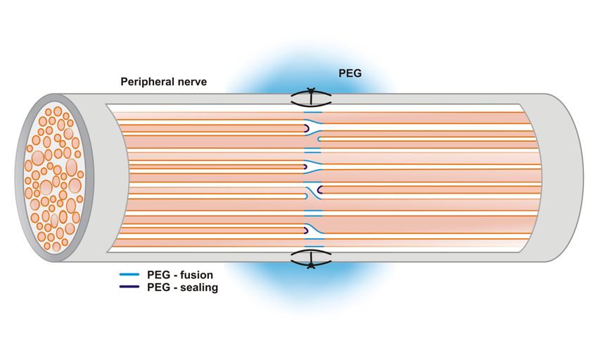
a crush injury), PEG mediates membrane sealing (self-fusion within the axon) (see Figure 1) [37]. diffusion tensor
tractography of PEG-fused fixed sciatic nerves [40]. When axon ends are distantly disconnected (e.g.,
a crush injury), PEG mediates membrane sealing (self-fusion within the axon) (see Figure 1) [37].
Figure 1. Mechanism of polyethylene glycol (PEG)-mediated fusion and sealing of axon ends.
Polyethylene glycol application at the site of a nerve cut may result in either membrane fusion or
Figure 1. Mechanism of polyethylene glycol (PEG)-mediated fusion and sealing of axon ends.
membrane sealing.
Polyethylene glycol application at the site of a nerve cut may result in either membrane fusion or
membrane
The sealing.
protocol of effective PEG-induced axonal fusion was invented and improved by
Bittner et al. [36,41,42]. It starts with a wash in a Ca2+ -free solution (in order to avoid Ca2+ -mediated
The protocol of effective PEG-induced axonal fusion was invented and improved by Bittner et
axolemmal sealing), then the nerve ends are coapted with epineurial microsutures. Next, washes are
al. [36,41,42]. It starts with a wash in a Ca2+-free solution (in order to avoid Ca2+-mediated axolemmalInt. J. Mol. Sci. 2019, 20, 1478 3 of 10
done with 1% methylene blue (antioxidant, for 1–2 min), 50% PEG (2–5 kDa, preferably 3.35 kDa)
for 1–2 min, and finally isotonic saline containing Ca2+ (e.g., Ringer’s Lactate) [13,43–47]. Worth
highlighting is the fact that protocols include no exact dose (volume) of PEG solution. The volume
of solution needed to cover the whole treated nerve varies each time. An isolated PEG application,
independent of the protocol, does not result in better PNI recovery [48]. Apart from the direct use of
a PEG solution on the nerve coaptation site, PEG is also used in bridging nerve gaps, as a conduit
material (solid PEG), or as a conduit filling (PEG solution) [49,50].
From the clinical point of view, it is worth mentioning that PEG is considered safe to use in humans.
Although no study included in this systemic review state any side effects of PEG treatment, a review
on its pharmaceutical applications by D’souza and Shegokar summarized that toxicity, mutagenicity,
as well as teratogenic tests in rats and human clinical trials have proven its safety via oral and non-oral
routes [51]. Still, PEG is often used as a drug carrier (PEGylated conjugates, surface-coating with
PEG) which can lead to the formation of anti-PEG antibodies [51]. However, a single intraoperative
exposition to a PEG solution causes a relatively small risk of antibodies formation compared with
chronic PEGylated drug administration.
2. Methods
We performed a systemic review of the literature according to PRISMA guidelines. The search
was carried out on 1 December, 2018, using PubMed and ScienceDirect as data sources. We searched
classical articles and clinical trials. The following keywords were used: “Polyethylene glycol” OR
“PEG” AND “nerve” AND “injury”. The inclusion criteria included articles which reported in vivo
PEG treatment of PNI in animal models and human subjects. Non-English articles were excluded.
3. Results
On the basis of the search criteria, eight studies were found that met the inclusion criteria. During
the reference analysis of the retrieved articles, nine additional articles were identified. Finally, 17 studies
on an animal models (1 study on a guinea pig model and 15 studies on a rat model) and 1 study
in human subjects were selected out of a total of 447 screened records. Excluded studies mainly
comprised PEG treatments in the spinal cord injury model and applications of solid PEG as a conduit
or scaffold material. Figure 2 presents a flow diagram of the literature search and selection process.
George D. Bittner is considered a pioneer of PEG therapy in PNI. His first report was published
in 1985 [36]. Until 2002, all studies were performed ex vivo. Table 1 summarizes the studies on PEG
treatment of PNI on animals from 2002 to 2018 (see Table S1, a comprehensive version of Table 1
available as supplementary data). The majority of experiments was conducted on the rat sciatic
nerve injury model. Different PEG application protocols were evaluated on crush and cut injuries.
The results were evaluated with electrophysiological recordings, behavioral (motor) testing (e.g., Sciatic
Functional Index, Foot Fault Test), histological tissue analysis, and axonal dye diffusion. In 14/16
experiments, electrophysiological recordings were carried out. Measured parameters included: CAPs,
CMAPs (compound muscle action potentials), and muscle contraction force. Postrepair restoration of
CAPs/CMAPs conduction through a lesion site is an indicator of successful PEG-mediated axonal
fusion. On the basis of the restoration of CAPs, the first 100% success rate of PEG treatment (n = 32)
was reported by Bittner et al. in a rat sciatic nerve cut injury model [52]. The behavioral (motor)
testing is the most relevant part of neuroregeneration outcomes evaluation, as it is the closest model
to a clinical setting. In most studies that implemented motor recovery analysis, PEG-treated animals
showed better performance than controls in the short term, which is consistent with a rapid nerve
conduction recovery caused by PEG. In long-term observations, this tendency persisted in all, except
two experiments [47,53]. Histological analysis is the most objective method from those listed, as tissue
harvesting is independent of animals’ generated disruptions, unlike electrophysiological recordings
and behavioral testing. PEG-treated animals had a significantly higher number of axons in nerve
parts distal to the injury site compared with controls (reported in five experiments, for details seeInt. J. Mol. Sci. 2019, 20, 1478 4 of 10
Table S1) [45,53–56]. Also, axonal diameters in the PEG-treated groups were significantly larger than
the respective controls [57–59]. Almost all studies, except one by Brown et al., reported some kind of
superiority of PEG treatments. The study by Brown et al. stated that PEG addition is not beneficial in
facial nerve cut injury with suture-based repair [60]. Other dissimilarities in the study by Brown et al.
are a lack of evaluation with electrophysiological recordings and a lack of nerve histological analysis.
The results were based on motor function evaluation (eye blink reflex and vibrissae movement), axonal
dye diffusion for motor neuron survival assessment, and histological muscle analysis. The authors
implied that the facial nerve is not as susceptible as the sciatic nerve to PEG fusion. However, after
taking into consideration the consistent physiology and histology within one species, that implication
may be false. Bittner et al. speculated that the unusual PEG fusion protocol used by Brown et al.
might not be optimal, and the lack of results of PEG fusion could be due to the use of incorrect
solutions [61]. Issacs et al. also did not observe enhanced nerve regeneration (measured by muscle
contraction force and nerve diameter assessment) [48]. It should be noted, however, that their study
was designed mainly to evaluate whether PEG application reduced scar formation compared to fibrin
glue (and indeed it proved PEG’s superiority in reducing a scar’s thickness). PEG was not applied in
accordance with the protocol considering the status of Ca2+ during nerve repair, therefore, a lack of
axon
Int. fusion
J. Mol. disproves
Sci. 2019, itsPEER
20, x FOR effectiveness
REVIEW reported elsewhere. 4 of 11
Figure
Figure2.2.PRISMA
PRISMAflow
flowdiagram
diagramof
ofthe
theliterature
literaturesearch
search for
for this
this systematic
systematic review.
review.
George D. Bittner is considered a pioneer of PEG therapy in PNI. His first report was published
in 1985 [36]. Until 2002, all studies were performed ex vivo. Table 1 summarizes the studies on PEG
treatment of PNI on animals from 2002 to 2018 (see Table S1, a comprehensive version of Table 1
available as supplementary data). The majority of experiments was conducted on the rat sciatic nerve
injury model. Different PEG application protocols were evaluated on crush and cut injuries. TheInt. J. Mol. Sci. 2019, 20, 1478 5 of 10
Table 1. Animal studies of polyethylene glycol therapy in the treatment of peripheral nerve injuries. + present, − absent.
Type of Injury Methods of Evaluation
PEG Therapy Superiority
Animal Model Electrophysiological Behavioral (Motor) Histological vs. Control Group Reference
Crush Cut
Recordings Testing Analysis
Guinea pig (sciatic nerve) + − + − − + [39]
Rat (sciatic nerve) + − + − − + [62]
Rat (sciatic nerve) − + + − + + [48]
Rat (sciatic nerve) + + + + − + [47]
Rat (sciatic nerve) + + + + − + [52]
Rat (sciatic nerve) − + + + + + [45]
Rat (sciatic nerve) − + + + + + [54]
Rat (sciatic nerve) − + + + + + [53]
Rat (sciatic nerve) + + + + + + [46]
Rat (femoral nerve) − + − − + − [63]
Rat (sciatic nerve) − + + + + + [55]
Rat (sciatic nerve) − + + + + + [56]
Rat (facial nerve) − + + − + + [57]
Rat (facial nerve) − + − + + − [60]
Rat (sciatic nerve) − + + + + + [58]
Rat (sciatic nerve) − + + + + + [59]Int. J. Mol. Sci. 2019, 20, 1478 6 of 10
Attempts to improve the PEG fusion protocol by supplementation with substances of supposed
pro-neuroregenerative potential, such as melatonin, methylprednisolone, methylene blue, protein
kinase A inhibitor (PKI), protein kinase C isozyme η pseudosubstrate fragment (ηPSF), and protein
kinase C isozyme θ pseudosubstrate fragment (θPSF), were largely unsuccessful [46,52,62]. Only a
methylene blue solution, when applied just before PEG treatment, significantly enhanced nerve
fusion, and consequently this compound was utilized in subsequent experiments [45,53–60]. Other
modifications of the fusion protocol were related to the technique of PEG application on the nerve
lesion site. If specified, in nearly all protocols, the solutions were administered via a hand-held
syringe. Riley et al. proposed the use of a device that minimizes the inconsistency of manual PEG
solution application. This device improved the successful postoperative CAPs recovery from 72%
(13/18 animals) in the standard PEG application method to 83% (15/18 animals). On the other hand,
according to the authors’ analyses, electrophysiological recordings (CAPs) and behavioral tests results
did not differ significantly between the groups [55].
The first study on PNI treated with PEG in an animal model investigated the effects of the treatment
depending on the time between injury and PEG application. When crushed sciatic nerve injury was
treated using the PEG protocol within 30 min from the injury, the success rate, based on recovery of
CAPs and muscle contractile force, reached 75% (6/8 animals). A 4 h delay after injury decreased the
success rate to 66.7% (4/6 animals) [39]. This shows that the longer the time from PNI to PEG application,
the lesser the benefits of PEG treatment. This raises the question of what the maximal reasonable time
delay from PNI to the application of PEG treatment is. Fifteen years later, Bamba et al. tried to answer this
question [56]. They evaluated nerve repair when PEG treatment was applied 1, 8, or 24 h after the sciatic
nerve cut injury. At all time points and in each PEG-treated rat, PEG application restored postrepair CAPs
conduction. Thus, the maximal time from injury to effective nerve fusion still remains undetermined.
Bamba et al. also incorporated PEG in human PNI therapy [64]. The enrolled patients met
the following inclusion criteria: sharp nerve injury and time to surgical repairInt. J. Mol. Sci. 2019, 20, 1478 7 of 10
analyses. A profound investigation into the recovery of all nerve functions (also autonomic) is crucial
for considering translating PEG therapy to the clinics. Rapid motor recovery indicates restoration of
orthodromic signal conduction through PEG-treated nerve lesion. Yet, no data on antidromic signal
conduction has been published so far. Also, none of the published studies discusses the effects of PEG
treatment on Schwann cells within the nerve injury region. Does reinnervation via PEG-mediated
axonal fusion prevent Schwann cells death? Animal studies assessing changes in the postinjury,
posttreatment survival rate of injured nerve Schwann cells should clarify this issue. The maximal
delay in successful PEG therapy implementation has not yet been determined. Bamba et al. reported
successful PEG-mediated fusion of the sciatic nerve 24 h after the initial nerve injury [56]. Establishment
of an injury-to-intervention time limit on an animal model will set a foundation to plan a randomized
clinical trial with objective tests for sensory and motor recovery evaluation in humans. A fact that
is worth mentioning in case of a human randomized controlled trial (RCT) design is blinding the
operator to the substance. Since PEG treatment in the described models incorporate intraoperative
washes of the operation field with a characteristic set of solutions, proper blinding should be done.
Supplementary Materials: Supplementary materials can be found at http://www.mdpi.com/1422-0067/20/6/
1478/s1.
Author Contributions: Conceptualization, A.M.P. and W.P.; methodology, A.M.P. and W.P.; validation, P.P. and
P.K.W.; data curation, A.M.P. and W.P.; writing—original draft preparation, A.M.P.; writing—review and editing,
A.M.P., W.P., P.P., P.K.W.; visualization, W.P.; supervision, P.P., P.K.W.; project administration, A.M.P.; funding
acquisition, A.M.P., W.P.
Funding: This research was funded by Grant No. 1MN/FS223 from the Polish Ministry of Science and
Higher Education.
Conflicts of Interest: The authors declare no conflict of interest. The funders had no role in the design of the
study; in the collection, analyses, or interpretation of data; in the writing of the manuscript, or in the decision to
publish the results.
Abbreviations
PRISMA Preferred Reporting Items for Systematic Reviews and Meta- Analyses
PNI Peripheral nerve injuries
PEG Polyethylene glycol
CAPs Compound action potentials
CMAPs Compound muscle action potentials
MB Methylene blue
2PD Static two-point discrimination test
SWM Semmes–Weinstein monofilament test
MRCC Medical Research Council Classification
RCT Randomized controlled trial
References
1. Lee, S.K.; Wolfe, S.W. Peripheral nerve injury and repair. J. Am. Acad. Orthop. Surg. 2000, 8, 243–252. [CrossRef]
2. Palispis, W.A.; Gupta, R. Surgical repair in humans after traumatic nerve injury provides limited functional
neural regeneration in adults. Exp. Neurol. 2017, 290, 106–114. [CrossRef] [PubMed]
3. Paprottka, F.J.; Wolf, P.; Harder, Y.; Kern, Y.; Paprottka, P.M.; Machens, H.G.; Lohmeyer, J.A. Sensory recovery
outcome after digital nerve repair in relation to different reconstructive techniques: Meta-analysis and
systematic review. Plast. Surg. Int. 2013, 2013, 704589. [CrossRef] [PubMed]
4. Hundepool, C.A.; Ultee, J.; Nijhuis, T.H.; Houpt, P.; Hovius, S.E. Prognostic factors for outcome after median,
ulnar, and combined median-ulnar nerve injuries: A prospective study. J. Plast. Reconstr. Aesthet. Surg. 2015,
68, 1–8. [CrossRef]
5. Devi, B.I.; Konar, S.K.; Bhat, D.I.; Shukla, D.P.; Bharath, R.; Gopalakrishnan, M.S. Predictors of Surgical
Outcomes of Traumatic Peripheral Nerve Injuries in Children: An Institutional Experience. Pediatr. Neurosurg.
2018, 53, 94–99. [CrossRef] [PubMed]Int. J. Mol. Sci. 2019, 20, 1478 8 of 10
6. Dahlin, L.B. The role of timing in nerve reconstruction. Int. Rev. Neurobiol. 2013, 109, 151–164. [CrossRef]
[PubMed]
7. Kouyoumdjian, J.A.; Graca, C.R.; Ferreira, V.F.M. Peripheral nerve injuries: A retrospective survey of 1124
cases. Neurol. India 2017, 65, 551–555. [CrossRef]
8. Kretschmer, T.; Antoniadis, G.; Braun, V.; Rath, S.A.; Richter, H.P. Evaluation of iatrogenic lesions in 722
surgically treated cases of peripheral nerve trauma. J. Neurosurg. 2001, 94, 905–912. [CrossRef] [PubMed]
9. Kretschmer, T.; Heinen, C.W.; Antoniadis, G.; Richter, H.P.; Konig, R.W. Iatrogenic nerve injuries.
Neurosurg. Clin. N. Am. 2009, 20, 73–90. [CrossRef]
10. Houdek, M.T.; Shin, A.Y. Management and complications of traumatic peripheral nerve injuries. Hand Clin.
2015, 31, 151–163. [CrossRef] [PubMed]
11. Yoo, S.; Nguyen, M.P.; Fukuda, M.; Bittner, G.D.; Fishman, H.M. Plasmalemmal sealing of transected
mammalian neurites is a gradual process mediated by Ca2+ -regulated proteins. J. Neurosci. Res. 2003,
74, 541–551. [CrossRef] [PubMed]
12. Fishman, H.M.; Bittner, G.D. Vesicle-mediated restoration of a plasmalemmal barrier in severed axons.
News Physiol. Sci. 2003, 18, 115–118. [CrossRef]
13. Spaeth, C.S.; Boydston, E.A.; Figard, L.R.; Zuzek, A.; Bittner, G.D. A model for sealing plasmalemmal
damage in neurons and other eukaryotic cells. J. Neurosci. 2010, 30, 15790–15800. [CrossRef]
14. Waller, A. Experiments on the Section of the Glosso-Pharyngeal and Hypoglossal Nerves of the Frog, and
Observations of the Alterations Produced Thereby in the Structure of Their Primitive Fibres. Edinb. Med.
Surg. J. 1851, 76, 369–376.
15. Stoll, G.; Muller, H.W. Nerve injury, axonal degeneration and neural regeneration: Basic insights. Brain Pathol.
1999, 9, 313–325. [CrossRef] [PubMed]
16. Beuche, W.; Friede, R.L. The role of non-resident cells in Wallerian degeneration. J. Neurocytol. 1984,
13, 767–796. [CrossRef] [PubMed]
17. Namgung, U. The role of Schwann cell-axon interaction in peripheral nerve regeneration. Cells Tissues Organs
2014, 200, 6–12. [CrossRef] [PubMed]
18. Allodi, I.; Udina, E.; Navarro, X. Specificity of peripheral nerve regeneration: Interactions at the axon level.
Prog. Neurobiol. 2012, 98, 16–37. [CrossRef]
19. Jessen, K.R.; Mirsky, R. The repair Schwann cell and its function in regenerating nerves. J. Physiol. 2016,
594, 3521–3531. [CrossRef]
20. Gutmann, E.; Guttmann, L.; Medawar, P.B.; Young, J.Z. The Rate of Regeneration of Nerve. J. Exp. Biol. 1942,
19, 14–44. [CrossRef]
21. Sunderland, S. Rate of regeneration in human peripheral nerves; analysis of the interval between injury and
onset of recovery. Arch. Neurol. Psychiatry 1947, 58, 251–295. [CrossRef] [PubMed]
22. Chiang, H.Y.; Huang, I.T.; Chen, W.P.; Chien, H.F.; Shun, C.T.; Chang, Y.C.; Hsieh, S.T. Regional difference in
epidermal thinning after skin denervation. Exp. Neurol. 1998, 154, 137–145. [CrossRef]
23. Frostick, S.P. The physiological and metabolic consequences of muscle denervation. Int. Angiol. J. Int.
Union Angiol. 1995, 14, 278–287.
24. Midrio, M. The denervated muscle: Facts and hypotheses. A historical review. Eur. J. Appl. Physiol. 2006,
98, 1–21. [CrossRef]
25. Vrbova, G.; Mehra, N.; Shanmuganathan, H.; Tyreman, N.; Schachner, M.; Gordon, T. Chemical
communication between regenerating motor axons and Schwann cells in the growth pathway. Eur. J. Neurosci.
2009, 30, 366–375. [CrossRef] [PubMed]
26. Sulaiman, O.A.; Gordon, T. Role of chronic Schwann cell denervation in poor functional recovery after nerve
injuries and experimental strategies to combat it. Neurosurgery 2009, 65, A105–A114. [CrossRef]
27. Jonsson, S.; Wiberg, R.; McGrath, A.M.; Novikov, L.N.; Wiberg, M.; Novikova, L.N.; Kingham, P.J. Effect of
delayed peripheral nerve repair on nerve regeneration, Schwann cell function and target muscle recovery.
PLoS ONE 2013, 8, e56484. [CrossRef] [PubMed]
28. Chan, K.M.; Gordon, T.; Zochodne, D.W.; Power, H.A. Improving peripheral nerve regeneration:
From molecular mechanisms to potential therapeutic targets. Exp. Neurol. 2014, 261, 826–835. [CrossRef]
29. Faroni, A.; Mobasseri, S.A.; Kingham, P.J.; Reid, A.J. Peripheral nerve regeneration: Experimental strategies
and future perspectives. Adv. Drug Deliv. Rev. 2015, 82–83, 160–167. [CrossRef]Int. J. Mol. Sci. 2019, 20, 1478 9 of 10
30. Christie, K.J.; Zochodne, D. Peripheral axon regrowth: New molecular approaches. Neuroscience 2013,
240, 310–324. [CrossRef]
31. Fowler, J.R.; Lavasani, M.; Huard, J.; Goitz, R.J. Biologic strategies to improve nerve regeneration after
peripheral nerve repair. J. Reconstr. Microsurg. 2015, 31, 243–248. [CrossRef] [PubMed]
32. Kubiak, C.A.; Kung, T.A.; Brown, D.L.; Cederna, P.S.; Kemp, S.W.P. State-of-the-Art Techniques in Treating
Peripheral Nerve Injury. Plast. Reconstr. Surg. 2018, 141, 702–710. [CrossRef]
33. Sullivan, R.; Dailey, T.; Duncan, K.; Abel, N.; Borlongan, C.V. Peripheral Nerve Injury: Stem Cell Therapy
and Peripheral Nerve Transfer. Int. J. Mol. Sci. 2016, 17. [CrossRef] [PubMed]
34. Gordon, T.; Chan, K.M.; Sulaiman, O.A.; Udina, E.; Amirjani, N.; Brushart, T.M. Accelerating axon growth to
overcome limitations in functional recovery after peripheral nerve injury. Neurosurgery 2009, 65, A132–A144.
[CrossRef] [PubMed]
35. Gordon, T.; English, A.W. Strategies to promote peripheral nerve regeneration: Electrical stimulation and/or
exercise. Eur. J. Neurosci. 2016, 43, 336–350. [CrossRef] [PubMed]
36. Bittner, G.D.; Ballinger, M.L.; Raymond, M.A. Reconnection of severed nerve axons with polyethylene glycol.
Brain Res. 1986, 367, 351–355. [CrossRef]
37. Bittner, G.D.; Sengelaub, D.R.; Ghergherehchi, C.L. Conundrums and confusions regarding how polyethylene
glycol-fusion produces excellent behavioral recovery after peripheral nerve injuries. Neural Regen. Res. 2018,
13, 53–57. [CrossRef]
38. Abdou, S.A.; Henderson, P.W. Fusogens: Chemical agents that can rapidly restore function after nerve injury.
J. Surg. Res. 2019, 233, 36–40. [CrossRef]
39. Donaldson, J.; Shi, R.; Borgens, R. Polyethylene glycol rapidly restores physiological functions in damaged
sciatic nerves of guinea pigs. Neurosurgery 2002, 50, 147–156.
40. Bamba, R.; Riley, D.C.; Kelm, N.D.; Does, M.D.; Dortch, R.D.; Thayer, W.P. A novel technique using
hydrophilic polymers to promote axonal fusion. Neural Regen. Res. 2016, 11, 525–528. [CrossRef] [PubMed]
41. Krause, T.L.; Bittner, G.D. Rapid morphological fusion of severed myelinated axons by polyethylene glycol.
Proc. Natl. Acad. Sci. USA 1990, 87, 1471–1475. [CrossRef] [PubMed]
42. Lore, A.B.; Hubbell, J.A.; Bobb, D.S., Jr.; Ballinger, M.L.; Loftin, K.L.; Smith, J.W.; Smyers, M.E.; Garcia, H.D.;
Bittner, G.D. Rapid induction of functional and morphological continuity between severed ends of
mammalian or earthworm myelinated axons. J. Neurosci. 1999, 19, 2442–2454. [CrossRef] [PubMed]
43. Spaeth, C.S.; Robison, T.; Fan, J.D.; Bittner, G.D. Cellular mechanisms of plasmalemmal sealing and axonal
repair by polyethylene glycol and methylene blue. J. Neurosci. Res. 2012, 90, 955–966. [CrossRef] [PubMed]
44. Spaeth, C.S.; Fan, J.D.; Spaeth, E.B.; Robison, T.; Wilcott, R.W.; Bittner, G.D. Neurite transection produces
cytosolic oxidation, which enhances plasmalemmal repair. J. Neurosci. Res. 2012, 90, 945–954. [CrossRef]
45. Sexton, K.W.; Pollins, A.C.; Cardwell, N.L.; Del Corral, G.A.; Bittner, G.D.; Shack, R.B.; Nanney, L.B.;
Thayer, W.P. Hydrophilic polymers enhance early functional outcomes after nerve autografting. J. Surg. Res.
2012, 177, 392–400. [CrossRef] [PubMed]
46. Ghergherehchi, C.L.; Bittner, G.D.; Hastings, R.L.; Mikesh, M.; Riley, D.C.; Trevino, R.C.; Schallert, T.;
Thayer, W.P.; Bhupanapadu Sunkesula, S.R.; Ha, T.A.; et al. Effects of extracellular calcium and surgical
techniques on restoration of axonal continuity by polyethylene glycol fusion following complete cut or crush
severance of rat sciatic nerves. J. Neurosci. Res. 2016, 94, 231–245. [CrossRef] [PubMed]
47. Britt, J.M.; Kane, J.R.; Spaeth, C.S.; Zuzek, A.; Robinson, G.L.; Gbanaglo, M.Y.; Estler, C.J.; Boydston, E.A.;
Schallert, T.; Bittner, G.D. Polyethylene glycol rapidly restores axonal integrity and improves the rate of
motor behavior recovery after sciatic nerve crush injury. J. Neurophysiol. 2010, 104, 695–703. [CrossRef]
48. Isaacs, J.; Klumb, I.; McDaniel, C. Preliminary investigation of a polyethylene glycol hydrogel “nerve glue”.
J. Brachial Plex. Peripher. Nerve Inj. 2009, 4, 16. [CrossRef]
49. Evangelista, M.S.; Perez, M.; Salibian, A.A.; Hassan, J.M.; Darcy, S.; Paydar, K.Z.; Wicker, R.B.; Arcaute, K.;
Mann, B.K.; Evans, G.R. Single-lumen and multi-lumen poly(ethylene glycol) nerve conduits fabricated
by stereolithography for peripheral nerve regeneration in vivo. J. Reconstr. Microsurg. 2015, 31, 327–335.
[CrossRef]
50. Mokarizadeh, A.; Mehrshad, A.; Mohammadi, R. Local Polyethylene Glycol in Combination with Chitosan
Based Hybrid Nanofiber Conduit Accelerates Transected Peripheral Nerve Regeneration. J. Invest. Surg.
2016, 29, 167–174. [CrossRef]Int. J. Mol. Sci. 2019, 20, 1478 10 of 10
51. D’Souza, A.A.; Shegokar, R. Polyethylene glycol (PEG): A versatile polymer for pharmaceutical applications.
Expert Opin. Drug Deliv. 2016, 13, 1257–1275. [CrossRef] [PubMed]
52. Bittner, G.D.; Keating, C.P.; Kane, J.R.; Britt, J.M.; Spaeth, C.S.; Fan, J.D.; Zuzek, A.; Wilcott, R.W.; Thayer, W.P.;
Winograd, J.M.; et al. Rapid, effective, and long-lasting behavioral recovery produced by microsutures,
methylene blue, and polyethylene glycol after completely cutting rat sciatic nerves. J. Neurosci. Res. 2012,
90, 967–980. [CrossRef] [PubMed]
53. Sexton, K.W.; Rodriguez-Feo, C.L.; Boyer, R.B.; Del Corral, G.A.; Riley, D.C.; Pollins, A.C.; Cardwell, N.L.;
Shack, R.B.; Nanney, L.B.; Thayer, W.P. Axonal fusion via conduit-based delivery of hydrophilic polymers.
Hand (N Y) 2015, 10, 688–694. [CrossRef] [PubMed]
54. Riley, D.C.; Bittner, G.D.; Mikesh, M.; Cardwell, N.L.; Pollins, A.C.; Ghergherehchi, C.L.; Bhupanapadu
Sunkesula, S.R.; Ha, T.N.; Hall, B.T.; Poon, A.D.; et al. Polyethylene glycol-fused allografts produce rapid
behavioral recovery after ablation of sciatic nerve segments. J. Neurosci. Res. 2015, 93, 572–583. [CrossRef]
55. Riley, D.C.; Boyer, R.B.; Deister, C.A.; Pollins, A.C.; Cardwell, N.L.; Kelm, N.D.; Does, M.D.; Dortch, R.D.;
Bamba, R.; Shack, R.B.; et al. Immediate Enhancement of Nerve Function Using a Novel Axonal Fusion
Device After Neurotmesis. Ann. Plastic Surg. 2017, 79, 590–599. [CrossRef] [PubMed]
56. Bamba, R.; Riley, D.C.; Kim, J.S.; Cardwell, N.L.; Pollins, A.C.; Shack, R.B.; Thayer, W.P. Evaluation of a
Nerve Fusion Technique With Polyethylene Glycol in a Delayed Setting After Nerve Injury. J. Hand Surg.
2018, 43, 82e81–82e87. [CrossRef]
57. Salomone, R.; Jacomo, A.L.; Nascimento, S.B.D.; Lezirovitz, K.; Hojaij, F.C.; Costa, H.; Bento, R.F. Polyethylene
glycol fusion associated with antioxidants: A new promise in the treatment of traumatic facial paralysis.
Head Neck 2018, 40, 1489–1497. [CrossRef]
58. Mikesh, M.; Ghergherehchi, C.L.; Hastings, R.L.; Ali, A.; Rahesh, S.; Jagannath, K.; Sengelaub, D.R.;
Trevino, R.C.; Jackson, D.M.; Bittner, G.D. Polyethylene glycol solutions rapidly restore and maintain
axonal continuity, neuromuscular structures, and behaviors lost after sciatic nerve transections in female
rats. J. Neurosci. Res. 2018, 96, 1223–1242. [CrossRef] [PubMed]
59. Mikesh, M.; Ghergherehchi, C.L.; Rahesh, S.; Jagannath, K.; Ali, A.; Sengelaub, D.R.; Trevino, R.C.;
Jackson, D.M.; Tucker, H.O.; Bittner, G.D. Polyethylene glycol treated allografts not tissue matched nor
immunosuppressed rapidly repair sciatic nerve gaps, maintain neuromuscular functions, and restore
voluntary behaviors in female rats. J. Neurosci. Res. 2018, 96, 1243–1264. [CrossRef]
60. Brown, B.L.; Asante, T.; Welch, H.R.; Sandelski, M.M.; Drejet, S.M.; Shah, K.; Runge, E.M.; Shipchandler, T.Z.;
Jones, K.J.; Walker, C.L. Functional and Anatomical Outcomes of Facial Nerve Injury with Application of
Polyethylene Glycol in a Rat Model. JAMA Facial Plast. Surg. 2018. [CrossRef]
61. Bittner, G.D.; Ghergherehchi, C.L.; Mikesh, M. Surgical Technique and Other Protocols Used Probably Did
Not Induce Polyethylene Glycol Fusion of Rat Facial Nerves. JAMA Facial Plast. Surg. 2018. [CrossRef]
[PubMed]
62. Stavisky, R.C.; Britt, J.M.; Zuzek, A.; Truong, E.; Bittner, G.D. Melatonin enhances the in vitro and in vivo
repair of severed rat sciatic axons. Neurosci. Lett. 2005, 376, 98–101. [CrossRef] [PubMed]
63. Robinson, G.A.; Madison, R.D. Polyethylene glycol fusion repair prevents reinnervation accuracy in rat
peripheral nerve. J. Neurosci. Res. 2016, 94, 636–644. [CrossRef] [PubMed]
64. Bamba, R.; Waitayawinyu, T.; Nookala, R.; Riley, D.C.; Boyer, R.B.; Sexton, K.W.; Boonyasirikool, C.;
Niempoog, S.; Kelm, N.D.; Does, M.D.; et al. A novel therapy to promote axonal fusion in human digital
nerves. J. Trauma Acute Care Surg. 2016, 81, S177–S183. [CrossRef] [PubMed]
65. De Ruiter, G.C.; Malessy, M.J.; Alaid, A.O.; Spinner, R.J.; Engelstad, J.K.; Sorenson, E.J.; Kaufman, K.R.;
Dyck, P.J.; Windebank, A.J. Misdirection of regenerating motor axons after nerve injury and repair in the rat
sciatic nerve model. Exp. Neurol. 2008, 211, 339–350. [CrossRef]
66. De Ruiter, G.C.; Spinner, R.J.; Verhaagen, J.; Malessy, M.J. Misdirection and guidance of regenerating axons
after experimental nerve injury and repair. J. Neurosurg. 2014, 120, 493–501. [CrossRef]
© 2019 by the authors. Licensee MDPI, Basel, Switzerland. This article is an open access
article distributed under the terms and conditions of the Creative Commons Attribution
(CC BY) license (http://creativecommons.org/licenses/by/4.0/).You can also read