Distinct Binding Dynamics, Sites and Interactions of Fullerene and Fullerenols with Amyloid-β Peptides Revealed by Molecular Dynamics Simulations ...
←
→
Page content transcription
If your browser does not render page correctly, please read the page content below
International Journal of
            Molecular Sciences

Article
Distinct Binding Dynamics, Sites and Interactions of
Fullerene and Fullerenols with Amyloid-β Peptides
Revealed by Molecular Dynamics Simulations
Zhiwei Liu 1,† , Yu Zou 2,† , Qingwen Zhang 2 , Peijie Chen 1 , Yu Liu 1 and Zhenyu Qian 1, *
 1    Key Laboratory of Exercise and Health Sciences (Ministry of Education), School of Kinesiology,
      Shanghai University of Sport, 399 Changhai Road, Shanghai 200438, China; 15266070790@163.com (Z.L.);
      chenpeijie@sus.edu.cn (P.C.); yuliu@sus.edu.cn (Y.L.)
 2    College of Physical Education and Training, Shanghai University of Sport, 399 Changhai Road,
      Shanghai 200438, China; zouyu_1993@163.com (Y.Z.); zqw@sus.edu.cn (Q.Z.)
 *    Correspondence: qianzhenyu@sus.edu.cn
 †    These authors contributed equally to this work.

 Received: 27 March 2019; Accepted: 24 April 2019; Published: 25 April 2019

 Abstract: The pathology Alzheimer’s disease (AD) is associated with the self-assembly of amyloid-β
 (Aβ) peptides into β-sheet enriched fibrillar aggregates. A promising treatment strategy is focused
 on the inhibition of amyloid fibrillization of Aβ peptide. Fullerene C60 is proved to effectively
 inhibit Aβ fibrillation while the poor water-solubility restricts its use as a biomedicine agent. In this
 work, we examined the interaction of fullerene C60 and water-soluble fullerenol C60 (OH)6 /C60 (OH)12
 (C60 carrying 6/12 hydroxyl groups) with preformed Aβ40/42 protofibrils by multiple molecular
 dynamics simulations. We found that when binding to the Aβ42 protofibril, C60 , C60 (OH)6 and
 C60 (OH)12 exhibit distinct binding dynamics, binding sites and peptide interaction. The increased
 number of hydroxyl groups C60 carries leads to slower binding dynamics and weaker binding
 strength. Binding free energy analysis demonstrates that the C60 /C60 (OH)6 molecule primarily
 binds to the C-terminal residues 31–41, whereas C60 (OH)12 favors to bind to N-terminal residues
 4–14. The hydrophobic interaction plays a critical role in the interplay between Aβ and all the
 three nanoparticles, and the π-stacking interaction gets weakened as C60 carries more hydroxyls.
 In addition, the C60 (OH)6 molecule has high affinity to form hydrogen bonds with protein backbones.
 The binding behaviors of C60 /C60 (OH)6 /C60 (OH)12 to the Aβ40 protofibril resemble with those to
 Aβ42 . Our work provides a detailed picture of fullerene/fullerenols binding to Aβ protofibril, and is
 helpful to understand the underlying inhibitory mechanism.

 Keywords: amyloid protofibril;                  fullerene;   binding site;   inhibitory mechanism;    molecular
 dynamics simulation

1. Introduction
     Amyloids are involved in a broad range of neurodegenerative diseases, including Alzheimer’s,
Huntington’s and Parkinson’s diseases [1–3]. The major constituents of amyloid plaques are associated
with fibrils formed by amyloid-β (Aβ) protein that display a cross-β structure characterized by β-strands
perpendicular to and inter-strand hydrogen bonds parallel to the fibril axis [4,5]. The fibrillation occurs
through a complex multistep process, involving the formation of soluble oligomers, protofibrils and
insoluble mature fibrils [6,7]. Small aggregates (soluble oligomers and protofibrils) in the early stage of
aggregation are suggested as primary neurotoxic agents [8–11]. Therefore, a promising strategy to
reduce the small toxic oligomer species is to inhibit Aβ peptide aggregation.

Int. J. Mol. Sci. 2019, 20, 2048; doi:10.3390/ijms20082048                               www.mdpi.com/journal/ijms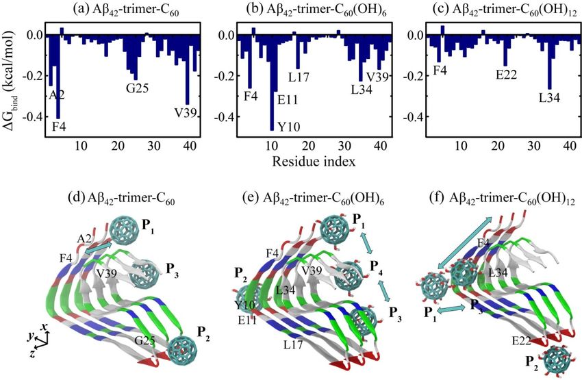
Int. J. Mol. Sci. 2019, 20, 2048                                                                     2 of 14

      The search for effective inhibitors has become an active area of research. An increasing number
of experimental and computational studies have reported that Aβ aggregation can be modulated
by nanoparticles [12–14], small molecules [15,16], short peptides [17,18], antibodies [19] and metal
ions [20]. Their findings provided new clues for the design of inhibitors targeting Aβ formation.
In recent years, the fullerenes have gained great attention, not only because of their antioxidant,
neuroprotective and antitumor properties [21], but also due to their promising ability of carrying
contrast agents, radiopharmaceuticals or drugs [22]. However, the poor solubility of fullerenes in water
restricts their potential biomedical applications. One of the common strategies to increase of their
solubility is to attach hydroxyl groups to the carbon cage, leading to the formation of hydroxylated
fullerene (i.e., fullerenol, C60 (OH)n ). Fullerenols have high solubility and ability to cross the blood
brain barriers [23]. Fullerene derivatives are reported to have remarkable anti-amyloid properties for
Alzheimer’s disease and other neurodegenerative diseases [24–28].
      Computational studies have investigated the molecular mechanism of fullerenes/fullerenols
binding and binding-induced protein remodeling using docking method and molecular dynamics
(MD) simulations [29–33]. For example, Li et al. examined the binding affinity of fullerenes in different
sizes and found that C60 destroys pentameric Aβ17–42 fibril structure to a greater extent with respect to
other fullerenes [29]. They also found that C60 (OH)16 is inclined to bind at the central hydrophobic and
the hydrophobic C-terminal region of monomer Aβ40 to prevent amyloid fibrillization [30]. Wei et al.
identified three primary binding sites of 1,2-(dimethoxymethano) fullerene (DMF) to Aβ1–42 protofibril:
the central hydrophobic residues 17–21, the residues 27–31 in turn region and the C-terminal residues
31–41 [31]. They also demonstrated that the fullerene nanoparticles – C60 and C180 exhibit stronger
inhibition on Aβ16–22 β-sheet formation [33]. Ding et al. found that different extent of hydroxylation
would significantly influence C60 (OH)n –protein interactions [32]. These results reveal the binding
modes and inhibitory/disruptive mechanisms of fullerenes/fullerenols, which greatly enhances our
understanding of fullerenes/fullerenols-protein interactions at atomic level.
      Our previous study examined the influence of DMF on Aβ42 dimerization by replica-exchange
MD simulations [34]. However, the interactions between Aβ protofibril and fullerenes with different
degree of hydroxylation remain elusive. Previous computational study showed that stable Aβ
trimer with well-preserved parallel β-strands could act as the smallest seed for Aβ polymerization
on self-assembled monolayers [35]. Following the work by Zheng and Wei [35,36], we chose a
trimer as Aβ protofibril model in our MD simulations. Here, we investigated the interaction of a
C60 /C60 (OH)6 /C60 (OH)12 molecule with Aβ42/40 protofibrillar trimer and the resulting protein structural
alterations by performing multiple MD simulations. We found that the increased hydroxylation extent
of C60 leads to slower binding dynamics and weaker binding strength. Binding sites and free energy
analyses demonstrate that the C60 /C60 (OH)6 molecule primarily binds to the C-terminal hydrophobic
region, whereas C60 (OH)12 favors to bind to N-terminal residues 4–14. Our simulations revealed the
dominant role of hydrophobic interaction in Aβ−nanoparticle interplay. Moreover, the water-soluble
C60 (OH)6 molecule has high affinity to form hydrogen bonds with protein backbones, which makes it
a more efficient inhibitor than C60 and C60 (OH)12 .

2. Results and Discussion
     We performed MD simulations to study the binding behavior of C60 /C60 (OH)6 / C60 (OH)12
to Aβ42 protofibrillar trimer (Aβ42 -trimer for short) and Aβ40 protofibrillar trimer (Aβ40 -trimer
for short), respectively. The systems are labeled as Aβ42 -trimer-C60 , Aβ42 -trimer-C60 (OH)6 ,
Aβ42 -trimer-C60 (OH)12 , Aβ40 -trimer-C60 , Aβ40 -trimer-C60 (OH)6 and Aβ40 -trimer-C60 (OH)12 .
The molecular structures of Aβ42 -trimer, Aβ40 -trimer and C60 /C60 (OH)6 /C60 (OH)12 are shown in
Figure 1. The initial state of Aβ42 -trimer-C60 system is also displayed, and the other systems are
constructed similarly. More details are given in Model and Methods section.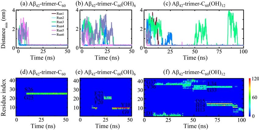
Int. J. Mol. Sci. 2019, 20, 2048                                                                                                    3 of 14

Int. J. Mol. Sci. 2019, 20, x FOR PEER REVIEW                                                                                       3 of 15

        Figure1.1. Molecular
      Figure         Molecular structures
                                    structures and
                                                 and simulation
                                                       simulation systemsystemsetup.
                                                                                  setup. (a–c)
                                                                                          (a–c) The
                                                                                                 Thestructures
                                                                                                      structuresofofAβ Aβ4242-trimer,
                                                                                                                              -trimer,
      AβAβ4040-trimer   andCC6060/C
              -trimer and            60(OH)6/C
                                  /C60        /C60  (OH)1212. .(d)
                                                 60(OH)         (d)The
                                                                     The initial conformation
                                                                                 conformation of  ofthe
                                                                                                     theAβ
                                                                                                         Aβ4242-trimer-C
                                                                                                                -trimer-C6060 system
                                                                                                                               system
      with    the C
        with the 60    molecule    placed   at three  different     positions (I-III). Color codes:  positively
                     C60 molecule placed at three different positions (I-III). Color codes: positively charged    charged   residues
      (blue),
        residuesnegatively  charged residues
                    (blue), negatively     charged (red), hydrophobic
                                                      residues              residues (white)
                                                                    (red), hydrophobic         and polar
                                                                                           residues       residues
                                                                                                     (white)         (green)
                                                                                                                and polar       in Aβ
                                                                                                                             residues
      peptides;
        (green) incarbon    atoms (cyan),
                      Aβ peptides;      carbon oxygen
                                                  atomsatoms
                                                           (cyan), (red) and hydrogen
                                                                      oxygen              atoms
                                                                                atoms (red)      (white)
                                                                                               and        in fullerene/fullerenol.
                                                                                                    hydrogen      atoms (white) in
      For   clarity, water molecules
        fullerene/fullerenol.              in thewater
                                   For clarity,    simulation
                                                         molecules box are  notsimulation
                                                                        in the   displayed; box
                                                                                             box are
                                                                                                  vectors  are shown,box
                                                                                                      not displayed;            z-axis
                                                                                                                        andvectors
      isare
          theshown,
               fibrillar elongation     direction.
                       and z-axis is the fibrillar elongation direction.

2.1. Dynamics of the Fullerene/Fullerenol Molecule Binding to Aβ42 -Trimer
 2.1. Dynamics of the Fullerene/Fullerenol Molecule Binding to Aβ42-Trimer
      To investigate the binding process of the fullerene/fullerenol molecule to Aβ42 -trimer, we first
       To investigate the binding process of the fullerene/fullerenol molecule to Aβ42-trimer, we first
monitored the time evolution of their minimum distance dmin (Figure 2a–c). As for the Aβ42 -trimer-C60
 monitored the time evolution of their minimum distance dmin (Figure 2a–c). As for the Aβ42-trimer-C60
system, the C60 molecule was initially placed 2 nm away from the Aβ42 -trimer. Once the MD simulations
 system, the C60 molecule was initially placed 2 nm away from the Aβ42-trimer. Once the MD
were initiated, dmin started to decrease or increase, depending on the initial velocity distributions.
 simulations were initiated, dmin started to decrease or increase, depending on the initial velocity
The minimum distances in Run 1, 2 and 4 were observed to decline to ~0.30 nm within the first 3 ns,
 distributions. The minimum distances in Run 1, 2 and 4 were observed to decline to ~0.30 nm within
while those in Run 3, 5 and 6 took ~10 ns to reach ~0.30 nm. Such fast and slow binding processes were also
 the first 3 ns, while those in Run 3, 5 and 6 took ~10 ns to reach ~0.30 nm. Such fast and slow binding
observed in Aβ42 -trimer-C60 (OH)6 and Aβ42 -trimer-C60 (OH)12 systems. Similar fast and slow processes
 processes were also observed in Aβ42-trimer-C60(OH)6 and Aβ42-trimer-C60(OH)12 systems. Similar
were reported in a previous MD study of DMF binding to Aβ fibril [31]. Moreover, we found that the slow
 fast and slow processes were reported in a previous MD study of DMF binding to Aβ fibril [31].
binding processes may last tens of nanoseconds for C60 (OH)6 and C60 (OH)12 , much longer than that for
 Moreover, we found that the slow binding processes may last tens of nanoseconds for C60(OH)6 and
C60 . It takes over 25 ns for two MD runs of Aβ-C60 (OH)6 system (Runs 3, 6) to reach a minimum distance
 C60(OH)12, much longer than that for C60. It takes over 25 ns for two MD runs of Aβ-C60(OH)6 system
of ~0.30 nm, and the situation was the same in Aβ-C60 (OH)12 system (Runs 3, 4). Specially, in Run 3
 (Runs 3, 6) to reach a minimum distance of ~0.30 nm, and the situation was the same in Aβ-C60(OH)12
of Aβ42 -trimer-C60 (OH)12 system, dmin increased sharply at 49.8 and 83.6 ns, and declined to ~0.30 nm
 system (Runs 3, 4). Specially, in Run 3 of Aβ42-trimer-C60(OH)12 system, dmin increased sharply at 49.8
in the next twenty nanoseconds. These indicate that the binding process of the C60 (OH)6 /C60 (OH)12
 and 83.6 ns, and declined to ~0.30 nm in the next twenty nanoseconds. These indicate that the
molecule to Aβ42 -trimer is slower than that of C60 .
 binding process of the C60(OH)6/C60(OH)12 molecule to Aβ42-trimer is slower than that of C60.
      To further examine the binding status of the fullerene/fullerenol molecule after the initial adsorption
       To further examine the binding status of the fullerene/fullerenol molecule after the initial
to Aβ42 -trimer, we monitored the time evolution of the number of contacts between individual residue
 adsorption to Aβ42-trimer, we monitored the time evolution of the number of contacts between
and the nanoparticle in a representative MD run for each simulated system in Figure 2d–f. The C60
 individual residue and the nanoparticle in a representative MD run for each simulated system in
molecule was observed to stay at a relatively fixed location during the remaining simulation time once
 Figure 2d–f. The C60 molecule was observed to stay at a relatively fixed location during the
stable contacts are formed. The C60 (OH)6 molecule also had a relatively fixed binding site, while it can
 remaining simulation time once stable contacts are formed. The C60(OH)6 molecule also had a
shift to other location transiently. As for the C60 (OH)12 molecule, its binding location kept changing
 relatively fixed binding site, while it can shift to other location transiently. As for the C60(OH)12
when simulation time increased, corresponding to a slow move on the protein surface. C60 (OH)12 also
 molecule, its binding location kept changing when simulation time increased, corresponding to a
contacted with more residues at the same time, which indicated a lower specificity of binding sites.
 slow move on the protein surface. C60(OH)12 also contacted with more residues at the same time,
These results reflect that with the hydroxylation extent of C60 increased, the binding strength between
 which indicated a lower specificity of binding sites. These results reflect that with the hydroxylation
Aβ42 -trimer and the nanoparticle molecule gets weaker.
 extent of C60 increased, the binding strength between Aβ42-trimer and the nanoparticle molecule gets
 weaker.
Int. J. Mol. Sci. 2019, 20, 2048 4 of 14
Int. J. Mol. Sci. 2019, 20, x FOR PEER REVIEW 4 of 15
Figure
Figure 2. 2. Dynamicsofofthe
Dynamics thefullerene/fullerenol
fullerene/fullerenol molecule
molecule binding
binding to
to Aβ -trimer.
Aβ4242 -trimer.(a–c) Time
(a–c) evolution
Time evolution
of the minimum distance between Aβ42-trimer and fullerene/fullerenol. Six independent molecular
of the minimum distance between Aβ42 -trimer and fullerene/fullerenol. Six independent molecular
dynamics (MD) runs are denoted in different colors. (d–f) Time evolution of the number of contacts
dynamics (MD) runs are denoted in different colors. (d–f) Time evolution of the number of contacts
between individual residue of Aβ42-trimer and fullerene/fullerenol in a representative MD run for
between individual residue of Aβ42 -trimer and fullerene/fullerenol in a representative MD run for each
each simulated system.
simulated system.
In order to quantify the binding strength, we calculated in Table 1 the binding free energy and
In order to quantify the binding strength, we calculated in Table 1 the binding free energy and its
its different components between Aβ42-trimer and the fullerene/fullerenol molecule using the
different components between Aβ42 -trimer and the fullerene/fullerenol molecule using the MM/PBSA
MM/PBSA (molecular mechanics/linear Poisson−Boltzmann surface area) method. The binding
(molecular mechanics/linear Poisson−Boltzmann surface area) method. The binding energy was
energy was calculated over all six MD runs for each simulated system using the last 20 ns data of
calculated over all six MD runs for each simulated system using the last 20 ns data of each MD trajectory.
each MD trajectory. The binding energy components show that the van der Waals interaction (ΔEvdW)
Thehas binding energy
a dominant components
contribution to theshow
total that
binding the energy
van der(ΔG Waals interaction (∆EvdW
bind). It is shown that ΔE
) has a dominant
vdW is -24.02 ± 0.74
contribution to the total binding energy
kcal/mol in the Aβ-C60 system, -24.02 ± 0.74bind (∆G ). It is shown that
kcal/mol in the Aβ-C60(OH) ∆E ±
vdW6 system and0.74
is -24.02 kcal/mol
-18.20 ± 1.02 in
thekcal/mol
Aβ-C60 in system,
the Aβ-C -24.02 ± 0.74 kcal/mol in the Aβ-C60 (OH)6 C
60(OH)12 system. Interestingly, although
system and -18.20 ± 1.02 kcal/mol in
60(OH)6 carries six more hydroxyl
thegroups
Aβ-C60 (OH)
than C60 system. Interestingly, although
12, their ΔEvdW is quite similar, and that C (OH) carries
60 of C606(OH)12 became six more hydroxyllarger.
~6 kcal/mol groups Thisthan
C60reveals ∆EvdW
, their that is quite similar, and that of
the increment of ΔEvdW is not in proportion C 60 (OH) became ~6 kcal/mol larger. This
12to the hydroxylation level of C60 surface. Due reveals that
thetoincrement of ∆EvdW
the additional is not
partial in proportion
charges that hydroxyls to the hydroxylation level of Cinteraction
bring, the electrostatic 60 surface. (ΔE Dueelec)toisthe
additional
strengthenedpartialascharges that hydroxyls
the hydroxyl number bring,increases. the electrostatic
The nonpolar interaction
solvation(∆E )
component
elec is strengthened
ΔG nonpolar
as contributes
the hydroxyl little to theincreases.
number free energy Thechange.
nonpolar Thesolvation
enhancedcomponent
hydrophilicity ∆Gnonpolar
with the addition little
contributes of
to hydroxyls resultschange.
the free energy in a positive value of ΔG
The enhanced solv (solvation effect),
hydrophilicity with theindicating
addition thatofwater is favorable
hydroxyls resultsfor in a
fullerenols
positive of ∆Gsolvation
value and solv effect
(solvation goes
effect), against
indicating the binding
that water of
is fullerenol
favorable forto Aβ. Our
fullerenols results
and are
solvation
consistent
effect with a the
goes against previous
bindingstudy on fullerenol
of fullerenol C60(OH)
to Aβ. interacting
Our16 results with Aβ40 [30].
are consistent withThey found that
a previous study
the electrostatics contribution is much increased in fullerenol with
on fullerenol C60 (OH)16 interacting with Aβ40 [30]. They found that the electrostatics contribution respect to that in fullerenes, yet
hydroxyl
is much groups in
increased contribute
fullerenol a positive amount
with respect to tothattheinbinding free energy.
fullerenes, Overall,
yet hydroxyl our free
groups energy a
contribute
calculation demonstrates that the total binding free energy rises
positive amount to the binding free energy. Overall, our free energy calculation demonstrates that the with more hydroxyl groups
attached to C60. This gives the explanation that higher hydroxylation level leads to slower binding
total binding free energy rises with more hydroxyl groups attached to C60 . This gives the explanation
dynamics and weaker binding strength.
that higher hydroxylation level leads to slower binding dynamics and weaker binding strength.
Table 1. Different components of binding free energy (in kcal/mol) between Aβ42 -trimer and the
fullerene/fullerenol molecule.
Systems ∆Evdw ∆Eelec ∆EMM ∆Gpolar ∆Gnonpolar ∆Gsolv ∆Gbind
Aβ42 -trimer-C60 −24.44 ± 0.69 0 −24.44 ± 0.69 0 −3.92 ± 0.16 −3.92 ± 0.16 −28.36 ± 0.71
Aβ42 -trimer-C60 (OH)6 −24.02 ± 0.74 −5.16 ± 0.69 −29.18 ± 0.25 15.27 ± 1.68 −3.61 ± 0.16 11.66 ± 1.69 −17.52 ± 1.71
Aβ42 -trimer-C60 (OH)12 −18.20 ± 1.02 −14.60 ± 1.45 −32.80 ± 1.77 27.06 ± 2.52 −3.30 ± 0.17 23.77 ± 2.53 −9.03 ± 3.09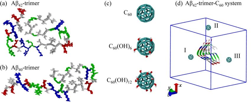
0                                0                       −3.92 ± 0.16      −28.36 ± 0.71
                                0.69                             0.69                           0.16

Aβ42-trimer-C60(OH)6          −24.02 ±                         −29.18 ±        15.27 ±        −3.61 ±
                                            −5.16 ± 0.69                                                   11.66 ± 1.69      −17.52 ± 1.71
                                0.74                             0.25            1.68           0.16

 Int.
Aβ    J. Mol. Sci. 2019, 20, 2048
   42-trimer-C60(OH)12
                               −18.20 ±       −14.60 ±         −32.80 ±        27.06 ±        −3.30 ±                               5 of 14
                                                                                                           23.77 ± 2.53       −9.03 ± 3.09
                                1.02            1.45             1.77            2.52           0.17

2.2. Binding Sites of The Fullerene/Fullerenol Molecule to Aβ42 -Trimer
2.2. Binding Sites of The Fullerene/Fullerenol Molecule to Aβ42-Trimer
      Identifying the binding sites of the C60 /C60 (OH)6 /C60 (OH)12 molecule to Aβ42 -trimer is the first
      Identifying the binding sites of the C60/C60(OH)6/C60(OH)12 molecule to Aβ42-trimer is the first
step to understand the underlying inhibition mechanism. To this aim, we calculated the residue-based
step to understand the underlying inhibition mechanism. To this aim, we calculated the
binding free energy of the nanoparticles to Aβ42 -trimer in Figure 3a–c using last 20 ns data of the
residue-based binding free energy of the nanoparticles to Aβ42-trimer in Figure 3a–c using last 20 ns
simulations. As shown, C60 has the lowest binding energy with aromatic residue F4, and hydrophobic
data of the simulations. As shown, C60 has the lowest binding energy with aromatic residue F4, and
residues V39 and A2, as well as G25 located in the turn region; C60 (OH)6 has the lowest binding
hydrophobic residues V39 and A2, as well as G25 located in the turn region; C60(OH)6 has the lowest
energy with aromatic Y10 and F4, negatively charged E11, and hydrophobic L34, L17 and V39;
binding energy with aromatic Y10 and F4, negatively charged E11, and hydrophobic L34, L17 and
C60 (OH)12 has the lowest binding energy with hydrophobic L34, negatively charged E22 and aromatic
V39; C60(OH)12 has the lowest binding energy with hydrophobic L34, negatively charged E22 and
F4. This indicates the critical roles of aromatic stacking and hydrophobic interactions in the interplay
aromatic F4. This indicates the critical roles of aromatic stacking and hydrophobic interactions in the
between Aβ and all the three nanoparticles. As for fullerenols C60 (OH)6 and C60 (OH)12 , their hydrogen
interplay between Aβ and all the three nanoparticles. As for fullerenols C60(OH)6 and C60(OH)12, their
bonding interaction with negatively charged residues of Aβ is also important.
hydrogen bonding interaction with negatively charged residues of Aβ is also important.

       Figure
       Figure 3.3.Analysis
                    Analysis    of binding
                            of binding          sites
                                         sites of       of the fullerene/fullerenol
                                                  the fullerene/fullerenol     molecule tomolecule        to (a–c)
                                                                                              Aβ42 -trimer.    Aβ42Residue-based
                                                                                                                    -trimer. (a–c)
       Residue-based    binding
       binding free energy.   Thefree   energy.
                                    binding        The was
                                              energy     binding     energyover
                                                              calculated      wasall
                                                                                   calculated
                                                                                     six MD runs overfor
                                                                                                       alleach
                                                                                                           six MD    runs for
                                                                                                                simulated      each
                                                                                                                             system
       simulated    system
       using the last       using
                        20 ns  data the   last 20
                                      of each   MDnstrajectory.
                                                         data of each  (d–f)MD   trajectory.
                                                                             Schematic          (d–f) Schematic
                                                                                            diagrams    for binding  diagrams    for
                                                                                                                        sites of the
       binding    sites  of  the   fullerene/fullerenol        molecule      to  Aβ     -trimer.   The    positions
       fullerene/fullerenol molecule to Aβ42 -trimer. The positions where the fullerene/fullerenol molecule has
                                                                                     42                                where     the
       fullerene/fullerenol
       high binding affinitymolecule
                               are named  haswith
                                               highP1binding
                                                       , P2 , etc.,affinity are named
                                                                    from N-termini      to with  P1, P2,and
                                                                                           C-termini,    etc.,z-axis
                                                                                                               from isN-termini    to
                                                                                                                        the fibrillar
       C-termini,   and z-axisThe
       elongation direction.     is the  fibrillar
                                     color  code iselongation
                                                     consistent withdirection.
                                                                          that inThe  color
                                                                                  Figure    1. code is consistent with that in
       Figure 1.
      According to the residue-based binding free energy, we found that C60 preferentially interacts
with Aβ42 -trimer at three different sites: 2AEF4, 23DVG25 and C-terminal residues 31–41. Through the
binding energy analysis at each site (Table 2), we found that C-terminal residues 31–41 and 2AEF4 have
the lowest binding energy, indicating these two regions are the most favorable binding sites for C60 .
This finding is in agreement with the binding sites (aromatic residues F4 and C-terminal hydrophobic
residues 31–40) identified in DMF interacting with Aβ dimer [34]. The C-terminal hydrophobic region
of residues 31–41 was also reported to be the dominant binding site in DMF interacting with Aβ
fibrillar hexamer [31]. As for C60 (OH)6 , it prefers to bind to Aβ42 -trimer at four sites: 2AEF4, 9GYE11,
17LVF19 and C-terminal residues 31–41, among which C-terminal residues 31–41 and 9GYE11 have
the lowest binding energy. As for C60 (OH)12 , it has three preferential sites: N-terminal residues 4–14,
22ED23 and 34LM35, among which N-terminal residues 4–14 are the most favorable. The hydrophobic
clusters A2-F4-L34-V36, L17-F19-I31 and A30-I32-M35-V40 play critical roles in the structural stability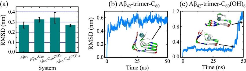
Int. J. Mol. Sci. 2019, 20, 2048                                                                              6 of 14

of Aβ42 fibril [5]. The strong binding of C60 /C60 (OH)6 to these clusters is expected to interfere with the
hydrophobic packing of Aβ side chains, and as a result goes against further fibrillization.

      Table 2. Free energy (in kcal/mol) of different binding sites for the fullerene/fullerenol molecule to
      Aβ42 -trimer.

           System              Aβ42 -trimer-C60        Aβ42 -trimer-C60 (OH)6       Aβ42 -trimer-C60 (OH)12
         Binding site        2–4    23–25   31–41    2–4    9–11    17–19   31–41   4–14    22–23   34–35
           ∆Gbind           −0.80   −0.57   −1.32   −0.45   −0.87   −0.28   −1.23   −0.73   −0.21   −0.38
          Deviation         0.09     0.02    0.01   0.06    0.03     0.04    0.05   0.07     0.04    0.05

      To exhibit the relation between binding dynamics and binding sites clearly, we presented the
schematic diagrams for binding sites of C60 /C60 (OH)6 /C60 (OH)12 to Aβ42 -trimer. The positions where the
fullerene/fullerenol molecule has high binding affinity are named with P1 , P2 , etc., from N-termini to
C-termini. As shown in Figure 3d, there are three positions P1 , P2 and P3 at which C60 prefers to stay
when binding to Aβ42 -trimer, mainly corresponding to the binding sties 2AEF4, 23DVG25 and C-terminal
residues 31–41, respectively. Note that C60 staying at P1 can interact with the region 2AEF4 and the
C-terminal residues 31–41 at the same time. Trajectory tracing shows that C60 binds mostly at P1 and P3
with a respective probability of 22.7% and 29.8%, in agreement with the free energy calculation, and the
location of C60 is relatively fixed. Moreover, the C60 molecule is able to wander on the surface groove
along z-axis of Aβ42 -trimer at P1 position. These preferential positions are near two hydrophobic clusters
A2-F4-L34-V36 and A30-I32-M35-V40, indicating that the binding of C60 to Aβ42 -trimer is dominantly
driven by the hydrophobic interaction. The importance of hydrophobic interaction was reported in other
studies on the binding processes of fullerene and other small molecules to Aβ [29,37,38]. With respect to
P1 and P3 , C60 has a relatively lower binding affinity to P2 . This binding site is facilitated by the groove
of a proper size in the 23DVGS26 region, where the side chains of D23 and S26 are in the outer side of
Aβ42 -trimer and G25 has no side chain. Similar concave-induced binding sites were observed in the
study of fullerenes with Aβ and other proteins [31,39,40].
      As for C60 (OH)6 , it has four preferential binding positions P1 , P2 , P3 and P4 (Figure 3e), mainly
corresponding to the binding sties 2AEF4, 9GYE11, 17LVF19 and C-terminal residues 31–41, respectively.
Interestingly, the C60 (OH)6 molecule is able to slip on the elongation surface (perpendicular to z-axis),
wandering between P1 and P4 or between P3 and P4 with a low probability. The P3 position is adjacent
to another hydrophobic cluster L17-F19-I31 of Aβ42 -trimer. Besides, C60 (OH)6 has high binding affinity
to 9GYE11 (P2 ), facilitated by the hydrogen bonds (H-bonds) formed in between. As for C60 (OH)12 ,
it prefers to bind to three positions P1 , P2 and P3 (Figure 3f), corresponding to the binding sties
N-terminal residues 4–14, 22ED23 and 34LM35, respectively. Different from the binding behaviors of
C60 and C60 (OH)6 , C60 (OH)12 is more likely to stay at the hydrophilic parts of protein surface. It is
able to move between positions P1 and P3 , or slip along the N-terminal β-strand, forming H-bonds
with main chain or side chain of amino acids. The C60 (OH)12 molecule may also contact with 22ED23
region. As the side chains of E22 and D23 are oriented to water solution, C60 (OH)12 is inclined to
form H-bonds with them. Considering the important roles of C-terminal hydrophobic residues in Aβ
aggregation and toxicity [41–43], it is conceivable that the binding of C60 and C60 (OH)6 molecules to
the C-terminal region can prevent Aβ fibrillization. In addition, the C60 (OH)6 molecule has higher
affinity to bind to elongation surfaces than C60 and C60 (OH)12 , which makes C60 (OH)6 a more effective
inhibitor. As previous computational and experimental studies suggested that binding at fibril ends
goes against fibrillar elongation [44–46], this binding would block the backbone amide sites for fibril
growth and as a result, slows down or inhibits the elongation process. It is noted that the bindings of
nanoparticles to protofibril and mature fibril are supposed to be distinct, because the relative area of
the exposed ends compared to the entire fibril surface will be greatly decreased in mature fibrils.
binding of C60 and C60(OH)6 molecules to the C-terminal region can prevent Aβ fibrillization. In
addition, the C60(OH)6 molecule has higher affinity to bind to elongation surfaces than C60 and
C60(OH)12, which makes C60(OH)6 a more effective inhibitor. As previous computational and
experimental studies suggested that binding at fibril ends goes against fibrillar elongation [44–46],
this binding would block the backbone amide sites for fibril growth and as a result, slows down7or
Int. J. Mol. Sci. 2019, 20, 2048 of 14
inhibits the elongation process. It is noted that the bindings of nanoparticles to protofibril and
mature fibril are supposed to be distinct, because the relative area of the exposed ends compared to
2.3.the entire fibril
Structural surfaceofwill
Influence Thebe greatly decreasedMolecule
Fullerene/Fullerenol in matureonfibrils.
Aβ42 -Trimer
2.3.InStructural
order to detect
Influence theofinfluence of fullerene/fullerenol
The Fullerene/Fullerenol Moleculebinding on the Aβ42 -trimer structure, we first
on Aβ42-Trimer
examined the secondary structural difference relative to the isolated Aβ42 -trimer. The β-sheet contents
of Aβ42In order Aβ
-trimer, to detect the influence of fullerene/fullerenol binding on the Aβ42-trimer structure, we
42 -trimer-C60 , Aβ42 -trimer-C60 (OH)6 and Aβ42 -trimer-C60 (OH)12 systems are 80.5%,
first examined the secondary structural difference relative to the isolated Aβ42-trimer. The β-sheet
83.3%, 81.1% and 80.4%, respectively, showing little difference. Then, we calculated the average
contents of Aβ42-trimer, Aβ42-trimer-C60, Aβ42-trimer-C60(OH)6 and Aβ42-trimer-C60(OH)12 systems are
Cα-root-mean-square deviation (Cα-RMSD) with respect to the initial coordinates of Aβ42 protofibrillar
80.5%, 83.3%, 81.1% and 80.4%, respectively, showing little difference. Then, we calculated the
trimer using the last 20 ns data of each MD trajectory. As shown in Figure 4a, the values of Cα-RMSD in
average Cα-root-mean-square deviation (Cα-RMSD) with respect to the initial coordinates of Aβ42
the absence and presence of the C60 (OH)12 molecule are 0.27 ± 0.03 nm and 0.27 ± 0.01 nm, showing
protofibrillar trimer using the last 20 ns data of each MD trajectory. As shown in Figure 4a, the
no statistically significant difference. In the presence of C60 /C60 (OH)6 , Aβ42 -trimer has an increased
values of Cα-RMSD in the absence and presence of the C60(OH)12 molecule are 0.27 ± 0.03 nm and
Cα-RMSD
0.27 ± 0.01 of 0.32 0.02 / 0.34
nm, ±showing no ±statistically
0.05 nm, while the values
significant are still
difference. Inwithin the errorofofCestimate
the presence with
60/C60(OH)6,
respect to that of isolated Aβ. These indicate
Aβ42-trimer has an increased Cα-RMSD of 0.32 ± 0.02 that the C /C
60 / 60 (OH)
0.34 ± 0.05 /C (OH)
6 60nm, while 12 molecule has a negligible
the values are still
influence
within on thethe structural
error stabilitywith
of estimate of Aβrespect
42 -trimer.to that of isolated Aβ. These indicate that the
C60Figure
/C60(OH) 4b,c
6/C60display the time has
(OH)12 molecule evolution of Cα-RMSD
a negligible influence of onthetheMD trajectory
structural contributing
stability most to
of Aβ42-trimer.
the total Cα-RMSD in
Figure 4b,c display the Aβ -trimer-C
42 time evolution
60 and Aβ -trimer-C
of Cα-RMSD
42 (OH)
of60the MD systems, respectively.
6 trajectory contributing most to With C60 ,
thethe
Cα-RMSD
total Cα-RMSDvalue of inAβAβ42 -trimer keeps
42-trimer-C 60 andrising in the first
Aβ42-trimer-C 20 ns
60(OH) and finally
6 systems, fluctuatesWith
respectively. at around 0.55
C60, the
nm. During this
Cα-RMSD value process,
of Aβ42the C60 molecule
-trimer keeps rising is observed
in the first to20
contact
ns and abundantly with side
finally fluctuates chains of
at around V39
0.55
andnm.I41,During
and leadthis toprocess,
twisted theC-termini.
C60 molecule is observed
In the to contact
Aβ42 -trimer-C 60 abundantly
(OH) 6 system, with
the side chains
Cα-RMSD of V39
value of
Aβand I41, and
42 -trimer keepsleadat to~0.30
twisted until t = 40.8
nmC-termini. In thens.Aβ 42-trimer-C
After that, it60(OH) system, and
rises 6sharply the Cα-RMSD
increases to >1.0ofnm.
value
Aβ42Cα-RMSD
When -trimer keeps at ~0.30
begins nm until
its quick t =the
rise, 40.8C60ns.(OH)
After6 that, it
molecule rises
is sharply
observed andto increases
bind at to
the >1.0 nm.
C-terminal
When Cα-RMSD begins its quick rise, the C (OH) molecule is observed
residues 31–41, and the hydrophobic cluster A2-F4-L34-V36 starts to collapse. Then, the sidechains of
60 6 to bind at the C-terminal
A2residues 31–41, andwith
and F4 dissociate the hydrophobic
those of L34cluster
and V36 A2-F4-L34-V36
one by one, and starts to collapse.
finally Then, theand
the N-termini sidechains
C-termini
of A2 and F4 dissociate with those of L34 and V36 one by one,
get separated far away. Note that it is the only MD trajectory among all the simulations we performed and finally the N-termini and
in C-termini
this studyget that separated far away. Note
N- and C-termini that it is is
dissociation theobserved.
only MD trajectory
It needs furtheramongstudying
all the simulations
to connect wethis
performed in this study that N- and C-termini dissociation is observed. It needs further studying to
dissociation with Aβ-C60 (OH)6 interaction explicitly.
connect this dissociation with Aβ-C60(OH)6 interaction explicitly.
Figure
4. 4. Influenceofoffullerene/fullerenol
Influence fullerene/fullerenol molecules
molecules on
on the
the Aβ
Aβ42-trimer structure. (a) The average
Figure 42 -trimer structure. (a) The average
Cα-root-mean-square deviation (Cα-RMSD) relative to the initial coordinatesofofAβ
Cα-root-mean-square deviation (Cα-RMSD) relative to the initial coordinates Aβ42 protofibrillar
42 protofibrillar
trimer. The values for Aβ-fullerene/fullerenol systems were calculated over all six MD runs for each
trimer. The values for Aβ-fullerene/fullerenol systems were calculated over all six MD runs for each
simulated system using the last 20 ns data of each MD trajectory, and those for isolated Aβ42-trimer
simulated system using the last 20 ns data of each MD trajectory, and those for isolated Aβ42 -trimer
systems were averaged over the last 50 ns data of two independent 200-ns MD runs. (b,c) Time
systems were averaged over the last 50 ns data of two independent 200-ns MD runs. (b,c) Time evolution
evolution of Cα-RMSD of the MD trajectory that contributes most to the total Cα-RMSD in
of Cα-RMSD of the MD trajectory that contributes most to the total Cα-RMSD in Aβ42 -trimer-C60 and
Aβ42 -trimer-C60 (OH)6 systems, respectively. The color code of the inset snapshots is consistent with
that in Figure 1.
The detailed interactions between Aβ42 -trimer and the fullerene/fullerenol molecule were also
investigated. As a previous study suggested that the salt bridges between H6, E11 and H13 stabilize the
kink in the N-terminal part of the β-sheets around Y10 [5], we examined the interplay of H6-E11-H13
and found that the interaction pairs stably stay together in all simulated systems except for one MD
trajectory of Aβ42 -trimer-C60 (OH)6 system. This trajectory corresponds to the MD run shown in
Figure 4c, and its snapshot of the final state is presented in Figure 5a. Even if the N- and C-termini areInt. J. Mol. Sci. 2019, 20, 2048 8 of 14
dissociated, the interaction pairs of H6, E11 and H13 mostly stay together, and the interactions of side
chains are weakened by excluding those of H6-3 (H6 in Chain 3) and E13-1 (E13 in Chain 1).
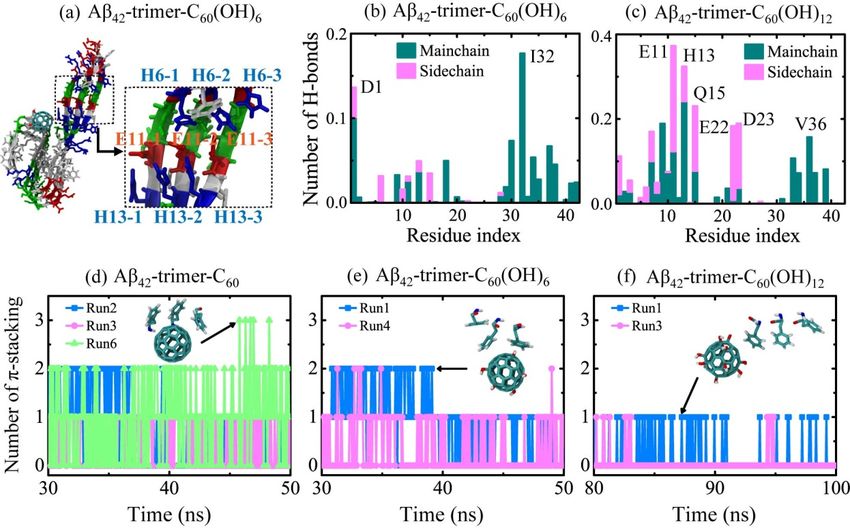
In Figure 5b,c, we calculated the number of H-bonds formed between individual residue and
fullerenols. It shows that C60 (OH)6 favors H-bonding with main chains of Aβ42 -trimer, and forms H-bonds
mostly with residues I32 and D1. The C60 (OH)12 molecule forms almost the same amount of H-bonds
with main chains and side chains, and it preferentially forms H-bonds with residues E11, H13, Q15, D23,
E22 and V36. Previous Thioflavin T (ThT) fluorescence and atomic force microscopy experiments showed
that fullerenol C60 (OH)16 can prevent Aβ40 fbrillization [30]. The recent study using ThT assay and
transmission electron microscope demonstrated that fullerenemalonate can inhibit Aβ42 aggregation [47].
Their computational results showed that the inhibition is attributed to the hydrogen bonding of the
fullerenemalonate carboxylate groups with Aβ. Here, the formation of H-bonds between main chains
and fullerenols is supposed to block the backbone amide sites for further addition of peptides in β-sheet
structure, which goes against the oligomerization or fibrillization of Aβ. The higher affinity of C60 (OH)6
bonding with main chains of Aβ peptides makes C60 (OH)6 a more efficient inhibitor than C60 (OH)12 .
Int. J. Mol. Sci. 2019, 20, x FOR PEER REVIEW 9 of 15
Figure
Figure 5.
5. Details
Detailsof of
interactions between
interactions betweenAβAβ42-trimer and the fullerene/fullerenol molecule. (a)
42 -trimer and the fullerene/fullerenol molecule.
Disturbance to the interplay of H6-E11-H13 observed
(a) Disturbance to the interplay of H6-E11-H13 observed in a trajectory of Aβ42of
in a trajectory -trimer-C 60(OH)6 system.
Aβ42 -trimer-C 60 (OH)6
(b,c) Number of H-bonds formed between Aβ 42 -trimer and fullerenols.
system. (b,c) Number of H-bonds formed between Aβ42 -trimer and fullerenols. (d–f) (d–f) Number of Number
π-stacking
of
structures
π-stackingbetween
structures 42-trimer Aβ
Aβbetween and fullerene/fullerenol. The geometrical criterions of H-bonding
42 -trimer and fullerene/fullerenol. The geometrical criterions of
and π-stacking
H-bonding andformations
π-stacking are defined are
formations in Model
defined and
in Methods
Model and section.
Methods section.
2.4. Dynamics, Sites and
The π-stacking Interactions
interaction is of The Fullerene/Fullerenol
important MoleculeofBinding
in the self-assembly amyloidto Aβ 40-Trimer
fibrils, with parallel,
T-shaped and herringbone (~50◦ ) orientations suggested for aromatic rings in proteins [48]. The binding
We also carried out multiple MD simulations to examine the binding dynamics, binding sites
energy analysis reveals the important role of F4 in the interaction between Aβ42 -trimer and
and interactions of the C60/C60(OH)6/C60(OH)12 molecule with Aβ40-trimer. Although the structure of
the fullerene/fullerenol molecule. To examine the aromatic stacking interaction between F4 and
Aβ40-trimer is different from that of Aβ42-trimer (see Figure 1), the binding behavior of nanoparticles
C /C60 (OH)6 /C60 (OH)12 , we calculated the number of π-stacking structures between Aβ42-trimer and
to60Aβ40-trimer was found to display a remarkable resemblance with that to Aβ42-trimer. As shown in
fullerene/fullerenol during the last 20 ns in Figure 5d–f. For the Aβ42 -trimer-C60 system, π-stacking
Figure 6, with the hydroxylation extent increased, the C60 molecule displays slower binding
structures were observed in three MD trajectories. Run 6 had the largest number of π-stacking structures,
dynamics, corresponding to weakened binding strength. The binding free energy analysis shows
and the maximum number was three. This means that the C60 molecule is able to have π-stacking
that the favorable residues of Aβ40-trimer with which the nanoparticle tend to interact are a little
interaction with all the aromatic rings of F4 in Aβ42 -trimer at the same time. The inset snapshot displays
different from those of Aβ42-trimer. Still, these residues are mostly hydrophobic or aromatic,
the corresponding structure, and the aromatic rings of F4 are oriented in parallel or herringbone
indicating the critical roles of hydrophobic and aromatic interactions in Aβ-nanoparticle
interactions. Moreover, the preferential binding regions of the nanoparticles interplaying with
Aβ40-trimer resemble with those of the nanoparticles binding to Aβ42-trimer. We also examined the
stability of the D23-K28 salt bridge, which is important for the structural stability of Aβ40 [4]. The salt
bridge would be interfered by the nanoparticle binding, whereas the connection between the salt
bridge disruption and the hydroxylation extent of C60 is not explicit.Int. J. Mol. Sci. 2019, 20, 2048 9 of 14
alignment relative to the C60 surface. As for C60 (OH)6 , it forms less π-stacking structures with
Aβ42 -trimer, and the maximum number of π-stacking decreases to two. For the Aβ42 -trimer-C60 (OH)12
system, π-stacking structures are observed in two trajectories and the total number of π-stacking
structures is the least. Only one aromatic ring of F4 can have π-stacking interaction with the C60 (OH)12
molecule at one moment, and the ring is mostly oriented in herringbone alignment relative to the
carbon surface of C60 (OH)12 . These results indicate that the more hydroxylated C60 is, the fewer and
weaker π-stacking interactions with Aβ42 -trimer the nanoparticle has.
2.4. Dynamics, Sites and Interactions of The Fullerene/Fullerenol Molecule Binding to Aβ40 -Trimer
We also carried out multiple MD simulations to examine the binding dynamics, binding sites
and interactions of the C60 /C60 (OH)6 /C60 (OH)12 molecule with Aβ40 -trimer. Although the structure of
Aβ40 -trimer is different from that of Aβ42 -trimer (see Figure 1), the binding behavior of nanoparticles
to Aβ40 -trimer was found to display a remarkable resemblance with that to Aβ42 -trimer. As shown in
Figure 6, with the hydroxylation extent increased, the C60 molecule displays slower binding dynamics,
corresponding to weakened binding strength. The binding free energy analysis shows that the favorable
residues of Aβ40 -trimer with which the nanoparticle tend to interact are a little different from those of
Aβ42 -trimer. Still, these residues are mostly hydrophobic or aromatic, indicating the critical roles of
hydrophobic and aromatic interactions in Aβ-nanoparticle interactions. Moreover, the preferential binding
regions of the nanoparticles interplaying with Aβ40 -trimer resemble with those of the nanoparticles
binding to Aβ42 -trimer. We also examined the stability of the D23-K28 salt bridge, which is important for
the structural stability of Aβ40 [4]. The salt bridge would be interfered by the nanoparticle binding, whereas
the connection between the salt bridge disruption and the hydroxylation extent of C60 is not explicit.
Int. J. Mol. Sci. 2019, 20, x FOR PEER REVIEW 10 of 15
Figure 6. 6.(a–c)
(a–c)
Time Time evolution
evolution of the distance
of the minimum minimumbetweendistance between
Aβ40 -trimer Aβ40-trimer and
and fullerene/fullerenol.
fullerene/fullerenol.
Six independent MDSix independent
runs are denotedMD runs are denoted
in different in different
colors. (d–f) colors. (d–f)
Residue-based Residue-based
binding free energy.
binding
The freeenergy
binding energy.was
The binding over
calculated energy wasMD
all six calculated
runs for over all six MDsystem
each simulated runs for each
using thesimulated
last 20 ns
system usingMD
data of each the trajectory.
last 20 ns data of each MD trajectory.
3. Materials
3. Materials and
and Methods
Methods
3.1. Aβ40/42 Protofibrillar Trimer and C60 /C60 (OH)6 /C60 (OH)12 Molecules
3.1. Aβ40/42 Protofibrillar Trimer and C60/C60(OH)6 /C60(OH)12 Molecules
The Aβ peptide (39–43-amino acid) is derived from the amyloid precursor protein (APP)
The Aβ peptide (39–43-amino acid) is derived from the amyloid precursor protein (APP)
through proteolytic cleavage by β- and γ-secretase, and the most abundant Aβ are Aβ (sequence:
through proteolytic cleavage by β- and γ-secretase, and the most abundant Aβ are Aβ42 42 (sequence:
DAEFRHDSGY EVHHQKLVFF AEDVGSNKGA IIGLMVGGVV IA) and Aβ40. The initial
10 20 30 40
coordinate of the Aβ42 protofibrillar trimer was taken from the Aβ42 fibril structure [5] (PDB ID:
5OQV) determined by cryo–electron microscopy (cryo-EM). The coordinate of the Aβ40 protofibrillar
trimer was taken from the Aβ40 fibril structure [4] (PDB ID: 2M4J) obtained from solid-state nuclearInt. J. Mol. Sci. 2019, 20, 2048 10 of 14
DAEFRHDSGY10 EVHHQKLVFF20 AEDVGSNKGA30 IIGLMVGGVV40 IA) and Aβ40 . The initial
coordinate of the Aβ42 protofibrillar trimer was taken from the Aβ42 fibril structure [5] (PDB ID: 5OQV)
determined by cryo–electron microscopy (cryo-EM). The coordinate of the Aβ40 protofibrillar trimer
was taken from the Aβ40 fibril structure [4] (PDB ID: 2M4J) obtained from solid-state nuclear magnetic
resonance (NMR) spectroscopic data. The protonation of the peptide was adjusted to the neutral pH.
The N- and C-termini were respectively capped by NH3 + and COO− in accordance with experiments.
The structure of C60 /C60 (OH)6 /C60 (OH)12 molecules used in this study is displayed in Figure 1.
The force field parameters were taken from a previous MD study on the interaction of Aβ
and hydroxylated carbon nanotube [49]. To simplify the modeling, the hydroxyl groups in
C60 (OH)6 /C60 (OH)12 molecules are distributed uniformly on the C60 surface.
The Aβ42 -trimer-C60 simulation system consists of an Aβ42 protofibrillar trimer and a C60 molecule
placed 2.0 nm (minimum distance) away from Aβ, as shown in Figure 1. To remove the bias of the
initial position of C60 on the binding site, the C60 molecule was initially placed at three different
locations (I, II, III). The other Aβ-fullerene/fullerenol systems were constructed similarly, and were
immersed in SPC [50] water. Counterions Na+ and Cl- were added to neutralize the system and
provide an additional 0.1 M salt concentration. Systems of isolated Aβ42 -trimer and Aβ40 -trimer in
water were run as control groups.
3.2. Details of MD Simulations
Atomistic MD simulations were performed in isothermal−isobaric (NPT) ensemble using
GROMACS-4.5.3 software package [51] with GROMOS96 53a6 force field [52], in accordance with
previous computational studies of Aβ peptides [31,33,34,49,53,54]. Periodic boundary conditions were
applied in all three directions. The temperature and pressure of the systems were coupled using the
Nose−Hoover algorithm [55,56] (310 K, τT = 0.2 ps) and Parinello–Rahman algorithm [57,58] (1 bar,
τP = 1.0 ps), respectively. The simulation time step was 2 fs with all bonds constrained using the
LINCS algorithm [59]. The electrostatic interactions were treated with the particle mesh Ewald (PME)
method [60] with a cutoff of 1.0 nm, and the van der Waals interactions were calculated using a cutoff of
1.4 nm. For Aβ40/42 -trimer-C60 and Aβ40/42 -trimer-C60 (OH)6 systems, six independent copies of each
system were carried out, each lasting 50 ns; for Aβ40/42 -trimer-C60 (OH)12 systems, six independent
100-ns MD runs were carried out; for isolated Aβ40/42 -trimer systems, two independent 200-ns MD
runs were carried out.
3.3. Analysis Methods
Trajectory analysis was performed using the GROMACS-4.5.3 package toolkits and in-house developed
codes. The secondary structure was calculated using the DSSP program [61]. Here, an atomic contact is
defined when two non-hydrogen atoms come within 0.54 nm. The H-bond is determined using geometrical
criteria: the distance between donor D and acceptor A is less than 0.35 nm and the D-H-A angle is larger than
150◦ . The π-stacking structure is defined when the centroid of residue aromatic ring is within 0.45 nm from
the spherical carbon surface of fullerene/fullerenol [62]. The binding energy between a ligand and a receptor
was estimated by means of (MM/PBSA) [63,64]: ∆Gbind = ∆EMM + ∆Gsolv − T∆S, ∆EMM = ∆EvdW + ∆Eelec ,
∆Gsolv = ∆Gpolar + ∆Gnonpolar , ∆Gnonpolar = γ· SASA + b. Here, EMM is the gas-phase energy, consisting of
electrostatic (∆Eelec ) and van der Waals (∆Evdw ) terms; ∆Gsolv is the sum of polar solvation energy ∆Gpolar
and nonpolar solvation component ∆Gnonpolar ; ∆Gpolar is estimated by solving the Poisson−Boltzmann
equation; ∆Gnonpolar is estimated by solvent accessible surface area (SASA). A water probe radius of
0.14 nm was used to calculate SASA, and γ (surface tension of the solvent) and b (fitting parameter)
were set to 0.542 kcal/mol/nm2 and 0.92 kcal/mol, respectively. As the binding free energy (∆Gbind )
reported here is the relative binding free energy, the contribution of conformational entropy of peptides
was ignored in accordance with a number of previous computational studies [33,34,65,66].Int. J. Mol. Sci. 2019, 20, 2048 11 of 14
4. Conclusions
We investigated the dynamics, sites and interactions of the C60 /C60 (OH)6 /C60 (OH)12 nanoparticle
binding to Aβ42/40 protofibrillar trimer by performing extensive atomistic MD simulations. To our
knowledge, this is the first atomistic explicit-solvent simulation study to investigate the binding behavior
of fullerenols to Aβ42/40 protofibril. Our simulations demonstrate that the higher hydroxylation
level of C60 leads to slower binding dynamics and weaker binding strength. When binding to
Aβ42 -trimer, C60 preferentially interacts with C-terminal residues 31–41 and 2AEF4; C60 (OH)6 prefers
to bind to C-terminal residues 31–41 and 9GYE11; C60 (OH)12 favors to bind to N-terminal residues
4–14. In addition, the C60 (OH)6 molecule has higher affinity to bind to elongation surfaces than
C60 and C60 (OH)12 . The binding of these nanoparticles has a slight influence on the secondary
structure and structural stability of Aβ42 -trimer during the simulation time. The hydrophobic
interaction plays a critical role in the interplay between Aβ42 and all three nanoparticles; π-stacking
interaction gets weakened as C60 carries more hydroxyls. The situations are quite similar when the
C60 /C60 (OH)6 /C60 (OH)12 nanoparticle binds to Aβ40 protofibrillar trimer. Overall, the proper binding
strength and high affinity to form hydrogen bonds with protein backbones make the water-soluble
C60 (OH)6 molecule an efficient inhibitor. This study provides a detailed picture of fullerene/fullerenols
binding to Aβ protofibril and expands the understanding of the underlying inhibitory mechanism,
which is helpful to the design of novel agents with anti-amyloid properties.
Author Contributions: Conceptualization, P.C. and Z.Q.; methodology, Y.Z. and Z.Q.; software, Y.Z. and Q.Z.;
validation, P.C. and Y.L.; formal analysis, Z.L. and Y.Z.; investigation, Q.Z. and Y.L.; resources, Q.Z.; data curation,
Z.L. and Y.Z.; writing—original draft preparation, Z.L.; writing—review and editing, Z.Q.
Funding: This research was funded by the National Natural Science Foundation of China, grant number 11704256.
Conflicts of Interest: The authors declare no conflict of interest.
References
1. Jucker, M.; Walker, L.C. Self-propagation of pathogenic protein aggregates in neurodegenerative diseases.
Nature 2013, 501, 45–51. [CrossRef]
2. Knowles, T.P.J.; Vendruscolo, M.; Dobson, C.M. The amyloid state and its association with protein
misfolding diseases. Nat. Rev. Mol. Cell Biol. 2014, 15, 384–396. [CrossRef]
3. Riek, R.; Eisenberg, D.S. The activities of amyloids from a structural perspective. Nature 2016, 539, 227–235.
[CrossRef]
4. Lu, J.X.; Qiang, W.; Yau, W.M.; Schwieters, C.D.; Meredith, S.C.; Tycko, R. Molecular structure of beta-amyloid
fibrils in Alzheimer’s disease brain tissue. Cell 2013, 154, 1257–1268. [CrossRef] [PubMed]
5. Gremer, L.; Schölzel, D.; Schenk, C.; Reinartz, E.; Labahn, J.; Ravelli, R.B.G.; Tusche, M.; Lopez-Iglesias, C.;
Hoyer, W.; Heise, H.; et al. Fibril structure of amyloid-β(1–42) by cryo–electron microscopy. Science 2017,
358, 116–119. [CrossRef]
6. Sun, Y.; Wang, B.; Ge, X.; Ding, F. Distinct oligomerization and fibrillization dynamics of amyloid core sequences
of amyloid-beta and islet amyloid polypeptide. Phys. Chem. Chem. Phys. 2017, 19, 28414–28423. [CrossRef]
7. Michaels, T.C.T.; Šarić, A.; Habchi, J.; Chia, S.; Meisl, G.; Vendruscolo, M.; Dobson, C.M.; Knowles, T.P.J.
Chemical kinetics for bridging molecular mechanisms and macroscopic measurements of amyloid fibril
formation. Annu. Rev. Phys. Chem. 2018, 69, 273–298. [CrossRef] [PubMed]
8. Kayed, R.; Head, E.; Thompson, J.L.; McIntire, T.M.; Milton, S.C.; Cotman, C.W.; Glabe, C.G. Common structure of
soluble amyloid oligomers implies common mechanism of pathogenesis. Science 2003, 300, 486–489. [CrossRef]
9. Haass, C.; Selkoe, D.J. Soluble protein oligomers in neurodegeneration: Lessons from the Alzheimer’s
amyloid β-peptide. Nat. Rev. Mol. Cell Biol. 2007, 8, 101–112. [CrossRef] [PubMed]
10. Straub, J.E.; Thirumalai, D. Toward a molecular theory of early and late events in monomer to amyloid
fibril formation. Annu. Rev. Phys. Chem. 2011, 62, 437–463. [CrossRef]
11. Nguyen, P.; Derreumaux, P. Understanding amyloid fibril nucleation and Aβ oligomer/drug interactions
from computer simulations. Acc. Chem. Res. 2014, 47, 603–611. [CrossRef] [PubMed]Int. J. Mol. Sci. 2019, 20, 2048 12 of 14
12. Zhang, M.; Mao, X.; Yu, Y.; Wang, C.-X.; Yang, Y.-L.; Wang, C. Nanomaterials for reducing amyloid cytotoxicity.
Adv. Mater. 2013, 25, 3780–3801. [CrossRef]
13. Radic, S.; Davis, T.P.; Ke, P.C.; Ding, F. Contrasting effects of nanoparticle-protein attraction on amyloid aggregation.
RSC Adv. 2015, 5, 105498. [CrossRef]
14. Wang, B.; Pilkington, E.H.; Sun, Y.; Davis, T.P.; Ke, P.C.; Ding, F. Modulating protein amyloid aggregation
with nanomaterials. Environ. Sci. Nano 2017, 4, 1772–1783. [CrossRef]
15. Young, L.M.; Saunders, J.C.; Mahood, R.A.; Revill, C.H.; Foster, R.J.; Tu, L.-H.; Raleigh, D.P.; Radford, S.E.;
Ashcroft, A.E. Screening and classifying small-molecule inhibitors of amyloid formation using ion mobility
spectrometry–mass spectrometry. Nat. Chem. 2014, 7, 73–81. [CrossRef]
16. Saunders, J.C.; Young, L.M.; Mahood, R.A.; Jackson, M.P.; Revill, C.H.; Foster, R.J.; Smith, D.A.; Ashcroft, A.E.;
Brockwell, D.J.; Radford, S.E. An in vivo platform for identifying inhibitors of protein aggregation.
Nat. Chem. Biol. 2016, 12, 94–101. [CrossRef]
17. Chen, Z.J.; Krause, G.; Reif, B. Structure and orientation of peptide inhibitors bound to β-amyloid fibrils.
J. Mol. Biol. 2005, 354, 760–776. [CrossRef] [PubMed]
18. Takahashi, T.; Mihara, H. Peptide and protein mimetics inhibiting amyloid β-peptide aggregation.
Acc. Chem. Res. 2008, 41, 1309–1318. [CrossRef] [PubMed]
19. Sevigny, J.; Chiao, P.; Bussière, T.; Weinreb, P.H.; Williams, L.; Maier, M.; Dunstan, R.; Salloway, S.; Chen, T.; Ling, Y.;
et al. The antibody aducanumab reduces Aβ plaques in Alzheimer’s disease. Nature 2016, 537, 50–56. [CrossRef]
20. Liao, Q.; Owen, M.C.; Bali, S.; Barz, B.; Strodel, B. Aβ under stress: The effects of acidosis, Cu2+-binding,
and oxidation on amyloid β-peptide dimers. Chem. Commun. 2018, 54, 7766–7769. [CrossRef]
21. Xiao, L.; Aoshima, H.; Saitoh, Y.; Miwa, N. Highly hydroxylated fullerene localizes at the cytoskeleton and
inhibits oxidative stress in adipocytes and a subcutaneous adipose-tissue equivalent. Free Radical Biol. Med. 2011,
51, 1376–1389. [CrossRef]
22. Grebowski, J.; Kazmierska, P.; Krokosz, A. Fullerenols as a new therapeutic approach in nanomedicine.
BioMed Res. Int. 2013, 2013, 9. [CrossRef]
23. Bosi, S.; Da Ros, T.; Spalluto, G.; Prato, M. Fullerene derivatives: An attractive tool for biological applications.
Eur. J. Med. Chem. 2003, 38, 913–923. [CrossRef] [PubMed]
24. Dugan, L.L.; Turetsky, D.M.; Du, C.; Lobner, D.; Wheeler, M.; Almli, C.R.; Shen, C.K.F.; Luh, T.Y.; Choi, D.W.;
Lin, T.S. Carboxyfullerenes as neuroprotective agents. Proc. Natl. Acad. Sci. USA 1997, 94, 9434–9439.
[CrossRef] [PubMed]
25. Kim, J.E.; Lee, M. Fullerene inhibits β-amyloid peptide aggregation. Biochem. Biophys. Res. Commun. 2003,
303, 576–579. [CrossRef]
26. Podolski, I.Y.; Podlubnaya, Z.A.; Kosenko, E.A.; Mugantseva, E.A.; Makarova, E.G.; Marsagishvili, L.G.;
Shpagina, M.D.; Kaminsky, Y.G.; Andrievsky, G.V.; Klochkov, V.K. Effects of hydrated forms of C60 fullerene
on amyloid 1-peptide fibrillization in vitro and performance of the cognitive task. J. Nanosci. Nanotechnol. 2007,
7, 1479–1485. [CrossRef]
27. Ye, S.; Zhou, T.; Pan, D.; Lai, Y.; Yang, P.; Chen, M.; Wang, Y.; Hou, Z.; Ren, L.; Jiang, Y. Fullerene C60 derivatives
attenuated microglia-mediated prion peptide neurotoxicity. J. Biomed. Nanotechnol. 2016, 12, 1820–1833. [CrossRef]
[PubMed]
28. Hsieh, F.-Y.; Zhilenkov, A.V.; Voronov, I.I.; Khakina, E.A.; Mischenko, D.V.; Troshin, P.A.; Hsu, S.-h.
Water-soluble fullerene derivatives as brain medicine: Surface chemistry determines if they are
neuroprotective and antitumor. ACS Appl. Mat. Interfaces 2017, 9, 11482–11492. [CrossRef]
29. Huy, P.D.Q.; Li, M.S. Binding of fullerenes to amyloid beta fibrils: Size matters. Phys. Chem. Chem. Phys. 2014,
16, 20030–20040. [CrossRef]
30. Bednarikova, Z.; Huy, P.D.Q.; Mocanu, M.-M.; Fedunova, D.; Li, M.S.; Gazova, Z. Fullerenol C60(OH)16 prevents
amyloid fibrillization of Aβ40 – in vitro and in silico approach. Phys. Chem. Chem. Phys. 2016, 18, 18855–18867.
[CrossRef]
31. Zhou, X.Y.; Xi, W.H.; Luo, Y.; Cao, S.Q.; Wei, G.H. Interactions of a water-soluble fullerene derivative with
amyloid-beta protofibrils: Dynamics, binding mechanism, and the resulting salt-bridge disruption. J. Phys.
Chem. B 2014, 118, 6733–6741. [CrossRef] [PubMed]
32. Radic, S.; Nedumpully-Govindan, P.; Chen, R.; Salonen, E.; Brown, J.M.; Ke, P.C.; Ding, F. Effect of fullerenol
surface chemistry on nanoparticle binding-induced protein misfolding. Nanoscale 2014, 6, 8340–8349. [CrossRef]
[PubMed]You can also read