Parametrizing the Spatial Dependence of 1H NMR Chemical Shifts in π-Stacked Molecular Fragments - MDPI
←
→
Page content transcription
If your browser does not render page correctly, please read the page content below
International Journal of
            Molecular Sciences

Article
Parametrizing the Spatial Dependence of 1H NMR
Chemical Shifts in π-Stacked Molecular Fragments
Jiří Czernek * and Jiří Brus
 Institute of Macromolecular Chemistry, Czech Academy of Sciences, Heyrovsky Square #2,
 16206 Prague, Czech Republic; brus@imc.cas.cz
 * Correspondence: czernek@imc.cas.cz; Tel.: +420-296-809-290

 Received: 14 October 2020; Accepted: 22 October 2020; Published: 24 October 2020

 Abstract: Most recently a renewed interest in several areas has arisen in factors governing the 1 H
 NMR chemical shift (1 H CS) of protons in aromatic systems. Therefore, it is important to describe how
 1 H CS values are affected by π-stacking intermolecular interactions. The parametrization of radial and

 angular dependences of the 1 H CS is proposed, which is based on conventional gauge-independent
 atomic orbital (GIAO) calculations of explicit molecular fragments. Such a parametrization is
 exemplified for a benzene dimer with intermonomer vertical and horizontal distances which are in
 the range of values often found in crystals of organic compounds. Results obtained by the GIAO
 calculations combined with B3LYP and MP2 methods were compared, and revealed qualitatively the
 same trends in the 1 H CS data. The parametrization was found to be quantitatively correct for the
 T-shaped benzene dimers, and its limitations were discussed. Parametrized 1 H CS surfaces should
 become useful for providing additional restraints in the search of site-specific information through an
 analysis of structurally induced 1 H CS changes.

 Keywords: noncovalent interactions; proton NMR; intermolecular stacking; GIAO; GIPAW

1. Introduction
     The proton nuclear magnetic resonance (1 H NMR) is a crucial technique for solving a vast variety
of chemical, physical, and biological problems in solution [1] and solid phases [2]. Some of those
problems concern the dispersive interactions [3] between aromatic moieties and the role they play
in various phenomena (see references [4–7] for the most recent examples). In such studies, the key
parameter is the 1 H chemical shift (1 H CS) perturbation caused by the presence of aromatic fragments
in the vicinity of an investigated proton [8,9]. It should be noted that the 1 H CS of protons in aromatic
molecules has received considerable interest, as most recently summarized in references [10,11].
Therefore, it is desirable to study factors governing the 1 H CS values in molecules which exhibit
C–H/π aromatic stacking interactions. Here, conventional gauge-independent atomic orbital (GIAO)
calculations (see Materials and Methods) are carried out for the proton sites in two types of stacked
molecular dimers. The first type are benzene dimers which serve for the purpose of a parametrization
of the 1 H CS landscape, as detailed in Section 2.1, and for checking the precision of the proposed
parametrization. The second type of investigated dimers are those taken from crystals of midsized
organic molecules [12,13] (see Section 2.2). For these systems, accurate 1 H CS data are available from the
solid-state NMR (SSNMR) measurements [12,14], and hence they are used in reliability considerations.
In particular, the GIAO 1 H CS estimates for the dimers are confronted with experiment and with results
of the gauge-including projector augmented wave (GIPAW) calculations performed in a plane-wave
density functional theory (PW DFT) scheme for periodic structures of aforementioned crystals [15].
In this way, it has been shown that it should be possible to successfully apply the parametrization
process to other ligands and to configurations relevant for binding of those ligands. At the same

Int. J. Mol. Sci. 2020, 21, 7908; doi:10.3390/ijms21217908                          www.mdpi.com/journal/ijms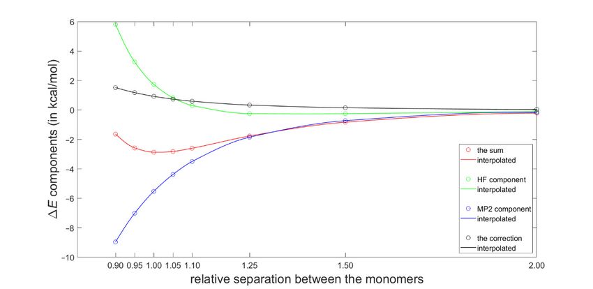
Int. J. Mol. Sci. 2020, 21, 7908                                                                             2 of 13

time, an inherent limitation of this approach should be noted, which is the presence of a generally
shaped contour line for a given 1 H CS value, as discussed in Section 3 and visualized. However, the
parametrized 1 H CS surfaces are expected to be applied together with other structural information
provided by NMR and possibly diffraction measurements, or by advanced computational methods [16],
which would constrain stacking fragments in the correct spatial arrangement compatible with some
specific 1 H CS value. Importantly, a set of 1 H CS values could, then, be used to elucidate structures of
complex systems, for instance, polydopamine [17–20]. It should also be noted that the 1 H CS surfaces
can be obtained with an inclusion of solvation at the interacting sites [21] in order to properly describe
binding between organic ligands and models of large biologically active molecules in solution.

2. Results

2.1. Development of the Proton Chemical Shift (1 H CS) Surface
     The energy parameters of clusters of benzene in various media have been intensely studied [22,23].
The potential energy surface (PES) of the gas-phase benzene dimer is quite well understood [24].
Here, for the purpose of describing the landscape of the 1 H CS values, the T-shaped dimer in “C2v
over atom” configuration was adopted from the work of Head-Gordon at al. [25] (the energetics
are discussed in Section 2.3 and put into context of other configurations). Using this structure, the
grid consisting of 7 × 7 points was constructed by simultaneously varying two parameters, without
any relaxation of the geometry. These parameters were (i) v, the vertical separation between the
investigated proton and the center of the other monomer and (ii) d, the lateral displacement in the
plane parallel to the monomer not containing that proton (see Figure 1). It should be mentioned that
one of the grid points corresponded to the geometry directly taken from reference [25], with v = 2.302
Å and d = 0.0 Å, and that all structures with nonzero values of d were of Cs symmetry. For the points
from intervals of v between 2.002 and 3.402 Å and of d between 0.0 and 1.5 Å, the 1 H CS isotropic
chemical shielding value of the investigated proton was provided by the GIAO and second-order
Møller–Plesset (GIAO-MP2)/6-311++G(2d,2p), and GIAO and Becke’s three-parameter together with
Lee–Yang–Parr functionals (GIAO-B3LYP)/6-311++G(2d,2p) approaches (see Materials and Methods).
These two methods were successfully applied to investigate trends in the chemical shielding [26–28].
Estimates of the 1 H CS values, denoted as δ, were obtained using the 1 H isotropic chemical shielding
calculated by the corresponding method for a proton of tetramethylsilane molecule optimized at the
MP2/aug-cc-pVTZ level, as detailed in Section 4.

      Figure 1. Schematic representation of the benzene dimer together with the Cartesian (v, d) and polar (ρ,
      φ) coordinate systems used in this work.

      An attempt was made to analytically describe the dependence of δ upon the variables v and d.
At first, however, geometries were expressed in the polar coordinates (ρ, φ), also depicted in Figure 1.
Numerical experiments were, then, performed in this coordinate system using relevant toolboxes of
Matlab® , and it was found that both sets of input δ data could be successfully fitted to a relatively
simple functional form given by Equation (1). Subsequently, the fitting was repeated in the (v, d)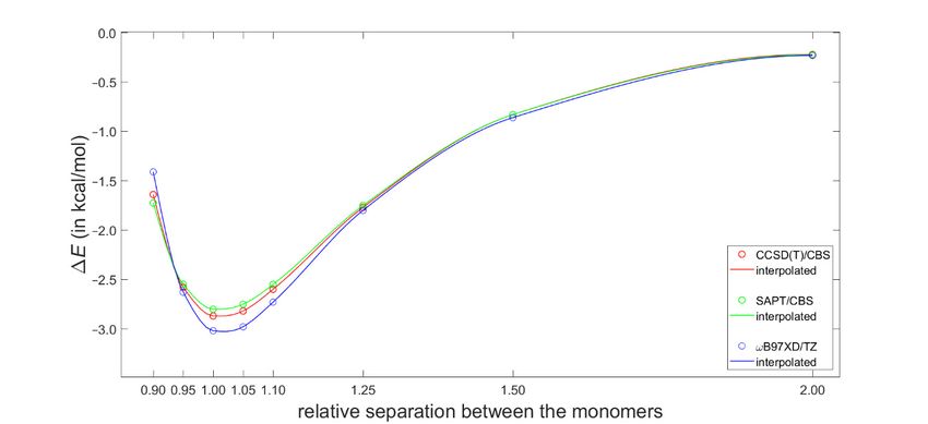
Int. J. Mol. Sci. 2020, 21, 7908                                                                           3 of 13

coordinate system while employing “e04fcf” subroutine from NAG® Library (related f90 program also
used “lsqgrd” subroutine to check that the gradients of δmodel , taken with respect to {A, B, C, D, E,
F} parameters of Equation (1), were sufficiently close to zero). The resulting parameter values were
considered to be final. They are listed in Supplementary Materials Table S1, and both sets of 49 δ points
are available from Table S4. The sole idea behind finding this analytic expression for δ is to obtain a
formula which is sufficiently accurate for numerically describing the 1 H CS surfaces, and Equation
(1) should not be interpreted in any other way. It follows from a comparison of the GIAO-MP2 and
GIAO-B3LYP surfaces that these methods provided qualitatively the same 1 H CS landscape. This is
significant, since the GIAO-B3LYP/6-311++G(2d,2p) approach, contrary to its MP2 counterpart, can be
routinely applied to large molecular fragments, possibly containing more than one hundred atoms.
Satisfactory precision of the fitting is also important, i.e., the maximum absolute deviation, average
absolute deviation, residual norm, and x2 are 0.28 ppm, 0.10 ppm, 0.83 ppm, and 0.1247, respectively,
for the GIAO-MP2 data, while these values accordingly amount to 0.32, 0.09, 0.61 ppm and 0.0993 for
the GIAO-B3LYP data.
                                                                     φ−F   ρ−E
                       δmodel (ρ, φ; A, B, C, D, E, F) = A + B sin C sin π D
                                                                                                      (1)
                      parametrized for ρ ∈ h2.0020; 3.7189i Å, φ ∈ h0.0; 0.6428i rad

     Figure 2 graphically presents an example 1 H CS surface and its fairly complex curvature and
other Int.
      issues
           J. Mol.are
                   Sci. discussed
                        2020, 21, x FORin Section
                                        PEER      3, while reliability of the model is addressed below. 2 of 7
                                             REVIEW

      Figure 2. The geometry dependence of the GIAO-MP2/6-311++G(2d,2p) predicted 1 H chemical shifts
      (magenta circles) and their parametrization (cyan stars are shown at the grid points) obtained for the
      T-shaped benzene dimer.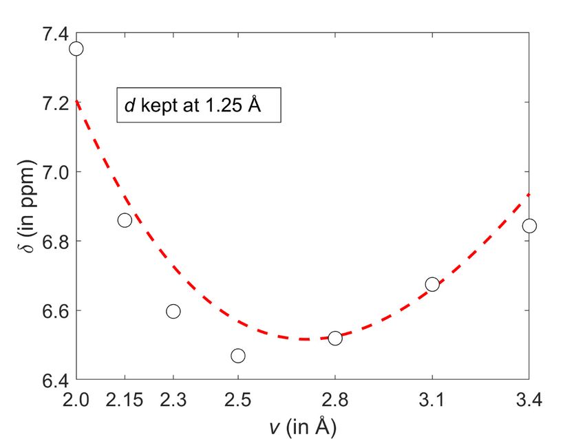
Int. J. Mol. Sci. 2020, 21, 7908                                                                    4 of 13

      This description of a spatial arrangement of benzene monomers using only two geometry variables
is, of course, simplified. Hence, it is of importance to assess how accurate the 1 H CS parametrization
would be if applied to the benzene dimer configurations with mutual orientation of planes other
than the one shown in Figure 1. This assessment was performed for the tilted T-shaped structures
from [29], which are available from the BEGDB database [30] (their dissociation curve is analyzed
in Section 2.3). The v, d values of five of those geometries fall into the parametrized interval, and
therefore their coordinates were used to obtain the 1 H CS from the model and directly from the GIAO
calculations. The predicted 1 H chemical shielding data are summarized in Table S2 and show only
small differences between the parametrized values and their counterparts provided explicitly by
quantum chemical calculations (maximum discrepancy amounts to 0.28 ppm and is found for the MP2
data of the structure with v = 2.6595 Å and d = 0.1361 Å). Moreover, these datasets exhibit the same
geometry dependence, namely, the 1 H chemical shielding values decrease with an increase in v and d
in this investigated region. The above two findings show that the presented model also works well for
the benzene dimers featuring interplanar angles which have not been included in the parameterization.
Therefore, it seems that it might be possible to use only two spatial coordinates to reliably quantify how
π-stacking interactions affect the 1 H CS of protons in aromatic systems in general. However, it should
be kept in mind that this model relies on the ability of quantum chemical calculations to accurately
describe the pair interactions influencing the 1 H CS value in a molecular cluster. Hence, in Section 2.2,
this ability is evaluated for organic solids whose structures pose additional challenges due to crystal
packing and due to heteroatoms or substituents present in the aromatic ring that also contains the
investigated proton.

2.2. Validation of the Dimer Model
     Older research on the 1 H CS of stacked proton sites in molecular crystals has been presented
in the excellent review article [31]. Here, two solid phase systems are analyzed, which have been
carefully studied by the groups of Kentgens [14] and Brown [12], and thus they provide the benchmark
data. Their data are considered to be fully reliable and cover a relatively large interval of pertinent
1 H CS values (see Table 1). The first system considers the protons, numbered H100 and H110 and

shown in Figure 11b of [14], of the crystalline isocyanoalanyl carbazole amid [13]. The second system
concerns the 1:1 cocrystal of dithianon and pyrimethanil with protons numbered H2 and H25 and
shown in Figure 5 of [12]. Experimental and theoretical data are collected in Table 1 (the following
values of the 1 H absolute isotropic chemical shielding of a proton in tetramethylsilane were used for
referencing: 30.8868 ppm for GIPAW and Perdew-Burke-Erzerhof (GIPAW-PBE) and 31.8308 ppm
for GIAO-B3LYP, respectively). The investigated protons are practically unaffected by the C–H . . .
X hydrogen bonding, which is not the case, for example, for aromatic protons in the well-known
1:1 cocrystal of indomethacin and nicotinamide [32,33], or in some systems previously studied by
this authors, in particular, L-tyrosine hydrochloride [34]. Consequently, the investigated protons are
supposed to probe an influence of the C–H/π stacking only and are used for further testing of the present
approach (see Figure 3; computational estimates of the 1 H CS are plotted against experimental values
in Figure S2). For this type of environment, it was ascertained that the GIAO-B3LYP/6-311++G(2d,2p)
dimer calculations could provide accurate 1 H chemical shielding values, i.e., the standard deviation of
residuals and adjusted R2 of the corresponding linear regression are 0.21 ppm and 0.93, respectively.
Accordingly, these results amount to 0.12 ppm and 0.98 for the periodic GIPAW calculations which are,
thus, highly successful, as was anticipated on the basis of previous work on structurally similar systems,
for instance, naproxen [35,36]. It has to be mentioned, however, that large errors of the PW DFT
predicted 1 H CS may occur for hydroxyl hydrogen-bonded sites [37–39], but those discrepancies are
caused mainly by the proton dynamics [40–42] that is unlikely to be significant in the molecular crystals
studied here. Nevertheless, the current results validate the approach which applies the GIAO-B3LYP
calculations of dimers for describing the spatial dependence of the 1 H CS in fragments affected by
π-stacking in molecular solids.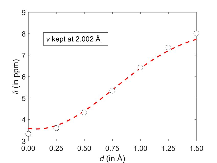
Int. J. Mol. Sci. 2020, 21, 7908 5 of 13
Table 1. Structural and spectral parameters of investigated solid-phase systems. In this table, the
measured chemical shift is denoted as δ, and its theoretical estimate as δ’. See the text for details.
σ/δ’ σ/δ’
σ/δ’ Fit
Crystal Site v (in Å) d (in Å) δ (in ppm) Periodic Cluster
(in ppm)
(in ppm) (in ppm)
H25 2.5238 0.5056 4.0 26.9417/3.9 27.2285/4.6 26.5599/5.3
Dithianon–pyrimethanil
H2 2.6975
Int. J. Mol. Sci. 2020, 21, x FOR PEER0 REVIEW 0.9521 6.2 24.4601/6.4 24.8389/7.0 25.6178/6.2 3 of 7
H10 2.6009 0.1846 4.8 26.2797/4.6 26.7231/5.1 26.7550/5.1
The isocyanide
H110 2.7557 0.2720 5.8 25.0655/5.8 25.6729/6.2 26.4137/5.4
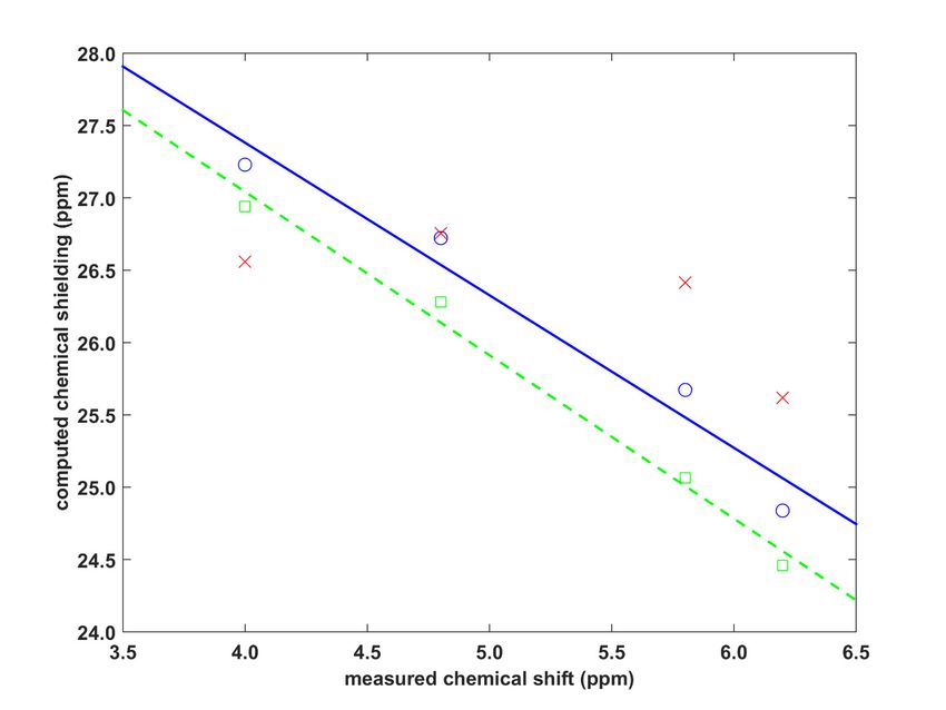
Figure 3. Comparison between theory and experiment for the proton solid-state nuclear magnetic
resonance (1 H SSNMR) parameters of two molecular crystals specified in the text. Green squares, blue
circles, and red crosses pertain to the GIPAW-PBE, GIAO-B3LYP, and parametrized data, respectively (the
green dashed line is y = −1.292*x + 31.56 ppm, while the blue straight line is y = −1.054*x + 31.60 ppm).
Corresponding results obtained from the model expressed by Equation (1) are also plotted in
Figure 3. Their quality is generally poor, as the parametrization was carried out using the benzene
dimer, not for fragments actually present in the investigated crystals. However, an inspection of
these results indicates that the parameters of Equation (1) may be transferable to structurally similar
residues. This can be seen for the proton site involved in the C–H/π interaction between fused (and
otherwise unsubstituted) benzene rings. Namely, the H110 site of the isocyanoalanyl carbazole amid
structure [13] is located on one such ring (denoted as “P” in [14]) of a carbazole unit, and is stacked
by another benzene ring (also “P”) of the neighboring molecule. As shown in Figure 3, at the 1 H CS
of 4.8 ppm, the difference between the parametrized result and the GIAO-B3LYP value is minimal
(0.04 ppm). The contacts of the remaining protons (H110 , H2, and H25 of the structures specified above)
involve substituted rings and are not detailed here. Separate parametrizations would be needed in
order to accurately describe the 1 H CS in these moieties. It should be kept in mind that the presented
analysis is implicitly influenced by the 1 H CS measurement uncertainties. Those uncertainties can beInt. J. Mol. Sci. 2020, 21, 7908 6 of 13
expected to be about 0.2 ppm, but they could be reduced, in particular, by using ultrafast magic-angle
spinning SSNMR techniques [43–45], and at very high magnetic fields.
2.3. Dimerization Energy Considerations
Most recently, Platzer et al. [5] discussed a possible connection between increasing binding enthalpy
and a higher 1 H CS perturbation for certain geometries of fragments involved in CH–π interactions in
protein–ligand complexes. Specifically, they scanned the PES of a T-shaped benzene dimer using the
ωB97X-D/cc-pVTZ dispersion corrected DFT approach (see Materials and Methods for referencing
of this and the subsequent computational techniques), which had been previously shown to reliably
describe the sandwich configuration [46]. Of practical significance is the fact that the ωB97X-D/cc-pVTZ
interaction energy can be obtained using a tiny fraction of computational resources needed to estimate
the corresponding “gold standard” coupled cluster singles and doubles with iterative inclusion of
triples (CCSD(T))/complete basis set (CBS) value (such a value would at present be inaccessible for
systems containing more than about 60 atoms) [47]. Moreover, the ωB97X-D method combined with a
midsized basis set have recently been used in important investigations of stacking [16,48]. Hence, the
ωB97X-D/cc-pVTZ approach was applied here to calculate interaction energies at the grid points for the
1 H CS calculations described above. These interaction energies are provided in Table S4 and graphically
presented in Figure S1. They are assumed to be quite accurate based on aforementioned references
and also based on a high quality of the data obtained for the dissociation curve of the tilted T-shaped
benzene dimer (see Figure 4 and Table S3). Namely, for its geometries reported in [29] and deposited in
the BEGDB database [30], the ωB97X-D/cc-pVTZ results were compared to fully reliable CCSD(T)/CBS
data and to their DFT-based symmetry-adapted intermolecular perturbation theory (DFT-SAPT)/CBS
counterparts. Figure 4 shows only insignificant differences between the ωB97X-D/cc-pVTZ and the
two sets of CBS-extrapolated values. The outstanding performance of the DFT-SAPT computational
protocol, specified in Section 4, and whose results closely match the CCSD(T)/CBS points of the
investigated curve are also noteworthy. As for the CCSD(T)/CBS values, they were reconstructed
according to the description provided in [29] in order to examine their breakdown into the ∆EaQZ HF
,
extrap.
∆E J. Mol., Sci.
Int.MP2 ∆Ecorrection
and2020, 21, x FOR PEERcomponents
CCSD(T)−MP2
REVIEW (see Equation (3) in Section 4). This breakdown is shown in7
4 of
Figure 5 and illustrates a delicate balance of contributions to the interaction energy.
Figure 4. Binding energies of the tilted T-shaped benzene dimer described in the text.Int. J. Mol. Sci. 2020, 21, x FOR PEER REVIEW 5 of 7
Int. J. Mol. Sci. 2020, 21, 7908 7 of 13
Figure 5. The CCSD(T)/CBS interaction energy components at points along the dissociation curve of
the tilted T-shaped benzene dimer.
3. Discussion
The GIAO-MP2 and GIAO-B3LYP methods were combined with the 6-311++G(2d,2p) basis set
and applied to a relatively large interval of radial and angular orientations of the benzene dimer
(both parametrizations are provided as Matlab m-files in Supplementary Materials). It is stressed
that a majority of these orientations fulfil the commonly used criteria for aromatic C–H/ π bonding.
Specifically, the Brandl–Weiss geometric system for identification of C–H/π interactions employs in a
lateral dimension the coordinate denoted as dHp −X [49] that is numerically equal to the coordinate d
used here. In structural database searches, a cut-off value for dHp −X of “1.0 or 1.2 Å for different sized π
-acceptor systems“ is typically used [50] and would have covered most of the grid points if it had been
applied in this work. Nevertheless, the GIAO calculations revealed a complicated landscape of the
1 H CS values of the proton directly involved in the C–H/π interaction. These values do not change
monotonically with increasing intermonomer separation for all lateral displacements considered here.
An illustration of this non-monotonic behavior is provided in Figure 6 which shows a slice of the 1 H
CS surface taken at d = 1.25 Å. Using the parametrization expressed by Equation (1), a minimum of
δmodel with respect to the vertical distance v can be easily obtained by taking the partial derivative and,
after the (ρ, φ) → (v, d) coordinate transformation, solving for zero the right hand side of Equation (2)
with d fixed at 1.25 Å.
∂ π(F − φ) π(E − ρ)
" ! !#
δ (ρ, φ; A, B, C, D, E, F) = − B π cos sin /C (2)
∂φ model C D
At such a minimum point, the 1 H CS is 6.5170 ppm and v is 2.7133 Å, apparently in agreement
∂2
with Figure 6. For d = 1.25 Å and v = 2.7133 Å, the second derivative ∂φ 2 δmodel has a negative value
(namely, −1.6529 ppm/Å2 ), of course confirming that the examined slice is convex. However, the 1 H
CS grows monotonically with increasing displacement in the whole interval of investigated vertical
distances (an example of this dependence is given in Figure 7). As a consequence of completely
different profiles in v and d dimensions, contour lines have an irregular shape, which would not be
the case if simple models were applied (such models were carefully compared in a relatively recent
study) [51]. Several contour lines are visualized in Figure 2, and numerical examples now follow.
For the lowest vertical separation considered here (v = 2.002 Å), it is immediately found through
Equation (1) that the particular values of 1 H CS of [5.5, 6.0, and 6.5 ppm] are, respectively, reached
at d = 0.7786, 0.9005, and 1.0305 Å. Then, using these displacement values, the 1 H CS of 5.5, 6.0, and
6.5 ppm are located at v = 2.3648, 2.7317, and 3.0944 Å, respectively. This analysis explicitly showsInt. J. Mol. Sci. 2020, 21, 7908 8 of 13
that the same 1 H CS value may occur in vastly different spatial arrangements of stacked molecules.
Additional information would obviously be needed to resolve such ambiguities during a structure
determination
Int. J.process, analogously
Mol. Sci. 2020, 21, x FOR PEERto the requirement for neutron diffraction data in distinguishing
REVIEW 6 of 7
hydrogen bond networks of some polymorphs [52].
Figure 6. A cut through the GIAO-MP2/6-311++G(2d,2p) 1 H chemical shift (CS) surface discussed in
Int. J. Mol. Sci. 2020, 21, x FOR PEER REVIEW 7 of 7
the text. Values obtained from regular calculations and from the corresponding parametrization are
shown as open black circles and the dashed red line, respectively.
Figure 7. A cut through the GIAO-MP2/6-311++G(2d,2p) 1 H CS surface discussed in the text. Values
obtained from regular calculations and from the corresponding parametrization are shown as open
black circles and the dashed red line, respectively.Int. J. Mol. Sci. 2020, 21, 7908 9 of 13
4. Materials and Methods
The standard second-order Møller–Plesset (MP2) approach and the standard Becke’s three-parameter,
Lee-Yang–Parr (B3LYP) combination of DFT functionals were used. In the chemical shielding calculations,
these methods were combined with the standard 6-311++G(2d,2p) basis set and with the GIAO strategy
to overcome the gauge problem [53,54]. Interaction energies corrected for the basis set superposition
error by the counterpoise (CP) scheme [55] were obtained using the empirically corrected ωB97X-D DFT
functional [56] and the standard cc-pVTZ (correlation-consistent polarized valence triple-ζ) basis set. In
the full geometry optimization of tetramethylsilane, the MP2 method was combined with the standard
aug-cc-pVTZ (the cc-pVTZ augmented with diffuse functions) basis set, and Td symmetry was imposed.
All these calculations were carried out using the Gaussian 09 suite of codes [57].
Crystal structures [12,13] served as input for periodic DFT calculations which adopt the
pseudopotential scheme [58–60] implemented in the CASTEP 16.1 program [60]. Unit-cell parameters
of these structures were kept at experimental values, while atomic positions were optimized with
respect to the crystal-lattice energy that was approximated using the Perdew–Burke–Erzerhof (PBE)
DFT functional [61]. For resulting geometries, the chemical shielding was predicted by applying the
GIPAW method [62,63] and the PBE functional. In all CASTEP calculations, the settings were consistent
with “Fine” accuracy level of the Materials Studio 2019 software [64]. In particular, the PW cut-off
value was 42.0 Ry. The ultrasoft on-the-fly generated pseudopotentials were adopted [65].
The CCSD(T) (coupled cluster singles and doubles with iterative inclusion of triples) interaction
energies extrapolated to the CBS (complete basis set) limit, ∆ECBS
CCSD(T)
, were estimated using Equation (3)
extrap.
∆ECBS
CCSD(T)
= ∆EaQZ
HF
+ ∆EMP2 + ∆Ecorrection
CCSD(T)−MP2
(3)
The ∆EaQZ
HF
term is the interaction energy obtained at the Hartree–Fock (HF) level using the
extrap.
aug-cc-pVQZ basis set. The ∆EMP2 denotes the MP2 correlation energy contribution, which was
obtained using the aug-cc-pVTZ and aug-cc-pVQZ values by their extrapolation that employed the
scheme of Halkier et al. (Equation (7) in [66]). The ∆Ecorrection
CCSD(T)−MP2
component, which aims at correctly
approximating higher-order correlation energy contributions to the interaction energy, was computed
using the aug-cc-pVDZ basis set in the underlying CCSD(T) and MP2 calculations. The CP correction
was applied throughout.
The DFT-based SAPT (symmetry-adapted intermolecular perturbation theory) [67] calculations
(abbreviated as DFT-SAPT) were performed in the density-fitting variant [68]. The procedure described
in [69] was followed except for an extrapolation of the interaction energy components. Namely, in this
work all the components were extrapolated to their CBS limit and summed up, while Hesselmann et
al. [69] extrapolated only the second-order dispersion contributions. The Molpro version 2008.1 [70]
was used to obtain energies leading to the CCSD(T)/CBS and DFT-SAPT/CBS results.
Supplementary Materials: The following are available online at http://www.mdpi.com/1422-0067/21/21/7908/s1,
Table S1: Values of parameters in Equation (1), Table S2: The 1 H chemical shielding data for the tilted T-shaped
dimer of benzene, Table S3: Interaction energies of the tilted T-shaped dimer of benzene, Table S4: Predicted
values of the 1 H CS and of the ωB97X-D/cc-pVTZ interaction energy at 49 grid points, Figure S1: A plot of
the ωB97X-D/cc-pVTZ interaction energies, Figure S2: A plot of measured and computed 1 H chemical shifts of
selected protons in two molecular crystals; script files ‘f_b3lyp.m’ and ‘f_mp2.m’ to obtain an estimate of the 1 H
CS on parametrized δ (v, d) surface.
Author Contributions: Conceptualization, J.C. and J.B.; Investigation, writing, J.C.; Validation, funding acquisition,
J.B. All authors have read and approved the final manuscript.
Funding: Ministry of Education, Youth and Sports of the Czech Republic project LTAUSA18011.
Acknowledgments: Computational resources were supplied by the project “e-Infrastruktura CZ” (e-INFRA
LM2018140) provided within the program Projects of Large Research, Development and Innovations Infrastructures.
J.C. is grateful for the excellent support he has been receiving from the staff of the MetaCentrum VO (https:
//metavo.metacentrum.cz/).Int. J. Mol. Sci. 2020, 21, 7908 10 of 13
Conflicts of Interest: The authors declare no conflict of interest.
References
1. Larive, C.K.; Barding, G.A.; Dinges, M.N. NMR spectroscopy for metabolomics and metabolic profiling.
Anal. Chem. 2015, 87, 133–146. [CrossRef] [PubMed]
2. Zhang, R.; Mroue, K.H.; Sun, P.; Ramamoorthy, A. High-Resolution Proton NMR Spectroscopy of Polymers
and Biological Solids. In Modern Magnetic Resonance, 2nd ed.; Webb, G.A., Ed.; Springer: Cham, Switzerland,
2018; pp. 521–536.
3. Stone, A.J. The Theory of Intermolecular Forces, 1st ed.; Clarendon Press: Oxford, UK, 2002; pp. 56–63.
4. Kudisch, B.; Maiuri, M.; Moretti, L.; Oviedo, M.B.; Wang, L.; Oblinsky, D.G.; Prud’homme, R.K.; Wong, B.M.;
McGill, S.A.; Scholes, G.D. Ring currents modulate optoelectronic properties of aromatic chromophores at 25
T. Proc. Natl. Acad. Sci. USA 2020, 117, 11289–11298. [CrossRef] [PubMed]
5. Platzer, G.; Mayer, M.; Beier, A.; Brüschweiler, S.; Fuchs, J.E.; Engelhardt, H.; Geist, L.; Bader, G.;
Schörghuber, J.; Lichtenecker, R.; et al. PI by NMR: Probing CH–π Interactions in Protein–Ligand Complexes
by NMR Spectroscopy. Angew. Chem. Int. Ed. 2020, 59, 14861–14868. [CrossRef] [PubMed]
6. Rickhaus, M.; Jirasek, M.; Tejerina, L.; Gotfredsen, H.; Peeks, M.D.; Haver, R.; Jiang, H.-W.; Claridge, T.D.W.;
Anderson, H.L. Global aromaticity at the nanoscale. Nat. Chem. 2020, 12, 236–241. [CrossRef]
7. Gabryelczyk, B.; Cai, H.; Shi, X.; Sun, Y.; Swinkels, P.J.M.; Salentinig, S.; Pervushin, K.; Miserez, A. Hydrogen
bond guidance and aromatic stacking drive liquid-liquid phase separation of intrinsically disordered
histidine-rich peptides. Nat. Commun. 2019, 10. [CrossRef]
8. Chaudhari, S.R.; Griffin, J.M.; Broch, K.; Lesage, A.; Lemaur, V.; Dudenko, D.; Olivier, Y.; Sirringhaus, H.;
Emsley, L.; Grey, C.P. Donor–acceptor stacking arrangements in bulk and thin-film high-mobility conjugated
polymers characterized using molecular modelling and MAS and surface-enhanced solid-state NMR
spectroscopy. Chem. Sci. 2017, 8, 3126–3136. [CrossRef]
9. Bass, T.M.; Carr, C.R.; Sherbow, T.J.; Fettinger, J.C.; Berben, L.A. Syntheses of Square Planar Galluim
Complexes and a Proton NMR Correlation Probing Metalloaromaticity. Inor. Chem. 2020, 59, 13517–13523.
[CrossRef]
10. Lampkin, B.J.; Karadakov, P.B.; VanVeller, B. Detailed Visualization of Aromaticity Using Isotropic Magnetic
Shielding. Angew. Chem. Int. Ed. 2020, 59, 2–9. [CrossRef]
11. Kilymis, D.; Bartók, A.P.; Pickard, C.J.; Forse, A.C.; Merlet, C. Efficient prediction of nucleus independent
chemical shifts for polycyclic aromatic hydrocarbons. Phys. Chem. Chem. Phys. 2020, 22, 13746–13755.
[CrossRef]
12. Pöppler, A.-C.; Corlett, E.K.; Pearce, H.; Seymour, M.P.; Reid, M.; Montgomery, M.G.; Brown, S.P. Single-crystal
X-ray diffraction and NMR crystallography of a 1:1 cocrystal of dithianon and pyrimethanil. Acta Cryst. C
2017, 73, 149–156. [CrossRef]
13. Schwartz, E.; Lim, E.; Gowda, C.M.; Liscio, A.; Fenwick, O.; Tu, G.; Palermo, V.; de Gelder, R.;
Cornelissen, J.J.L.M.; Van Eck, E.R.H.; et al. Synthesis, Characterization, and Surface Initiated Polymerization
of Carbazole Functionalized Isocyanides. Chem. Mater. 2010, 8, 2597–2607. [CrossRef]
14. Gowda, C.M.; Vasconcelos, F.; Schwartz, E.; Van Eck, E.R.H.; Marsman, M.; Cornelissen, J.J.L.M.; Rowan, A.E.;
De Wijs, G.A.; Kentgens, A.P.M. Hydrogen bonding and chemical shifts assignments in carbazole functionalized
isocyanides from solid-state NMR and first-principles calculations. Phys. Chem. Chem. Phys. 2011, 13, 13082–13095.
[CrossRef] [PubMed]
15. Bonhomme, C.; Gervais, C.; Babonneau, F.; Coelho, C.; Pourpoint, F.; Azais, T.; Asbrook, S.E.; Griffin, J.M.;
Yates, J.R.; Pickard, J.C. First-Principles Calculation of NMR Parameters Using the Gauge Including Projector
Augmented Wave Method: A Chemist’s Point of View. Chem. Rev. 2012, 112, 5733–5779. [CrossRef]
[PubMed]
16. Bootsma, A.N.; Doney, A.C.; Wheeler, S.E. Predicting the Strength of Stacking Interactions between
Heterocycles and Aromatic Amino Acid Side Chain. J. Am. Chem. Soc. 2019, 141, 11027–11035. [CrossRef]
17. D’Ischia, M.; Napolitano, A.; Pezzella, A.; Meredith, P.; Buehler, M. Melanin biopolymers: Tailoring chemical
complexity for materials design. Angew. Chem. Int. Ed. 2020, 59, 11196–11205. [CrossRef]
18. Lyu, Q.; Hsueh, N.; Chai, C.L.L. Unravelling the polydopamine mystery: Is the end in sight? Polym. Chem.
2019, 10, 5771–5777. [CrossRef]Int. J. Mol. Sci. 2020, 21, 7908 11 of 13
19. Proks, V.; Brus, J.; Pop-Georgievski, O.; Večerníková, E.; Wiśniewski, W.; Kotek, J.; Urbanová, M.; Rypáček, F.
Thermal-Induced Transformation of Polydopamine Structures: An Efficient Route for the Stabilization of the
Polydopamine Surfaces. Macromol. Chem. Phys. 2013, 214, 499–507. [CrossRef]
20. Circu, M.; Filip, C. Closer to the polydopamine structure: New insights from a combined 13 C/1 H/2 H
solid-state NMR study on deuterated samples. Polym. Chem. 2018, 9, 3379–3387. [CrossRef]
21. Loeffler, J.R.; Fernández-Quintero, M.L.; Schauperl, M.; Liedl, K.R. STACKED – Solvation Theory of Aromatic
Complexes as Key for Estimating Drug Binding. J. Chem. Inf. Model. 2020, 60, 2304–2313. [CrossRef]
22. Bartolomei, M.; Pirani, F.; Marques, J.M.C. Low-energy structures of benzene clusters with a novel accurate
potential surface. J. Comput. Chem. 2015, 36, 2291–2301. [CrossRef]
23. Kennedy, M.R.; McDonald, A.R.; DePrince, A.E., III; Marshall, M.S.; Podeszwa, R.; Sherrill, C.D. Resolving the
three-body contribution to the lattice energy of crystalline benzene: Benchmark results from coupled-cluster
theory. J. Chem. Phys. 2014, 140. [CrossRef]
24. Miliordos, E.; Apra, E.; Xantheas, S.S. Benchmark Theoretical Study of the π−π Binding Energy in the
Benzene Dimer. J. Phys. Chem. A 2014, 118, 7568–7578. [CrossRef]
25. DiStasio, R.A., Jr.; Von Helden, G.; Steele, R.P.; Head-Gordon, M. On the T-shaped structures of the benzene
dimer. Chem. Phys. Lett. 2007, 437, 277–283. [CrossRef]
26. Czernek, J.; Brus, J. Exploring Accuracy Limits of Predictions of the 1 H NMR Chemical Shielding Anisotropy
in the Solid State. Molecules 2019, 24, 1731. [CrossRef] [PubMed]
27. Czernek, J.; Brus, J. Monitoring the Site-Specific Solid-State NMR Data in Oligopeptides. Int. J. Mol. Sci.
2020, 21, 2700. [CrossRef]
28. Czernek, J.; Brus, J. Polymorphic Forms of Valinomycin Investigated by NMR Crystallography. Int. J. Mol. Sci.
2020, 21, 4907. [CrossRef]
29. Řezáč, J.; Riley, K.E.; Hobza, P. S66: A Well-balanced Database of Benchmark Interaction Energies Relevant
to Biomolecular Structures. J. Chem. Theory Comput. 2011, 7, 2427–2438. [CrossRef]
30. Řezáč, J.; Jurečka, P.; Riley, K.E.; Černý, J.; Valdes, H.; Pluháčková, K.; Berka, K.; Řezáč, T.; Pitoňák, M.;
Vondrášek, J.; et al. Quantum Chemical Benchmark Energy and Geometry Database for Molecular Clusters
and Complex Molecular Systems (www.begdb.com): A Users Manual and Examples. Collect. Czech.
Chem. Commun. 2008, 73, 1261–1270. [CrossRef]
31. Brown, S.P. Applications of high-resolution 1 H solid-state NMR. Solid State Nucl. Magn. Reson. 2012, 41,
1–27. [CrossRef]
32. Dudenko, D.V.; Yates, J.R.; Harris, K.D.M.; Brown, S.P. An NMR crystallography DFT-D approach to analyse
the role of intermolecular hydrogen bonding and π–π interactions in driving cocrystallisation of indomethacin
and nicotinamide. CrystEngComm 2013, 15, 8797–8807. [CrossRef]
33. Dudenko, D.V.; Williams, P.A.; Hughes, C.E.; Antzutkin, O.N.; Velaga, S.P.; Brown, S.P.; Harris, K.D.M.
Exploiting the Synergy of Powder X-ray Diffraction and Solid-State NMR Spectroscopy in Structure
Determination of Organic Molecular Solids. J. Phys. Chem. C 2013, 117, 12258–12265. [CrossRef] [PubMed]
34. Czernek, J.; Brus, J. The covariance of the differences between experimental and theoretical chemical shifts as
an aid for assigning two-dimensional heteronuclear correlation solid-state NMR spectra. Chem. Phys. Lett.
2014, 608, 334–339. [CrossRef]
35. Carignani, E.; Borsacchi, S.; Bradley, J.P.; Brown, S.P.; Geppi, M. Strong Intermolecular Ring Current Influence
on 1 H Chemical Shifts in Two Crystalline Forms of Naproxen: A Combined Solid-State NMR and DFT Study.
J. Phys. Chem. C 2013, 117, 17731–17740. [CrossRef]
36. Czernek, J. On the solid-state NMR spectra of naproxen. Chem. Phys. Lett. 2015, 619, 230–235. [CrossRef]
37. Corlett, E.K.; Blade, H.; Hughes, L.P.; Sidebottom, P.J.; Walker, D.; Walton, R.I.; Brown, S.P. Investigating
discrepancies between experimental solid-state NMR and GIPAW calculation: N=C–N 13 C and OH· · · O
1 H chemical shifts in pyridinium fumarates and their cocrystals. Solid State Nucl. Magn. Reson. 2020, 108.
[CrossRef]
38. Hušák, M.; Jegorov, A.; Rohlíček, J.; Fitch, A.; Czernek, J.; Kobera, L.; Brus, J. Determining the Crystal
Structures of Peptide Analogs of Boronic Acid in the Absence of Single Crystals: Intricate Motifs of Ixazomib
Citrate Revealed by XRPD Guided by ss-NMR. Cryst. Growth Des. 2018, 18, 3616–3625. [CrossRef]
39. Harris, R.K.; Hodgkinson, P.; Zorin, V.; Dumez, J.N.; Elena-Herrmann, B.; Emsley, L.; Salager, E.; Stein, R.S.
Computation and NMR crystallography of terbutaline sulfate. Magn. Reson. Chem. 2010, 48, S103–S112.
[CrossRef]Int. J. Mol. Sci. 2020, 21, 7908 12 of 13
40. Kerr, H.E.; Softley, L.K.; Suresh, K.; Nangia, A.; Hodgkinson, P.; Radosavjlevic Evans, I. A
furosemide–isonicotinamide cocrystal: An investigation of properties and extensive structural disorder.
CrystEngComm 2015, 17, 6707–6715. [CrossRef]
41. Frantsuzov, I.; Ford, S.J.; Radosavjlevic Evans, I.; Horsewill, A.J.; Trommsdorff, H.P.; Johnson, M.R.
Measurement of Proton Tunneling in Short Hydrogen Bonds in Single Crystals of 3,5 Pyridinedicarboxylic
Acid Using Nuclear Magnetic Resonance Spectroscopy. Phys. Rev. Lett. 2014, 113. [CrossRef]
42. Dračínský, M.; Hodgkinson, P. A molecular dynamics study of the effects of fast molecular motions on
solid-state NMR parameters. CrystEngComm 2013, 15, 8705–8712. [CrossRef]
43. Nishiyama, Y.; Malon, M.; Potrzebowski, M.J.; Paluch, P.; Amoreux, J.P. Accurate NMR determination of
C–H or N–H distances for unlabeled molecules. Solid State Nucl. Magn. Reson. 2016, 73, 15–21. [CrossRef]
[PubMed]
44. Zhang, R.; Mroue, K.H.; Ramamoorthy, A. Proton-Based Ultrafast Magic Angle Spinning Solid-State NMR
Spectroscopy. Acc. Chem. Res. 2017, 50, 1105–1113. [CrossRef] [PubMed]
45. Li, M.; Lu, X.; Xu, W.; Troup, G.M.; McNevin, M.J.; Nie, H.; Su, Y. Quantifying Pharmaceutical Formulations
from Proton Detected Solid-State NMR under Ultrafast Magic Angle Spinning. J. Pharm. Sci. 2020, 109,
3045–3053. [CrossRef] [PubMed]
46. Huber, R.G.; Margreiter, M.A.; Fuchs, J.E.; Von Grefenstein, S.; Tuatermann, C.S.; Liedl, K.R.; Fox, T.
Heteroaromatic π-Stacking Energy Landscapes. J. Chem. Inf. Model. 2014, 54, 1371–1379. [CrossRef]
47. Gyevy-Nagy, L.; Kállay, M.; Nagy, P.R. Integral-Direct and Parallel Implementation of the CCSD(T) Method:
Algorithmic Developments and Large-Scale Applications. J. Chem. Theory Comput. 2020, 16, 366–384.
[CrossRef]
48. Bootsma, A.N.; Doney, A.C.; Wheeler, S.E. Tuning Stacking Interactions between Asp–Arg Salt Bridges and
Heterocyclic Drug Fragments. J. Chem. Inf. Model. 2019, 59, 149–158. [CrossRef]
49. Brandl, M.; Weiss, M.S.; Jabs, A.; Sühnel, J.; Hilgenfeld, R. C–H . . . π-Interaction in Proteins. J. Mol. Biol.
2001, 307, 357–377. [CrossRef]
50. Nishiyo, M.; Umezawa, Y.; Fantini, J.; Weiss, M.S.; Chakrabarti, P. CH–π hydrogen bonds in biological
macromolecules. Phys. Chem. Chem. Phys. 2014, 16, 12648–12683. [CrossRef]
51. Sahakyan, A.B.; Vendruscolo, M. Analysis of the Contributions of Ring Current and Electric Field Effects to
the Chemical Shifts of RNA Bases. J. Phys. Chem. B 2013, 117, 1989–1998. [CrossRef]
52. Widdifield, C.M.; Farrell, J.D.; Cole, J.C.; Howard, J.A.K.; Hodgkinson, P. Resolving alternative organic
crystal structures using density functional theory and NMR chemical shifts. Chem. Sci. 2020, 11, 2987–2992.
[CrossRef]
53. Ditchfield, R. Self-consistent perturbation theory of diamagnetism. Mol. Phys. 1974, 27, 789–807. [CrossRef]
54. Wolinski, K.; Hinton, J.F.; Pulay, P. Efficient implementation of the gauge-independent atomic orbital method
for NMR chemical shift calculations. J. Am. Chem. Soc. 1990, 112, 8251–8260. [CrossRef]
55. Boys, S.; Bernardi, F. The calculation of small molecular interactions by the differences of separate total
energies. Some procedures with reduced errors. Mol. Phys. 1970, 19, 553–566. [CrossRef]
56. Chai, J.-D.; Head-Gordon, M. Long-range corrected hybrid density functionals with damped atom-atom
dispersion corrections. Phys. Chem. Chem. Phys. 2008, 10, 6615–6620. [CrossRef]
57. Frish, M.J.; Trucks, J.W.; Schlegel, H.B.; Scuseria, G.E.; Robb, M.A.; Cheeseman, J.R.; Scalmani, G.; Barone, V.;
Mennucci, B.; Petersson, G.A.; et al. Gaussian 09; Revision D.01; Gaussian, Inc.: Wallingford, UK, 2013.
58. Kresse, G.; Joubert, D. From ultrasoft pseudopotentials to the projector augmented-wave method. Phys. Rev. B
1999, 59, 1758–1775. [CrossRef]
59. Segall, M.D.; Lindan, P.J.D.; Probert, M.J.; Pickard, C.J.; Hasnip, P.J.; Clark, S.J.; Payne, M.C. First principles
simulation: Ideas, illustrations, and the CASTEP code. J. Phys. Condens. Matter 2002, 14, 2717–2744.
[CrossRef]
60. Clark, S.J.; Segall, M.D.; Pickard, C.J.; Hasnip, P.J.; Probert, M.J.; Refson, K.; Payne, M.C. First principles
methods using CASTEP. Z. Kristallogr. 2005, 220, 567–570. [CrossRef]
61. Perdew, J.P.; Burke, K.; Ernzerhof, M. Generalized gradient approximation made simple. Phys. Rev. Lett.
1996, 77, 3865–3868. [CrossRef]
62. Pickard, C.J.; Mauri, F. All-electron magnetic response with pseudopotentials: NMR chemical shifts.
Phys. Rev. B 2001, 63. [CrossRef]Int. J. Mol. Sci. 2020, 21, 7908 13 of 13
63. Yates, J.R.; Pickard, C.J.; Mauri, F. Calculations of NMR chemical shifts for extended systems using ultrasoft
pseudopotentials. Phys. Rev. B 2007, 76. [CrossRef]
64. BIOVIA Materials Studio. Dassault Systèmes, Vélizy-Villacoublay: Paris, France. Available online: https://www.
3ds.com/products-services/biovia/products/molecular-modeling-simulation/biovia-materials-studio/ (accessed on
22 September 2020).
65. Gao, S.-P.; Pickard, C.J.; Perlov, A.; Milman, V. Core-Level Spectroscopy Calculation and the Plane Wave
Pseudopotential Method. J. Phys. Condens. Matter 2009, 21. [CrossRef] [PubMed]
66. Halkier, A.; Helgaker, T.; Jørgensen, P.; Klopper, W.; Koch, H.; Olsen, J.; Wilson, A.K. Basis-set convergence in
correlated calculations on Ne, N2 , and H2 O. Chem. Phys. Lett. 1998, 286, 243–252. [CrossRef]
67. Patkowski, K. Recent developments in symmetry-adapted perturbation theory. Wiley Interdiscip. Rev. Comput.
Mol. Phys. 2019, 10. [CrossRef]
68. Hesselmann, A.; Jansen, G.; Schütz, M. Density-functional theory-symmetry-adapted intermolecular
perturbation theory with density fitting: A new efficient method to study intermolecular interaction
energies. J. Chem. Phys. 2005, 122. [CrossRef]
69. Hesselmann, A.; Jansen, G.; Schütz, M. Interaction Energy Contributions of H-Bonded and Stacked Structures
of the AT and GC DNA Base Pairs from the Combined Density Functional Theoryand Intermolecular
Perturbation Theory Approach. J. Am. Chem. Soc. 2006, 128, 11730–11731. [CrossRef] [PubMed]
70. Werner, H.J.; Knowles, P.J.; Manby, F.R.; Black, J.A.; Doll, K.; Hesselmann, A.; Kats, D.; Kohn, A.; Korona, T.;
Kreplin, D.A.; et al. The Molpro quantum chemistry package. J. Chem. Phys. 2020, 152. [CrossRef]
Publisher’s Note: MDPI stays neutral with regard to jurisdictional claims in published maps and institutional
affiliations.
© 2020 by the authors. Licensee MDPI, Basel, Switzerland. This article is an open access
article distributed under the terms and conditions of the Creative Commons Attribution
(CC BY) license (http://creativecommons.org/licenses/by/4.0/).You can also read