NICOTINIC CHOLINERGIC SYSTEM AND COVID-19: IN SILICO IDENTIFICATION OF AN INTERACTION BETWEEN SARS-COV-2 AND NICOTINIC RECEPTORS WITH POTENTIAL ...
←
→
Page content transcription
If your browser does not render page correctly, please read the page content below
International Journal of
            Molecular Sciences

Article
Nicotinic Cholinergic System and COVID-19:
In Silico Identification of an Interaction between
SARS-CoV-2 and Nicotinic Receptors with Potential
Therapeutic Targeting Implications
Konstantinos Farsalinos 1,† , Elias Eliopoulos 2,† , Demetres D. Leonidas 3,† ,
Georgios E. Papadopoulos 3 , Socrates Tzartos 1 and Konstantinos Poulas 1, *
 1    Laboratory of Molecular Biology and Immunology, Department of Pharmacy, University of Patras,
      Panepistimiopolis, 26500 Rio-Patras, Greece; kfarsalinos@gmail.com (K.F.); stzartos@gmail.com (S.T.)
 2    Laboratory of Genetics, Department of Biotechnology, Agricultural University of Athens, Iera Odos 75,
      11855 Athens, Greece; eliop@aua.gr
 3    Department of Biochemistry and Biotechnology, University of Thessaly, Biopolis, 41500 Larissa, Greece;
      ddleonidas@bio.uth.gr (D.D.L.); geopap@bio.uth.gr (G.E.P.)
 *    Correspondence: kpoulas@upatras.gr; Tel.: +30-2610-962353
 †    These authors contributed equally to this work.

 Received: 17 June 2020; Accepted: 11 August 2020; Published: 13 August 2020

 Abstract: While SARS-CoV-2 uses angiotensin converting enzyme 2 (ACE2) as the receptor for cell
 entry, it is important to examine other potential interactions between the virus and other cell receptors.
 Based on the clinical observation of low prevalence of smoking among hospitalized COVID-19
 patients, we examined and identified a “toxin-like” amino acid (aa) sequence in the Receptor Binding
 Domain of the Spike Glycoprotein of SARS-CoV-2 (aa 375–390), which is homologous to a sequence
 of the Neurotoxin homolog NL1, one of the many snake venom toxins that are known to interact with
 nicotinic acetylcholine receptors (nAChRs). We present the 3D structural location of this “toxin-like”
 sequence on the Spike Glycoprotein and the superposition of the modelled structure of the Neurotoxin
 homolog NL1 and the SARS-CoV-2 Spike Glycoprotein. We also performed computational molecular
 modelling and docking experiments using 3D structures of the SARS-CoV-2 Spike Glycoprotein and
 the extracellular domain of the nAChR α9 subunit. We identified a main interaction between the
 aa 381–386 of the SARS-CoV-2 Spike Glycoprotein and the aa 189–192 of the extracellular domain
 of the nAChR α9 subunit, a region which forms the core of the “toxin-binding site” of the nAChRs.
 The mode of interaction is very similar to the interaction between the α9 nAChR and α-bungarotoxin.
 A similar interaction was observed between the pentameric α7 AChR chimera and SARS-CoV-2
 Spike Glycoprotein. The findings raise the possibility that SARS-CoV-2 may interact with nAChRs,
 supporting the hypothesis of dysregulation of the nicotinic cholinergic system being implicated in
 the pathophysiology of COVID-19. Nicotine and other nicotinic cholinergic agonists may protect
 nAChRs and thus have therapeutic value in COVID-19 patients.

 Keywords: COVID-19; SARS-CoV-2; smoking; nicotine; nicotinic cholinergic system; inflammation;
 acetylcholine receptors

1. Introduction
     As the global pandemic of corona virus disease 2019 (COVID-19) was spreading, it was recognized
early that the virus (SARS-COV-2) uses the angiotensin converting enzyme 2 (ACE2) as a receptor
for cell entry [1]. The 3-D structure and the function of SARS-CoV-2 Spike Glycoprotein has been

Int. J. Mol. Sci. 2020, 21, 5807; doi:10.3390/ijms21165807                           www.mdpi.com/journal/ijms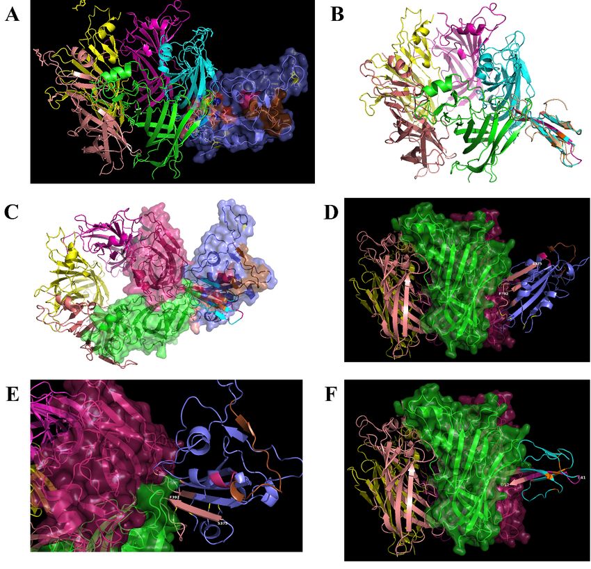
Int. J. Mol. Sci. 2020, 21, 5807                                                                   2 of 15

published [2] and the structure of the complex of SARS-CoV-2 Spike Glycoprotein with the ACE2 has
been solved by cryo-EM experiments [3]. This has been the focus of the global research community,
examining the interactions between disease conditions or medications and ACE2 expression [4,5].
Among the risk factors that have been examined is smoking, considering that it is a major risk factor
for respiratory infections [6]. Until recently, smoking was associated with the down-regulation of
ACE2 [7]. However, recent studies show that it may cause ACE2 up-regulation, and this may propagate
viral spread and disease severity [8]. However, the association between smoking (and nicotine) and
COVID-19 appears to be much more complex. Consistent clinical observations from retrospective cases
series of COVID-19 patients have shown a low prevalence of smoking among hospitalized COVID-19
patients [9].
      In April 2020, we hypothesized for the first time that the nicotinic cholinergic system (NCS) may
be implicated in the pathophysiology of severe COVID-19, and we recently expanded our research
on this hypothesis [9,10]. Immune dysregulation and cytokine storm appear to play a major role
in the pathophysiology of severe COVID-19 [11]. The “cholinergic anti-inflammatory pathway” is
an important regulator of the inflammatory response [12]. Its effects are mediated mainly by the
vagus nerve and by α7 nicotinic acetylcholine receptors (nAChRs). The highest concentration of α7
nAChRs is observed in the hippocampus, the thalamic reticular nucleus, as well as the lateral and
medial geniculate nuclei. On the cellular level, they are expressed both pre- and postsynaptically [13].
Presynaptically, they are positioned to allow Ca2+ influx into the synaptic bouton, and in this way,
they play a major role in modulating the release of many neurotransmitters. Alpha7 nAChRs are
also observed on macrophages and other immune cells and have been implicated in preventing
sepsis and acute respiratory distress syndrome (ARDS) in animal models [13,14]. Alpha7 nAChRs are
expressed, also, in human bronchial epithelial and endothelial cells [15], which are the major targets of
SARS-CoV-2. Dysfunction of the NCS could explain other clinical manifestations of COVID-19, and we
therefore hypothesized that there may be a direct interaction between SARS-CoV-2 and the NCS [10].
      Considering the above, we focused on the potential interaction between SARS-CoV-2 and
the nicotinic cholinergic system, particularly α7 nAChRs [10]. Any such interaction could have
potential therapeutic implications considering that there are pharmacological agents, such as nicotine,
which could protect these receptors through competitive binding to the receptors. The identification
of a potential interaction between any virus protein and the cholinergic pathway could promote
a better understanding of the large spectrum of COVID-19 clinical manifestations and could lead
to the identification of potentially therapeutic compounds among the many cholinergic agonists.
Additionally, such interactions could explain the previously mentioned clinical observations of
under-representation of smokers among hospitalized COVID-19 patients. This would establish the
hypothesis that SARS-CoV-2 disrupts the cholinergic anti-inflammatory pathway and causes a variety
of clinical manifestations by interacting with nAChRs, and nicotine could prevent this by restoring the
function of these receptors.
      While tobacco cigarette smoke contains thousands of compounds, many of which are highly toxic
and are associated with combustion, we focused on nicotine which has well-established pharmacological
effects and has been available for years as an approved medication in various forms. Therefore, nicotine
represented a compound with promising prospects if proven to be associated with therapeutic benefits,
while additional approved compounds act on nicotinic cholinergic receptors and could also be examined
for potential effects.
      In this study, we compared amino acid sequences between SARS-CoV-2 and snake venom
neurotoxins. The latter are well-established inhibitors of the NCS [16,17]. Subsequently, molecular
modelling and docking experiments were performed to examine if there may be an interaction between
SARS-CoV-2 Spike Glycoprotein and nAChRs. Based on these findings, we also present the hypothesis
that nicotine and other nicotinic cholinergic agonists may have a therapeutic value in COVID-19.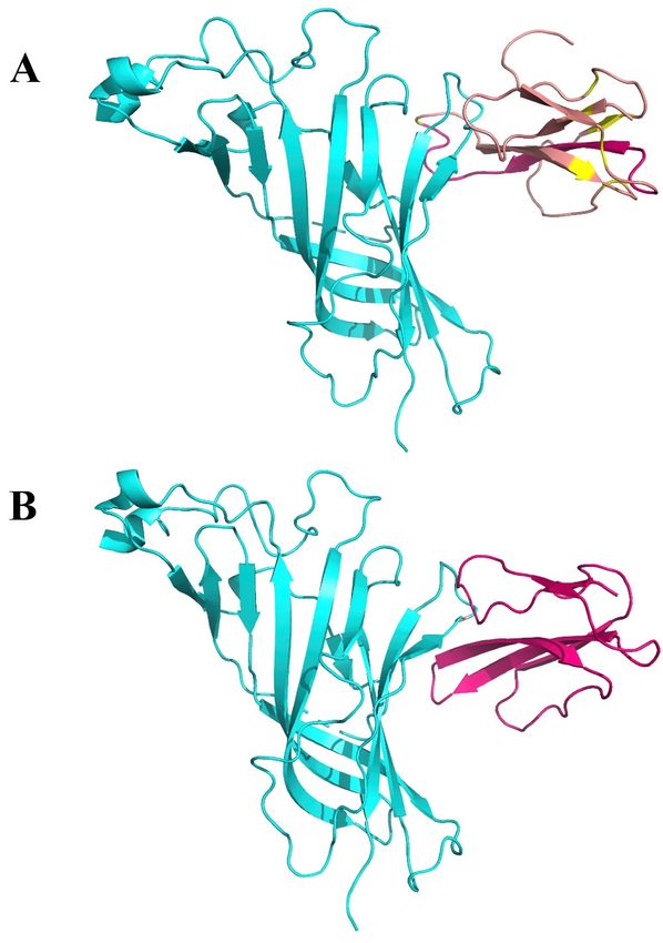
Int. J. Mol. Sci. 2020, 21, 5807 3 of 15
2. Results
Int. J. Mol. Sci. 2020, 21, x FOR PEER REVIEW 3 of 15
2.1. Sequence Alignment
2.1. Sequence Alignment
Figure 1A presents the sequence alignment between the SARS-CoV-2 Spike Glycoprotein
Figure 1A presents the sequence alignment between the SARS-CoV-2 Spike Glycoprotein
(P0DTC2—upper lines) and Neurotoxin homolog NL1 (Q9DEQ3—lower lines). We identified a
(P0DTC2—upper lines) and Neurotoxin homolog NL1 (Q9DEQ3—lower lines). We identified a
double “recombination” within the same sequence of S protein (aa 375–390) which is homologous in
double “recombination” within the same sequence of S protein (aa 375–390) which is homologous in
sequence to an equivalent of the neurotoxin homolog NL1, part of the toxin’s “three-finger” interacting
sequence to an equivalent of the neurotoxin homolog NL1, part of the toxin’s “three-finger”
motif. Figure 1B presents the amino acids within this sequence which are identical (red) or functionally
interacting motif. Figure 1B presents the amino acids within this sequence which are identical (red)
equivalent (conservative replacement, having similar biochemical properties—yellow) to Neurotoxin
or functionally equivalent (conservative replacement, having similar biochemical properties—
homolog NL1 toxin. This peptide fragment (aa 375–390) is part of the Receptor Binding Domain
yellow) to Neurotoxin homolog NL1 toxin. This peptide fragment (aa 375–390) is part of the Receptor
(aa 319–541) of the SARS-COV-2 Spike Glycoprotein (the domain through which the Spike protein
Binding Domain (aa 319–541) of the SARS-COV-2 Spike Glycoprotein (the domain through which the
recognizes the ACE2 on the host’s cell surface) neighboring to the ACE2 Receptor Binding Motif
Spike protein recognizes the ACE2 on the host’s cell surface) neighboring to the ACE2 Receptor
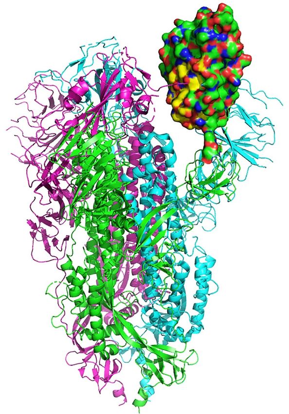
(aa 437–508). Figure 2 displays the structural location of the toxin-like sequence (aa 375–390) within
Binding Motif (aa 437–508). Figure 2 displays the structural location of the toxin-like sequence (aa
the SARS-CoV-2 Spike Glycoprotein. The sequence is located in the Receptor Binding Domain (next to
375–390) within the SARS-CoV-2 Spike Glycoprotein. The sequence is located in the Receptor Binding
the Receptor Binding Motif) of the SARS-CoV-2 Spike Glycoprotein.
Domain (next to the Receptor Binding Motif) of the SARS-CoV-2 Spike Glycoprotein.
Figure 1. (A). Sequence alignment between the SARS-CoV-2 Spike (S1) Glycoprotein (P0DTC2—upper
lines) and Neurotoxin homolog NL1 (Q9DEQ3—lower lines). (B) Amino acids, within this sequence,
Figure are
which 1. (A). Sequence
identical (red)alignment between
or functionally the SARS-CoV-2
equivalent (yellow) Spike (S1) Glycoprotein
to Neurotoxin homolog (P0DTC2—
NL1 toxin
upper lines)
are shown. and Neurotoxin homolog NL1 (Q9DEQ3—lower lines). (B) Amino acids, within this
sequence, which are identical (red) or functionally equivalent (yellow) to Neurotoxin homolog NL1
toxin are shown.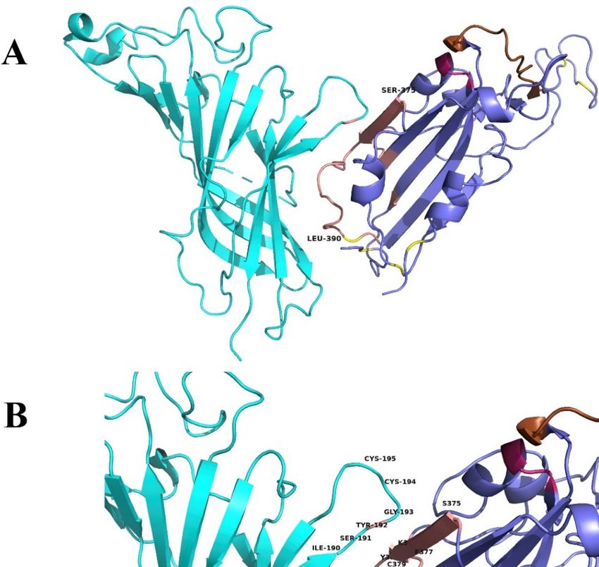
Int. J. Mol. Sci. 2020, 21, 5807                                                                           4 of 15
Int. J. Mol. Sci. 2020, 21, x FOR PEER REVIEW                                                              4 of 15

     Figure
     Figure 2.
            2. Structural
               Structural location
                           location of
                                    of the
                                        the toxin-like
                                            toxin-like sequence
                                                        sequence (aa
                                                                  (aa 375–390)
                                                                      375–390) within
                                                                               within the
                                                                                      the SARS-CoV-2
                                                                                           SARS-CoV-2 Spike
                                                                                                      Spike
     Glycoprotein.
     Glycoprotein. Receptor
                    Receptor Binding
                              Binding Domain
                                       Domain is is in
                                                    in green
                                                       green and
                                                             and the
                                                                 the aa
                                                                     aa 375–390
                                                                        375–390 peptide
                                                                                peptide is
                                                                                        is in
                                                                                           in yellow.
                                                                                              yellow.

2.2. Interaction
2.2.  Interaction between
                  between SARS-CoV-2
                          SARS-CoV-2 and
                                     and nAChRs
                                         nAChRs
     The proposed
     The  proposed interface
                       interface region
                                 region (Figure
                                        (Figure 3)3) is
                                                     is formed
                                                         formed between
                                                                 between the
                                                                          the aa
                                                                               aa 381–386
                                                                                  381–386 ofof the
                                                                                               the SARS-CoV-2
                                                                                                   SARS-CoV-2
Spike Glycoprotein
Spike  Glycoprotein andand the
                            the aa
                                aa 189–192
                                   189–192 of
                                           of the
                                               the extracellular
                                                    extracellular domain
                                                                  domain of
                                                                          of the
                                                                             the nAChR
                                                                                  nAChR α9     subunit, aa region
                                                                                           α9 subunit,      region
which  forms   the core  of the “toxin-binding   site”  of the nAChRs  [18]. The  interaction   between
which forms the core of the “toxin-binding site” of the nAChRs [18]. The interaction between the two     the   two
proteins is
proteins is caused
             caused byby hydrogen
                          hydrogen bonds
                                     bonds and
                                            and shape
                                                  shape complementarity.
                                                           complementarity. The
                                                                             The mode
                                                                                   mode ofof interaction
                                                                                             interaction isis very
                                                                                                              very
similar with   the interaction   between  α9  nAChR      and  α-bungarotoxin   and  neurotoxin
similar with the interaction between α9 nAChR and α-bungarotoxin and neurotoxin homolog NL1       homolog     NL1
(Figures 4  and  5), two  snake  venom  toxins  which     are known to inhibit nAChRs.    A  similar
(Figures 4 and 5), two snake venom toxins which are known to inhibit nAChRs. A similar interaction   interaction
was found
was  found between
             between thethe ligand
                            ligand binding
                                    binding domain
                                            domain of   of the
                                                           the pentameric
                                                               pentameric α7   nicotinic receptor
                                                                           α7 nicotinic  receptor chimera
                                                                                                   chimera and and
the SARS-CoV-2     Spike   Glycoprotein  (Figure
the SARS-CoV-2 Spike Glycoprotein (Figure 6).      6).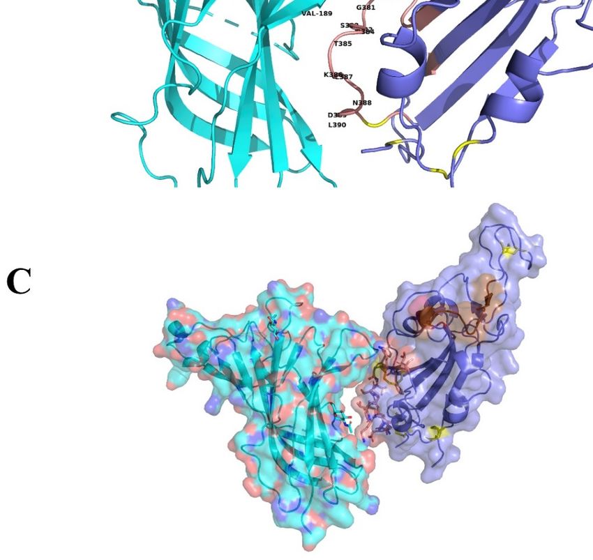
Int. J. Mol. Sci. 2020, 21, 5807                                                                                       5 of 15
   Int. J. Mol. Sci. 2020, 21, x FOR PEER REVIEW                                                                    5 of 15

      Figure 3. (A)
        Figure       Molecular
                 3. (A)          docking
                         Molecular         of SARS-CoV-2
                                      docking              Spike Glycoprotein
                                                of SARS-CoV-2                    (purple)(purple)
                                                                   Spike Glycoprotein      with α9 AChR   extracellular
                                                                                                     with α9   AChR
      subunit  (green).
        extracellular       The(green).
                       subunit    interaction  between between
                                         The interaction  the two theproteins  is caused
                                                                        two proteins       by hydrogen
                                                                                      is caused            bonds
                                                                                                 by hydrogen   bondsand
      shape
        and complementarity.
             shape complementarity. SpikeSpike
                                          glycoprotein   is interacting
                                                glycoprotein              with
                                                              is interacting   thethe
                                                                             with   toxin-like  sequence
                                                                                       toxin-like sequence(aa
                                                                                                            (aa375–390)
                                                                                                                 375–
        390) (brown
      (brown  color).color).  (B) interaction
                        (B) The   The interaction  between
                                               between   α9α9   nAChRsubunit
                                                             nAChR      subunit(aa
                                                                                 (aa 189–195
                                                                                     189–195 are
                                                                                               areforming
                                                                                                   formingthe
                                                                                                            the“toxin
                                                                                                                  “toxin
        binding
      binding    site’)
               site’)   andthe
                      and    thebinding
                                 bindingsite
                                          sitewith
                                               withmore
                                                    more details.
                                                         details. (C)
                                                                   (C) Surface
                                                                       Surfacemodels.
                                                                                models.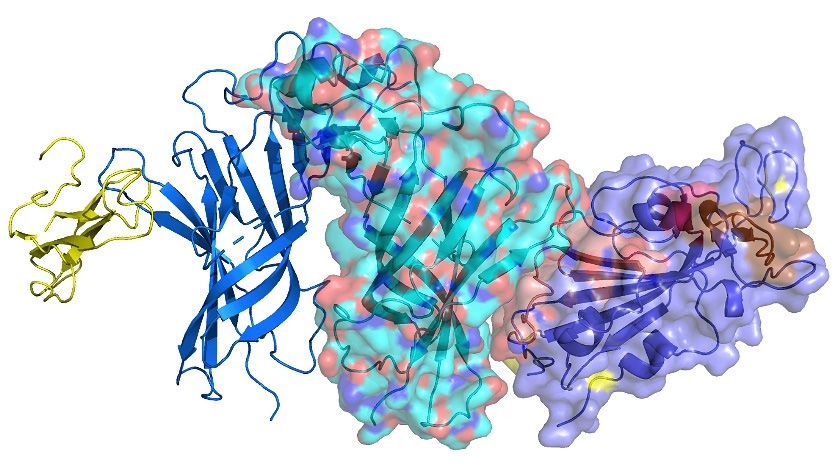
Int. J. Mol. Sci. 2020, 21, x FOR PEER REVIEW                                                                         6 of 15

Int. J. Mol. Sci. 2020, 21, 5807                                                                                          6 of 15
      Int. J. Mol. Sci. 2020, 21, x FOR PEER REVIEW                                                             6 of 15

      Figure 4. (A). Interaction between α9 AChR (green—on the left) and α-bungarotoxin (brown—on the
      Figure
      right).  4. (A).
           Figure
              (B).       Interaction
                    4. (A). Interaction
                   Interaction         between
                                 betweenbetween
                                           α9 AChR  AChR
                                                α9AChR
                                                α9          (green—on
                                                          (green—on
                                                      (green—on     thethe theand
                                                                           left)
                                                                        left)    left) and α-bungarotoxin
                                                                                 andneurotoxin
                                                                                      α-bungarotoxin        (brown—on
                                                                                                     (brown—on
                                                                                                 homolog  NL1   the
                                                                                                              (purple—
      the right).
           right).   (B).
                   (B).
      on the right).       Interaction
                        Interaction      between
                                     between α9    α9
                                                AChR   AChR    (green—on
                                                       (green—on   the left)  the
                                                                             and    left) and
                                                                                  neurotoxin   neurotoxin
                                                                                              homolog NL1 homolog
                                                                                                          (purple—  NL1
      (purple—on      the right).
           on the right).

      Figure 5. Interaction between two (of the five) α9 nAChR subunits (green—in the middle),
      alpha-bungarotoxin (yellow—on the left) and SARS-CoV-2 Spike Glycoprotein (purple—on the right).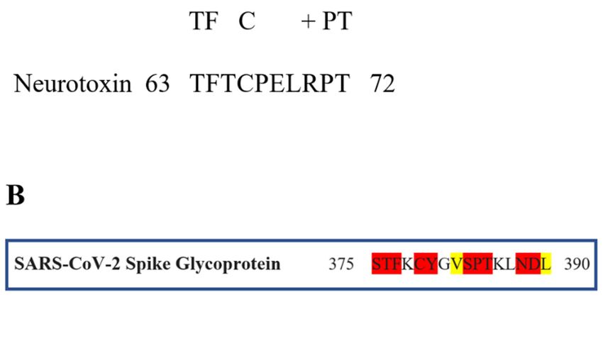
Int. J. Mol. Sci. 2020, 21, x FOR PEER REVIEW                                                                  7 of 15

Int. J.Figure
       Mol. Sci.5.2020,
                   Interaction
                        21, 5807 between two (of the five) α9 nAChR subunits (green—in the middle), alpha- 7 of 15
     bungarotoxin (yellow—on the left) and SARS-CoV-2 Spike Glycoprotein (purple—on the right).

      Figure 6. (A) Interaction between the ligand binding domain of a pentameric α7 nicotinic receptor
     Figure 6. (A). Interaction between the ligand binding domain of a pentameric α7 nicotinic receptor
      chimera (left) and SARS-CoV-2 Spike Glycoprotein (purple—on the right). (B) α-bungarotoxin
     chimera (left) and SARS-CoV-2 Spike Glycoprotein (purple—on the right). (B). α-bungarotoxin and
      and Neurotoxin homolog NL1 bound to the pentameric nicotinic receptor. (C) SARS-CoV-2 Spike
     Neurotoxin homolog NL1 bound to the pentameric nicotinic receptor. (C). SARS-CoV-2 Spike
      Glycoprotein (purple—on the right) bound to the pentameric nicotinic receptor (top view) and side
     Glycoprotein (purple—on the right) bound to the pentameric nicotinic receptor (top view) and side
      view. (D) The beginning of the toxin-like sequence (S375) and the top of the finger-like fragment (F392)
     view. (D). The beginning of the toxin-like sequence (S375) and the top of the finger-like fragment
      are shown. (E) Close view of the interacting surfaces. (F) Neurotoxin homolog NL1 bound to the
     (F392) are shown. (E). Close view of the interacting surfaces. (F). Neurotoxin homolog NL1 bound to
      pentameric nicotinic receptor in close view, emphasizing the interaction between the toxin and the
     the pentameric nicotinic receptor in close view, emphasizing the interaction between the toxin and
      nicotinic receptor.
     the nicotinic receptor.
 2.3. Potential Therapeutic Targeting Implications for COVID-19
2.3. Potential Therapeutic Targeting Implications for COVID-19
      The in silico experiments of this study identified a potential interaction between SARS-CoV-2
 and The   in silico
      nAChRs.        experiments
                  If verified        of and
                               in vivo   this study   identified
                                              in vitro,           a potential
                                                        these findings    couldinteraction   between
                                                                                  have significant      SARS-CoV-2
                                                                                                     implications    in
and   nAChRs. If the
 understanding      verified  in vivo and in
                        pathophysiology       of vitro, these findings
                                                  COVID-19               could have
                                                               and its clinical          significant implications
                                                                                   manifestations,   and could also in
understanding
 result in novelthe     pathophysiology
                    therapeutic               of COVID-19
                                   interventions.              and its of
                                                       The hallmark     clinical
                                                                           severemanifestations,
                                                                                     COVID-19 is and      couldstorm.
                                                                                                     cytokine     also
result  in novel therapeutic
 This represents                 interventions.
                   a form of immune                The hallmark
                                           dysregulation           of severe
                                                            and also          COVID-19
                                                                      a failure             is cytokine storm.
                                                                                  of the inflammatory    responseThisto
represents   a form  of immune     dysregulation     and  also a failure of the  inflammatory     response
 return to homeostasis [18]. While the initial response is important in controlling inflammation and        to return
to homeostasis
 infection,       [18]. While
            uncontrolled        the initial
                            cytokine        response
                                       release  can beisdetrimental,
                                                         important inleading
                                                                        controlling    inflammation
                                                                                 to tissue             and infection,
                                                                                            damage including    acute
uncontrolled
 lung injury andcytokine
                    ARDS release
                            [19,20]. can
                                      Casebeseries
                                               detrimental,   leading
                                                    of COVID-19        to tissue
                                                                    patients  havedamage      including
                                                                                      shown that         acute
                                                                                                   elevated      lung
                                                                                                             levels  of
injury   and  ARDS    [19,20].   Case   series  of  COVID-19     patients  have    shown    that
 cytokines and inflammatory markers predicted adverse outcomes such as mechanical ventilation and elevated  levels  of
cytokines and inflammatory markers predicted adverse outcomes such as mechanical ventilation and
Int. J. Mol. Sci. 2020, 21, 5807 8 of 15
death [21–23]. Autopsy findings also suggested that severe COVID-19 is associated with immune
dysregulation [24].
The nicotinic cholinergic system has been identified as an important modulator of the inflammatory
response. The “cholinergic anti-inflammatory pathway” represents a neural pathway of immune
homeostasis and cytokine synthesis control [25]. This is a reflex, bi-directional interaction between the
nervous and immune system. Sensory input about the inflammatory status transmits information to
the central nervous system through the afferent fibers of the vagus nerve, while appropriate responses
generate from the efferent fibers to subsequently modulate the inflammatory response and cytokine
release [12,26]. This is a rapid reflex mechanism. In 2000, Borovikova et al. first identified that vagus
nerve stimulation attenuated the inflammatory response to endotoxin and reduced the production of
TNF-α in a rat model of septic shock [27]. Surgical vagotomy significantly enhanced TNF-α response
to inflammatory stimuli [12]. Subsequently, several experimental studies identified the α7 nAChR as a
key mediator of the neural modulation of inflammation. For example, Wang et al., reported that α7
nAChR knockout mice showed elevated TNF-α production in response to endotoxin, while electrical
vagus stimulation did not attenuate this response [13]. Alpha7 nAChRs are present in macrophages and
B- and T-lymphocytes [28,29]. Additionally, they are present in human bronchial epithelial cells [16],
alveolar epithelial type II cells [30], endothelial cells [15] and neutrophils [31].
The cholinergic anti-inflammatory pathway is particularly active in the lungs. Parasympathetic
innervation exists in the airway wall and stimuli are transmitted to the central nervous system through
afferent vagus fibers. Pulmonary nociceptors (chemosensitive receptors) were found to be activated by
pro-inflammatory cytokines and transmit signals through afferent vagus fibers to the central nervous
system [30,32]. Postganglionic cholinergic neurons, which innervate lung tissue, are stimulated through
the activation of the efferent vagus nerve fibers, resulting in the activation of α7 nAChR on infiltrated
inflammatory cells during acute lung injury, suppressing the production of pro-inflammatory cytokines
and attenuating lung injury [33,34].
This study identified that there may be a direct interaction between SARS-CoV-2 and nAChRs.
This could potentially result in the dysfunction of these receptors and disruption of the cholinergic
anti-inflammatory pathway. Nicotinic agonists could protect from this disruption by activating
nAChRs. Experimental data have shown that several α7 nAChR agonists, including nicotine, decreased
inflammatory manifestations in an acid-induced acute lung injury mouse model [31]. A 60% reduction
in excess lung water and extravascular plasma equivalents was observed in nicotine-treated compared
to saline-treated groups, while histological examination revealed less pulmonary edema and neutrophil
infiltration in the nicotine-treated group. The effects were counteracted by an α7 nAChR antagonist [31].
Additionally, protein concentration, neutrophil counts, cytokine levels and epithelial cell injury were
reduced in bronchoalveolar lavage of nicotine-treated mice. In another study examining a model
of acute lung inflammatory injury induced by Gram-negative sepsis, nicotine promoted the local
suppression of inflammatory mediator production by regulating pro-inflammatory cell transmigration
and trans-alveolar permeability [32]. Activation of nicotine also increased survival of mice with
Gram-negative pneumonia [32]. Similar protective effects were observed in lipopolysaccharide-induced
acute lung injury, with nicotine suppressing the release of several pro-inflammatory cytokines [32,35,36].
These effects appear to be mediated through inhibition of NF-κB activity [31,37–39].
Nicotine is an approved medication that has been available for years in different forms,
specifically transdermal patches, gums, nasal sprays and oral inhalers and sublingual tablets/lozenges.
These formulations are mainly used as smoking cessation interventions and are generally well tolerated
and with minimal side effects [40,41]. While nicotine use is currently confined to smokers, prescribed
as a smoking substitute, it has been administered therapeutically for the treatment of neurologic or
gastrointestinal disorders in non-smoking patients for several weeks, with minimal side effects [42–45].
In one study, a daily dose of up to 90mg was administered [42]. No nicotine dependence was reported
among non-smokers when nicotine was withdrawn after study completion. Therefore, administering
nicotine for few days, as is expected to be the treatment duration, for COVID-19 appears to be feasibleInt. J. Mol. Sci. 2020, 21, 5807 9 of 15
even for non-smokers—with specific precautions, such as in patients with recent myocardial injury or
unstable coronary artery disease.
Besides nicotine, other nicotinic agonists may have a therapeutic role by protecting nAChRs and
activating the cholinergic anti-inflammatory pathway. Choline, a precursor of acetylcholine, was found
to suppress murine endotoxemia and sepsis [31,46]. Galantamine is a central acetylcholinesterase
inhibitor approved for the treatment of mild to moderate dementia and Alzheimer’s disease. It is also
a positive allosteric ligand of nAChRs42 and has been shown to stimulate peripheral α7 nAChRs in an
experimental model of colitis showing potent anti-inflammatory effects [47]. Experimental studies have
shown that it protects against lipopolysaccharide-induced acute lung injury in rats and acid-induced
ARDS in rabbits, suggesting that it acts on the cholinergic anti-inflammatory pathway [48]. Varenicline
is an approved smoking cessation medication that exhibits strong α7 nAChR agonist activity [49].
One study showed that it exhibits anti-inflammatory property in the lung tissue of mice, mediated via
α7 nAChRs [50]. These medications could be proposed for clinical trials, although nicotine has been
tested far more extensively in relation to the cholinergic anti-inflammatory pathway.
An overview of the nicotinic agonists presented above is shown in Table 1. The proposed
hypothesis for the interaction between SARS-CoV-2 and nAChRs and the therapeutic implications are
displayed in Figure 7.
Table 1. Approved nicotinic cholinergic agonists that could be tested in in vitro and in vivo studies for
their effects on COVID-19.
FDA Approved Brand
Compound Mode of Action Dose
Approval Indication Name
Choline Acetylcholine (1) Dietary 550 mg/day Various
synthesis precursor supplement (men)
425 mg/day
(women) (2)
Nicotine Alpha7 AChR agonist Yes Smoking Maximum Nicorette
cessation dose (3) Nicoderm
21 mg/day Nicotrol
(patch) Others
40 mg/day
(nasal
spray)
64 mg/day
(inhaler)
96 mg/day
(gum)
Galantamine Weak Yes Alzheimer’s 16–24 Reminyl
acetylcholinesterase disease mg/day Razadyne
inhibitor
Allosteric agonist for
nicotinic acetylcholine
receptors
Varenicline Alpha7 AChR agonist Yes Smoking 2 mg/day Champix
Alpha4 beta2 AChR cessation Chantix
partial agonist
(1) Choline is an essential nutrient and is available as a dietary supplement in various formulations. (2) The dose
refers to Adequate Intake (AI). (3) Nicotine dose refers to smokers who are nicotine users (through smoking) and,
thus, have developed tolerance.cessation Chantix
Alpha4 beta2 AChR partial
agonist
(1) Choline is an essential nutrient and is available as a dietary supplement in various formulations.
(2) The dose refers to Adequate Intake (AI). (3) Nicotine dose refers to smokers who are nicotine users
Int. J. Mol. Sci. 2020, 21, 5807 10 of 15
(through smoking) and, thus, have developed tolerance.
Figure 7. Graphic
Figure Graphicdisplay
displayof of
thethe
interaction between
interaction SARS-CoV-2
between and α7and
SARS-CoV-2 nicotinic acetylcholine
α7 nicotinic acetylcholine
receptors (nAChRs)
receptors (nAChRs)and
andthe potential
the therapeutic
potential implications.
therapeutic implications.
3.3.Discussion
Discussion
Animal venoms
Animal venomsand andespecially
especially snake
snakevenoms havehave
venoms evolved to contain
evolved a wide adiversity
to contain of
wide diversity of
proteins that induce inflammatory and toxic effects [17]. Their pharmacological properties
proteins that induce inflammatory and toxic effects [17]. Their pharmacological properties have been have been
well-studied, revealing a complex mode of action. Many of these toxins exert their action by binding
well-studied, revealing a complex mode of action. Many of these toxins exert their action by binding
to the muscle or the neuronal type nAChRs [16,20]. Neurotoxins, such as α-bungarotoxin, interact
to the muscle or the neuronal type nAChRs [16,20]. Neurotoxins, such as α-bungarotoxin, interact
with the ACh binding site of nAChRs with the sequence aa185–200 being of great importance for
with
suchthe ACh binding
binding sitemolecular
[51,52].The of nAChRs with theand
modelling sequence
dockingaa185–200
experiments being of greatinimportance
presented this study for such
binding [51,52].The molecular modelling and docking experiments presented
suggest an interaction between nAChRs and SARS-CoV-2 Spike Glycoprotein, with the sequence in this study suggest an
interaction
aa189–195 of between
the nAChRnAChRs
being and
at theSARS-CoV-2 Spike Glycoprotein,
core of this interaction. with the sequence
This could compromise the NCSaa189–195
and of
the
thenAChR being
cholinergic at the core of this
anti-inflammatory interaction.
pathway, leading toThis could compromise
a hyper-immune responsethe
andNCS and the
cytokine cholinergic
storm.
The consistent pathway,
anti-inflammatory observations of a low
leading to arate of smoking among
hyper-immune hospitalized
response COVID-19
and cytokine storm.patients
(despite the limitations and perplexities), the potential links between dysfunction
The consistent observations of a low rate of smoking among hospitalized COVID-19 of the NCS and patients
clinical manifestations of COVID-19 and the indications for a direct interaction between
(despite the limitations and perplexities), the potential links between dysfunction of the NCS and SARS-CoV-
2 and nAChRs leading to NCS dysregulation generate the hypothesis for a novel therapeutic
clinical manifestations of COVID-19 and the indications for a direct interaction between SARS-CoV-2
intervention aiming at restoring the function of the cholinergic anti-inflammatory pathway and
and nAChRs leading to NCS dysregulation generate the hypothesis for a novel therapeutic intervention
promoting immune homeostasis [10]. Therapeutic interventions to reduce the hyper-immune
aiming
responseat restoring
have already the been
function of theand
suggested cholinergic anti-inflammatory
are currently underway, in some pathway
cases and promoting immune
for medications
homeostasis [10]. Therapeutic interventions to reduce the hyper-immune response have already been
suggested and are currently underway, in some cases for medications with warnings and precautions
for their use in active infections [53–57]. While our hypothesis is similarly oriented to controlling the
cytokine storm, a different pathway is proposed with medications that are relatively safe and not
contraindicated for use in active infections. Furthermore, it should be clarified that the interaction and
potential implications presented in this study are not linked to the renin-angiotensin system which,
through ACE2, is involved in viral cell entry and replication. We postulate that the pathophysiological
mechanisms through which the virus causes severe disease, relevant to an uncontrolled response of the
immune system to viral invasion and failure to return to homeostasis, are at least partly different from
the mode of cell entry and replication. The findings presented herein suggest that a different pathway
may be targeted as a mediator for COVID-19 progression and associated symptoms, the nicotinic
cholinergic system. We also provide insight about the potential therapeutic role of already approvedInt. J. Mol. Sci. 2020, 21, 5807 11 of 15
medications, which can be used through repurposing, in alleviating symptoms and preventing disease
progression without hindering viral replication.
A limitation of this study is that it is based on a theoretical model, and there is currently no in vitro
or in vivo study that has examined the possibility of an interaction between SARS-Cov-2 and nAChRs.
The study was initiated based on clinical observations about the association between smoking and
COVID-19 among hospitalized patients. Tobacco cigarette smoke contains thousands of compounds,
most of which have known toxic effects. The possibility that other chemicals besides nicotine may
be associated with a potentially beneficial effect in COVID-19 cannot be excluded. For example, low
levels of carbon monoxide may have anti-inflammatory properties and have been used experimentally
for lung sepsis [58]. The results of this in silico study suggest a plausible mechanism through which
nicotine may be implicated in the course of COVID-19 but does not examine the effects of other
chemicals present in tobacco cigarette smoke.
The activation of nAChRs by either endogenous (acetylcholine) or exogenous agonists is induced
by opening the ion channel in the receptor, allowing the flow of cations, and results in a variety of
biological responses. nAChR antagonists, such as α-neurotoxins, compete with typical agonists for
binding, and their binding is restricted to nAChR α-subunits. Nicotine and other nicotinic cholinergic
agonists (choline, varenicline and galantamine) are FDAapproved drugs for a number of pathologies
(including for smoking cessation) and may reverse this binding, by competing for binding with
the SARS-CoV-2 Spike Glycoprotein, and promote the activity of the cholinergic anti-inflammatory
pathway. This needs to be studied further and eventually verified in in vitro and in vivo studies.
4. Materials and Methods
4.1. Sequence Retrieval and Alignment
To examine this hypothesis, we compared amino acid sequences between SARS-CoV-2 and snake
venom neurotoxins. The latter are well-established inhibitors of the NCS [17]. The protein sequences of
the “three finger” neurotoxin and the SARS-CoV-2 Spike Glycoprotein were retrieved from the National
Center for Biotechnology Information (NCBI, Bethesda, MD, USA) database with details (designation
and accession numbers) listed in Figure 1. Mega BLAST [59] was used for Blastp (protein-protein
BLASTS) searches at the UNIPROT database (and PDB and SwissProt). ClustalOmega (Clustal-O) [60]
was used to perform all the multiple sequence alignment programs. Default parameters were used for
the alignment.
4.2. Structure Retrieval, Alignment and Modelling
The three dimensional structures of the SARS-CoV-2 Spike Glycoprotein (S1) in complex with
the human angiotensin converting enzyme 2 (hACE2) (PDB id: 6LZG), the hACE2 (1R41, 1R42),
the cryo-EM determined complex of spike protein S-ACE2-B0 AT1 neutral amino acid transporter (PDB
id: 6M18), the structure of a neutralizing to SARS-CoV monoclonal antibody that also cross reacts
with the S protein of SARS-CoV-2 when the latter is in complex with the ACE2 receptor (PDB id:
6NB7) and the extracellular domain of the nAChR α9 subunit in complex with α-bungarotoxin (PDB
id: 3U8M) were downloaded from the Protein Data Bank [61] and used to analyze the consequences
to structure and function of the interaction between S1 and nAChR. The crystal structures of the
ligand-binding domain of a pentameric α7 nicotinic receptor chimera (PDB id: 3SQ9) were used
for additional structural comparisons [62]. All figures depicting 3D models were created using the
molecular graphics program PyMOL V.2.2 [63]. Molecular modelling and molecular docking studies
were performed using the software packages MOE and PyMol [63,64]. Since the structure of the
human α7 nAChR is not solved yet, we used the crystal structure of the extracellular domain of the
homologous α9 nAChR bound to α-bungarotoxin.Int. J. Mol. Sci. 2020, 21, 5807 12 of 15
5. Conclusions
This in silico study identified a potential interaction between SARS-CoV-2 and nAChRs,
which could result in dysregulation of the cholinergic anti-inflammatory pathway and could adversely
affect immune homeostasis in COVID-19. These findings have potential therapeutic implications
since cholinergic agonists could protect and restore the function of nAChRs. Nicotine, varenicline,
and galantamine—examples of FDA-approved cholinergic agonists for various pathologies—are
proposed as therapeutic interventions for COVID-19, targeting the NCS. Since these proposals
are based on theoretical models, further in vitro and in vivo studies are needed to explore these
pathophysiological mechanisms.
Author Contributions: K.F. and K.P. conceived the study. E.E., D.D.L. and G.E.P. performed the in silico
experiments and modelling studies. K.F., S.T. and K.P. interpreted the data. K.P., E.E., D.D.L. and S.T. wrote the
first draft of the manuscript. K.F. and K.P. revised the manuscript and authored the final version. All authors have
read and approved the content of the manuscript submitted for publication.
Funding: Authors are grateful to the “National Research Infrastructures on Integrated Structural Biology,
Drug Screening Efforts and Drug Target Functional Characterization (INSPIRED)” for financial support of
the personnel.
Conflicts of Interest: The authors declare no conflicts of interest.
References
1. Hoffmann, M.; Kleine-Weber, H.; Schroeder, S.; Krüger, N.; Herrler, T.; Erichsen, S.; Schiergens, T.S.; Herrler, G.;
Wu, N.-H.; Nitsche, A.; et al. SARS-CoV-2 Cell Entry Depends on ACE2 and TMPRSS2 and Is Blocked by a
Clinically Proven Protease Inhibitor. Cell 2020, 181, 271–280.e8. [CrossRef]
2. Wang, Q.; Zhang, Y.; Wu, L.; Niu, S.; Song, C.; Zhang, Z.; Lu, G.; Qiao, C.; Hu, Y.; Yuen, K.Y.; et al. Structural
and Functional Basis of SARS-CoV-2 Entry by Using Human ACE2. Cell 2020, 181, 894–904.e9. [CrossRef]
[PubMed]
3. Walls, A.C.; Park, Y.J.; Tortorici, M.A.; Wall, A.; McGuire, A.T.; Veesler, D. Structure, Function, and Antigenicity
of the SARS-CoV-2 Spike Glycoprotein. Cell 2020, 181, 281–292.e6. [CrossRef] [PubMed]
4. Vaduganathan, M.; Vardeny, O.; Michel, T.; McMurray, J.J.V.; Pfeffer, M.A.; Solomon, S.D. Renin-Angiotensin-
Aldosterone System Inhibitors in Patients with Covid-19. N. Engl. J. Med. 2020, 382, 1653–1659. [CrossRef]
5. Verdecchia, P.; Cavallini, C.; Spanevello, A.; Angeli, F. The pivotal link between ACE2 deficiency and
SARS-CoV-2 infection. Eur. J. Intern. Med. 2020, 76, 14–20. [CrossRef]
6. Arcavi, L.; Benowitz, N.L. Cigarette smoking and infection. Arch. Intern. Med. 2004, 164, 2206–2216.
[CrossRef]
7. Oakes, J.M.; Fuchs, R.M.; Gardner, J.D.; Lazartigues, E.; Yue, X. Nicotine and the renin-angiotensin system.
Am. J. Physiol. Regul. Integr. Comp. Physiol. 2018, 315, R895–R906. [CrossRef]
8. Brake, S.J.; Barnsley, K.; Lu, W.; McAlinden, K.D.; Eapen, M.S.; Sohal, S.S. Smoking Upregulates
Angiotensin-Converting Enzyme-2 Receptor: A Potential Adhesion Site for Novel Coronavirus SARS-CoV-2
(Covid-19). J. Clin. Med. 2020, 9, 841. [CrossRef]
9. Farsalinos, K.; Barbouni, A.; Niaura, R. Systematic review of the prevalence of current smoking among
hospitalized COVID-19 patients in China: Could nicotine be a therapeutic option? Intern. Emerg. Med. 2020.
[CrossRef]
10. Farsalinos, K.; Niaura, R.; Le Houezec, J.; Barbouni, A.; Tsatsakis, A.; Kouretas, D.; Vantarakis, A.; Poulas, K.
Editorial: Nicotine and SARS-CoV-2: COVID-19 may be a disease of the nicotinic cholinergic system.
Toxicol. Rep. 2020, 7, 658–663. [CrossRef]
11. Mehta, P.; McAuley, D.F.; Brown, M.; Sanchez, E.; Tattersall, R.S.; Manson, J.J.; HLH Across Speciality
Collaboration, UK. COVID-19: Consider cytokine storm syndromes and immunosuppression. Lancet
2020, 395, 1033–1034. [CrossRef]
12. Tracey, K.J. The inflammatory reflex. Nature 2002, 420, 853–859. [CrossRef] [PubMed]
13. Wang, H.; Yu, M.; Ochani, M.; Amella, C.A.; Tanovic, M.; Susarla, S.; Li, J.H.; Wang, H.; Yang, H.; Ulloa, L.; et al.
Nicotinic acetylcholine receptor alpha7 subunit is an essential regulator of inflammation. Nature 2003, 421,
384–388. [CrossRef] [PubMed]Int. J. Mol. Sci. 2020, 21, 5807 13 of 15
14. Fujii, T.; Mashimo, M.; Moriwaki, Y.; Misawa, H.; Ono, S.; Horiguchi, K.; Kawashima, K. Expression and
Function of the Cholinergic System in Immune Cells. Front Immunol. 2017, 8, 1085. [CrossRef] [PubMed]
15. Wang, Y.; Pereira, E.F.; Maus, A.D.; Ostlie, N.S.; Navaneetham, D.; Lei, S.; Albuquerque, E.X.; Conti-Fine, B.M.
Human bronchial epithelial and endothelial cells express alpha7 nicotinic acetylcholine receptors.
Mol. Pharmacol. 2001, 60, 1201–1209. [CrossRef] [PubMed]
16. Kalamida, D.; Poulas, K.; Avramopoulou, V.; Fostieri, E.; Lagoumintzis, G.; Lazaridis, K.; Sideri, A.; Zouridakis, M.;
Tzartos, S.J. Muscle and neuronal nicotinic acetylcholine receptors. FEBS J. 2007, 274, 3799–3845. [CrossRef]
17. Tsetlin, V.I.; Hucho, F. Snake and snail toxins acting on nicotinic acetylcholine receptors: Fundamental
aspects and medical applications. FEBS Lett. 2004, 557, 9–13. [CrossRef]
18. Shimabukuro-Vornhagen, A.; Gödel, P.; Subklewe, M.; Stemmler, H.J.; Schlößer, H.A.; Schlaak, M.; Kochanek, M.;
Böll, B.; von Bergwelt-Baildon, M.S. Cytokine release syndrome. J. Immunother. Cancer 2018, 6, 56. [CrossRef]
19. Turner, M.D.; Nedjai, B.; Hurst, T.; Pennington, D.J. Cytokines and chemokines: At the crossroads of cell
signalling and inflammatory disease. Biochim. Biophys. Acta 2014, 1843, 2563–2582. [CrossRef]
20. Dinarello, C.A. Infection, fever, and exogenous and endogenous pyrogens: Some concepts have changed.
J. Endotoxin Res. 2004, 10, 201–222.
21. Ruan, Q.; Yang, K.; Wang, W.; Jiang, L.; Song, J. Clinical predictors of mortality due to COVID-19 based on
an analysis of data of 150 patients from Wuhan, China. Intensive Care Med. 2020, 46, 846–848. [CrossRef]
[PubMed]
22. Qin, C.; Zhou, L.; Hu, Z.; Zhang, S.; Yang, S.; Tao, Y.; Xie, C.; Ma, K.; Shang, K.; Wang, W.; et al. Dysregulation
of immune response in patients with COVID-19 in Wuhan, China. Clin. Infect. Dis. 2020, ciaa248. [CrossRef]
23. Zhou, F.; Yu, T.; Du, R.; Fan, G.; Liu, Y.; Liu, Z.; Xiang, J.; Wang, Y.; Song, B.; Gu, X.; et al. Clinical course and
risk factors for mortality of adult inpatients with COVID-19 in Wuhan, China: A retrospective cohort study.
Lancet 2020, 395, 1054–1062. [CrossRef]
24. Xu, Z.; Shi, L.; Wang, Y.; Zhang, J.; Huang, L.; Zhang, C.; Liu, S.; Zhao, P.; Liu, H.; Zhu, L.; et al. Pathological
findings of COVID-19 associated with acute respiratory distress syndrome. Lancet Respir. Med. 2020, 8,
420–422. [CrossRef]
25. Gallowitsch-Puerta, M.; Tracey, K.J. Immunologic role of the cholinergic anti-inflammatory pathway and the
nicotinic acetylcholine alpha 7 receptor. Ann. N. Y. Acad. Sci. 2005, 1062, 209–219. [CrossRef] [PubMed]
26. Pavlov, V.A.; Wang, H.; Czura, C.J.; Friedman, S.G.; Tracey, K.J. The cholinergic anti-inflammatory pathway:
A missing link in neuroimmunomodulation. Mol. Med. 2003, 9, 125–134. [CrossRef] [PubMed]
27. Borovikova, L.V.; Ivanova, S.; Zhang, M.; Yang, H.; Botchkina, G.I.; Watkins, L.R.; Wang, H.; Abumrad, N.;
Eaton, J.W.; Tracey, K.J. Vagus nerve stimulation attenuates the systemic inflammatory response to endotoxin.
Nature 2000, 405, 458–462. [CrossRef]
28. Skok, M.V.; Grailhe, R.; Agenes, F.; Changeux, J.P. The role of nicotinic receptors in B-lymphocyte development
and activation. Life Sci. 2007, 80, 2334–2336. [CrossRef]
29. Fujii, T.; Mashimo, M.; Moriwaki, Y.; Misawa, H.; Ono, S.; Horiguchi, K.; Kawashima, K. Physiological
functions of the cholinergic system in immune cells. J. Pharmacol. Sci. 2017, 134, 1–21. [CrossRef]
30. Li, H.F.; Yu, J. Airway chemosensitive receptors in vagus nerve perform neuro-immune interaction for
lung-brain communication. Adv. Exp. Med. Biol. 2009, 648, 421–426. [CrossRef]
31. Su, X.; Lee, J.W.; Matthay, Z.A.; Mednick, G.; Uchida, T.; Fang, X.; Gupta, N.; Matthay, M.A. Activation of
the alpha7 nAChR reduces acid-induced acute lung injury in mice and rats. Am. J. Respir. Cell Mol. Biol.
2007, 37, 186–192. [CrossRef] [PubMed]
32. Su, X.; Matthay, M.A.; Malik, A.B. Requisite role of the cholinergic alpha7 nicotinic acetylcholine receptor
pathway in suppressing Gram-negative sepsis-induced acute lung inflammatory injury. J. Immunol.
2010, 184, 401–410. [CrossRef] [PubMed]
33. Yu, J.; Lin, S.; Zhang, J.; Otmishi, P.; Guardiola, J.J. Airway nociceptors activated by pro-inflammatory
cytokines. Respir. Physiol. Neurobiol. 2007, 156, 116–119. [CrossRef] [PubMed]
34. Yamada, M.; Ichinose, M. The cholinergic anti-inflammatory pathway: An innovative treatment strategy for
respiratory diseases and their comorbidities. Curr. Opin. Pharmacol. 2018, 40, 18–25. [CrossRef] [PubMed]
35. Mabley, J.; Gordon, S.; Pacher, P. Nicotine exerts an anti-inflammatory effect in a murine model of acute
lung injury. Inflammation 2011, 34, 231–237. [CrossRef]Int. J. Mol. Sci. 2020, 21, 5807 14 of 15
36. Ni, Y.F.; Tian, F.; Lu, Z.F.; Yang, G.D.; Fu, H.Y.; Wang, J.; Yan, X.L.; Zhao, Y.C.; Wang, Y.J.; Jiang, T. Protective
effect of nicotine on lipopolysaccharide-induced acute lung injury in mice. Respiration 2011, 81, 39–46.
[CrossRef]
37. Li, Q.; Zhou, X.D.; Kolosov, V.P.; Perelman, J.M. Nicotine reduces TNF-α expression through a α7
nAChR/MyD88/NF-kB pathway in HBE16 airway epithelial cells. Cell. Physiol. Biochem. 2011, 27, 605–612.
[CrossRef]
38. Yoshikawa, H.; Kurokawa, M.; Ozaki, N.; Nara, K.; Atou, K.; Takada, E.; Kamochi, H.; Suzuki, N.
Nicotine inhibits the production of proinflammatory mediators in human monocytes by suppression of
I-kappaB phosphorylation and nuclear factor-kappaB transcriptional activity through nicotinic acetylcholine
receptor alpha7. Clin. Exp. Immunol. 2006, 146, 116–123. [CrossRef]
39. Marrero, M.B.; Bencherif, M. Convergence of alpha 7 nicotinic acetylcholine receptor-activated pathways for
anti-apoptosis and anti-inflammation: Central role for JAK2 activation of STAT3 and NF-kappaB. Brain Res.
2009, 1256, 1–7. [CrossRef]
40. Wadgave, U.; Nagesh, L. Nicotine Replacement Therapy: An Overview. Int. J. Health Sci. (Qassim) 2016, 10,
425–435. [CrossRef]
41. Lee, P.N.; Fariss, M.W. A systematic review of possible serious adverse health effects of nicotine replacement
therapy. Arch. Toxicol. 2017, 91, 1565–1594. [CrossRef] [PubMed]
42. Villafane, G.; Cesaro, P.; Rialland, A.; Baloul, S.; Azimi, S.; Bourdet, C.; Le Houezec, J.; Macquin-Mavier, I.;
Maison, P. Chronic high dose transdermal nicotine in Parkinson’s disease: An open trial. Eur. J. Neurol.
2007, 14, 1313–1316. [CrossRef] [PubMed]
43. Pullan, R.D.; Rhodes, J.; Ganesh, S.; Mani, V.; Morris, J.S.; Williams, G.T.; Newcombe, R.G.; Russell, M.A.;
Feyerabend, C.; Thomas, G.A.; et al. Transdermal nicotine for active ulcerative colitis. N. Engl. J. Med.
1994, 330, 811–815. [CrossRef] [PubMed]
44. Sandborn, W.J.; Tremaine, W.J.; Offord, K.P.; Lawson, G.M.; Petersen, B.T.; Batts, K.P.; Croghan, I.T.;
Dale, L.C.; Schroeder, D.R.; Hurt, R.D. Transdermal nicotine for mildly to moderately active ulcerative colitis.
A randomized, double-blind, placebo-controlled trial. Ann. Intern. Med. 1997, 126, 364–371. [CrossRef]
[PubMed]
45. Newhouse, P.; Kellar, K.; Aisen, P.; White, H.; Wesnes, K.; Coderre, E.; Pfaff, A.; Wilkins, H.; Howard, D.;
Levin, E.D. Nicotine treatment of mild cognitive impairment: A 6-month double-blind pilot clinical trial.
Neurology 2012, 78, 91–101. [CrossRef]
46. Parrish, W.R.; Rosas-Ballina, M.; Gallowitsch-Puerta, M.; Ochani, M.; Ochani, K.; Yang, L.H.; Hudson, L.;
Lin, X.; Patel, N.; Johnson, S.M.; et al. Modulation of TNF release by choline requires alpha7 subunit nicotinic
acetylcholine receptor-mediated signaling. Version 2. Mol. Med. 2008, 14, 567–574. [CrossRef]
47. Wazea, S.A.; Wadie, W.; Bahgat, A.K.; El-Abhar, H.S. Galantamine anti-colitic effect: Role of alpha-7 nicotinic
acetylcholine receptor in modulating Jak/STAT3, NF-κB/HMGB1/RAGE and p-AKT/Bcl-2 pathways. Sci. Rep.
2018, 8, 5110. [CrossRef]
48. Li, G.; Zhou, C.L.; Zhou, Q.S.; Zou, H.D. Galantamine protects against lipopolysaccharide-induced acute
lung injury in rats. Braz. J. Med. Biol. Res. 2016, 49, e5008. [CrossRef]
49. Arias, H.R.; Feuerbach, D.; Targowska-Duda, K.; Kaczor, A.A.; Poso, A.; Jozwiak, K. Pharmacological and
molecular studies on the interaction of varenicline with different nicotinic acetylcholine receptor subtypes.
Potential mechanism underlying partial agonism at human α4β2 and α3β4 subtypes. Biochim. Biophys. Acta
2015, 1848, 731–741. [CrossRef]
50. Koga, M.; Kanaoka, Y.; Tashiro, T.; Hashidume, N.; Kataoka, Y.; Yamauchi, A. Varenicline is a smoking
cessation drug that blocks alveolar expansion in mice intratracheally administrated porcine pancreatic
elastase. J. Pharmacol. Sci. 2018, 137, 224–229. [CrossRef]
51. Zouridakis, M.; Zisimopoulou, P.; Poulas, K.; Tzartos, S.J. Recent Advances in Understanding the Structure
of Nicotinic Acetylcholine Receptors. IUBMB Life 2009, 61, 407–423. [CrossRef] [PubMed]
52. Marinou, M.; Tzartos, S.J. Identification of regions involved in the binding of α-bungarotoxin to the human α7
neuronal nicotinic acetylcholine receptor using synthetic peptides. Biochem. J. 2003, 372, 543–554. [CrossRef]
[PubMed]
53. Zhang, C.; Wu, Z.; Li, J.W.; Zhao, H.; Wang, G.Q. The cytokine release syndrome (CRS) of severe COVID-19
and Interleukin-6 receptor (IL-6R) antagonist Tocilizumab may be the key to reduce the mortality. Int. J.
Antimicrob. Agents 2020, 55, 105954. [CrossRef] [PubMed]Int. J. Mol. Sci. 2020, 21, 5807 15 of 15
54. Georgiev, T. Coronavirus disease 2019 (COVID-19) and anti-rheumatic drugs. Rheumatol. Int. 2020, 40, 825–826.
[CrossRef] [PubMed]
55. Slatter, H. FDA Approves Phase III Clinical Trial of Tocilizumab for COVID-19 Pneumonia. Cancer Network.
26 March 2020. Available online: https://www.cancernetwork.com/news/fda-approves-phase-iii-clinical-
trial-tocilizumab-covid-19-pneumonia (accessed on 4 April 2020).
56. Feldmann, M.; Maini, R.N.; Woody, J.N.; Holgate, S.T.; Winter, G.; Rowland, M.; Richards, D.; Hussell, T.
Trials of anti-tumour necrosis factor therapy for COVID-19 are urgently needed. Lancet 2020, 395, 1407–1409.
[CrossRef]
57. US FDA. Highlights of Prescribing Information. ACTERMA (Tocilizumab). Available online: https://www.
accessdata.fda.gov/drugsatfda_docs/label/2016/125276s107_125472s018lbl.pdf (accessed on 14 May 2020).
58. Ryter, S.W.; Choi, A.M. Carbon monoxide in exhaled breath testing and therapeutics. J. Breath Res.
2013, 7, 017111. [CrossRef] [PubMed]
59. Basic Local Alignment Search Tool. Available online: http://www.ncbi.nlm.nih.gov/BLAST/ (accessed on
14 May 2020).
60. Sievers, F.; Wilm, A.; Dineen, D.; Gibson, T.J.; Karplus, K.; Li, W.; Lopez, R.; McWilliam, H.; Remmert, M.;
Söding, J.; et al. Fast, scalable generation of high-quality protein multiple sequence alignments using Clustal
Omega. Mol. Syst. Biol. 2011, 7, 539. [CrossRef]
61. Berman, H.M.; Westbrook, J.; Feng, Z.; Gilliland, G.; Bhat, T.N.; Weissig, H.; Shindyalov, L.N.; Bourne, P.E.
The Protein Data Bank. Nucleic Acids Res. 2000, 28, 235–242. [CrossRef]
62. Li, S.X.; Huang, S.; Bren, N.; Noridomi, K.; Dellisanti, C.D.; Sine, S.M.; Chen, L. Ligand-binding domain
of an alpha 7-nicotinic receptor chimera and its complex with agonist. Nat. Neurosci. 2011, 14, 1253–1259.
[CrossRef]
63. Schrödinger LLC: The PyMOL Molecular Graphics System. 2016 Version 2.2. Available online: pymol.org/2/
support.html (accessed on 5 March 2019).
64. Molecular Operating Environment (MOE). The Chemical Computing Group (CCG). Available online:
www.chemcomp.com (accessed on 14 May 2020).
© 2020 by the authors. Licensee MDPI, Basel, Switzerland. This article is an open access
article distributed under the terms and conditions of the Creative Commons Attribution
(CC BY) license (http://creativecommons.org/licenses/by/4.0/).You can also read