REDUCED SENSITIVITY OF SARS-COV-2 VARIANT DELTA TO ANTIBODY NEUTRALIZATION
←
→
Page content transcription
If your browser does not render page correctly, please read the page content below
Article

Reduced sensitivity of SARS-CoV-2 variant
Delta to antibody neutralization

https://doi.org/10.1038/s41586-021-03777-9                      Delphine Planas1,2, David Veyer3,4, Artem Baidaliuk5, Isabelle Staropoli1,
                                                                Florence Guivel-Benhassine1, Maaran Michael Rajah1,6, Cyril Planchais7, Françoise Porrot1,
Received: 26 May 2021
                                                                Nicolas Robillard4, Julien Puech4, Matthieu Prot5, Floriane Gallais8,9, Pierre Gantner8,9,
Accepted: 29 June 2021                                          Aurélie Velay8,9, Julien Le Guen10, Najiby Kassis-Chikhani11, Dhiaeddine Edriss4,
                                                                Laurent Belec4, Aymeric Seve12, Laura Courtellemont12, Hélène Péré3, Laurent Hocqueloux12,
Published online: 8 July 2021
                                                                Samira Fafi-Kremer8,9, Thierry Prazuck12, Hugo Mouquet7, Timothée Bruel1,2,14 ✉,
    Check for updates                                           Etienne Simon-Lorière5,14, Felix A. Rey13,14 & Olivier Schwartz1,2,14 ✉

                                                                The SARS-CoV-2 B.1.617 lineage was identified in October 2020 in India1–5. Since then, it
                                                                has become dominant in some regions of India and in the UK, and has spread to many
                                                                other countries6. The lineage includes three main subtypes (B1.617.1, B.1.617.2 and
                                                                B.1.617.3), which contain diverse mutations in the N-terminal domain (NTD) and the
                                                                receptor-binding domain (RBD) of the SARS-CoV-2 spike protein that may increase the
                                                                immune evasion potential of these variants. B.1.617.2—also termed the Delta variant—
                                                                is believed to spread faster than other variants. Here we isolated an infectious strain of
                                                                the Delta variant from an individual with COVID-19 who had returned to France from
                                                                India. We examined the sensitivity of this strain to monoclonal antibodies and to
                                                                antibodies present in sera from individuals who had recovered from COVID-19
                                                                (hereafter referred to as convalescent individuals) or who had received a COVID-19
                                                                vaccine, and then compared this strain with other strains of SARS-CoV-2. The Delta
                                                                variant was resistant to neutralization by some anti-NTD and anti-RBD monoclonal
                                                                antibodies, including bamlanivimab, and these antibodies showed impaired binding
                                                                to the spike protein. Sera collected from convalescent individuals up to 12 months
                                                                after the onset of symptoms were fourfold less potent against the Delta variant
                                                                relative to the Alpha variant (B.1.1.7). Sera from individuals who had received one dose
                                                                of the Pfizer or the AstraZeneca vaccine had a barely discernible inhibitory effect on
                                                                the Delta variant. Administration of two doses of the vaccine generated a neutralizing
                                                                response in 95% of individuals, with titres three- to fivefold lower against the Delta
                                                                variant than against the Alpha variant. Thus, the spread of the Delta variant is
                                                                associated with an escape from antibodies that target non-RBD and RBD epitopes of
                                                                the spike protein.

The SARS-CoV-2 Delta variant (B.1.617.2) has been detected in many                               certain monoclonal and polyclonal antibodies compared to the Alpha
countries. It has become predominant in the Indian state of Maha-                                variant1–5,7–9.
rashtra and probably other Indian regions4, and represented 77%
of the sequenced viruses circulating in the UK between 2 June and
9 June 20216. It has been classified as a variant of concern (VOC)                               Isolation and characterization of the Delta variant
and is believed to be 60% more transmissible than the Alpha vari-                                We isolated the Delta variant from a nasopharyngeal swab of a symp-
ant (B.1.1.7). Little is known about the sensitivity of the Delta variant                        tomatic individual a few days after his return to France from India. The
to the humoral immune response. Recent reports have indicated                                    virus was amplified by two passages on Vero E6 cells. Sequences of the
that members of the B.1.617 lineage exhibit a reduced sensitivity to                             swab and the outgrown virus were identical, and identified the Delta

1
 Virus and Immunity Unit, Department of Virology, Institut Pasteur, CNRS UMR 3569, Paris, France. 2Vaccine Research Institute, Creteil, France. 3INSERM, Functional Genomics of Solid Tumors
(FunGeST), Centre de Recherche des Cordeliers, Université de Paris and Sorbonne Université, Paris, France. 4Laboratoire de Virologie, Service de Microbiologie, Hôpital Européen Georges
Pompidou, Assistance Publique des Hôpitaux de Paris, Paris, France. 5G5 Evolutionary Genomics of RNA Viruses, Department of Virology, Institut Pasteur, Paris, France. 6Université de Paris,
Sorbonne Paris Cité, Paris, France. 7Laboratory of Humoral Immunology, Department of Immunology, Institut Pasteur, INSERM U1222, Paris, France. 8CHU de Strasbourg, Laboratoire de
Virologie, Strasbourg, France. 9Université de Strasbourg, INSERM, IRM UMR_S 1109, Strasbourg, France. 10Service de Gériatrie, Hôpital Européen Georges Pompidou, Assistance Publique des
Hôpitaux de Paris, Paris, France. 11Unité d’Hygiène Hospitalière, Service de Microbiologie, Hôpital Européen Georges Pompidou, Assistance Publique des Hôpitaux de Paris, Paris, France. 12CHR
d’Orléans, Service de Maladies Infectieuses, Orléans, France. 13Structural Virology Unit, Department of Virology, Institut Pasteur, CNRS UMR 3569, Paris, France. 14These authors contributed
equally: Timothée Bruel, Etienne Simon-Lorière, Felix A. Rey, Olivier Schwartz. ✉e-mail: timothee.bruel@pasteur.fr; olivier.schwartz@pasteur.fr

276 | Nature | Vol 596 | 12 August 2021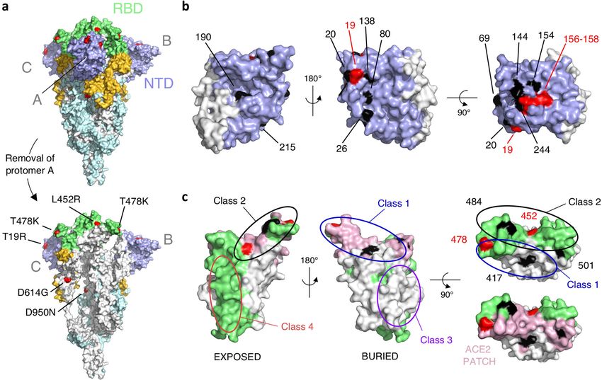
Bamlanivimab Etesivimab Casirivimab Imdevimab
120 120 120 120
100 100 100 100
Neutralization (%)
Neutralization (%)
Neutralization (%)
Neutralization (%)
80 80 80 80
60 60 60 60
D614G
40 40 40 40 Alpha
Beta
20 20 20 20
Delta
0 0 0 0
10–4 10–3 10–2 10–1 100 101 10–4 10–3 10–2 10–1 100 101 10–4 10–3 10–2 10–1 100 101 10–4 10–3 10–2 10–1 100 101
Concentration (μg ml–1) Concentration (μg ml–1) Concentration (μg ml–1) Concentration (μg ml–1)
Fig. 1 | Neutralization of the SARS-CoV-2 variants D614G, Alpha, Beta and strain and the Alpha, Beta and Delta variants by four therapeutic monoclonal
Delta by therapeutic monoclonal antibodies. Neutralization curves of antibodies (bamlanivimab, etesivimab, casirivimab and imdevimab). Data are
monoclonal antibodies. Dose–response analysis of neutralization of the D614G mean ± s.d. of four independent experiments.
variant (GISAID accession ID: EPI_ISL_2029113) (Extended Data Fig. 1). trimer interface (Extended Data Fig. 4a), suggesting that this mutation
When compared to the D614G strain (belonging to the basal B.1 line- may contribute to the regulation of spike protein dynamics, as shown
age), which was used here as a reference, the spike protein contained for the D614G mutation13. As with other VOCs, some mutations in the
eight mutations, including four mutations in the NTD (T19R, G142D, Delta variant cluster in the NTD (Extended Data Fig. 4b). The 156–157
Δ156–157 and R158G), two in the RBD (L452R and T478K), one muta- deletion and G158R mutation in the Delta variant map to the same sur-
tion close to the furin-cleavage site (P681R) and one in the S2 region face as the 144 and 241–243 deletions in the Alpha and Beta (B.1.351)
(D950N) (Extended Data Fig. 1). This set of mutations was different variants, respectively. The T19R mutation maps to a surface patch that
from that observed in other members of the B.1.617 lineage and other has several mutations in the Alpha variant. These altered residues are
VOCs (Extended Data Fig. 1). Viral stocks were titrated using S-Fuse found in the NTD ‘supersite’ that is targeted by most anti-NTD neutral-
reporter cells and Vero cells10,11. The viral titres were similar in the two izing antibodies15. In the RBD, mutations appearing in VOCs map to
target cells and reached 105–106 infectious units per ml. Large syncytia the periphery of the ACE2-binding surface (Extended Data Fig. 4c),
expressing the spike protein were observed in cells that were infected suggesting that the virus accumulates mutations there to reduce or
with the Delta variant (Extended Data Fig. 2). Future work will help to avoid recognition by antibodies while maintaining binding to ACE2.
determine whether Delta is more fusogenic than other variants, as For example, the L452R mutation found in the Delta variant impairs
suggested here by the large size of Delta-induced syncytia. neutralization by antibodies16 and is located at this periphery. The only
mutation within the ACE2 patch is at location 501, which increases the
affinity of the RBD for ACE2 and is also involved in antibody escape13.
Phylogenetic analysis of the B.1.617 lineage The T478K mutation in the RBD is unique to the Delta variant and falls
To contextualize the isolate of the Delta variant reported here, we within the epitope region of potent neutralizing monoclonal antibodies
inferred a global phylogeny by subsampling the diversity of SARS-CoV-2 categorized as ‘Class 1’17 (Extended Data Fig. 4c). This mutation is close
sequences available on the GISAID EpiCoV database (Extended Data to the E484K mutation that facilitates antibody escape13. These observa-
Fig. 3). The B.1.617 lineage, which is divided into three sublineages tions prompted us to analyse the neutralization potential, against the
according to the PANGO classification12, derives from the B.1 lineage Delta variant, of monoclonal antibodies and sera from convalescent
(D614G). The three sublineages show multiple changes in the spike individuals who had recovered from COVID-19 and from individuals
protein, including the L452R substitution in the RBD, which has already who had been vaccinated against COVID-19.
been observed in other variants such as B.1.429, and P681R. This substi-
tution is located in the furin-cleavage site and may enhance the fuso-
genic activity of the spike protein13. The E484Q substitution—which Neutralization of Delta by antibodies
may be functionally similar to the antibody-escape mutation E484K We assessed the sensitivity of the Delta variant to a panel of human
that is found in the Beta and Gamma variants (B.1.351 and P.1, respec- monoclonal antibodies using the S-Fuse assay. We tested four clinically
tively)—is present in the B.1.617.1 and B.1.617.3 subtypes, and is likely to approved monoclonal antibodies that target the RBD18,19 (bamlanivimab
have reverted in the Delta sublineage, as it was present in a sequence (LY-CoV555), etesevimab (LY-CoV016), casirivimab (REGN10933) and
(B.1.617) ancestral to the three sublineages14 (Extended Data Fig. 1). imdevimab (REGN10987)), as well as eight anti-RBD (RBD-48, RBD-85,
Whether the absence of E484Q, the presence of T478K or other changes RBD-98 and RBD-109) and anti-NTD (NTD-18, NTD-20, NTD-69 and NTD-
in the spike protein or elsewhere may facilitate viral replication and 71) monoclonal antibodies derived from convalescent individuals (C.P.
transmissibility remains unknown. Notably, the B.1.617 lineage is not et al., manuscript in preparation). Neutralizing anti-RBD monoclonal
homogeneous, with multiple mutations that are fixed in a sublineage antibodies can be classified into four main categories17,20. RBD-48 and
(for example, the spike protein mutations T19R, G142D or D950N) also RBD-85 belong to the first category (Class 1) and act by blocking the
being detected at lower frequencies in other sublineages. This may binding of the ‘up’ conformation of RBD to ACE217. The precise epitopes
reflect founder effects or similar selective pressures acting on these of RBD-98 and RBD-109 are not yet defined but overlap with those of
emerging variants. RBD-48 and RBD-85. The anti-NTD antibodies bind to uncharacterized
epitopes.
We measured the potency of the four therapeutic antibodies against
Mutational changes in the Delta variant the Delta variant and included as a comparison D614G (B.1) and the
The locations of the spike protein mutations in the Delta variant showed Alpha and Beta variants. The antibodies neutralized D614G with a
a similar overall distribution to those that appeared in other VOCs. In half-maximal inhibitory concentration (IC50) that ranged from 1.2 × 10⁻³
particular, in addition to D614G, the D950N mutation mapped to the to 6.5 × 10⁻² μg ml−1 (Fig. 1). Etesivimab exhibited a 200-fold increase in
Nature | Vol 596 | 12 August 2021 | 277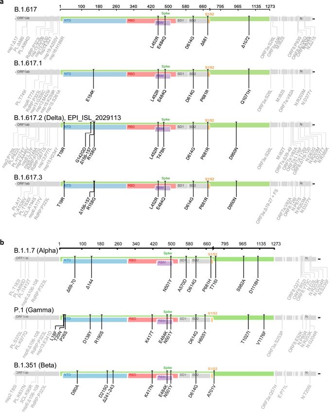
Article
a Strasbourg cohort b Pfizer c AstraZeneca
Unvaccinated (M12) Vaccinated (M12) W3 W8 W10 W16
** **** ****
105 105 **** *** ***
**** **** 104 104 ** 104 104 ****
** **** **** ****
104 **** 104 *** ****
103 ** 103 103 *** 103
ED50
ED50
ED50
ED50
ED50
ED50
**
103 103
102 102 102 102
102 102
101 101 101 101
101 101
G
a
ta
lta
G
a
ta
lta
G
a
ta
lta
G
a
ta
lta
G
a
ta
lta
G
a
ta
lta
ph
ph
ph
ph
ph
ph
14
14
14
14
14
14
Be
Be
Be
Be
Be
Be
De
De
De
De
De
De
Al
Al
Al
Al
Al
Al
D6
D6
D6
D6
D6
D6
Fig. 2 | Sensitivity of the SARS-CoV-2 variants D614G, Alpha, Beta and Delta independent experiments. A two-sided Friedman test with Dunn’s multiple
to sera from convalescent individuals and vaccinated individuals. comparison was performed between each of the viral strains. *P < 0.05,
Neutralization titres of the sera against the indicated viral isolates are **P < 0.01, ***P < 0.001, ****P < 0.0001. Strasbourg cohort, unvaccinated (M12):
expressed as ED50 values. a, Neutralizing activity of sera from the Strasbourg D614G versus Beta, P = 0.0052; D614G versus Delta, P = 0.0052; Alpha versus
cohort of convalescent individuals (n = 26; left) and convalescent individuals Beta, P < 0.0001; Alpha versus Delta, P < 0.0001. Strasbourg cohort, vaccinated
who had been vaccinated (n = 21; right). Samples were collected at month 12 (M12): D614G versus Beta, P < 0.0001; Alpha versus Beta, P < 0.0001; Alpha
(M12) after the onset of symptoms. b, Neutralizing activity of sera from versus Delta, P < 0.0001. Pfizer (W3): D614G versus Beta, P = 0.0001; D614G
recipients of the Pfizer vaccine, sampled at week 3 (W3) after vaccination versus Delta, P = 0.0013. Pfizer (W8): D614G versus Beta, P = 0.0002; Alpha
(n = 16; left) and week 8 (W8) after vaccination (week 5 after the second dose) versus Beta, P < 0.0001; Alpha versus Delta, P = 0.0098. AstraZeneca (W10):
(n = 16; right). c, Neutralizing activity of sera from recipients of the AstraZeneca D614G versus Beta, P < 0.0001; D614G versus Delta, P < 0.0001; Alpha versus
vaccine, sampled at week 10 (W10) after vaccination (n = 23; left) and week 16 Beta, P = 0.0006; Alpha versus Delta, P = 0.0056. AstraZeneca (W16): D614G
(W16) after vaccination (week 4 after the second dose) (n = 20; right). The versus Beta, P < 0.0001; D614G versus Delta, P = 0.0005, Alpha versus Beta,
dotted line indicates the limit of detection (ED50 = 30). Data are mean from two P < 0.0001; Alpha versus Delta, P < 0.0001.
IC50 against the Alpha variant. As previously reported, bamlanivimab mild-to-moderate and asymptomatic cases of COVID-19 (Extended
and etesivimab did not neutralize the Beta variant21. Bamlanivimab Data Table 1). The individuals were not vaccinated at the time at which
lost antiviral activity against the Delta variant, in line with previous samples were taken. We have previously characterized the potency
results that showed that L452R is an escape mutation for this antibody16. of these sera against D614G, Alpha and Beta isolates11. We analysed
Etesivimab, casirivimab and imdevimab remained active against the samples taken from individuals a median of 188 days after the onset
Delta variant (Fig. 1). of symptoms (referred to as month-6 samples), and calculated the
The four other anti-RBD monoclonal antibodies neutralized D614G. median half-maximal effective dilution (ED50) for each combination
The IC50 values of RBD-48 and RBD-98 were about 15–100-fold higher for of serum and virus (Extended Data Fig. 7a). With the Alpha variant, we
the Alpha variant than for D614G, whereas RBD-85 exhibited increased obtained similar ED50 values in this series of experiments to those in
activity against the Alpha variant. Three monoclonal antibodies inhibited our previous analysis11 (Extended Data Fig. 7b). We thus included our
the Delta variant, whereas RBD-85 was inactive (Extended Data Fig. 5). published data for D614G and Beta in the comparison. With the Delta
The four anti-NTD monoclonal antibodies were globally less efficient variant, neutralization titres were significantly decreased by four- to
than anti-RBD monoclonal antibodies. They inhibited D614G with a sixfold when compared to the Alpha and D614G strains, respectively
high IC50 value (1–60 μg ml−1) (Extended Data Fig. 5). Three anti-NTD (Extended Data Fig. 7a). This reduction in neutralizing titres was similar
antibodies lost activity against the Alpha and Delta variants, whereas against the Delta and Beta variants (Extended Data Fig. 7a).
the fourth (NTD-18) inhibited the two variants to some extent. Thus, We asked whether this neutralization profile was maintained for
the Delta variant escapes neutralization by some antibodies that target longer periods of time. We analysed sera from 47 individuals from
the RBD or NTD. another cohort consisting of healthcare workers from Strasbourg Uni-
We examined by flow cytometry the binding of each monoclonal versity Hospitals who had a qRT–PCR-confirmed diagnosis of COVID-19
antibody to Vero cells infected with the different variants, and used and who experienced mild disease22,23. Twenty-six individuals were
radar plots to show the binding of all antibodies tested (Extended Data unvaccinated, and 21 had received a single dose of vaccine 7–81 days
Fig. 6). D614G was recognized by the 12 monoclonal antibodies tested. before sampling. The samples were collected at a later time point (month
The Alpha and Delta variants were recognized by nine and by seven 12), with a median of 330 and 359 days for unvaccinated and vaccinated
monoclonal antibodies, respectively. Bamlanivimab no longer bound to individuals, respectively23 (Extended Data Table 1). As observed pre-
the Delta variant. We also analysed the binding of the 12 monoclonal viously23, the neutralization activity was globally low at month 12 in
antibodies to the Beta variant, which is more resistant to neutralization. unvaccinated individuals (Fig. 2a). There was a fourfold decrease in
Bamlanivimab and etesivimab lost their binding to the Beta variant, ED50 against the Beta and Delta variants relative to the Alpha variant
and only five of the antibodies bound to this variant (Extended Data (Fig. 2a). The 21 individuals within the month-12 cohort who had had
Fig. 6). Thus, the escape of the Delta variant and other variants from a single dose of vaccine included 9 individuals who had received the
neutralization is due to a reduction or loss of binding of the antibodies. AstraZeneca vaccine, 9 the Pfizer vaccine and 3 the Moderna vaccine.
Sera from these vaccinated participants showed a marked increase in
neutralizing antibody titres against the Alpha, Beta and Delta variants,
Sensitivity of Delta to convalescent sera as compared to convalescent individuals who had not been vaccinated
We examined the neutralization ability of sera from convalescent (Fig. 2a). Therefore, as shown with other variants23,24, a single dose of vac-
individuals. We first selected samples from 56 donors in a cohort of cine boosts cross-neutralizing antibody responses to the Delta variant.
individuals from the French city of Orléans. All individuals had tested We then classified the individuals as neutralizers (individuals whose
positive for SARS-CoV-2 infection by quantitative PCR with reverse serum contained neutralizing antibodies that were detectable at the
transcription (qRT–PCR) or serology and included critical, severe, first serum dilution of 1/30) and non-neutralizers, for the viral variants
278 | Nature | Vol 596 | 12 August 2021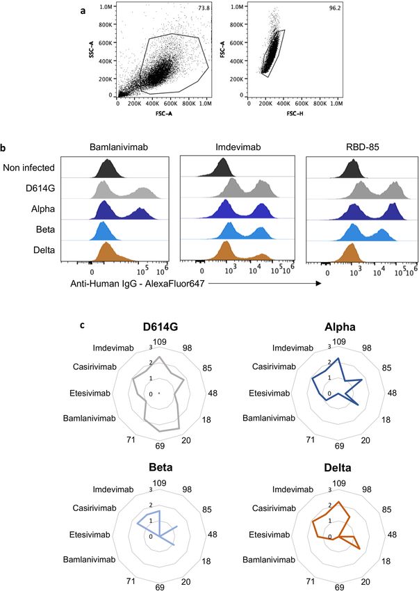
and the two cohorts (Extended Data Fig. 7c). Sera from between 76%            from recent recipients of a COVID-19 vaccine against an infectious
and 92% of individuals neutralized the four strains at month 6. The           Delta isolate. Some monoclonal antibodies, including bamlanivimab,
fraction of neutralizers was lower in the second cohort at month 12,          lost their ability to bind to the spike protein and no longer neutralized
and this effect was particularly marked for the Beta and Delta variants.      the Delta variant. We also showed that the Delta variant is less sensitive
Sera from 88% of individuals neutralized the Alpha variant, whereas           to sera from naturally immunized individuals. Vaccination of conva-
sera from only 47% of individuals neutralized the Delta variant. After        lescent individuals boosted the humoral immune response to well
vaccination, sera from 100% of the convalescent individuals neutral-          above the threshold of neutralization. These results strongly suggest
ized the four strains (Extended Data Fig. 7c).                                that vaccination of previously infected individuals is likely to be pro-
  Thus, the Delta variant exhibits enhanced resistance to neutralization      tective against a large array of circulating viral strains, including the
by sera from convalescent individuals who have not been vaccinated,           Delta variant.
particularly by one year after the infection.                                    In individuals who had not previously been infected with SARS-CoV-2,
                                                                              a single dose of either the Pfizer or the AstraZeneca vaccine induced
                                                                              a barely detectable level of neutralizing antibodies against the Delta
Sensitivity of Delta to sera from vaccinated individuals                      variant. About 10% of the sera neutralized this variant. However, a
We next asked whether vaccine-elicited antibodies neutralized the             two-dose regimen generated high sero-neutralization levels against
Delta variant in individuals who had not previously been infected with        the Alpha, Beta and Delta variants in individuals sampled at week 8 to
SARS-CoV-2. We randomly selected 59 individuals from a cohort of vac-         week 16 after vaccination. Levels of neutralizing antibodies are highly
cinated individuals in Orléans. The characteristics of these individuals      predictive of immune protection from symptomatic SARS-CoV-2 infec-
are outlined in Extended Data Table 2. Sixteen individuals received the       tion25. A recent report analysing all sequenced symptomatic cases of
Pfizer vaccine. They were sampled at week 3 after the first dose and week     COVID-19 in England was used to estimate the effect of vaccination
8 (corresponding to week 5 after the second dose). Thirteen individu-         on infection26. Effectiveness was notably lower for the Delta variant
als were also sampled at week 16. Forty-three individuals received the        than for the Alpha variant after one dose of the AstraZeneca or the
AstraZeneca vaccine. Sera from 23 individuals were sampled after 1 dose       Pfizer vaccine. The two-dose effectiveness against the Delta variant
(week 10) and from 20 other individuals after 2 doses (week 16, corre-        was estimated to be 60% and 88% for the AstraZeneca and the Pfizer
sponding to week 4 after the second dose). We measured the potency of         vaccine, respectively26. Our neutralization experiments indicate
the sera against D614G and the Alpha, Beta and Delta variants (Fig. 2b, c).   that antibodies elicited by the Pfizer and AstraZeneca vaccines are
   With the Pfizer vaccine, after a single dose (at week 3) the levels of     efficacious against the Delta variant, but about three- to fivefold less
neutralizing antibodies were low against D614G, and almost undetect-          potent than they are against the Alpha variant. There was no major
able against the Alpha, Beta and Delta variants (Fig. 2b). Titres signifi-    difference in the levels of antibodies elicited by the Pfizer or the
cantly increased after the second dose. We observed a threefold and           AstraZeneca vaccines.
a sixteenfold reduction in the neutralization titres against the Delta           Potential limitations of our work include the low number of vac-
and the Beta variants, respectively, when compared to the Alpha vari-         cinated individuals analysed and the lack of characterization of cel-
ant (Fig. 2b). Similar differences between strains were observed at a         lular immunity, which may be more cross-reactive than the humoral
later time point (week 16), although titres were globally slightly lower      response. Future work with more individuals and longer survey periods
(Extended Data Fig. 7b).                                                      will help to characterize the role of humoral responses in the efficacy
   A similar pattern was observed with the AstraZeneca vaccine. A single      of vaccines against circulating variants.
dose (week 10) induced low levels of antibodies neutralizing the Delta           Our results demonstrate that the emerging Delta variant partially—
and Beta variants, when compared to the D614G and Alpha strains               but notably—escapes neutralizing monoclonal antibodies and poly-
(Fig. 2c). Four weeks after the second dose (week 16), neutralizing titres    clonal antibodies elicited by previous infection with SARS-CoV-2 or
were strongly increased. There was, however, a fivefold and a ninefold        by vaccination.
reduction in neutralization titres against the Delta and the Beta variants,
respectively, relative to the Alpha variant (Fig. 2c).
   We then classified the vaccine recipients as neutralizers and              Online content
non-neutralizers, for the four viral strains (Extended Data Fig. 7d, e).      Any methods, additional references, Nature Research reporting sum-
For the Pfizer vaccine, sera from 13% of individuals neutralized the Delta    maries, source data, extended data, supplementary information,
variant after a single dose. Sera from 81 to 100% of individuals neutral-     acknowledgements, peer review information; details of author con-
ized any of the four stains after the second dose, at week 8. This fraction   tributions and competing interests; and statements of data and code
remained stable at week 16, with the exception of the Beta variant, which     availability are available at https://doi.org/10.1038/s41586-021-03777-9.
was neutralized by sera from only 46% of the individuals. Sera from 74%
and 61% of individuals that received a single dose of the AstraZeneca         1.   Yadav, P. D. et al. Neutralization of variant under investigation B.1.617 with sera of BBV152
vaccine neutralized the D614G and the Alpha strains, respectively. This            vaccinees. Clin. Infect. Dis. https://doi.org/10.1093/cid/ciab411 (2021).
                                                                              2.   Ferreira, I. et al. SARS-CoV-2 B.1.617 emergence and sensitivity to vaccine-elicited
fraction sharply dropped with the Beta and the Delta variants, which               antibodies. Preprint at https://doi.org/10.1101/2021.05.08.443253 (2021).
were inhibited by only 4 and 9% of the sera, respectively. Four weeks         3.   Hoffmann, M. et al. SARS-CoV-2 variant B.1.617 is resistant to Bamlanivimab and evades
after the second dose of the AstraZeneca vaccine, sera from 95–100%                antibodies induced by infection and vaccination. Cell Rep. https://doi.org/10.1016/
                                                                                   j.celrep.2021.109415 (2021).
of individuals neutralized the four strains.                                  4.   Cherian, S. et al. Convergent evolution of SARS-CoV-2 spike mutations, L452R, E484Q
   Therefore, a single dose of Pfizer or AstraZeneca either showed low             and P681R, in the second wave of COVID-19 in Maharashtra, India. Preprint at https://doi.
or no efficiency against the Beta and the Delta variants. Both vaccines            org/10.1101/2021.04.22.440932 (2021).
                                                                              5.   Edara, V.-V. et al. Infection and vaccine-induced neutralizing-antibody responses to the
generated a neutralizing response that efficiently targeted the Delta              SARS-CoV-2 B.1.617 variants. New Eng. J. Med. https://doi.org/10.1056/NEJMc2107799
variant only after the second dose.                                                (2021).
                                                                              6.   Public Health England. Variants: distribution of case data, 11 June 2021. https://www.gov.
                                                                                   uk/government/publications/covid-19-variants-genomically-confirmed-case-numbers/
                                                                                   variants-distribution-of-case-data-11-june-2021 (2021).
Discussion                                                                    7.   Tada, T. et al. The spike proteins of SARS-CoV-2 B.1.617 and B.1.618 variants identified in
We studied the cross-reactivity of monoclonal antibodies to                        India provide partial resistance to vaccine-elicited and therapeutic monoclonal
                                                                                   antibodies. Preprint at https://doi.org/10.1101/2021.05.14.444076 (2021).
pre-existing SARS-CoV-2 strains, sera from convalescent individuals           8.   Liu, J. et al. BNT162b2-elicited neutralization of B.1.617 and other SARS-CoV-2 variants.
six or twelve months after the onset of COVID-19 symptoms and sera                 Nature https://doi.org/10.1038/s41586-021-03693-y (2021).

                                                                                                                       Nature | Vol 596 | 12 August 2021 | 279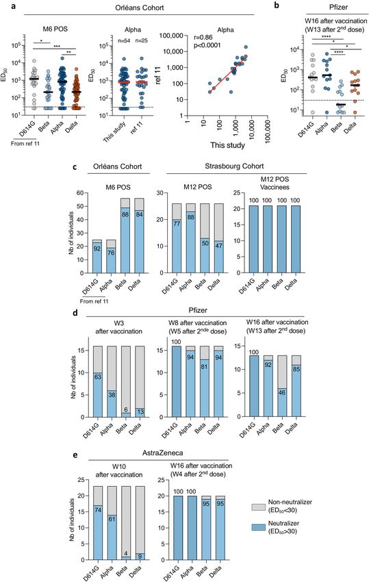
Article
9.    Wall, E. C. et al. Neutralising antibody activity against SARS-CoV-2 VOCs B.1.617.2 and        19.   Starr, T. N. et al. Prospective mapping of viral mutations that escape antibodies used to
      B.1.351 by BNT162b2 vaccination. Lancet 397, 2331–2333 (2021).                                       treat COVID-19. Science 371, 850–854 (2021).
10.   Buchrieser, J. et al. Syncytia formation by SARS-CoV-2-infected cells. EMBO J. 39,             20.   Liu, L. et al. Potent neutralizing antibodies against multiple epitopes on SARS-CoV-2
      e106267 (2020).                                                                                      spike. Nature 584, 450–456 (2020).
11.   Planas, D. et al. Sensitivity of infectious SARS-CoV-2 B.1.1.7 and B.1.351 variants to         21.   Wang, P. et al. Antibody resistance of SARS-CoV-2 variants B.1.351 and B.1.1.7. Nature 593,
      neutralizing antibodies. Nat. Med. 27, 917–924 (2021).                                               130–135 (2021).
12.   Rambaut, A. et al. A dynamic nomenclature proposal for SARS-CoV-2 lineages to assist           22.   Fafi-Kremer, S. et al. Serologic responses to SARS-CoV-2 infection among hospital staff
      genomic epidemiology. Nat. Microbiol. 5, 1403–1407 (2020).                                           with mild disease in eastern France. EBioMedicine 59, 102915 (2020).
13.   Plante, J. A. et al. The variant gambit: COVID-19’s next move. Cell Host Microbe 29, 508–      23.   Gallais, F. et al. Anti-SARS-CoV-2 antibodies persist for up to 13 months and reduce risk of
      515 (2021).                                                                                          reinfection. Preprint at https://doi.org/10.1101/2021.05.07.21256823 (2021).
14.   Centers for Disease Control and Prevention. SARS-CoV-2 variant classifications and             24.   Krammer, F. et al. Antibody responses in seropositive persons after a single dose of
      definitions. https://www.cdc.gov/coronavirus/2019-ncov/cases-updates/                                SARS-CoV-2 mRNA vaccine. N. Engl. J. Med. 384, 1372–1374 (2021).
      variant-surveillance/variant-info.html (2021).                                                 25.   Khoury, D. S. et al. Neutralizing antibody levels are highly predictive of immune protection
15.   McCallum, M. et al. N-terminal domain antigenic mapping reveals a site of vulnerability              from symptomatic SARS-CoV-2 infection. Nat. Med. (2021).
      for SARS-CoV-2. Cell 184, 2332–2347 (2021).                                                    26.   Bernal, J. L. et al. Effectiveness of COVID-19 vaccines against the B.1.617.2 variant. Preprint
16.   Starr, T. N., Greaney, A. J., Dingens, A. S. & Bloom, J. D. Complete map of SARS-CoV-2 RBD           at https://doi.org/10.1101/2021.05.22.21257658 (2021).
      mutations that escape the monoclonal antibody LY-CoV555 and its cocktail with
      LY-CoV016. Cell Rep. Med. 2, 100255 (2021).
                                                                                                     Publisher’s note Springer Nature remains neutral with regard to jurisdictional claims in
17.   Barnes, C. O. et al. SARS-CoV-2 neutralizing antibody structures inform therapeutic
                                                                                                     published maps and institutional affiliations.
      strategies. Nature 588, 682–687 (2020).
18.   Taylor, P. C. et al. Neutralizing monoclonal antibodies for treatment of COVID-19. Nat. Rev.
      Immunol. 21, 382–393 (2021).                                                                   © The Author(s), under exclusive licence to Springer Nature Limited 2021

280 | Nature | Vol 596 | 12 August 2021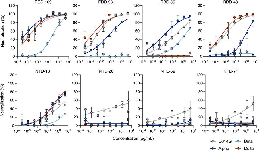
Methods                                                                    with augur and visualized with auspice as implemented in the Nextstrain
                                                                           pipeline (https://github.com/nextstrain/ncov; version from 21 May
Data reporting                                                             2021)27. Within Nextstrain, a random subsampling approach capping
No statistical methods were used to predetermine sample size. The          a maximum number of sequences per global region was used for the
experiments were not randomized and the investigators were not             contextual non-B.1.617 sequences. Acknowledgement of the contribut-
blinded to allocation during experiments and outcome assessment.           ing and originating laboratories for all sequences used in the analysis
Our research complies with all relevant ethical regulations.               is provided in Supplementary Table 1.

Orléans cohort of convalescent and vaccinated individuals                  Three-dimensional mapping of mutations on B1.617.2 and other
Since 27 August 2020, a prospective, monocentric, longitudinal,            variants to the surface of the spike protein
interventional cohort clinical study enrolling 170 individuals with        Panels in Extended Data Fig. 4 were prepared with the PyMOL Molecular
SARS-CoV-2 infection (with different disease severities) and 59 healthy    Graphics System, v.2.1 (Schrödinger). The atomic model used (Protein
control individuals has been ongoing, and aims to describe the persis-     Data Bank code: 6XR8) has been previously described28.
tence of specific and neutralizing antibodies over a 24-month period.
This study was approved by the Ile de France IV ethical committee. At      S-Fuse neutralization assay
enrolment, written informed consent was collected and participants         U2OS-ACE2 GFP1-10 or GFP 11 cells, also termed S-Fuse cells, become
completed a questionnaire that covered sociodemographic character-         GFP+ when they are productively infected by SARS-CoV-210,11. Cells
istics, virological findings (SARS-CoV-2 qRT–PCR results, including date   tested negative for mycoplasma. Cells were mixed (ratio 1:1) and
of testing), clinical data (date of symptom onset, type of symptoms,       plated at 8 × 10³ per well in a μClear 96-well plate (Greiner Bio-One).
hospitalization) and data related to anti-SARS-CoV-2 vaccination if ever   The indicated SARS-CoV-2 strains were incubated with serially diluted
(brand product, date of first and second doses). The serological status    monoclonal antibodies or sera for 15 min at room temperature and
of participants was assessed every three months. Those who underwent       added to S-Fuse cells. The sera were heat-inactivated 30 min at 56 °C
anti-SARS-CoV-2 vaccination had regular blood sampling after the           before use. Eighteen hours later, cells were fixed with 2% paraform-
first dose of vaccine (ClinicalTrials.gov Identifier: NCT04750720). The    aldehyde (PFA), washed and stained with Hoechst (dilution 1:1,000,
primary outcome was the presence of antibodies to SARS-CoV-2 spike         Invitrogen). Images were acquired with an Opera Phenix high-content
protein as measured with the S-Flow assay. The secondary outcome was       confocal microscope (PerkinElmer). The GFP area and the number
the presence of neutralizing antibodies as measured with the S-Fuse        of nuclei were quantified using Harmony software (PerkinElmer).
assay. For the present study, we selected 56 convalescent individuals      The percentage of neutralization was calculated using the number
and 59 vaccinated individuals (16 with Pfizer and 43 with AstraZeneca).    of syncytia as value with the following formula: 100 × (1 – (value
Study participants did not receive any compensation.                       with serum − value in ‘non-infected’)/(value in ‘no serum’ − value in
                                                                           ‘non-infected’)). The neutralizing activity of each serum was expressed
Strasbourg cohort of convalescent individuals                              as the ED50 value. ED50 values (in μg ml−1 for monoclonal antibodies
Since April 2020, a prospective, monocentric, longitudinal, inter-         and in dilution values for sera) were calculated with a reconstructed
ventional cohort clinical study enrolling 308 hospital staff from the      curve using the percentage of the neutralization at the different
Strasbourg University Hospitals who had a qRT–PCR-confirmed diag-          concentrations.
nosis of SARS-CoV-2 infection has been ongoing (ClinicalTrials.gov
Identifier: NCT04441684). At enrolment (from 17 April 2020), written       Clinical history of the patient infected with B.1.617.2
informed consent was collected and participants completed a ques-          A 54-year-old man was admitted on 27 April 2021 to the emergency
tionnaire that covered sociodemographic characteristics, virological       department of the Hôpital Européen Georges Pompidou hospital in
findings (SARS-CoV-2 qRT–PCR results, including date of testing) and       Paris, France, for an acute respiratory distress syndrome with fever. He
clinical data (date of symptom onset, type of symptoms, hospitaliza-       had no medical background and came back from a trip to India (West
tion). This study was approved by the Institutional Review Board of        Bengal and a few days spent in Delhi) 10 days before (17 April 2021),
Strasbourg University Hospital. The serological status of the partici-     where he stayed 15 days for his work. The onset of symptoms (abdominal
pants has been described at months 3 and 6 after the onset of symp-        pain and fever) was approximately 18 April 2021. The nasopharyngeal
toms22,23. Laboratory identification of SARS-CoV-2 was performed at        swab tested positive for SARS-CoV-2 at his date of admission. Lung
least 10 days before inclusion by qRT–PCR testing on nasopharyngeal        tomo-densitometry showed a mild (10–25%) COVID-19 pneumonia
swab specimens according to current guidelines (Institut Pasteur;          without pulmonary embolism. He initially received oxygen therapy
WHO technical guidance). The assay targets two regions of the viral        (2 l min−1), dexamethasone (6 mg per day) and enoxaparin (0.4 ml twice
RNA-dependent RNA polymerase (RdRp) gene with a threshold of detec-        a day). His respiratory state worsened on day 3 (30 April 2021). He was
tion of 10 copies per reaction. The primary outcome was the presence       transferred to an intensive care unit, in which he received high-flow oxy-
of antibodies to the SARS-CoV-2 spike protein as measured with the         gen therapy (maximum 12 l min−1). His respiratory condition improved,
S-Flow assay. The secondary outcome was the presence of neutralizing       and he was transferred back to a conventional unit on day 8 (5 May 2021).
antibodies as measured with the S-Fuse assay. For the present study,       He was discharged from hospital on day 15 (May 10 2021).
we randomly selected 47 convalescent individuals at month 12 (26
unvaccinated and 21 vaccinated). Study participants did not receive any    Virus strains
compensation.                                                              The reference D614G strain (hCoV-19/France/GE1973/2020) was sup-
                                                                           plied by the National Reference Centre for Respiratory Viruses hosted by
Phylogenetic analysis                                                      the Institut Pasteur and headed by S. van der Werf. This viral strain was
All SARS-CoV-2 sequences available on the GISAID EpiCov database as        supplied through the European Virus Archive – Global (EVAg) platform,
of 21 May 2021 were retrieved. A subset of complete and high-coverage      a project that has received funding from the European Union’s Horizon
sequences, as indicated in GISAID, assigned to lineages B.1.617.1,         2020 research and innovation program under grant agreement no.
B.1.617.2 and B.1.617.3 were randomly subsampled to contain up to five     653316. The variant strains were isolated from nasal swabs using Vero
sequences per country and epidemiological week in R with the packages      E6 cells and amplified by one or two passages. B.1.1.7 originated from
tidyverse and lubridate. Together with a single B.1.617 sequence this      an individual in Tours (France) who had returned from the UK. B.1.351
subset was included in the global SARS-CoV-2 phylogeny reconstructed       (hCoV-19/France/IDF-IPP00078/2021) originated from an individual in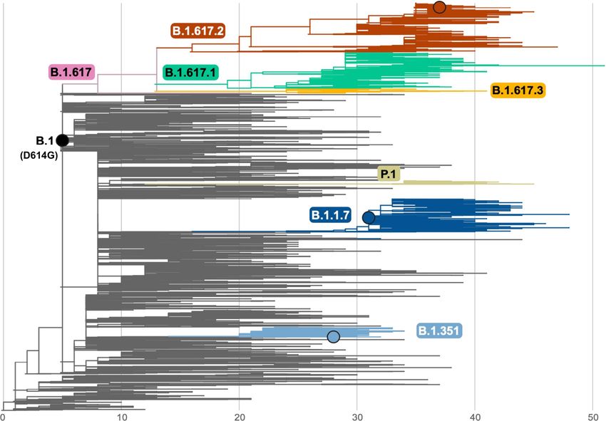
Article
Creteil (France). B.1.617.2 was isolated from a nasopharyngeal swab of a
hospitalized patient who had returned from India, as described above.       Data availability
The swab was provided and sequenced by the Laboratoire de Virologie         All data supporting the findings of this study are available within
of the Hopital Européen Georges Pompidou (Assistance Publique des           the Article or from the corresponding authors upon request. Viral
Hôpitaux de Paris). All individuals provided informed consent for the       sequences are available upon request and were deposited at GISAID
use of the biological materials. Titration of viral stocks was performed    (https://www.gisaid.org/) under the following numbers: hCoV-19/
on Vero E6, with a limiting dilution technique allowing a calculation of    France/GE1973/2020 (D614G): EPI_ISL_414631; Alpha (B.1.1.7): EPI_
TCID50, or on S-Fuse cells. Viruses were sequenced directly on nasal        ISL_735391; Beta (B.1.351): EPI_ISL_964916 and Delta (B.1.617.2): EPI_
swabs, and after one or two passages on Vero cells. Sequences were          ISL_2029113. Source data are provided with this paper.
deposited in the GISAID database immediately after their generation,
with the following IDs: D614G: EPI_ISL_414631; B.1.1.7: EPI_ISL_735391;     27. Hadfield, J. et al. Nextstrain: real-time tracking of pathogen evolution. Bioinformatics 34,
                                                                                4121–4123 (2018).
B.1.1.351: EPI_ISL_964916; B.1.617.2: ID: EPI_ISL_2029113.                  28. Cai, Y. et al. Distinct conformational states of SARS-CoV-2 spike protein. Science 369,
                                                                                1586–1592 (2020).
Flow cytometry                                                              29. Lorin, V. & Mouquet, H. Efficient generation of human IgA monoclonal antibodies. J.
                                                                                Immunol. Methods 422, 102–110 (2015).
Vero cells were infected with the indicated viral strains at a multiplic-   30. Tzou, P. L. et al. Coronavirus antiviral research database (CoV-RDB): an online database
ity of infection (MOI) of 0.1. Two days after, cells were detached using        designed to facilitate comparisons between candidate anti-coronavirus compounds.
                                                                                Viruses 12, 1006 (2020).
PBS-EDTA and transferred into U-bottom 96-well plates (50,000 cells
per well). Cells were fixed in 4% PFA for 15–30 min at room tempera-
ture. Cells were then incubated for 15–30 min at room temperature           Acknowledgements We thank N. Casartelli for critical reading of the manuscript; P. Guardado
                                                                            Calvo for discussion; the individuals who participated in the study; members of the Virus and
with the indicated monoclonal antibodies (1 μg ml−1) in PBS, 1% BSA,        Immunity Unit for discussions and help; N. Aulner and the UTechS Photonic BioImaging (UPBI)
0.05% sodium azide and 0.05% saponin. Cells were washed with PBS            core facility (Institut Pasteur), a member of the France BioImaging network, for image
and stained using anti-IgG AF647 (1:600 dilution) (Thermo Fisher Sci-       acquisition and analysis; and the DRCI, CIC, Médecine du Travail and Pôle de Biologie teams
                                                                            (CHU de Strasbourg) for the management of the Strasbourg cohort and serology testing. The
entific). Stainings were also performed on control uninfected cells.        Opera system was co-funded by the Institut Pasteur and the Région Île de France (DIM1Health).
Data were acquired on an Attune Nxt instrument using Attune Nxt             Work in the O.S. laboratory is funded by the Institut Pasteur, Urgence COVID-19 Fundraising
Software v.3.2.1 (Life Technologies) and analysed with FlowJo v.10.7.1      Campaign of Institut Pasteur, Fondation pour la Recherche Médicale (FRM), ANRS, the Vaccine
                                                                            Research Institute (ANR-10-LABX-77), Labex IBEID (ANR-10-LABX-62-IBEID), ANR/FRM Flash
(Becton Dickinson).                                                         Covid PROTEO-SARS-CoV-2 and IDISCOVR. Work in UPBI is funded by grant ANR-
                                                                            10-INSB-04-01 and the Région Île-de-France program DIM1-Health. D.P. is supported by the
Antibodies                                                                  Vaccine Research Institute. The H.M. laboratory is funded by the Institut Pasteur, the Milieu
                                                                            Intérieur Program (ANR-10-LABX-69-01), INSERM, REACTing, EU (RECOVER) and Fondation de
The four therapeutic antibodies were provided by CHR Orleans. Human         France (00106077) grants. The S.F.K. laboratory is funded by Strasbourg University Hospitals
anti-SARS-CoV2 monoclonal antibodies were cloned from S-specific            (SeroCoV-HUS; PRI 7782), the Programme Hospitalier de Recherche Clinique (PHRC N 2017–
                                                                            HUS no. 6997), the Agence Nationale de la Recherche (ANR-18-CE17-0028), Laboratoire
blood memory B cells of individuals who were convalescing from
                                                                            d’Excellence TRANSPLANTEX (ANR-11-LABX-0070_TRANSPLANTEX) and the Institut National
COVID-19 (C.P. et al., manuscript in preparation). Recombinant human        de la Santé et de la Recherche Médicale (UMR_S 1109). The E.S.-L. laboratory is funded by the
IgG1 monoclonal antibodies were produced by co-transfection of Free-        Institut Pasteur and the French Government’s Investissement d’Avenir programme, Laboratoire
                                                                            d’Excellence ‘Integrative Biology of Emerging Infectious Diseases’ (grant no. ANR-10-LABX-
style 293‐F suspension cells (Thermo Fisher Scientific) as previously
                                                                            62-IBEID). The funders of this study had no role in study design, data collection, analysis and
described29, purified by affinity chromatography using protein G sepha-     interpretation, or writing of the Article.
rose 4 fast flow beads (GE Healthcare) and validated by enzyme-linked
immunosorbent assay (ELISA) against the trimeric S, RBD, S2 and NTD         Author contributions Experimental strategy design, experiments: D.P., D.V., A.B., I.S., F.G.-B.,
                                                                            M.M.R., F.P., T.B., E.S.-L. and F.A.R. Vital materials: D.V., C.P., N.R., J.P., M.P., F.G., P.G., A.V., J.L.G.,
proteins (C.P. et al., manuscript in preparation).                          L.C., N.K.-C., D.E., L.B., A.S., H.P., L.H., S.F.-K., T.P. and H.M. Manuscript writing: D.P., T.B., E.S.-L.,
                                                                            F.A.R. and O.S. Manuscript editing: D.V., M.M.R., H.P., L.H., S.F.K., T.P. and H.M.
Statistical analysis
                                                                            Competing interests C.P., H.M., O.S, T.B. and F.A.R. have a pending patent application for the
Flow cytometry data were analysed with FlowJo v.10 software (TriStar).
                                                                            anti-RBD monoclonal antibodies described in the present study (PCT/FR2021/070522). F.A.R. is
Calculations were performed using Excel 365 (Microsoft). Figures were       a member of the Board of MELETIOS Therapeutics and of the Scientific Advisory Board of
drawn using GraphPad Prism 9. Statistical analysis was conducted using      eureKARE. The remaining authors declare no competing interests.
GraphPad Prism 9. Statistical significance between different groups was
                                                                            Additional information
calculated using the tests indicated in each figure legend.                 Supplementary information The online version contains supplementary material available at
                                                                            https://doi.org/10.1038/s41586-021-03777-9.
Reporting summary                                                           Correspondence and requests for materials should be addressed to T.B. or O.S.
                                                                            Peer review information Nature thanks the anonymous reviewers for their contribution to the
Further information on research design is available in the Nature           peer review of this work.
Research Reporting Summary linked to this paper.                            Reprints and permissions information is available at http://www.nature.com/reprints.
Extended Data Fig. 1 | Schematic overview of the B.1.617 sublineage and focus on the spike protein were built with the Sierra tool30. Amino acid VOCs. a, b, Schematic overview of the B.1.617 sublineage (a) and the VOCs modifications in comparison to the ancestral Wuhan-Hu-1 sequence B.1.1.7 (Alpha), P1 (Gamma) and B.1.351 (Beta) (b). Consensus sequences with a (NC_045512) are indicated.
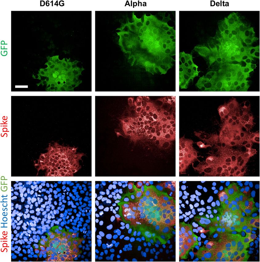
Article

Extended Data Fig. 2 | SARS-CoV-2 variants induce syncytia in S-Fuse cells.         stained with anti-spike antibodies and Hoechst to visualize nuclei. Syncytia
S-Fuse cells were exposed to the indicated SARS-CoV-2 strain (at a MOI of 10 −3).   (green), spike protein (red) and nuclei (blue) are shown. Representative images
The cells become GFP+ when they fuse together. After 20 h, infected cells were      from three independent experiments are shown. Scale bar, 50 μm.
Extended Data Fig. 3 | Global phylogeny of SARS-CoV-2 highlighting the        nucleotide substitutions from the root of the tree. The branches
B.1.617 lineage. The maximum likelihood tree was inferred using IQ-Tree, as   corresponding to key lineages are coloured: B.1.1.7, dark blue; B.1.351, light
implemented in the Nextstrain pipeline on a subsampled dataset of 3,794       blue; P.1, beige; B.1.617, pink; B.1.617.1, green; B.1.617.2, red; and B.1.617.3,
complete genomes. Branch lengths are scaled according to the number of        orange. A black circle indicates the position of the viruses studied here.
Article

Extended Data Fig. 4 | Mapping mutations of the Delta variant and other             middle panel shows a view from the back. The right panel shows a view from the
variants of concern to the surface of the spike protein. a, The spike protein       top of the trimer. Mutations found in the main variants of concern are
trimer (Protein Data Bank code: 6XR8, corresponding to a closed spike trimer        indicated. The mutations found in the Delta variant are in red. c, RBD shown in
with all three RBDs in the ‘down’ conformation) is shown with its surface           three orthogonal views, coloured according to solvent exposure in the context
coloured according to domains: NTD in dark blue, RBD in green, the remainder        of the closed spike: green and white indicate exposed and buried surfaces, as in
of S1 in yellow and S2 in light blue. Interfaces between protomers were left        a. The ACE2-binding surface is coloured in pink. The left panel shows a view
white to help visualize the boundaries of the protomers. The three polypeptide      from the top of the trimer, and the middle panel a view from below. The right
chains in the trimer were arbitrarily defined as A, B and C. Surface patches        panels show a view down the ACE2-binding surface, highlighted in pink in the
corresponding to residues mutated in the Delta variant are coloured in red. The     bottom panel. Mutations found in the main variants of concern are indicated.
bottom panel has the front protomer (chain A) removed to show the trimer            The mutations found in the Delta variant are in red. The ovals indicate the
interface (buried regions in the trimer are in white). The mutations in Delta are   epitope regions of the four main classes of anti-RBD neutralizing antibodies.
labelled in the bottom panel. b, NTD shown in three orthogonal views. The left      Note that the mutations on the RBD cluster all around the ACE2 patch. Panels
panel corresponds roughly to the orientation seen in chain B in a, and the          were prepared with the PyMOL Molecular Graphics System, v.2.1 (Schrödinger).
Extended Data Fig. 5 | Neutralization of the SARS-CoV-2 variants D614G,      antibodies on the D614G strain (grey) and the Alpha (dark blue), Beta (light
Alpha, Beta and Delta by monoclonal antibodies that target the RBD and       blue) and Delta (orange) variants. Data are mean ± s.d. of three independent
the NTD domains. Neutralization curves of monoclonal antibodies. Dose–       experiments.
response analysis of the neutralization by four anti-RBD and four anti-NTD
Article

Extended Data Fig. 6 | Binding of anti-SARS-CoV-2 monoclonal antibodies              binding of bamlanivimab, imdevimab and RBD-85 to Vero cells infected with
to Vero cells infected with the SARS-CoV-2 variants D614G, Alpha, Beta and           the indicated variants. c, Radar charts represent for each antibody the
Delta. Vero cells were infected with the indicated variants at a MOI of 0.1. After   logarithm of the mean of fluorescent intensity of the staining, relative to the
48 h, cells were stained with anti-SARS-CoV-2 monoclonal antibodies (1 μg ml−1)      non-infected condition. Data are representative of three independent
and analysed by flow-cytometry. a, Gating strategy. b, Histograms show the           experiments.
Extended Data Fig. 7 | See next page for caption.
Article
Extended Data Fig. 7 | Sensitivity of the SARS-CoV-2 variants D614G, Alpha,           Data are mean from two independent experiments. The dotted line indicates
Beta and Delta to sera from convalescent individuals and vaccinated                   the limit of detection (ED50 = 30). A two-sided Kruskal–Wallis test with Dunn’s
individuals. a, ED50 values for the neutralizing activity of sera from                multiple comparison was performed between each viral strain. *P < 0.05,
convalescent individuals from the Orléans cohort against the four viral               **P < 0.01, ***P < 0.001, ****P < 0.0001. D614G versus Beta, P < 0.0001; D614G
variants. Samples were collected 6 months after the onset of symptoms (M6             versus Delta, P = 0.0375; Alpha versus Beta, P < 0.0001; Alpha versus Delta,
POS). The sensitivity of the D614G and Alpha variants to sera from 25                 P = 0.0375. c–e, Fraction of neutralizers in the cohorts of convalescent or
individuals has been described previously11. Fifty-six sera (including the 25         vaccinated individuals. Individuals with an ED50 of neutralization above 30
previous sera) were tested against the Beta and Delta variants. Neutralization        were categorized as neutralizers and are indicated in blue. Non-neutralizers are
data obtained in this study and in ref. 11 were compared (middle) and correlated      in grey. c, Analysis of convalescent individuals from the Orléans cohort
(right). Similar results were obtained, allowing the datasets to be bridged. Data     collected at month 6 (left; related to a), and unvaccinated (middle; related to
are mean from two independent experiments. The dotted line indicates the              Fig. 2a) or vaccinated (right, related to Fig. 2a) individuals from the Strasbourg
limit of detection (ED50 = 30). A two-sided Kruskal–Wallis test with Dunn’s           cohort collected at month 12. d, Sera from recipients of the Pfizer vaccine were
multiple comparison was performed between each of the viral strains.                  sampled at week 3 (left), week 8 (middle) (both related to Fig. 2c) and week 16
*P < 0.05, **P < 0.01, ***P < 0.001, ****P < 0.0001. D614G versus Beta, P = 0.0153;   (right; related to b) after vaccination. e, Sera from recipients of the
D614G versus Delta, P = 0.0008; Alpha versus Delta, P = 0.0014. b, ED50 values        AstraZeneca vaccine were sampled at week 10 (left) and week 16 (right) after
for the neutralizing activity of sera from individuals who received a Pfizer          vaccination (related to Fig. 2c). The numbers indicate the percentage of
vaccine, sampled at week 16 (corresponding to week 13 after the second dose).         neutralizers.
Extended Data Table 1 | Characteristics of the two cohorts of convalescent individuals
Article
Extended Data Table 2 | Characteristics of the cohort of vaccinated individuals
nature research | reporting summary
                                                                                                     Corresponding author(s): Timothée Bruel, Olivier Schwartz
                                                                                                     Last updated by author(s): Jun 17, 2021

Reporting Summary
Nature Research wishes to improve the reproducibility of the work that we publish. This form provides structure for consistency and transparency
in reporting. For further information on Nature Research policies, see our Editorial Policies and the Editorial Policy Checklist.

Statistics
For all statistical analyses, confirm that the following items are present in the figure legend, table legend, main text, or Methods section.
n/a Confirmed
           The exact sample size (n) for each experimental group/condition, given as a discrete number and unit of measurement
           A statement on whether measurements were taken from distinct samples or whether the same sample was measured repeatedly
           The statistical test(s) used AND whether they are one- or two-sided
           Only common tests should be described solely by name; describe more complex techniques in the Methods section.

           A description of all covariates tested
           A description of any assumptions or corrections, such as tests of normality and adjustment for multiple comparisons
           A full description of the statistical parameters including central tendency (e.g. means) or other basic estimates (e.g. regression coefficient)
           AND variation (e.g. standard deviation) or associated estimates of uncertainty (e.g. confidence intervals)

           For null hypothesis testing, the test statistic (e.g. F, t, r) with confidence intervals, effect sizes, degrees of freedom and P value noted
           Give P values as exact values whenever suitable.

           For Bayesian analysis, information on the choice of priors and Markov chain Monte Carlo settings
           For hierarchical and complex designs, identification of the appropriate level for tests and full reporting of outcomes
           Estimates of effect sizes (e.g. Cohen's d, Pearson's r), indicating how they were calculated
                                                Our web collection on statistics for biologists contains articles on many of the points above.

Software and code
Policy information about availability of computer code
  Data collection        Harmony Software v4.9 (Perkin-Elmer), Attune Nxt Software v3.2.1 (ThermoFischer), Flowjo Software v10.7.1, R v4.1.0, tidyverse v1.3.1,
                         Lubridate v1.7.10

  Data analysis          Excel 365 v16.46 (Microsoft), Prism v9.0.2 (GraphPad Software)
For manuscripts utilizing custom algorithms or software that are central to the research but not yet described in published literature, software must be made available to editors and
reviewers. We strongly encourage code deposition in a community repository (e.g. GitHub). See the Nature Research guidelines for submitting code & software for further information.

Data
Policy information about availability of data
 All manuscripts must include a data availability statement. This statement should provide the following information, where applicable:
    - Accession codes, unique identifiers, or web links for publicly available datasets
    - A list of figures that have associated raw data
                                                                                                                                                                                         April 2020

    - A description of any restrictions on data availability

 All data are provided as supplementary tables

                                                                                                                                                                                               1
You can also read