COMPARATIVE GENOMICS AND DIVERSITY OF SARS-COV-2 SUGGEST POTENTIAL REGIONAL VIRULENCE
←
→
Page content transcription
If your browser does not render page correctly, please read the page content below
Comparative genomics and diversity of SARS-CoV-2
suggest potential regional virulence
Sundru Manjulata Devi ( manjusundru@gmail.com )
Bioinformatics Section, SVR BIOSCIENCE RESEARCH SERVICES, Salboni, West Bengal, India
Annapurna Pamreddy
Division of Nephrology, Department of Medicine, University of Texas Health, Long School of Medicine,
San Antonio, Texas, USA
Balakuntalam Kasinath
Division of Nephrology, Department of Medicine, University of Texas Health, Long School of Medicine,
San Antonio, Texas, USA
Kumar Sharma ( SharmaK3@uthscsa.edu )
Division of Nephrology, Department of Medicine, University of Texas Health, Long School of Medicine,
San Antonio, Texas, USA
Research Article
Keywords: SARS-CoV-2, comparative genomics, diversity, mutational analysis, pathogenesis
DOI: https://doi.org/10.21203/rs.3.rs-29557/v1
License: This work is licensed under a Creative Commons Attribution 4.0 International License.
Read Full License
Page 1/20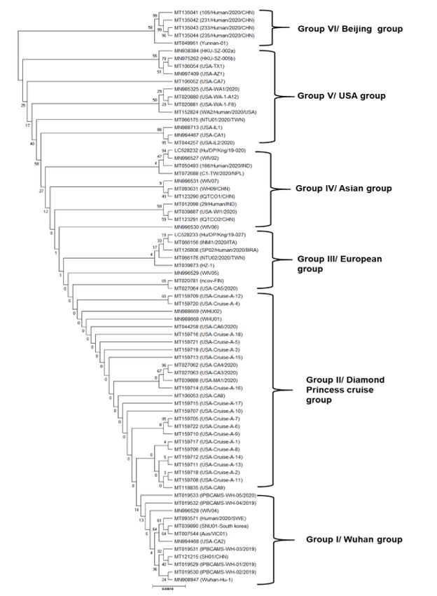
Abstract
It is widely known fact about the global pandemic caused by Severe Acute Respiratory Syndrome
Coronavirus -2 (SARS-CoV-2) to humans, which imposed immediate lockdown of effected territories in the
prevailing provinces. However, few provinces were able to control infection severity with lower death
rates. Interestingly three types of genomic features were noticed through comparative genomics in the
available genome sequences SARS-CoV-2, due to the insertion/deletions of orf3a, orf6, orf7a and orf7b.
Whole genome phylogeny (n=75 genomes) revealed a large diversity within the SARS-CoV-2, and
distributed in 6 clusters namely China, Diamond princess, Asian, European, USA and Beijing. This study
asserts diversity in the genome with high mutation rate and migration of carriers over the world. Here, we
describe the polymorphic loci of Spike glycoprotein and its putative mechanism for pathogenicity, which
unveiled the presence of GPI anchor amidation, PPI hotspot, O-linked glycosylation, catalytic site, Iron
binding site, signal cleavage, disulphide linkage, sulfation, transmembrane region, and C-terminal signal
sites. Mutational changes at spike glycoprotein of South Korea, India, Greece, Spain, Australia, Sweden
and Yunnan samples possibly suggest the prevalence of mutated strains with either low or high
virulence. The regions at the spike glycoprotein also have high binding capacity to angiotensin converting
enzyme 2 (ACE2) suggesting a key link for explaining damage to multiple organs including lungs, kidney
and heart. Factors in uencing the mutations at the spike glycoprotein region will need to be investigated
to understand and neutralize the upsurge of the alarming Pandemic and to control the global spread of
the disease.

Introduction
In the history of global infections, COVID–19 (Corona Virus disease) has left its dangerous,
uncontrollable outbreak footprint. Towards the end of the year 2019, people of Wuhan, the capital city of
Hubei province in China, developed a strange pneumonia-like infection due to an unknown aetiology. It
was later recognized to be a part of coronavirus family1. This pandemic was spread over 210 countries
with over 3,248,685 con rmed cases and 229,399 deaths world-wide till date (01 May, 2020)
(https://www.worldmeters.info/coronavirus). As of the date of manuscript preparation, the US now has
almost 1/3 of all COVID–19 cases worldwide. With a high mortality rate of about 3–6% across the world,
the havoc created by COVID–19 has been massive. The transmission was vast and understanding the
genome variation has been in priority. Till date, no speci c drugs or vaccines are available to control the
infection, and symptomatic treatments to block the viral replication is in early trials. Considering COVID–
19 as a major public health emergency, globally several countries have suspended their trade and called
off social events to prevent community transmission. Furthermore, to battle the virus, countries world-
wide have resorted to self-quarantine and social-isolation as containment strategy for the bene t of the
mankind. Medical supplies, protective agents and hand hygiene are the only resort to prevent the
transmission dynamics of this deadly disease. Ian M. Jones had suggested that the SARS-CoV–2
mutates rapidly in the respiratory tract2. The data sharing among collaborators or investigators had made
the analysis more accurate and easy. Moreover, certain strains prevailing at few provinces had shown low

                                                 Page 2/20
mortality rates. Whereas, countries like Italy, Spain, USA and a few more had high mortality rates
indicating the presence of evolved virulent strain when compared to the original strain from Wuhan.
Understanding the genetically distinct variants in a phenotype is very important in analysing the
pathogenic mechanism of infected hosts.
As RNA viruses tend to evolve rapidly among large populations with short generation times 3, monitoring
evolutionary patterns in “real time” is important. This leads to the emergence of many new novel strains
of COVID–19, it has become an important aspect to differentiate between the virulent strains of SARS-
CoVs in the current scenario. The virus was found more related to the betacoronavirus of bats (RaTG13)
and pangolins (Manis javanica) with 96.2% and 91.02% homology respectively. The spike gene of SARS-
CoV–2 has shown slight variation with polybasic cleavage site (PCS)4,5. The PCS of Spike protein gets
cleaved by furin leaving its infection to different organs of the host 4–6. The whole-genome sequence
(WGS) data would probably show its evolution and reasons for its mutation rates1,7. Laboratories from
many countries have deposited over 2400 genome sequences of SARS-CoV–2 at the NCBI, GISAID and
Nextstrain databases (https://nextstrain.org/ncov), which allowed us to analyze this novel virus.
Acknowledging the importance of the spread and the evolution of the virulent pathogens, the NEXTstrain
database provided the necessary information related to the phylodynamics, genomes and the
surveillance data 8.
It has been hypothesized that the pathogenesis of disease is possibly due to alveolar damage followed
by spleen atrophy, enlarged liver, injury to kidney and neuronal dysfunction in patients9,10. The ability of
SARS-CoV–2 to interact with the kidneys of host and shed of viral particles through faecal and urine of
patients suggests the multiple organ damage with increased severity11. Though, the target organ reported
was lungs due to the speci c binding of SARS-CoV–2 to ACE2 receptors, the presence of other sites
responsible for effective binding of the spike protein in other organs remains an enigma. Hence, the
present study focused on the variations in the genomes of COVID–19, which were distributed worldwide.
Further investigation on the mutated strains pathogenesis in biopsy samples of different human organs
will be investigated.
Results And Discussion
Genome diversity and comparative genomics among SARS-CoV–2
In the present investigation, SARS-COV–2 genome sequences were retrieved from NCBI and GISAID
database (till April 25th 2020). Among the 520 complete genome data of SARS-CoV–2, the genomes
which showed variations in their size and their geographic region were targeted. A total of 75 complete
sequences of SARS-CoV–2 that were prevalent in countries like China, USA, France, Australia, Spain, Italy,
India, Nepal, Taiwan, South Korea, South Africa, Greece, Sweden, Pakistan, Peru, Brazil, Iraq, Turkey and
Israel were collected (Table S1). The knowledge of patient’s ethnicity and racial background were not
readily available for all the samples. The genome analysis revealed that the SARS-CoV–2 is a 30 Kbp
genome with over 10 to 12 genes (Table 1). The largest genome size was noticed in a Shanghai patient
Page 3/20(SH01) of China (Accession number MT121215) with 29945 bp (reported on 2nd Feb 2020). While the
smaller genome size of 29852 bp was detected in a USA patient (CA6) (Accession number MT044258)
(isolated on 27th Jan 2020). These two samples were compared to Wuhan-Hu–1 (MN908947 or
NC045512), which is of 29,903 bp genome size and serves as a reference sample (Fig. 1a). Comparative
genomics of these three isolates revealed that the SH01 sample had a deletion of ORF3a, ORF6, ORF7a,
and 7b, while CA6 isolate had a deletion of ORF7b. However, the genome size of SH01 was noticed to be
larger when compared to the other two samples. Though the function of these genes was not known,
their absence had revealed diversity at strain level (Table 1, Fig. 1b).
Most of the coronaviruses (CoVs) of the Coronaviridae family possess two overlapping ORF1a and
ORF1b polypeptides and other structural proteins like Spike (S), Envelope (E), membrane (M) and
nucleocapsid (N)12. Among the samples that were analysed, ORF7b was present only in 8 samples of
ncov-FIN, Yunnan–01, WH09/CHN, WIV02, WIV04, WIV05, WIV06 and WIV07. Subsequently, ORF3a,
ORF7a, ORF7b and ORF8 were found to be deleted in HU/DP/Kng/19–20, SH01/CHN, WHU01 and
WHU02 samples. However, the severity of the infection in these variants associated with the
corresponding patient is not yet known, as the case history details are not available. These mutations
could make an impact on the immunogenic changes that would either suppress or become more virulent
than the wild type strain. The prevalence of more virulent strains may increase the severity of outbreak.
However, extensive research has to be conducted to correlate the nature of mutations with the outbreak
severity.
Table 1: General features of genes involved in SARS-CoV-2 of three isolates, i.e., Wuhan-Hu-
1, CA6 and SH01 (in this study Wuhan-Hu-1 served as a reference sample)
S.NO Gene name Description Gene length (bp)
Wuhan-Hu-1 CA6 SH01
1 orf1ab Polyprotein 21291 21267 21271
2 orf S Surface glycoprotein 3822 3822 3822
3 orf 3a Hypothetical protein 828 828 -
4 orf E Envelope protein 228 228 228
5 orf M Membrane protein 669 669 669
6 orf 6 Hypothetical protein 186 186 -
7 orf 7a Hypothetical protein 366 366 -
8 orf 7b Hypothetical protein 132 - -
9 orf 8 Hypothetical protein 366 366 -
10 orf N Nucleocapsid phosphoprotein 1260 1260 1260
11 orf 10 Hypothetical protein 117 117 117
absence of genes
A recent work published by Tang and his co-workers2 suggested the prevalence of two types of COVID–
19, named as L and S type, based on their SNPs at ORF1ab and ORF8. L type was prevalent and
accounted for about 70% of infected China population during Jan-Feb 2020. In the same study, they have
noticed many nonsynonymous mutations in the 103 samples analyzed. However, the factors behind the
emergence of L and S type are still ambiguous. In the current study, around 75 samples from different
Page 4/20parts of the world were considered to study their evolutionary patterns. The genome size is diverse and
shows many deletions and insertions. In any case, the genetic information indicates that SARS-CoV–2 is
not derived from available virus data in a laboratory, as it shares homology towards SARS,
betacoronavirus of bats, and pangolins14.
Phylogenetic evolution among COVID–19 positive samples
Due to the variations in the genome sequences of SARS-CoV–2, a genome phylogeny was constructed to
understand the evolution and transmission pattern. The dendrogram suggested six groups (Fig. 2), of
which group 1 had, isolates from Wuhan (IPBCAMS-WH–1/2019, WH–2/2019, and WH–3/2019),
Shanghai (SH01/CHN), USA (USA-CA2), Australia (AUS/VIC01), South Korea (SNU01) and Sweden
(Human/2020/SWE). These eight samples had a close resemblance with Wuhan-Hu–1 (reference isolate)
and hence this group is described as Wuhan group. Group 2 is almost a clone where one international
conveyance, i.e., Diamond Princess Cruise from Japan, and had over 700 coronavirus cases, with patients
from the different parts of the world such as USA, Hongkong, Japan and China, the spread of the
infection was vast in other countries. This group 2 is named as Diamond Princess cruise group, and the
patients were quarantined in the ship for two weeks. The third European group had patients from Italy,
Finland, Brazil, and a few cases from China, Japan, Taiwan and USA were grouped together, suggesting
transmission from the Wuhan epicentre. Next is the Asian group (group IV), isolated in subjects from
China, Japan, Taiwan, Nepal, India, and Hongkong. Group V is the USA group with the samples clubbed
together with the patients from California (CA), Texas (TX), Washington (WA), and Illinois (IL). However,
WA had a close cluster indicating the community transfer at USA. Also, a few patients of the USA had a
travel history to China and other COVID–19 affected areas. Group VI is the Beijing group of China, which
emerged from a patient from Yunnan. Phylogenomics suggests the high rate of either co-infection or
recombination between different strains of SARS-CoV–2, showing its diversity. However, multiple
samples have to be studied to ascertain sustainable facts, given their uncertain nature of genome
variations. Tang et al.,2 could differentiate the SARS-CoV–2 by phylogenetics into L and S types based on
their aggressiveness. It was perceived that L type was more virulent than S type and mainly possessed
isolates from Wuhan, France, Australia, Singapore, USA, Hongkong, Taiwan, Japan and other countries. It
will be always interesting to study the transmission of these mutations and its pathogenesis in any
prevalent area.
Notable changes at Spike glycoprotein
Regardless of critical advances in cutting edge sequencing innovations, which have encouraged the
disclosure of thousands of novel zoonotic viruses, methods for downstream evaluation of these novel
sequences are de cient. Hence, an approach to determine the functional viromics in a more applicable
way to understand the host-protein interactions is obligatory. The Spike (S) protein plays a role in the
entry of virus into host cells, by binding to angiotensin converting enzyme 2 (ACE–2). The motif nder
programme of S protein in Wuhan-Hu–1 showed 9 Pfam motifs (Fig. S1a)
(https://www.genome.jp/tools/motif/). The S protein of CoVs isolated from bats and infected humans
Page 5/20had >98% homology with few mutations. Basically, the S protein had an identical ribosome binding
domain (RBD) and an O-linked glycan residue domain with polybasic cleavage site (RRARS) which was
analysed through multiple alignment by Geneious Prime programme15. In the current investigation, we
nd that the RBD of all 75 samples is highly conserved with 9 amino acid variations when compared to
Bat-RaTG13 (Fig. S1b). Similarly, the O-linked glycan residue domain had an insertion of four nucleotides
PRRA (Fig. S1b). Later on, the samples which showed mutations at Spike glycoprotein were retrieved
from Nextstrain database. Among the 358 samples analysed (data not shown), over 33 samples showed
variations and suggested strain variation (Table 2). In samples from Peru (1), Israel (1), Greece (3), Spain
(2), France (1), India (10) showed a common mutational site at D614G. However, these samples had
variations in other ORFs of their genome, suggesting strain diversity. It was found that most of the strains
possessed a unique pattern showing its strain-speci city. However, the immunological aspects of various
strains and analysis is still lacking and need to be investigated. In the entire study, the structural genes of
SARS-CoV–2 were mutated more rapidly than the non-structural genes.
It has been reported that Human angiotensin converting enzyme II (ACE2) receptor is the binding site for
most SARS-CoV11. This was supported by another study which asserted that the novel SARS-CoV–2
utilizes the ACE2 to bind and nd its entry in to the host cell4. ACE2 expression in organs like kidney and
heart has been reported, providing a mechanism for the multi-organ dysfunction that can be seen with
SARS-CoV–2 infection16,17. Interspecies diversity within different bat species harbouring the coronavirus
was found18. In the same study, a surveillance of bat-CoV’s revealed the presence of SARS-like
coronavirus, unclassi ed betacoronavirus and new betacoronavirus species. The co-infection of these
CoVs in mineshaft bat species showed potential infection in the host. Further, the RBD of pangolin CoVs
are indistinguishable from that of SARS-CoV–2 at 6 of 6 key amino acids examined previously18,19. This
observation shows that entry of CoV in a host with human-like ACE2 could choose for a RBD with high-
a nity15. Whether the ACE2 expression in these organs affects the SARS-CoV–2 infectivity remains
unclear. Majority of the scienti c reports state that acute kidney injury (AKI), abdominal discomfort and
cardiac damage are the most commonly reported symptoms of COVID–1920,21 suggesting that SARS-
CoV–2 may have a tropism for these organs. Such recombination’s and transmission could likewise
choose for the insertion of the polybasic cleavage site (PCS), which is absent in pangolin and bats
coronaviruses12. These PCS are highly conserved in a particular strain and shows their high
pathogenicity, leading to a possible pandemic outbreak with high mortality or morbidity rate22, as
observed in H5N1 virus. A putative recognition motif i.e., PRRARSV is present in all the sample analysed
and is the active site for furin recognition14,23. The natural selection of virus with cleavage site would
probably have taken when such virus similar to the existing SARS-CoV had undergone several passages
under in vitro cell-line models. It is improbable that the O-linked glycan site would have triggered without
immune pressure, which was not present in the cell-lines. The insertion of PRRA amino acids, make the
SARS-CoV–2 novel and more pathogenic than SARS and MERS.
Table 2: Mutational changes observed different ORF’s of SARS-CoV-2 (mutations at spike
glycoprotein (S) was represented in separate column)
Page 6/20Prevalence of mutant strains in certain provinces as biological markers
The samples from Brazil (3), France (1), Greece (4), Spain (3), Turkey (1), Peru (1), Israel (1), Sweden (7),
India (12), China (3), USA (1), South Korea (1), South Africa (1) and Australia (2) possessed mutational
changes at the Spike protein of SARS-CoV–2 and followed by their countries mortality rate was assessed
(Table 3). Deletion of an amino acid tyrosine (Y), lysine (K) and Guanine (G) at 144, 528 and 107
positions was noticed in subjects of Indian (MT012098), Spain (MT233521) and France (MT320538),
respectively, who had a travel history from Wuhan, China (https://www.covid19india.org/,
www.nextstrain.org). Though the spike protein had no variation at the ribosome binding site, the
mutations noticed in these 42 samples would either increase or decrease the severity of the outbreak.
However, further analysis is required to prove the severity of these samples. The prevalence of these
strains in different geographical regions has to be assessed, as these might serve as biomarkers in
understanding the antigenic and immunogenic changes.
Table 3: Mutational changes in the spike protein of SARS-CoV-2 with their countries
mortality rates
Page 7/20Accession Sample ID Country Mutations at S No. of No. of Mortality
Number protein confirmed Deaths rate (%)
cases
MT350282 BRA/SP02cc/2020 Brazil N74K 79695 5513 6.91
MT263074 PER/Peru-10/2020 Peru D614G 33931 943 2.77
MT233521 Valencia6/2020 Spain K528del 239639 24543 10.24
MT292569 Valencia13/2020 Spain D614G 239639 24543 10.24
MT292575 Valencia16/2020 Spain D614G 239639 24543 10.24
MT276598 ISR/ISR-IT0320 Israel D614G 15870 219 1.37
MT328032 GRC/10/2020 Greece D614G 2576 136 5.39
MT328035 GRC/13/2020 Greece D614G 2576 136 5.39
MT328034 GRC/16/2020 Greece I197Y 2576 136 5.39
MT328033 GRC/12/2020 Greece D614G 2576 136 5.39
MT093571 210/human/2020/SWE Sweden F797C 21092 2586 12.26
MT049951 Yunnan- China Y28N 82862 4633 5.59
01/human/2020
MT039890 SNU01/South Korea South S221W 10765 247 2.29
Korea
MT007544 Australia/VIC01/2020 Australia S247R 6753 91 1.34
MT327745 TUR/ERAGEM- Turkey V772I 117589 3081 2.62
001/2020
MT320538 FRA/KRA-ROB/2020 France G107del, 166420 24087 14.47
D614G
MT300186 USA-CA United D614G 1067382 61849 5.79
States of
America
MT324062 ZAF/R03006/2020 South D614G 5350 103 2.42
Africa
MT012098 29/human/2020/IND India Y144del, 33610 1079 3.21
R408I
MT050493 166/human/2020/IND India A930V 33610 1079 3.21
EPI_ISL_426414 India/GBRC1/2020 India Q271R, D614G 33610 1079 3.21
EPI_ISL_426415 India/GBRC1s/2020 India Q271R, D614G 33610 1079 3.21
EPI_ISL_430468 India/S2/2020/West India D614G, 33610 1079 3.21
Bengal G1124V
EPI_ISL_430464 India/S3/2020/West India D614G, 33610 1079 3.21
Bengal G1124V
EPI_ISL_424365 India/3239/2020 India D614G 33610 1079 3.21
EPI_ISL_428482 India/nimh-0182/2020 India D614G, 33610 1079 3.21
C1250F
Page 8/20EPI_ISL_426424 USA/IN_92003/2020 India D614G, 33610 1079 3.21
L1203F
EPI_ISL_426423 USA/IN_82003/2020 India D614G, 33610 1079 3.21
L1203F
EPI_ISL_426422 USA/IN_72003/2020 India D614G, 33610 1079 3.21
L1203F
EPI_ISL_420551 India/777/2020 India D614G 33610 1079 3.21
EPI_ISL_429691 BRA/CV35/2020 Brazil Y695S, 79695 5513 6.91
G832D,
H1088N
EPI_ISL_429677 BRA/CV17/2020 Brazil K776T 79695 5513 6.91
EPI_ISL_426882 Australia/VIC913/2020 Australia G446V, 6753 91 1.34
G1124V
EPI_ISL_429129 Sweden/20-08681 Sweden D80Y 21092 2586 12.26
EPI_ISL_430859 Sweden/20-08717 Sweden K1073N 21092 2586 12.26
EPI_ISL_429152 Sweden/20-50179 Sweden V62F 21092 2586 12.26
EPI_ISL_429157 Sweden/20-50234 Sweden M1237I 21092 2586 12.26
EPI_ISL_429116 Sweden/20-08143 Sweden Y917H 21092 2586 12.26
EPI_ISL_411951 Sweden/01/2020 Sweden F797C 21092 2586 12.26
EPI_ISL_415709 Hanghou/ZJU-01 China R682Q 82862 4633 5.56
EPI_ISL_421259 Pingxiang/JX151 China S254F 82862 4633 5.56
While analysing the cases in these provinces, it was noticed that the death rate was low in South Korea,
Greece, Brazil, Israel, Peru, Turkey, South Africa and Australia, thus COVID–19 cases curve has declined.
However, these states also followed many measures in controlling the outbreak such as early lockdowns,
self-isolation, social distancing, hygienic practicesas instructed by their governments. However, in
Sweden, and India the COVID–19 cases are being analysed and the graph is up surging due to a hike in
the con rmed cases (https://www.worldometers.info/coronavirus/). It can be seen that the death toll is
comparatively low in these areas when compared to the other areas such as Wuhan, Italy, Spain, France,
United States of America and Germany. This might indicate that the prevalence of mutated strains which
might have emerged during coinfection within the provinces, would have either reduced or increased its
severity. Furthermore, the pathogenicity probabilistically was assessed in the putative neutral variants.
The MutPred Indel software could assess the sites responsible for its virulence (Table 4). The sample
Human/2020/SWE from Sweden had not shown any pathogenic sites when compared to Wuhan-Hu–1
(reference strain). However, most of the subjects analysed had putative variants of S protein showing
several post-translational mechanisms such as, catalytic site, proteolytic cleavage, Iron binding site,
glycosyl-phosphatidylinositol (GPI) anchor amidation, PPI hotspot, sulfation, transmembrane region,
copper binding, signal cleavage, cytoplasmic loop, C-terminal signal, and O-linked glycosylation sites,
Page 9/20suggesting probability of more virulence in these samples (P>0.5). Most of the isolates had catalytic site,
PPI hotspot and Iron binding as their common mechanism for pathogenesis. However, including these
mechanisms many other isolates possessed extra mechanisms for their mode of action. For example, the
mechanism of palmitoylation was noted only in a sample from an Indian subject
(166/Human/2020/IND). The subjects from Turkey and Brazil had disul te linkage and sulfation as their
mechanism. Considering the prevailing situation in India, the presence of pathogenic variant of spike
protein it can be postulated that the rate of COVID–19 cases would increase eventually during the next
few days. Hence, every citizen has to be abide to the preventive measures.
Table 4: Pathogenicity prediction with MutPred-Indel model in the putative Spike protein
variants of SARS-CoV-2 samples
Page 10/20Sample ID Country Site P-score Mechanism for pathogenicity
Wuhan-Hu-1 China S221 0.515 Catalytic site, Iron binding
(MN908947)
(reference sample)
Human/2020/SWE Sweden - - -
29/Human/2020/IND India H145 0.385 PPI hotspot, Catalytic site, Iron binding, O-linked
glycosylation, C-terminal signal
166/Human/2020/IND India S247 0.36415 GPI anchor amidation, PPI hotspot,, Catalytic site,
signal cleavage, Iron binding, palmitoylation
Yunnan-01 Yunnan N28 0.449 Iron binding
SNU-01 South S221 0.354 Catalytic site, Iron binding
Korea
Aus/VIC01 Australia R247 0.4547 PPI hotspot, Proteolytic cleavage, Copper binding,
Catalytic site, Iron binding
TUR/ERAGEM- Turkey V3F 0.35058 Catalytic site, PPI hotspot, Iron binding, Disulfide
001/2020 linkage, Sulfation
BRA/SP02cc/2020 Brazil K83 0.346 Catalytic site, PPI hotspot, Iron binding, Disulfide
linkage, Sulfation
ZAF/R03006/2020 South G623 0.3458 Catalytic site, Iron binding, PPI hotspot, Disulfide
Africa linkage, GPI anchor amidation
GRC/13/2020 Greece I206 0.35209 PPI hotspot, Catalytic site, Iron binding, Disulfide
linkage, GPI anchor amidation
India/GBRC1s/2020 India R271 0.36268 PPI hotspot, Catalytic site, Iron binding, Disulfide
linkage, GPI anchor amidation
FRA/KRA-ROB/2020 France L1203 0.541 PPI hotshot, Catalytic site, Iron binding
USA/IN_82003/2020 USA F1203 0.453 Signal cleavage, Iron binding, Transmembrane region,
signal helix, PPI hotspot
Valencia6_ESP Spain S529 0.370 Iron binding, GPI anchor amidation, Catalytic site,
Signal cleavage, C-terminal signal
SWE/20-08681 Sweden Y80 0.589 PPI hotspot, Iron binding
SWE/20-08717 Sweden Y80 0.389 Cytoplasmic loop, PPI hotspot, O-linked glycosylation,
catalytic site, signal helix
SWE/20-50179 Sweden V62 0.517 Iron binding, PPI hotspot, catalytic site, cytoplasmic
loop, O-linked glycosylation
SWE/20-50234 Sweden Y80 0.389 PPI hotspot, cytoplasmic loop, C-terminal signal, Signal
cleavage, Iron binding
SWE/20-08143 Sweden Y80 0.390 PPI hotspot, O-linked glycosylation, catalytic site, Iron
binding, C-terminal signal
SWE/20-01/2020 Sweden Y80 0.391 PPI hotspot, Catalytic site, Iron binding, C-terminal
signal, signal helix
- no mechanism of pathogenicity was detected
Petit et al. 24suggested that palmitoylation aids in providing anchoring ability during cell fusion and
receptor binding in SARS-CoV, this mechanism noted in COVID–19 sample suggest conformational
changes during palmitoylation process leading to signal transduction mechanism at both intra- and-
extracellular domains. Sulfation is a process for protein-protein interaction and found to play a role in
extracellular extension for high a nity towards binding, leading to the activation of receptors and
Page 11/20stability of proteins by correct protein folding mechanism 25. Hence, mutational changes in the spike
glycoprotein may instigate its conformational changes, which is most likely to prompt the evolving
antigenicity26. Studies pertaining to the localization of amino acids associated with this protein among
different variants of SARS-CoV–2, are readily not available. A recent study on protein-protein interactions
(PPI) by Gordon et al.27 had suggested that the spike protein has the ability to interact with GOLGA7-
ZDHHC5 acyl transferase complex and can be a therapeutic target. GPI anchor sites are also found to
target host innate defense system, which allows functions in tra cking, cell adhesion and metabolism. It
was reported that, Bone marrow stroma antigen 2 (BST2), also called as CD317 or tetherin has a capacity
to inhibit the enveloped virus release into the host, hence such sites can be targeted for therapeutics28. It
will be important to explore these mutational changes. Along these lines, reinforcing SARS-CoV–2
surveillance among different geographical regions can provide scienti c evidence for its more
pathogenicity and allows in taking preventive and controlling measures in the transmission of disease.
ACE2 expression in human organs targeted in kidney
SARS-CoV–2 infection starts by binding of the viral surface spike protein to the human angiotensin-
converting enzyme 2 (ACE2) receptor following modi cation of the spike protein by transmembrane
protease serine 2 (TMPRSS2)29. Initially, ACE2 is expressed in the lung (principally Type II alveolar cells7)
and seems to be the predominant portal of entry. Considering SARS entry into target human cells, it can
be observed that the expression of ACE2 protein is signi cantly found in the epithelial cells of the lung
alveoli and small intestine and endothelial cells of organs including spleen, kidney, liver, lymph nodes,
brain30,31. Burgeoning data con rm association of COVID–19 infection with increased morbidity and
mortality from kidney disease. It is important to investigate whether SARS-CoV–2 replication occurs in
these organs contributing to the virus disseminating throughout the body.
High expression of ACE2 was noticed in proximal tubular cells and to a lesser extent in podocytes,
however, kidney glomerular endothelial and mesangial cells were not affected17. It was perceived that
only 6% of patients infected with SARS-CoV experienced Acute Kidney Injury (AKI) during SARS outbreak
during 2003 32. Furthermore, AKI was identi ed as a serious complication of SARS, with mortality of 92%
in patients 32. During post-mortem from SARS patients, SARS-CoV viral particles were noticed in renal
specimens, suggesting AKI was caused by active replication of SARS-CoV in tubular cells 32. They
suggested that renal impairment was likely associated with multi-organ failure as SARS-CoV was not
demonstrable in any of the examined patients. Further, AKI (including cytokine release syndrome and
SARS patients) might be a speci c pathogenic condition, and might not be due to the active replication of
virus at kidneys 32, 33. An increased viral infection in alveolar cells leads to the production of large amount
of cytokines, causing multiple-organ failure. Previously a study had reported that release of interferon-
gamma-related cytokine increased the severity of organ damage in SARS patients34. Recently, a study
described that the human kidney is a speci c target for SARS-CoV–2 infection35. The difference between
the higher renal tropism of SARS-CoV–2 versus SARS-CoV can be assessed by the increased a nity of
Page 12/20SARSCoV–2 for ACE2, contributing towards pronounced infection of the kidney, leading to viral reservoir
36
.
In addition, a small survey on COVID–19 patients has revealed that, proteinuria and haematuria are
common features that were noticed in 40% of patients post hospitalization 37. A reduced density of
in ammation and edema was observed in CT scan reports of kidneys samples infected with SARS-CoV–
238. Furthermore, SARS-CoV–2 seems to be affected more by AKI frequently than subjects infected with
SARS-CoV37. A very recent study by Yao et al.39 con rms that SARS-CoV–2 infection damages vessels,
kidney and other organs, in addition to the lungs,. Hyaline thrombi are found in small vessels in different
organs. It is of utmost importance to investigate pathological changes in autopsy material. Before organ
donation is considered in future, it will be important to investigate whether the SARS-CoV–2 has infected
the kidneys; the risk of such organ grafts has not been reported as yet. In any case, it has been indicated
that SARSCoV–2 has a high tropism for the kidney, where it has been shown to reproduce in practically
30% of COVID–19 patients40. Consequently, screening for COVID–19 in kidney donors is probably more
important during screening time and need to be quarantined for 14–28 days who possess either
symptoms or had a travel history to high-risk regions 31. A research study demonstrated that more than
66% of patients had died with COVID–19 infection who had diabetes or cardiovascular disease41. As a
rst-line treatment, angiotensin-receptor blockers (ARBs) were given to COVID–19 patients. Certain
reports revealed that ARBs were found to express ACE2 by nearly 2 to 5 fold in kidney and heart samples
42–45
. Since SARSCoV–2 has a high tropism for the kidney35,40, investigating how ARBs affect the
infection rate and renal and cardiac injury in COVID–19 infection is of great importance.
Conclusion
Bat coronavirus remains a considerable worldwide risk to general wellbeing of humans. The genomic
highlights depicted in the present study might clarify the transmissibility of SARS-CoV–2 in human race,
yet its inception is a question. Despite the fact that the nCoV–2019 had close genetic relatedness
towards RaTG13 of bat coronavirus, which was isolated in 2013 (however, the genome data of this strain
was made open just after the outbreak of COVID–19). The current investigation provided three types of
variants in the SARS-CoV–2 genome. The phylogeny showed six clusters which includes Wuhan,
Diamond princess, European, Asian, USA and Beijing group. The polybasic cleavage site in the Spike
protein of COVID–19 isolates is very conserved and different from bat and pangolins CoV’s, suggesting
its novel pathogenic nature. Mutations in the spike protein could either reduce or increase the severity of
the outbreak. The mechanism of pathogenicity among putative variants of spike surface glycoprotein
suggested more virulence in few samples of India, Australia, Greece, South Korea and Yunnan. The
available clinical data have con rmed that AKI is one of the main risk factors in the prognosis of COVID–
19. Patients with diabetic nephropathy, end-stage renal disease, and, renal transplantation may be at high
risk of the SAR-CoV–2. However, spread of SARS-like infections from various intermediate animals may
assist in explaining its emergence or outbreaks. The recognizable proof of a potential intermediate host
Page 13/20of SARS-CoV–2, along with their genome sequence data of the virus at early stage, would also be
profoundly useful.
Materials And Methods
Information related to daily cases of COVID–19 and SARS-
CoV–2 genome data
The genome data of SARS-CoV–2 was retrieved from the public repositories like NCBI data and the global
information on COVID–19 cases was obtained through worldometers
(https://www.worldometers.info/coronavirus/) and NEXTstrain (https://nextstrain.org/ncov) websites.
Totally 75 genomes were considered based on the variation in their genome size, country and divergence.
The samples used in the current study are enlisted in Table S1. However, the genome ethnicity and racial
inheritance of all the samples are not available.
Comparative genome analysis
Three genomes MT121215 and MT044528 were considered in the study which possessed highest and
lowest genome size, and were compared to the reference sample (MN908947). The comparative genome
analysis was performed by using Geneious Prime Software Version 2019.2.113.
Phylogenetic evolution
To further analyze the evolution of isolates, a phylogenetic tree (n = 75) was constructed using the
complete genome data of SARS-CoV–2 by using MEGA-X (Molecular Evolutionary Genetic Analysis)
software46. The evolutionary history was deduced by using the Neighbor-Joining method with 500
bootstrap replicates47. Further, the evolutionary distances were computed using the Maximum Composite
Likelihood method. The analysis involved 75 nucleotide sequences. There was a total of 39547 positions
in the nal dataset.
Bioinformatics tools used in the analysis of spike protein
Multiple sequence analysis of the spike protein among the 75 isolates was performed by using Clustal W
programme48. The VIPR database is used to analyse the single nucleotide polymorphism (SNP) at spike
glycoprotein as described by Pickett et al.49. Further Genome Detective Virus Tools was also used to look
at the mutational analysis (https://www.genomedetective.com/app/typingtool/virus/). The pfam motifs
were analysed by using genome motifs database (https://www.genome.jp/tools/motif/). Further, the
ribosome binding region and the polybasic cleavage site was determined as described by Andersen et
al.15.
Page 14/20Pathogenicity prediction in the phenotypes
To further assess the pathogenicity of the variants (putatively neutral) a machine learning-based method
software package was employed for the spike protein phenotypes. The MutPred-Indel software assess
the probabilistically the pathogenicity of the neutral variants and suggests the features affecting the
phenotypes50.
Declarations
Authors Contribution
SMD performed the bioinformatics analysis on the collected data, planned the work and wrote the
manuscript, AP had collected the data and worked on kidney biopsy samples of COVID-19, BK edited the
manuscript, and KS monitored the analysis and edited the manuscript.
Acknowledgment
The authors thank all family members and friends for all the support during the crisis period of
COVID-19. The work was not supported by any fund, the article was written to analyse the genome data
of COVID-19 considering the present awareness.
Con ict of Interest
The author claims no con ict of interest.
References
1. Wu, F. et al. A new coronavirus associated with human respiratory disease in China. Nature 579, 265-
269 (2020).
2. Tang, X. et al. On the origin and continuing evolution of SARS-CoV-2. National Science Review
(2020).
3. Duffy, S., Shackelton, L.A. & Holmes, E.C. Rates of evolutionary change in viruses: patterns and
determinants. Nature Reviews Genetics 9, 267-276 (2008).
4. Zhou, P. et al. A pneumonia outbreak associated with a new coronavirus of probable bat origin.
Nature 579, 270-273 (2020).
5. Wong, M.C., Cregeen, S.J.J., Ajami, N.J. & Petrosino, J.F. Evidence of recombination in coronaviruses
implicating pangolin origins of nCoV-2019. Biorxiv (2020).
6. Tsai, H.J., Chi, L.-A. & Alice, L.Y. Monoclonal antibodies targeting the synthetic peptide corresponding
to the polybasic cleavage site on H5N1 in uenza hemagglutinin. Journal of biomedical science 19,
37 (2012).
Page 15/207. Perrella, A. et al. Editorial–Novel Coronavirus 2019 (Sars-CoV2): a global emergency that needs new
approaches. Eur Rev Med Pharmaco 24, 2162-2164 (2020).
8. Had eld, J. et al. Nextstrain: real-time tracking of pathogen evolution. Bioinformatics 34, 4121-4123
(2018).
9. Yao, X. et al. A pathological report of three COVID-19 cases by minimally invasive autopsies.
Zhonghua bing li xue za zhi= Chinese journal of pathology 49, E009-E009 (2020).
10. Perico, L., Benigni, A. & Remuzzi, G. Should COVID-19 concern nephrologists? Why and to what
extent? The emerging impasse of angiotensin blockade. Nephron, 1-9 (2020).
11. Li H, L.L., Zhang D, Xu J, Dai H, Tang N, Su X, Cao B. SARS-CoV-2 and viral sepsis: observations and
hypotheses. The Lancet (2020 Apr 17).
12. Fan, Y., Zhao, K., Shi, Z. & Zhou, P. Bat Coronaviruses in China. Viruses, 11 (3), 210. (2019).
13. Kearse, M. et al. Geneious Basic: an integrated and extendable desktop software platform for the
organization and analysis of sequence data. Bioinformatics 28, 1647-1649 (2012).
14. Zhang, T., Wu, Q. & Zhang, Z. Probable pangolin origin of SARS-CoV-2 associated with the COVID-19
outbreak. Current Biology (2020).
15. Andersen, K.G., Rambaut, A., Lipkin, W.I., Holmes, E.C. & Garry, R.F. The proximal origin of SARS-CoV-
2. Nature medicine 26, 450-452 (2020).
16. Zhang, H., Penninger, J.M., Li, Y., Zhong, N. & Slutsky, A.S. Angiotensin-converting enzyme 2 (ACE2)
as a SARS-CoV-2 receptor: molecular mechanisms and potential therapeutic target. Intensive Care
Medicine 46, 586-590 (2020).
17. Ye, M. et al. Glomerular Localization and Expression of Angiotensin-Converting Enzyme 2 and
Angiotensin-Converting Enzyme: Implications for Albuminuria in Diabetes. Journal of the American
Society of Nephrology 17, 3067-3075 (2006).
18. Ge, X.-Y. et al. Coexistence of multiple coronaviruses in several bat colonies in an abandoned
mineshaft. Virologica Sinica 31, 31-40 (2016).
19. Hu, B. et al. Discovery of a rich gene pool of bat SARS-related coronaviruses provides new insights
into the origin of SARS coronavirus. PLoS pathogens 13(2017).
20. Gallagher, P.E., Ferrario, C.M. & Tallant, E.A. Regulation of ACE2 in cardiac myocytes and broblasts.
American Journal of Physiology-Heart and Circulatory Physiology 295, H2373-H2379 (2008).
21. G, C. Bulk and single-cell transcriptomics identify tobacco-use disparity in lung gene expression of
ACE2, the receptor of 2019-nCov. MedRxiv (2020 Jan 1).
22. Hatta, M., Gao, P., Halfmann, P. & Kawaoka, Y. Molecular basis for high virulence of Hong Kong H5N1
in uenza A viruses. Science 293, 1840-1842 (2001).
23. Yamada, Y. & Liu, D.X. Proteolytic activation of the spike protein at a novel RRRR/S motif is
implicated in furin-dependent entry, syncytium formation, and infectivity of coronavirus infectious
bronchitis virus in cultured cells. Journal of virology 83, 8744-8758 (2009).
Page 16/2024. Petit, C.M. et al. Palmitoylation of the cysteine-rich endodomain of the SARS–coronavirus spike
glycoprotein is important for spike-mediated cell fusion. Virology 360, 264-274 (2007).
25. Ngounou Wetie, A.G. et al. Investigation of stable and transient protein-protein interactions: Past,
present, and future. Proteomics 13, 538-557 (2013).
26. Phan, T. Genetic diversity and evolution of SARS-CoV-2. Infection, Genetics and Evolution 81, 104260
(2020).
27. Gordon, D.E. et al. A SARS-CoV-2-human protein-protein interaction map reveals drug targets and
potential drug-repurposing. BioRxiv (2020).
28. Wang, S.M., Huang, K.J. & Wang, C.T. Severe acute respiratory syndrome coronavirus spike protein
counteracts BST2‐mediated restriction of virus‐like particle release. Journal of medical virology 91,
1743-1750 (2019).
29. Hoffmann, M. et al. SARS-CoV-2 Cell Entry Depends on ACE2 and TMPRSS2 and Is Blocked by a
Clinically Proven Protease Inhibitor. Cell 181, 271-280.e8 (2020).
30. Hamming, I. et al. Tissue distribution of ACE2 protein, the functional receptor for SARS coronavirus.
A rst step in understanding SARS pathogenesis. The Journal of Pathology 203, 631-637 (2004).
31. Perico, L., Benigni, A. & Remuzzi, G. Should COVID-19 Concern Nephrologists? Why and to What
Extent? The Emerging Impasse of Angiotensin Blockade. Nephron (2020).
32. Chu, K.H. et al. Acute renal impairment in coronavirus-associated severe acute respiratory syndrome.
Kidney International 67, 698-705 (2005).
33. Tisoncik, J.R. et al. Into the Eye of the Cytokine Storm. Microbiology and Molecular Biology Reviews
76, 16-32 (2012).
34. Huang, K.-J. et al. An interferon-γ-related cytokine storm in SARS patients. Journal of Medical
Virology 75, 185-194 (2005).
35. Diao, B. et al. Human Kidney is a Target for Novel Severe Acute Respiratory Syndrome Coronavirus 2
(SARS-CoV-2) Infection. medRxiv, 2020.03.04.20031120 (2020).
36. Wan, Y., Shang, J., Graham, R., Baric, R.S. & Li, F. Receptor Recognition by the Novel Coronavirus from
Wuhan: an Analysis Based on Decade-Long Structural Studies of SARS Coronavirus. Journal of
Virology 94, e00127-20 (2020).
37. Wang T, H.M., Chen X, Fu Y, Lei C, Dong H, Zhou Y, Jia H, Chen X, Yan J. Caution on Kidney
Dysfunctions of 2019-nCoV Patients. medRxiv. (February 7th 2020).
38. Cheng, Y. et al. Kidney impairment is associated with in-hospital death of COVID-19 patients.
medRxiv, 2020.02.18.20023242 (2020).
39. Yao, X.H. et al. [A pathological report of three COVID-19 cases by minimally invasive autopsies].
Zhonghua bing li xue za zhi = Chinese journal of pathology 49, E009 (2020).
40. Pan, X.-W. et al. Identi cation of a potential mechanism of acute kidney injury during the COVID-19
outbreak: a study based on single-cell transcriptome analysis. Intensive care medicine, 1-3 (2020).
41. Remuzzi, A. & Remuzzi, G. COVID-19 and Italy: what next? The Lancet 395, 1225-1228 (2020).
Page 17/2042. Gallagher, P.E., Ferrario, C.M. & Tallant, E.A. MAP kinase/phosphatase pathway mediates the
regulation of ACE2 by angiotensin peptides. American Journal of Physiology-Cell Physiology 295,
C1169-C1174 (2008).
43. Ishiyama, Y. et al. Upregulation of Angiotensin-Converting Enzyme 2 After Myocardial Infarction by
Blockade of Angiotensin II Receptors. Hypertension 43, 970-976 (2004).
44. Ferrario, C.M. et al. Effect of Angiotensin-Converting Enzyme Inhibition and Angiotensin II Receptor
Blockers on Cardiac Angiotensin-Converting Enzyme 2. Circulation 111, 2605-2610 (2005).
45. Jessup, J.A. et al. Effect of angiotensin II blockade on a new congenic model of hypertension derived
from transgenic Ren-2 rats. American Journal of Physiology-Heart and Circulatory Physiology 291,
H2166-H2172 (2006).
46. Kumar, S., Stecher, G., Li, M., Knyaz, C. & Tamura, K. MEGA X: molecular evolutionary genetics
analysis across computing platforms. Molecular biology and evolution 35, 1547-1549 (2018).
47. Tamura, K., Nei, M. & Kumar, S. Prospects for inferring very large phylogenies by using the neighbor-
joining method. Proceedings of the National Academy of Sciences 101, 11030-11035 (2004).
48. Altschul, S.F. et al. Gapped BLAST and PSI-BLAST: a new generation of protein database search
programs. Nucleic acids research 25, 3389-3402 (1997).
49. Pickett, B.E. et al. ViPR: an open bioinformatics database and analysis resource for virology research.
Nucleic acids research 40, D593-D598 (2012).
50. Pagel, K.A. et al. Pathogenicity and functional impact of non-frameshifting insertion/deletion
variation in the human genome. PLoS computational biology 15, e1007112 (2019).
Figures
Page 18/20Figure 1
a: Circular maps of the SARS-CoV-2 samples analysed by using Geneious Prime Software Version
2019.2.113 (Yellow color indicates gene, Green color is for CDS and orange color represents nascent
peptide). b: Comparative genome analysis of SARS-CoV-2 isolates Wuhan-Hu-1 (NC045512), CA-6
(MT044258) and SH01 (MT121215)
Page 19/20Figure 2
Phylogenetic emergence of COVID-19 among 75 prevalent samples globally In this study, each sample
was given an ID, however, the ethnicity and geographical location of most of the patients details were not
available.
Page 20/20You can also read