Prospective mapping of viral mutations that escape antibodies used to treat COVID-19
←
→
Page content transcription
If your browser does not render page correctly, please read the page content below
REPORTS

                                                                                                                              Cite as: T. N. Starr et al., Science
                                                                                                                              10.1126/science.abf9302 (2021).

Prospective mapping of viral mutations that escape
antibodies used to treat COVID-19
Tyler N. Starr1*, Allison J. Greaney1,2,3*, Amin Addetia1,4, William W. Hannon1,4, Manish C. Choudhary5,
Adam S. Dingens1, Jonathan Z. Li5, Jesse D. Bloom1,2,6†
1
 Basic Sciences and Computational Biology, Fred Hutchinson Cancer Research Center, Seattle, WA 98109, USA. 2Department of Genome Sciences, University of
Washington, Seattle, WA 98109, USA. 3Medical Scientist Training Program, University of Washington, Seattle, WA 98109, USA. 4Molecular and Cellular Biology Graduate
Program, University of Washington, Seattle, WA 98109, USA. 5Brigham and Women’s Hospital, Harvard Medical School, Boston, MA 02115, USA. 6Howard Hughes Medical
Institute, Seattle, WA 98109, USA.
*These authors contributed equally to this work.
†Corresponding author. Email: jbloom@fredhutch.org

                                                                                                                                                                      Downloaded from http://science.sciencemag.org/ on February 2, 2021
Antibodies are a potential therapy for SARS-CoV-2, but the risk of the virus evolving to escape them
remains unclear. Here we map how all mutations to SARS-CoV-2’s receptor-binding domain (RBD) affect
binding by the antibodies in the REGN-COV2 cocktail and the antibody LY-CoV016. These complete maps
uncover a single amino-acid mutation that fully escapes the REGN-COV2 cocktail, which consists of two
antibodies targeting distinct structural epitopes. The maps also identify viral mutations that are selected in
a persistently infected patient treated with REGN-COV2, as well as during in vitro viral escape selections.
Finally, the maps reveal that mutations escaping the individual antibodies are already present in circulating
SARS-CoV-2 strains. Overall, these complete escape maps enable interpretation of the consequences of
mutations observed during viral surveillance.

Antibodies are being developed as therapeutics to combat                             binding (7) affected binding by recombinant forms of the two
SARS-CoV-2 (1). Antibodies against some other viruses can be                         antibodies in Regeneron’s REGN-COV2 cocktail (REGN10933
rendered ineffective by viral mutations that are selected dur-                       and REGN10987) (11, 12), and Eli Lilly’s LY-CoV016 antibody
ing treatment of infected patients (2, 3) or that spread glob-                       (also known as CB6 or JS016) (13) (fig. S1B). REGN-COV2 was
ally to confer resistance on entire viral clades (4). Therefore,                     recently granted an emergency use authorization for treat-
determining which SARS-CoV-2 mutations escape key anti-                              ment of COVID-19 (14), while LY-CoV016 is currently in phase
bodies is essential for assessing how mutations observed dur-                        3 clinical trials (15).
ing viral surveillance may impact the efficacy of antibody                               We completely mapped RBD mutations that escape bind-
treatments.                                                                          ing by the three individual antibodies as well as the
    Most leading anti-SARS-CoV-2 antibodies target the viral                         REGN10933 + REGN10987 cocktail (Fig. 1, A and B, and
receptor-binding domain (RBD), which mediates binding to                             zoomable maps at https://jbloomlab.github.io/SARS-CoV-2-
the ACE2 receptor (5, 6). We recently developed a deep mu-                           RBD_MAP_clinical_Abs/). REGN10933 and REGN10987 are
tational scanning method to map how all mutations to the                             escaped by largely nonoverlapping sets of mutations in the
RBD affect its function and recognition by antiviral antibod-                        RBD’s receptor-binding motif (Fig. 1A), consistent with struc-
ies (7, 8). This method involves creating libraries of RBD mu-                       tural work showing that these antibodies target distinct
tants, expressing them on the surface of yeast, and using                            epitopes in this motif (11). But surprisingly, one mutation
fluorescence-activated cell sorting and deep sequencing to                           (E406W) strongly escapes the cocktail of both antibodies (Fig.
quantify how each mutation affects RBD folding, ACE2 affin-                          1A). The escape map for LY-CoV016 also reveals escape muta-
ity (measured across a titration series), and antibody binding                       tions at a number of different sites in the RBD (Fig. 1B). Alt-
(fig. S1A). Here we used the duplicate mutant libraries de-                          hough some escape mutations impair the RBD’s ability to
scribed in (7), which consist of barcoded RBD variants that                          bind ACE2 or be expressed in properly folded form, many
cover 3804 of the 3819 possible amino acid mutations. Our                            come at little or no cost to these functional properties accord-
libraries were made in the context of the RBD from the early                         ing to prior deep mutational scanning measurements using
isolate Wuhan-Hu-1, which still represents the most common                           yeast-displayed RBD (7) (color gradient in Fig. 1, A and B, in-
RBD sequence although several mutants are currently in-                              dicates loss of ACE2 affinity and in fig. S2 indicates decrease
creasing in frequency (9, 10). We mapped how the 2034 mu-                            in RBD expression).
tations that do not strongly disrupt RBD folding and ACE                                 To validate the antigenic effects of key mutations, we

First release: 25 January 2021                                        www.sciencemag.org                (Page numbers not final at time of first release) 1
performed neutralization assays using spike-pseudotyped          competition among viral lineages. Both these forces appear
lentiviral particles, and found concordance between the anti-    to be at play in the persistently infected patient (Fig. 2C and
body-binding escape maps and neutralization assays (Fig. 1C      fig. S4C): E484A (not an escape mutation in our maps) hitch-
and fig. S3). As expected from the maps for the REGN-COV2        hikes with F486I (which escapes REGN10933) after treat-
antibodies, a mutation at site 486 escaped neutralization only   ment, and the viral lineage carrying N440D and Q493K
by REGN10933, whereas mutations at sites 439 and 444 es-         (which escape REGN10987 and REGN10933, respectively)
caped neutralization only by REGN10987—and so none of            competes first with the REGN10933 escape-mutant Y489H,
these mutations escaped the cocktail. But E406W escaped          and then with the E484A/F486I lineage and Q493K-alone lin-
both individual REGN-COV2 antibodies, and thus also              eage.
strongly escaped the cocktail. Structural analyses and viral-        Three of the four escape mutations in the REGN-COV2-
escape selections led Regeneron to posit that no single          treated patient were not identified in Regeneron’s viral cell-
amino-acid mutation could escape both antibodies in the          culture selections (Fig. 2B), illustrating an advantage of com-
cocktail (11, 12), but our complete maps identify E406W as a     plete maps. Viral selections are “incomplete” in the sense that
cocktail escape mutation. Importantly, E406W impacts the         they only identify whatever mutations are stochastically se-
REGN-COV2 antibodies in a relatively specific way and does       lected in that particular cell-culture experiment. In contrast,

                                                                                                                                      Downloaded from http://science.sciencemag.org/ on February 2, 2021
not grossly perturb the function of the RBD, since it only       complete maps annotate all mutations, which could include
mildly reduces neutralization by LY-CoV016 (Fig. 1C) and the     mutations that arise for reasons unrelated to treatment but
titers of spike-pseudotyped lentiviral particles (fig. S3F).     incidentally affect antibody binding.
    To explore if our escape maps are consistent with how the        Of course, viral evolution is shaped by functional con-
virus evolves under antibody selection, we first examined        straints as well as pressure to evade antibodies. The muta-
data from Regeneron’s viral escape-selection experiments in      tions selected in cell culture and the patient consistently met
which spike-expressing VSV was grown in cell culture in the      the following criteria: they escaped antibody binding, were
presence of REGN10933, REGN10987, or the cocktail (12).          accessible via a single-nucleotide change, and imposed little
That work identified five escape mutations from REGN10933,       or no cost on ACE2 affinity [as measured by prior deep mu-
two from REGN10987, and none from the cocktail (Fig. 2A).        tational scanning using yeast-displayed RBD (7)] (Fig. 2D and
All seven cell-culture-selected mutations were prominent in      fig. S5). Therefore, complete maps of how mutations affect
our escape maps while also being accessible by just a single-    key biochemical phenotypes of the RBD (e.g., ACE and anti-
nucleotide change to the wildtype codon in the Wuhan-Hu-1        body binding) can be used to assess likely paths of viral evo-
RBD sequence (Fig. 2B), demonstrating concordance between        lution. A caveat is that over longer evolutionary timeframes,
the escape maps and viral evolution under antibody pressure      the space of tolerated mutations could shift due to epistatic
in cell culture. Notably, E406W is not accessible by a single-   interactions, as has been observed in viral immune and drug
nucleotide change, which may explain why it was not identi-      escape (19–21).
fied by the Regeneron cocktail selections despite being rela-        The complete maps enable us to assess what escape mu-
tively well tolerated for RBD folding and ACE2 affinity.         tations are already present among circulating SARS-CoV-2.
    To determine if the escape maps could inform analysis of     We examined all human-derived SARS-CoV-2 sequences
viral evolution in infected humans, we examined deep se-         available as of 11 January 2021, and found a substantial num-
quencing data from a persistently infected immunocompro-         ber of RBD mutations that escaped one or more of the anti-
mised patient who was treated with REGN-COV2 at day 145          bodies (Fig. 3). However, the only escape mutations present
after diagnosis with COVID-19 (16). The late timing of treat-    in >0.1% of sequences were the REGN10933 escape-mutant
ment allowed ample time for the patient’s viral population to    Y453F [0.3% of sequences; see also (12)], the REGN10987 es-
accumulate genetic diversity, some of which could have been      cape-mutant N439K [1.7% of sequences; see also Fig. 1C and
driven by immune pressure since the patient mounted a weak       (22)], and the LY-CoV016 escape-mutant K417N (0.1% of se-
autologous neutralizing antibody response prior to treatment     quences; see also Fig. 1C). Y453F is associated with independ-
(16). Administration of REGN-COV2 was followed by rapid          ent outbreaks linked to mink farms in the Netherlands and
changes in the frequencies of five amino-acid mutations in       Denmark (23, 24); notably the mink sequences themselves
the RBD (Fig. 2C and fig. S4). Our escape maps showed that       sometimes also contain other escape mutations such as
three of these mutations escaped REGN10933, and one es-          F486L (24). N439K is prevalent in Europe, where it has com-
caped REGN10987 (Fig. 2B). Notably, the mutations did not        prised a large percentage of sequences from regions includ-
all sweep to fixation after antibody treatment: instead, there   ing Scotland and Ireland (22, 25). K417N is present in the
were competing rises and falls (Fig. 2C). This pattern has       B.1.351 lineage first identified in South Africa (10). Another
been observed in the adaptive within-host evolution of other     mutation of current interest is N501Y, which is present in
viruses (17, 18), and can arise from genetic hitchhiking and     B.1.351 and also the B.1.1.7 lineage originally identified in the

First release: 25 January 2021                       www.sciencemag.org       (Page numbers not final at time of first release)   2
United Kingdom (9). Our maps indicate that N501Y has no                                           J. Schlesinger, M. C. Nussenzweig, Viraemia suppressed in HIV-1-infected humans
effect on either REGN-COV2 antibody and only a modest ef-                                         by broadly neutralizing antibody 3BNC117. Nature 522, 487–491 (2015).
                                                                                                  doi:10.1038/nature14411 Medline
fect on LY-CoV016 (Fig. 3).                                                                  3. J. R. Kugelman, J. Kugelman-Tonos, J. T. Ladner, J. Pettit, C. M. Keeton, E. R. Nagle,
     To determine if the escape maps could be rationalized                                        K. Y. Garcia, J. W. Froude, A. I. Kuehne, J. H. Kuhn, S. Bavari, L. Zeitlin, J. M. Dye,
from the structural interfaces of the antibodies and RBD, we                                      G. G. Olinger, M. Sanchez-Lockhart, G. F. Palacios, Emergence of Ebola virus
projected the maps onto crystal or cryo-EM structures (Fig.                                       escape variants in infected nonhuman primates treated with the MB-003 antibody
                                                                                                  cocktail. Cell Rep. 12, 2111–2120 (2015). doi:10.1016/j.celrep.2015.08.038
4A; interactive versions at https://jbloomlab.github.io/SARS-                                     Medline
CoV-2-RBD_MAP_clinical_Abs/). As might be expected, es-                                      4. E. A. F. Simões, E. Forleo-Neto, G. P. Geba, M. Kamal, F. Yang, H. Cicirello, M. R.
cape mutations generally occur in the antibody-RBD inter-                                         Houghton, R. Rideman, Q. Zhao, S. L. Benvin, A. Hawes, E. D. Fuller, E. Wloga, J. M.
face. However, structures alone are insufficient to predict                                       N. Pizarro, F. M. Munoz, S. A. Rush, J. S. McLellan, L. Lipsich, N. Stahl, G. D.
                                                                                                  Yancopoulos, D. M. Weinreich, C. A. Kyratsous, S. Sivapalasingam, Suptavumab
which mutations mediate escape. For example, LY-CoV016                                            for the prevention of medically attended respiratory syncytial virus infection in
uses both its heavy and light chains to bind a wide epitope                                       preterm infants. Clin. Infect. Dis. ciaa951 (2020). doi:10.1093/cid/ciaa951
overlapping the ACE2-binding surface, but escape is domi-                                         Medline
nated by mutations at RBD residues that contact the heavy                                    5. D. Wrapp, N. Wang, K. S. Corbett, J. A. Goldsmith, C.-L. Hsieh, O. Abiona, B. S.
                                                                                                  Graham, J. S. McLellan, Cryo-EM structure of the 2019-nCoV spike in the
chain CDRs (Fig. 4A and fig. S6, E to G). In contrast, escape

                                                                                                                                                                                              Downloaded from http://science.sciencemag.org/ on February 2, 2021
                                                                                                  prefusion        conformation.       Science         367,      1260–1263        (2020).
from REGN10933 and REGN10987 mostly occurs at RBD res-                                            doi:10.1126/science.abb2507 Medline
idues that pack at the antibody heavy/light-chain interface                                  6. A. C. Walls, Y.-J. Park, M. A. Tortorici, A. Wall, A. T. McGuire, D. Veesler, Structure,
                                                                                                  function, and antigenicity of the sars-CoV-2 spike glycoprotein. Cell 181, 281–
(Fig. 4A and fig. S6, A to D). The E406W mutation that es-
                                                                                                  292.e6 (2020). doi:10.1016/j.cell.2020.02.058 Medline
capes the REGN-COV2 cocktail occurs at a residue not in con-                                 7. T. N. Starr, A. J. Greaney, S. K. Hilton, D. Ellis, K. H. D. Crawford, A. S. Dingens, M. J.
tact with either antibody (Fig. 4, A and B). Although E406 is                                     Navarro, J. E. Bowen, M. A. Tortorici, A. C. Walls, N. P. King, D. Veesler, J. D. Bloom,
in closer structural proximity to LY-CoV016 (Fig. 4B and fig.                                     Deep mutational scanning of SARS-CoV-2 receptor binding domain reveals
                                                                                                  constraints on folding and ACE2 binding. Cell 182, 1295–1310.e20 (2020).
S6H), the E406W mutation has a much smaller impact on
                                                                                                  doi:10.1016/j.cell.2020.08.012 Medline
this antibody (Fig. 1, B and C), suggesting a long-range struc-                              8. A. J. Greaney, T. N. Starr, P. Gilchuk, S. J. Zost, E. Binshtein, A. N. Loes, S. K. Hilton,
tural mechanism specific to the REGN-COV2 antibodies (fig.                                        J. Huddleston, R. Eguia, K. H. D. Crawford, A. S. Dingens, R. S. Nargi, R. E. Sutton,
S6I). Taken together, mutations at RBD residues that contact                                      N. Suryadevara, P. W. Rothlauf, Z. Liu, S. P. J. Whelan, R. H. Carnahan, J. E. Crowe
                                                                                                  Jr., J. D. Bloom, Complete mapping of mutations to the SARS-CoV-2 spike
antibody do not always mediate escape, and several promi-
                                                                                                  receptor-binding domain that escape antibody recognition. Cell Host Microbe 29,
nent escape mutations occur at residues not in contact with                                       44–57.e9 (2021). doi:10.1016/j.chom.2020.11.007 Medline
antibody (Fig. 4B and fig. S6, D and G).                                                     9. A. Rambaut, N. Loman, O. Pybus, W. Barclay, J. Barrett, A. Carabelli, T. Connor, T.
     Overall, we have completely mapped mutations that es-                                        Peacock, D. Robertson, E. Volz, “Preliminary genomic characterisation of an
cape three leading anti-SARS-CoV-2 antibodies. These maps                                         emergent SARS-CoV-2 lineage in the UK defined by a novel set of spike
                                                                                                  mutations,” Virological.org (2020); https://virological.org/t/preliminary-
demonstrate that prior characterization of escape mutations                                       genomic-characterisation-of-an-emergent-sars-cov-2-lineage-in-the-uk-
was incomplete, identifying neither a single amino-acid mu-                                       defined-by-a-novel-set-of-spike-mutations/563.
tation that escapes both antibodies in the REGN-COV2 cock-                                   10. H. Tegally, E. Wilkinson, M. Giovanetti, A. Iranzadeh, V. Fonseca, J. Giandhari, D.
tail nor most mutations that arose in a persistently infected                                     Doolabh, S. Pillay, E. J. San, N. Msomi, K. Mlisana, A. von Gottberg, S. Walaza, M.
                                                                                                  Allam, A. Ismail, T. Mohale, A. J. Glass, S. Engelbrecht, G. V. Zyl, W. Preiser, F.
patient treated with the cocktail. Of course, our maps still do                                   Petruccione, A. Sigal, D. Hardie, G. Marais, M. Hsiao, S. Korsman, M.-A. Davies, L.
not answer the most pressing question: will SARS-CoV-2                                            Tyers, I. Mudau, D. York, C. Maslo, D. Goedhals, S. Abrahams, O. Laguda-Akingba,
evolve widespread resistance to these antibodies? But cer-                                        A. Alisoltani-Dehkordi, A. Godzik, C. K. Wibmer, B. T. Sewell, J. Lourenço, L. C. J.
                                                                                                  Alcantara, S. L. K. Pond, S. Weaver, D. Martin, R. J. Lessells, J. N. Bhiman, C.
tainly, it is concerning that so many escape mutations impose
                                                                                                  Williamson, T. de Oliveira, Emergence and rapid spread of a new severe acute
little cost on RBD folding or receptor affinity, and that some                                    respiratory syndrome-related coronavirus 2 (SARS-CoV-2) lineage with multiple
are already present at low levels among circulating viruses.                                      spike mutations in South Africa. medRxiv 2020.12.21.20248640 [Preprint]. 22
Ultimately, it will be necessary to wait and see what muta-                                       December 2020. https://doi.org/10.1101/2020.12.21.20248640.
                                                                                             11. J. Hansen, A. Baum, K. E. Pascal, V. Russo, S. Giordano, E. Wloga, B. O. Fulton, Y.
tions spread as SARS-CoV-2 circulates in the human popula-
                                                                                                  Yan, K. Koon, K. Patel, K. M. Chung, A. Hermann, E. Ullman, J. Cruz, A. Rafique, T.
tion. Our work will help with the “seeing,” by enabling                                           Huang, J. Fairhurst, C. Libertiny, M. Malbec, W. Y. Lee, R. Welsh, G. Farr, S.
immediate interpretation of the effects of the mutations cat-                                     Pennington, D. Deshpande, J. Cheng, A. Watty, P. Bouffard, R. Babb, N.
aloged by viral genomic surveillance.                                                             Levenkova, C. Chen, B. Zhang, A. Romero Hernandez, K. Saotome, Y. Zhou, M.
                                                                                                  Franklin, S. Sivapalasingam, D. C. Lye, S. Weston, J. Logue, R. Haupt, M. Frieman,
REFERENCES AND NOTES                                                                              G. Chen, W. Olson, A. J. Murphy, N. Stahl, G. D. Yancopoulos, C. A. Kyratsous,
1. A. Renn, Y. Fu, X. Hu, M. D. Hall, A. Simeonov, Fruitful neutralizing antibody pipeline        Studies in humanized mice and convalescent humans yield a SARS-CoV-2
     brings hope to defeat SARS-Cov-2. Trends Pharmacol. Sci. 41, 815–829 (2020).                 antibody cocktail. Science 369, 1010–1014 (2020). doi:10.1126/science.abd0827
     doi:10.1016/j.tips.2020.07.004 Medline                                                       Medline
2. M. Caskey, F. Klein, J. C. C. Lorenzi, M. S. Seaman, A. P. West Jr., N. Buckley, G.       12. A. Baum, B. O. Fulton, E. Wloga, R. Copin, K. E. Pascal, V. Russo, S. Giordano, K.
     Kremer, L. Nogueira, M. Braunschweig, J. F. Scheid, J. A. Horwitz, I. Shimeliovich,          Lanza, N. Negron, M. Ni, Y. Wei, G. S. Atwal, A. J. Murphy, N. Stahl, G. D.
     S. Ben-Avraham, M. Witmer-Pack, M. Platten, C. Lehmann, L. A. Burke, T.                      Yancopoulos, C. A. Kyratsous, Antibody cocktail to SARS-CoV-2 spike protein
     Hawthorne, R. J. Gorelick, B. D. Walker, T. Keler, R. M. Gulick, G. Fätkenheuer, S.          prevents rapid mutational escape seen with individual antibodies. Science 369,

First release: 25 January 2021                                               www.sciencemag.org                 (Page numbers not final at time of first release)                        3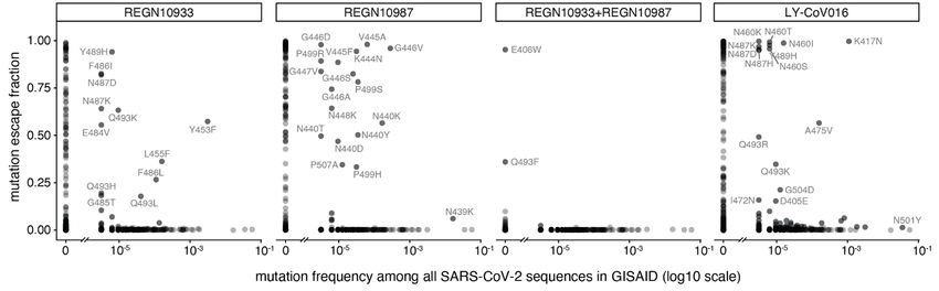
1014–1018 (2020). doi:10.1126/science.abd0831 Medline                                         Tracking SARS-CoV-2 mutations by locations and dates of interest. bioRxiv
13. R. Shi, C. Shan, X. Duan, Z. Chen, P. Liu, J. Song, T. Song, X. Bi, C. Han, L. Wu, G.          2020.09.23.310565             [Preprint].        28         September          2020.
     Gao, X. Hu, Y. Zhang, Z. Tong, W. Huang, W. J. Liu, G. Wu, B. Zhang, L. Wang, J. Qi,          https://doi.org/10.1101/2020.09.23.310565.
     H. Feng, F.-S. Wang, Q. Wang, G. F. Gao, Z. Yuan, J. Yan, A human neutralizing            26. S. Elbe, G. Buckland-Merrett, Data, disease and diplomacy: GISAID’s innovative
     antibody targets the receptor-binding site of SARS-CoV-2. Nature 584, 120–124                 contribution to global health. Glob. Chall. 1, 33–46 (2017). doi:10.1002/gch2.1018
     (2020). doi:10.1038/s41586-020-2381-y Medline                                                 Medline
14. Regeneron Pharmaceuticals, Inc., “Regeneron’s casirivimab and imdevimab                    27. 27. T. Starr, J. Bloom, A. Greaney, A. Addetia, jbloomlab/SARS-CoV-2-
     antibody cocktail for COVID-19 is first combination therapy to receive FDA                    RBD_MAP_clinical_Abs:            Science        revision,      Zenodo         (2021);
     emergency use authorization,” press release (21 November 2020);                               https://doi.org/10.5281/zenodo.4443311.
     https://investor.regeneron.com/news-releases/news-release-                                28. W. Hannon, jbloomlab/SARS-CoV-2_chronic-infection-seq: Archived for
     details/regenerons-regen-cov2-first-antibody-cocktail-covid-19-receive/.                      publication,             Version             1,            Zenodo             (2021);
15. Eli Lilly and Company, “A phase 3 randomized, double-blind, placebo-controlled                 https://doi.org/10.5281/zenodo.4433185.
     trial to evaluate the efficacy and safety of LY3819253 alone and in combination           29. A. E. Wentz, E. V. Shusta, A novel high-throughput screen reveals yeast genes that
     with LY3832479 in preventing SARS-CoV-2 infection and COVID-19 in skilled                     increase secretion of heterologous proteins. Appl. Environ. Microbiol. 73, 1189–
     nursing and assisted living facility residents and staff; a NIAID and Lilly                   1198 (2007). doi:10.1128/AEM.02427-06 Medline
     Collaborative Study” (Clinical trial registration NCT04497987, clinicaltrials.gov,        30. J. Shang, G. Ye, K. Shi, Y. Wan, C. Luo, H. Aihara, Q. Geng, A. Auerbach, F. Li,
     2020); https://clinicaltrials.gov/ct2/show/NCT04497987.                                       Structural basis of receptor recognition by SARS-CoV-2. Nature 581, 221–224
16. B. Choi, M. C. Choudhary, J. Regan, J. A. Sparks, R. F. Padera, X. Qiu, I. H. Solomon,         (2020). doi:10.1038/s41586-020-2179-y Medline

                                                                                                                                                                                             Downloaded from http://science.sciencemag.org/ on February 2, 2021
     H.-H. Kuo, J. Boucau, K. Bowman, U. D. Adhikari, M. L. Winkler, A. A. Mueller, T. Y.-     31. J. Otwinowski, D. M. McCandlish, J. B. Plotkin, Inferring the shape of global
     T. Hsu, M. Desjardins, L. R. Baden, B. T. Chan, B. D. Walker, M. Lichterfeld, M. Brigl,       epistasis. Proc. Natl. Acad. Sci. U.S.A. 115, E7550–E7558 (2018).
     D. S. Kwon, S. Kanjilal, E. T. Richardson, A. H. Jonsson, G. Alter, A. K. Barczak, W.         doi:10.1073/pnas.1804015115 Medline
     P. Hanage, X. G. Yu, G. D. Gaiha, M. S. Seaman, M. Cernadas, J. Z. Li, Persistence        32. K. H. D. Crawford, R. Eguia, A. S. Dingens, A. N. Loes, K. D. Malone, C. R. Wolf, H. Y.
     and evolution of SARS-CoV-2 in an immunocompromised host. N. Engl. J. Med.                    Chu, M. A. Tortorici, D. Veesler, M. Murphy, D. Pettie, N. P. King, A. B. Balazs, J. D.
     383, 2291–2293 (2020). doi:10.1056/NEJMc2031364 Medline                                       Bloom, Protocol and reagents for pseudotyping lentiviral particles with SARS-
17. K. S. Xue, T. Stevens-Ayers, A. P. Campbell, J. A. Englund, S. A. Pergam, M. Boeckh,           CoV-2 spike protein for neutralization assays. Viruses 12, 513 (2020).
     J. D. Bloom, Parallel evolution of influenza across multiple spatiotemporal scales.           doi:10.3390/v12050513 Medline
     eLife 6, e26875 (2017). doi:10.7554/eLife.26875 Medline                                   33. K. H. D. Crawford, A. S. Dingens, R. Eguia, C. R. Wolf, N. Wilcox, J. K. Logue, K.
18. A. F. Feder, S.-Y. Rhee, S. P. Holmes, R. W. Shafer, D. A. Petrov, P. S. Pennings,             Shuey, A. M. Casto, B. Fiala, S. Wrenn, D. Pettie, N. P. King, A. L. Greninger, H. Y.
     More effective drugs lead to harder selective sweeps in the evolution of drug                 Chu, J. D. Bloom, Dynamics of neutralizing antibody titers in the months after
     resistance in HIV-1. eLife 5, e10670 (2016). doi:10.7554/eLife.10670 Medline                  severe acute respiratory syndrome coronavirus 2 infection. J. Infect. Dis. jiaa618
19. J. D. Bloom, L. I. Gong, D. Baltimore, Permissive secondary mutations enable the               (2020). doi:10.1093/infdis/jiaa618 Medline
     evolution of influenza oseltamivir resistance. Science 328, 1272–1275 (2010).             34. J. Köster, S. Rahmann, Snakemake—A scalable bioinformatics workflow engine.
     doi:10.1126/science.1187816 Medline                                                           Bioinformatics 28, 2520–2522 (2012). doi:10.1093/bioinformatics/bts480
20. L. I. Gong, M. A. Suchard, J. D. Bloom, Stability-mediated epistasis constrains the            Medline
     evolution of an influenza protein. eLife 2, e00631 (2013). doi:10.7554/eLife.00631        35. S. Chen, Y. Zhou, Y. Chen, J. Gu, fastp: An ultra-fast all-in-one FASTQ
     Medline                                                                                       preprocessor.          Bioinformatics           34,        i884–i890          (2018).
21. T. H. Zhang, L. Dai, J. P. Barton, Y. Du, Y. Tan, W. Pang, A. K. Chakraborty, J. O.            doi:10.1093/bioinformatics/bty560 Medline
     Lloyd-Smith, R. Sun, Predominance of positive epistasis among drug resistance-            36. H. Li, Aligning sequence reads, clone sequences and assembly contigs with BWA-
     associated mutations in HIV-1 protease. PLOS Genet. 16, e1009009 (2020).                      MEM. arXiv:1303.3997 [q-bio.GN] (26 May 2013).
     doi:10.1371/journal.pgen.1009009 Medline                                                  37. K. Katoh, D. M. Standley, MAFFT multiple sequence alignment software version 7:
22. E. C. Thomson, L. E. Rosen, J. G. Shepherd, R. Spreafico, A. da Silva Filipe, J. A.            Improvements in performance and usability. Mol. Biol. Evol. 30, 772–780 (2013).
     Wojcechowskyj, C. Davis, L. Piccoli, D. J. Pascall, J. Dillen, S. Lytras, N.                  doi:10.1093/molbev/mst010 Medline
     Czudnochowski, R. Shah, M. Meury, N. Jesudason, A. De Marco, K. Li, J. Bassi, A.          38. S. K. Hilton, J. Huddleston, A. Black, K. North, A. S. Dingens, T. Bedford, J. D.
     O’Toole, D. Pinto, R. M. Colquhoun, K. Culap, B. Jackson, F. Zatta, A. Rambaut, S.            Bloom, dms-view: Interactive visualization tool for deep mutational scanning data.
     Jaconi, V. B. Sreenu, J. Nix, R. F. Jarrett, M. Beltramello, K. Nomikou, M. Pizzuto,          J. Open Source Softw. 5, 2353 (2020). doi:10.21105/joss.02353
     L. Tong, E. Cameroni, N. Johnson, A. Wickenhagen, A. Ceschi, D. Mair, P. Ferrari,         39. B. J. Grant, A. P. C. Rodrigues, K. M. ElSawy, J. A. McCammon, L. S. D. Caves,
     K. Smollett, F. Sallusto, S. Carmichael, C. Garzoni, J. Nichols, M. Galli, J. Hughes,         Bio3d: An R package for the comparative analysis of protein structures.
     A. Riva, A. Ho, M. G. Semple, P. J. M. Openshaw, J. K. Baillie, The ISARIC4C                  Bioinformatics 22, 2695–2696 (2006). doi:10.1093/bioinformatics/btl461
     Investigators, the COVID-19 Genomics UK (COG-UK) consortium, S. J. Rihn, S. J.                Medline
     Lycett, H. W. Virgin, A. Telenti, D. Corti, D. L. Robertson, G. Snell, The circulating
     SARS-CoV-2 spike variant N439K maintains fitness while evading antibody-                  ACKNOWLEDGMENTS
     mediated immunity. bioRxiv 2020.11.04.355842 [Preprint]. 5 November 2020.                 We thank K. Crawford for help with neutralization assays, A. Feder, D. Veesler,
     https://doi.org/10.1101/2020.11.04.355842.                                                   N. King, and D. Ellis for helpful comments, and the Fred Hutch Flow Cytometry
23. European Centre for Disease Prevention and Control, Rapid Risk Assessment:                    and Genomics facilities for assistance. Funding: This work was supported by the
     Detection of new SARS-CoV-2 variants related to mink, ECDC, Stockholm, 12                    NIAID (R01AI127893 and R01AI141707 to J.D.B. and T32AI083203 to A.J.G.), the
     November 2020; www.ecdc.europa.eu/en/publications-data/detection-new-                        Gates Foundation (INV-004949 to J.D.B.), and the Massachusetts Consortium
     sars-cov-2-variants-mink.                                                                    for Pathogen Readiness through grants from the Evergrande Fund (to J.Z.L.).
24. B. B. Oude Munnink, R. S. Sikkema, D. F. Nieuwenhuijse, R. J. Molenaar, E. Munger,            Scientific computing at the Fred Hutch is supported by ORIP grant
     R. Molenkamp, A. van der Spek, P. Tolsma, A. Rietveld, M. Brouwer, N.                        S10OD028685. T.N.S. is a Washington Research Foundation Innovation Fellow at
     Bouwmeester-Vincken, F. Harders, R. Hakze-van der Honing, M. C. A. Wegdam-                   the University of Washington Institute for Protein Design and an HHMI Fellow of
     Blans, R. J. Bouwstra, C. GeurtsvanKessel, A. A. van der Eijk, F. C. Velkers, L. A. M.       the Damon Runyon Cancer Research Foundation (DRG-2381-19). J.D.B. is an
     Smit, A. Stegeman, W. H. M. van der Poel, M. P. G. Koopmans, Transmission of                 Investigator of the Howard Hughes Medical Institute. Author contributions:
     SARS-CoV-2 on mink farms between humans and mink and back to humans.                         T.N.S., A.J.G., A.S.D., and J.D.B. designed the study. T.N.S., A.J.G., A.A., and
     Science 371, 172–177 (2021). doi:10.1126/science.abe5901 Medline                             A.S.D. performed the experiments. T.N.S., A.J.G., and J.D.B. analyzed the
25. A. T. Chen, K. Altschuler, S. H. Zhan, Y. A. Chan, B. E. Deverman, COVID-19 CG:               experimental data. J.Z.L. and M.C.C. sequenced the persistent infection, and

First release: 25 January 2021                                                 www.sciencemag.org                 (Page numbers not final at time of first release)                     4
W.W.H. analyzed that data. T.N.S., A.A., W.W.H., and J.D.B. wrote the initial draft,
and all authors edited the final version. Competing interests: J.Z.L. has
consulted for Abbvie and Jan Biotech. The other authors declare no competing
interests. Data and materials availability: Raw sequencing data are on the NCBI
SRA under BioProject PRJNA639956/BioSample SAMN16850904 (escape
mapping) and Bioproject PRJNA681234 (patient sequencing). Computer code
and processed data for the escape mapping are at (27) and
https://github.com/jbloomlab/SARS-CoV-2-RBD_MAP_clinical_Abs. Code and
data for the patient sequencing are at (28) and
https://github.com/jbloomlab/SARS-CoV-2_chronic-infection-seq. The
sequences of the antibodies are provided via citations in the Material and
Methods, the cells and plasmids for the neutralization assays are available in BEI
Resources and AddGene (see Materials and Methods for details), and the yeast
mutant libraries are available to academic researchers upon request with a
completed Materials Transfer Agreement. This work is licensed under a Creative
Commons Attribution 4.0 International (CC BY 4.0) license, which permits
unrestricted use, distribution, and reproduction in any medium, provided the
original work is properly cited. To view a copy of this license, visit
Downloaded from http://science.sciencemag.org/ on February 2, 2021
https://creativecommons.org/licenses/by/4.0/. This license does not apply to
figures/photos/artwork or other content included in the article that is credited
to a third party; obtain authorization from the rights holder before using such
material.
SUPPLEMENTARY MATERIALS
science.sciencemag.org/cgi/content/full/science.abf9302/DC1
Materials and Methods
Figs. S1 to S6
References (29–39)
MDAR Reproducibility Checklist
Data S1
1 December 2020; accepted 19 January 2021
Published online 25 January 2021
10.1126/science.abf9302
First release: 25 January 2021 www.sciencemag.org (Page numbers not final at time of first release) 5Downloaded from http://science.sciencemag.org/ on February 2, 2021 First release: 25 January 2021 www.sciencemag.org (Page numbers not final at time of first release) 6
Fig. 1. Complete maps of mutations that escape binding by the REGN-COV2 antibodies and Ly-CoV016.
    (A) Maps for antibodies in REGN-COV2. Line plots at left show escape at each site in the RBD (summed effects
    of all mutations at each site). Sites of strong escape (purple underlines) are shown in logo plots at right. The
    height of each letter is proportional to how strongly that amino-acid mutation mediates escape, with a per-
    mutation “escape fraction” of 1 corresponding to complete escape. The y-axis scale is different for each row, so
    for instance E406W escapes all REGN antibodies but it is most visible for the cocktail as it is swamped out by
    other sites of escape for the individual antibodies. See https://jbloomlab.github.io/SARS-CoV-2-
    RBD_MAP_clinical_Abs/ for zoomable versions. Letters are colored by how mutations affect the RBD’s affinity
    for ACE2 as measured via yeast display (7), with yellow indicating poor affinity and brown indicating good
    affinity; see fig. S2, A and B, for maps colored by how mutations affect expression of folded RBD and fig. S2, C
    and D, for distribution of effects on ACE2 affinity and RBD expression across all mutations observed among
    circulating viral isolates. (B) Map for LY-CoV016. (C) Validation of key mutations in neutralization assays using
    spike-pseudotyped lentiviral particles. We chose to validate mutations predicted to have large effects or that are
    present at high frequency among circulating SARS-CoV-2 isolates (e.g., N439K). Each point indicates the fold-
    increase in inhibitory concentration 50% (IC50) for a mutation relative to the unmutated “wildtype” (WT) spike,
    which contains D614G. The dotted blue line at 1 indicates wildtype-like neutralization, and values >1 indicate
    increased neutralization resistance. Point colors indicate if escape was expected at that site from the maps.

                                                                                                                                  Downloaded from http://science.sciencemag.org/ on February 2, 2021
    Point shapes indicate that the fold change is censored (an upper or lower limit) due to the IC50 being outside
    the dilution series used. Most mutants were tested in duplicate, and so have two points. Full neutralization
    curves are presented in fig. S3.

First release: 25 January 2021                     www.sciencemag.org     (Page numbers not final at time of first release)   7
Downloaded from http://science.sciencemag.org/ on February 2, 2021
Fig. 2. Escape maps are consistent with viral mutations selected in cell culture and a persistently infected
patient. (A) Viral escape mutations selected by Regeneron with spike-pseudotyped VSV in cell culture in the
presence of antibody (12). (B) Escape maps like those in Fig. 1A but showing only mutations accessible by single-
nucleotide changes to the Wuhan-Hu-1 sequence, with non-gray colors indicating mutations in cell culture (red),
in the infected patient (blue), or both (purple). Figure S5 shows these maps colored by how mutations affect
ACE2 affinity or RBD expression. (C) Dynamics of RBD mutations in a patient treated with REGN-COV2 at day
145 of his infection (black dashed vertical line). E484A rose in frequency in linkage with F486I, but since E484A
is not an escape mutation in our maps it is not shown in other panels. See also fig. S4. (D) The escape mutations
that arise in cell culture and the infected patient are single-nucleotide accessible and escape antibody binding
without imposing a large cost on ACE2 affinity [as measured using yeast display (7)]. Each point is a mutation
with shape/color indicating whether it is accessible and selected during viral growth. Points further to the right
on the x-axis indicate stronger escape from antibody binding; points further up on the y-axis indicate higher
ACE2 affinity.
First release: 25 January 2021 www.sciencemag.org (Page numbers not final at time of first release) 8Fig. 3. Antibody escape mutations in circulating SARS-CoV-2. For each antibody or antibody combination, the
escape score for each mutation is plotted versus its frequency among the 317,866 high-quality human-derived
Downloaded from http://science.sciencemag.org/ on February 2, 2021
SARS-CoV-2 sequences on GISAID (26) as of January 11, 2021. Escape mutations with notable GISAID
frequencies are labeled. The REGN-COV2 cocktail escape mutation E406W requires multiple nucleotide
changes from the Wuhan-Hu-1 RBD sequence and is not observed among sequences in GISAID. Other mutations
to residue E406 (E406Q and E406D) are observed with low frequency counts, but neither of these mutant amino
acids is a single-nucleotide mutation away from W, either.
First release: 25 January 2021 www.sciencemag.org (Page numbers not final at time of first release) 9Downloaded from http://science.sciencemag.org/ on February 2, 2021
Fig. 4. Structural context of escape mutations. (A) Escape maps projected on antibody-bound RBD structures.
[REGN10933 and REGN10987: PDB 6XDG (11); LY-CoV016: PDB 7C01 (13)]. Antibody heavy- and light-chain
variable domains are shown as blue cartoons, and the RBD surface is colored to indicate how strongly mutations
at that site mediate escape (white indicates no escape, red indicates strongest escape site for that
antibody/cocktail). Sites where no mutations are functionally tolerated are colored gray. (B) For each antibody,
sites were classified as direct antibody contacts (non-hydrogen atoms within 4 Å of antibody), antibody-
proximal (4–8 Å), or antibody-distal (>8 Å). Each point indicates a site, classified as escape (red) or non-escape
(black). The dashed gray line indicates the cutoff used to classify sites as escape or non-escape (see Methods
for details). Red and black numbers indicate how many sites in each category are escape or non-escape sites,
respectively. Interactive visualizations are at https://jbloomlab.github.io/SARS-CoV-2-
RBD_MAP_clinical_Abs/ and hypothesized mechanisms of escape and additional structural details for labeled
points are shown in fig. S6.
First release: 25 January 2021 www.sciencemag.org (Page numbers not final at time of first release) 10Prospective mapping of viral mutations that escape antibodies used to treat COVID-19
Tyler N. Starr, Allison J. Greaney, Amin Addetia, William W. Hannon, Manish C. Choudhary, Adam S. Dingens, Jonathan Z. Li and
Jesse D. Bloom
published online January 25, 2021
Downloaded from http://science.sciencemag.org/ on February 2, 2021
ARTICLE TOOLS http://science.sciencemag.org/content/early/2021/01/22/science.abf9302
SUPPLEMENTARY http://science.sciencemag.org/content/suppl/2021/01/25/science.abf9302.DC1
MATERIALS
RELATED http://stm.sciencemag.org/content/scitransmed/13/577/eabf1555.full
CONTENT
http://stm.sciencemag.org/content/scitransmed/13/577/eabd2223.full
http://stm.sciencemag.org/content/scitransmed/12/564/eabd5487.full
http://stm.sciencemag.org/content/scitransmed/12/570/eabd3876.full
REFERENCES This article cites 29 articles, 7 of which you can access for free
http://science.sciencemag.org/content/early/2021/01/22/science.abf9302#BIBL
PERMISSIONS http://www.sciencemag.org/help/reprints-and-permissions
Use of this article is subject to the Terms of Service
Science (print ISSN 0036-8075; online ISSN 1095-9203) is published by the American Association for the Advancement of
Science, 1200 New York Avenue NW, Washington, DC 20005. The title Science is a registered trademark of AAAS.
Copyright © 2021 The Authors, some rights reserved; exclusive licensee American Association for the Advancement of Science.
No claim to original U.S. Government Works. Distributed under a Creative Commons Attribution License 4.0 (CC BY).You can also read