Biology of the black rot pathogen, Guignardia bidwellii, its development in susceptible leaves of grapevine Vitis vinifera
←
→
Page content transcription
If your browser does not render page correctly, please read the page content below
JOURNAL FÜR KULTURFLANZEN, 61 (3). S. 82–90, 2009, ISSN 0027-7479   VERLAG EUGEN ULMER KG, STUTTGART
Originalarbeit

                                                                            Cornelia I. Ullrich, Regina G. Kleespies, Melanie Enders, Eckhard Koch

                                                Biology of the black rot pathogen, Guignardia bidwellii, its
                                              development in susceptible leaves of grapevine Vitis vinifera
                                                                                               Biologie des Schwarzfäuleerregers Guignardia bidwellii,
                                                                                           seine Entwicklung in anfälligen Weinblättern (Vitis vinifera)
82

                 Abstract                                                             heit, ursprünglich aus den USA kommend, schon seit
                                                                                      über 100 Jahren in Südeuropa bekannt ist, ist das Wis-
                 Since 2002 / 2003 black rot caused by Guignardia bid-                sen über die Biologie von G. bidwellii noch immer lü-
                 wellii is regularly occurring in organic viticulture in the          ckenhaft. In der vorliegenden Arbeit wird der Lebenszyk-
                 wine growing areas at the Mosel and Nahe River and in                lus von G. bidwellii mit histologisch-mikroskopischen
                 the Middle Rhine valley in Germany. Though the disease               Methoden (Durchlicht-, Fluoreszenz- und Elektronen-
                 originates from the USA and is known in Europe already               mikroskopie) untersucht. Die Entwicklung des Pilzes in
                 for more than 100 years, the knowledge about the biolo-              einer anfälligen Rebsorte (Riesling) wird von der Spo-
                 gy of its causal organism is still scanty. In the present            renkeimung bis zur Ausbildung der Pyknidien und
                 study the life cycle of G. bidwellii is analysed with histo-         Pseudothezien verfolgt. Der Schwerpunkt der Arbeit
                 logical microscopical methods (bright field, phase con-              liegt auf der Entwicklung nach dem Eindringen in den
                 trast, fluorescence and electron microscopy). The devel-             Wirt, die durch subcuticuläres Wachstum charakteri-
                 opment of the fungus on a susceptible grapevine variety              siert ist. Ziel ist die Verbreiterung der Wissensbasis über
                 (Riesling) is followed from spore germination up to the              die Biologie von G. bidwelli sowie die Erarbeitung einer
                 development of pycnidia and pseudothecia. The study is               Grundlage für die Entwicklung effizienter Bekämpfungs-
                 focused on the phase after penetration of the fungus                 methoden und eine histologische Charakterisierung der
                 which is characterized by subcuticular spread. The aim is            Sortenresistenz.
                 to broaden the knowledge of the biology of G. bidwellii,
                 to provide the basis for efficient control measures and to           Stichwörter: Cuticula-Isolierung, Elektronenmikroskopie,
                 enable the histological characterization of varietal resis-          Leitbündelbewuchs, Lichtmikroskopie, Pseudothecien,
                 tance.                                                               Sporentypen, subcuticuläre Hyphennetze, Wachstums-
                                                                                      muster
                 Key words: Cuticle isolation, electron microscopy, growth
                 on vascular bundles, light microscopy, pseudothecia,
                 spore types, staining methods, subcuticular hyphal nets              Introduction

                                                                                      The fungal pathogen Guignardia bidwellii (Ellis) Viala &
                 Zusammenfassung                                                      Ravaz [Gb; anamorph: Phyllosticta ampelicida (Engle-
                                                                                      man) Van der Aa] originates from North America and is
                 Seit 2002 / 2003 tritt die durch den Ascomyceten Guig-               known as severe pathogen causing black rot of grapevine
                 nardia bidwellii hervorgerufene Schwarzfäule regelmä-                Vitis vinifera (SCRIBNER and VIALA, 1888; PRUNET, 1898;
                 ßig in ökologisch bewirtschafteten Weinbergen an Mo-                 REDDICK, 1911). The disease appeared in southern Europe
                 sel, Nahe und im Mittelrheintal auf. Obwohl die Krank-               (France and Italy) more than 100 years ago (in 1885),

                   Institute
                   Julius Kühn-Institut – Bundesforschungsinstitut für Kulturpflanzen, Institut für Biologischen Pflanzenschutz, Darmstadt
                   Correspondence
                   Dr. Eckhard Koch, Julius Kühn-Institut – Bundesforschungsinstitut für Kulturpflanzen, Institut für Biologischen Pflanzen-
                   schutz, Heinrichstr. 243, 64287 Darmstadt, Germany. E-Mail: eckhard.koch@jki.bund.de
                   Accepted
                   July 2008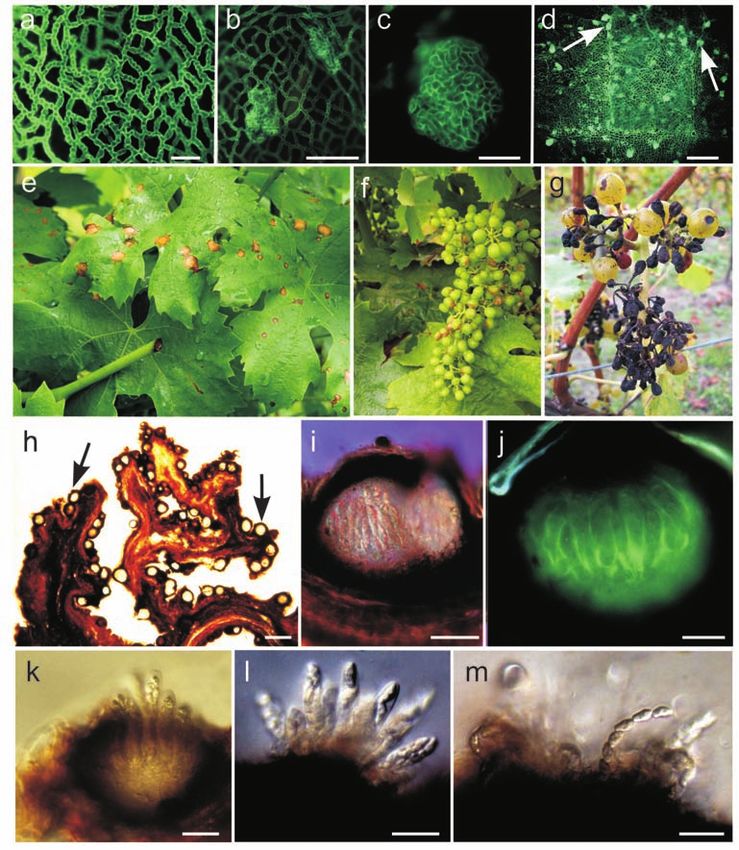
CORNELIA I. ULLRICH et al., Biology of the black rot pathogen, Guignardia bidwellii…

and gradually spread to the northern wine growing re-           Material and Methods
gions (BESSELAT and BOUCHET, 1984; MAURI and KOBEL,

                                                                                                                                      Originalarbeit
1988). Since 2002 / 2003 it is regularly occurring in vine-     Plants
yards of the wine growing areas at the Mosel and Nahe           Cuttings of Vitis vinifera L. cv. Riesling were provided by
River and in the Middle Rhine valley. The increased             the State Research Institute of Viticulture and Horticulture
spring temperatures of the last decades are supposed to         in Geisenheim, Germany. After rooting in Perlite for 4
contribute to this phenomenon (BMELV, 2004). Aban-              weeks, they were transplanted in a commercial potting
doned vineyards (Drieschen) serve as reservoirs of the          substrate (Fruhstorfer Erde Typ P) and further grown un-
pathogen. Because certain fungicides against powdery            der greenhouse and daylight conditions at 25°C. General-
and downy mildew have also effects against black rot, the       ly, 4-week-old shoots of 40 cm length with about 10 fully
disease is less problematic in integrated viticulture. How-     expanded leaves (Fig. 1a) were used for the experiments.
ever, severe damage has been recorded in organic viticul-
ture, where synthetic fungicides are not applied (HOLZ et       Fungal material
al., 2005). Differences in susceptibility between grape         The fungal isolate of Phyllosticta ampelicida (Engleman)
cultivars have been observed (M. HARMS, personal com-           Van der Aa (spore line 8088) used for production of pyc-
munication), but all commercially important Vitis vin-          nidiospores was obtained from the Agricultural Service
ifera cultivars are susceptible to the disease (WILCOX,         Center in Neustadt/Weinstrasse (DLR, Rheinland-Pfalz,
2003; BMELV, 2004).                                             Germany). The fungus was propagated in plates (Petri
   Detailed knowledge of the biology of the pathogen is a       dishes) on malt extract agar at 25°C under permanent                           83
prerequisite for the targeted development of control            light of fluorescent lamps with white light (Osram L36W
strategies. A number of detailed studies related to the bi-     Biolux) and dark blue light (Sylvania Blacklight-Blue,
ology of the black rot pathogen and the infection process       F36/BLB-T8, Japan). The plates were inoculated by plac-
on susceptible grapes were performed by Hoch and                ing agar discs carrying actively growing mycelium in
co-workers. As in many other fungi, pycnidiospores, ger-        their centre. Two weeks after inoculation, the plates were
mlings and appressoria of P. ampelicida are surrounded          flooded with tap water, and the resulting conidial sus-
by an extracellular matrix suspected to be involved in ad-      pensions adjusted to a density of 105 ml-1. The plants
hesion to the substratum and host recognition (KUO and          were inoculated by spraying the conidial suspensions
HOCH, 1995). Firm attachment of P. ampelicida pycnid-           with a glass atomizer on the upper (adaxial) leaf surface
iospores to the substratum is a prerequisite for germina-       until run-off. Alternatively, 10 µl drops of inoculum sus-
tion. It occurs only on poorly wettable surfaces such as        pension were deposited on the upper or lower leaf sur-
grape leaves, polystyrene and teflon (KUO and HOCH,             face. The inoculated plants were incubated in a humid
1996a). Within 6 hours of attachment to a hydrophobic           chamber for 24 h and then transferred to the greenhouse.
surface, pycnidiospores germinate, and mature mela-                Ascospores were obtained from fruit mummies collect-
nized appressoria are formed (SHAW et al., 1998). A pen-        ed in the vineyard in winter. Fruit mummies carrying
etration peg breaches the cuticle, giving rise to hyphae        pseudothecia were buried 1-2 cm deep in Perlite, incu-
that colonize the leaf by growing strictly subcuticularly.      bated at 20°C and kept humid by watering. After ten to
Their growth can therefore be followed by scanning elec-        15 days the fruit mummies were shortly soaked in water,
tron microscopy (KUO and HOCH, 1996b).                          dried with paper towel and stuck singly under the lid of a
   Schematic drawings of the life cycle of G. bidwelli exist    Petri dish using Vaseline. The bottom part of the Petri
(ALEXOPOULOS, 1966; SIVANESAN and HOLLIDAY, 1981; PEAR-         dish was filled with water agar, and before solidification
SON and GOHEEN, 1988), and photographs of the typical           of the agar a bottle screw cap (about 2 cm diameter)
symptoms of black rot are depicted in various publi-            filled with 1 ml of water was positioned just below the
cations (PEARSON and GOHEEN, 1988; HOLZ et al., 2005).          fruit mummy. After ejection of ascospores the suspension
However, other aspects of the biology of the fungus             obtained was used for plant inoculation. In some experi-
such as the different spore types, spread of hyphae in          ments, a mixture of suspensions of pycnidiospores and
susceptible tissues and the development of the spore            ascospores was applied. For the production of spermatia,
forming structures are less well documented. The work           autoclaved grape leaves were spread on water agar in
reported in this paper was initiated to fill this gap.          Petri dishes, inoculated with mycelial plugs, and incubat-
Upon artificial inoculation, growth and development of          ed at room temperature.
the pathogen was followed histologically. In order to
assess their specific advantages and disadvantages,             Staining procedures
several microscopical methods (bright field microsco-           Aniline blue staining. Samples were taken at different
py, fluorescence and electron microscopy) together              intervals after inoculation. For the preparation of whole
with a series of stains (aniline blue, lactophenol-trypan       leaf mounts, segments of 2 x 2 mm were cut from the
blue, Blankophor, Calcofluor, safranin, and chlorazol           leaves and stained immediately by incubation for 5 to
black) were used. Because, to our knowledge, the infec-         15 min or overnight in 0.1% aniline blue (Merck, Darm-
tion process by ascospores has not been studied before,         stadt, Germany) in 1M glycine buffer pH 9.4, or after
we decided to include both pycnidiospores and asco-             clearing with 100% chloralhydrate/90% lactic acid (2:1)
pores as inoculum.                                              for 3 days and thoroughly rinsing with H2Odest.

Journal für Kulturflanzen 61. 2009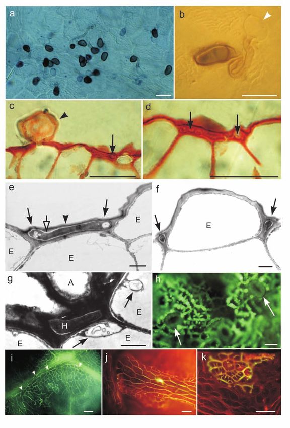
CORNELIA I. ULLRICH et al., Biology of the black rot pathogen, Guignardia bidwellii…
Originalarbeit

84
                                                                                                                    Fig. 1.    (a). Grapevine plant (4
                                                                                                                    week-old) at the time of inocula-
                                                                                                                    tion. The most susceptible leaves
                                                                                                                    were those at positions 2-4. (b-f)
                                                                                                                    Whole leaf mounts with subcu-
                                                                                                                    ticular hyphal nets; b. aniline
                                                                                                                    blue staining; (c) Blankophor
                                                                                                                    staining. (d) Detail, aniline blue
                                                                                                                    staining; (e) initial development
                                                                                                                    of subcuticular hyphae (arrow-
                                                                                                                    head) over anticlinal epidermal
                                                                                                                    cell walls originating from an ap-
                                                                                                                    pressorium (arrow) close to a vas-
                                                                                                                    cular bundle (*), trypan blue
                                                                                                                    staining; (f) detail of established
                                                                                                                    hyphal net with finger-like
                                                                                                                    branches of hyphae (arrowhead),
                                                                                                                    trypan blue staining. (g-k) Cross
                                                                                                                    sections stained with different
                                                                                                                    dyes; (g) infected stem with ma-
                                                                                                                    ture subcuticular hyphae (arrow-
                                                                                                                    head) and few inter-parenchyma
                                                                                                                    hyphae (arrow), aniline blue. (h)
                                                                                                                    Infected leaf with subcuticular
                                                                                                                    hyphae (arrowhead), aniline blue
                                                                                                                    staining, and (i) uninfected leaf
                                                                                                                    with smooth cuticle (arrowhead),
                                                                                                                    aniline blue staining; (j) infected
                                                                                                                    leaf with subcuticular hyphae
                                                                                                                    (arrowheads), chlorazol black
                                                                                                                    staining, and (k) uninfected leaf
                                                                                                                    with smooth cuticle (arrowhead).
                                                                                                                    Bars = 20 µm (d-f, h, i-k); 50 µm
                                                                                                                    (b,c,g).

                 Lactophenol-trypan blue staining. The leaf segments                   Germany) or Calcofluor White (syn. Fluorescent Bright-
                 were boiled in the staining solution (10 ml lactic acid, 10           ener 28 and Tinopal UNPA-GX; Sigma) in 0.1 M Tris buff-
                 ml glycerol, 10 ml water, 10 g phenol, 10 mg trypan blue,             er pH 9.0 for 5 min or overnight, with 0.05% chlorazol
                 Merck) for two to four min. After clearing in chloral hy-             black (Sigma) in 90% lactic acid, glycerol and H2Odest
                 drate (2.5 g in 1 ml H2Odest) for at least 30 min, the sam-           (1:1:1) at 90°C for 1 h (modified after BRUNDRETT et al.,
                 ples were mounted in the same solution, and viewed un-                1984), or with 0.02% safranin (Merck) in H2Odest. for at
                 der a compound microscope equipped with interference                  least 30 min.
                 optics.
                                                                                       Staining of hand sections and semi-thin sections. Semi-
                 Further dyes. Leaf segments were also stained with                    thin sections (2 µm) were cut with glass on a LKB ul-
                 0.01% Blankophor (syn. Diethanol; Bayer, Leverkusen,                  tratome III. microtome using the material embedded for

                                                                                                                    Journal für Kulturflanzen 61. 2009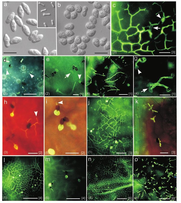
CORNELIA I. ULLRICH et al., Biology of the black rot pathogen, Guignardia bidwellii…

electron microscopy (see below). Hand sections and               shown in Fig. 1d (aniline blue staining) and Fig. 1f (try-
semi-thin sections were viewed with the light microscope         pan blue staining). The diameter of hyphae was 0.8 to 1.0

                                                                                                                                       Originalarbeit
after staining with safranin, aniline or unstained using         µm at 4 dpi (Fig. 2e, f). Mature subcuticular hyphae often
the differential-interference contrast (DIC).                    had finger-like short branches that grew on the periclinal
                                                                 walls of the epidermal cells (Fig. 1f). However, these
Light microscopy                                                 branches always covered only part of the periclinal walls,
After thoroughly rinsing in the corresponding buffer the         and anastomoses of their tips were only rarely observed.
stained samples were mounted in these buffers and                Fig. 1e shows the initial spreading of subcuticular hyphae
viewed under UV (filter block A), in transmitted light or        strictly over the anticlinal cell walls.
under differential-interference contrast (DIC) with an              Aniline blue or chlorazol black staining of cross sec-
Aristoplan epifluorescence microscope (Leica, Wetzlar,           tions confirmed that growth of the hyphae is strictly sub-
Germany). Images were taken by an Orthomat E camera              cuticular (Fig. 1h-k). Cross sections of the grapevine
system (Leica) on Kodachrome Elite 100 ASA daylight co-          stem also showed the subcuticular location of the hy-
lour slide films or digitally with a CCD camera (Colour          phae. However, at this advanced state (four weeks after
view II, Olympus, using the software Analysis Five).             inoculation) a few hyphae could be detected in the corti-
                                                                 cal parenchyma below the epidermis, probably already in
Electron microscopy                                              the early saprobic state (Fig. 1g).
For ultrastructural studies, infected leaf tissue (4 dpi) was       Precondition for infection of the plant is the successful
pre-fixed at 4°C with 3.0% glutaraldehyde in Veronal             formation of appressoria on the leaf surface. In the                           85
buffer (pH 7.2) for 24 h and stained with 2.0% osmium            present study, the melanized appressoria of G. bidwellii
tetroxide in the same buffer for 3 h. After dehydration, the     were frequently observed by light microscopy of whole
tissues were embedded in methacrylate. Thin sections             leaf mounts (Fig. 2a,b) and also found by light- and elec-
were obtained with a Leica Ultracut S microtome and con-         tron microscopy of semi-thin (Fig. 2c) and ultra-thin sec-
trasted using uranyl acetate and lead citrate. The sections      tions, respectively (Fig. 2g). Location of the hyphae
were examined and photographed using a Zeiss EM 902              strictly between the cuticle and the epidermal cell wall
electron microscope (Zeiss, Oberkochen, Germany).                was also confirmed by light- and electron microscopy of
                                                                 leaf sections (Fig. 2c-f). Electron micrographs revealed
Cuticle isolation                                                that hyphae were surrounded by electron-lucent fungal
Leaf segments of 2 x 2 mm were infiltrated with a solu-          cell walls. Furthermore, paramural-like bodies with vesi-
tion containing 25 mM MES buffer pH 5.0, 125 mM man-             cles accumulated in epidermal cells adjacent to the fun-
nitol, 0.5% toluene and 0.5% cellulase of Trichoderma            gal structures. Both symptoms indicate a response of the
viride (Sigma C9422) and 0.5% pectolyase of Aspergillus          plant to the presence of the parasite (Fig. 2g).
japonicus (Sigma P3026) (modified according to G. WOLF              Spores germinated and appressoria formed readily on
and C. HENKE, Univ. Göttingen, Germany, personal com-            both the upper (adaxial) and the lower (abaxial) leaf sur-
munication; JOCHEM, 1986). The samples were shaken in            face. Subcuticular hyphae developed well below the
the same solution at 37°C at 120 rpm for at least 18 h.          abaxial cuticle, also above the anticlinal cell walls around
The isolated pieces of cuticle were stained with 0.1%            the guard cells and subsidiary cells (Fig. 2h). Appressoria
aniline blue and viewed under fluorescent light.                 were mostly formed above anticlinal walls and preferen-
                                                                 tially but not obligatorily induced at or close to the leaf
                                                                 veins. Development of the fungal hyphae appeared to be
Results                                                          faster in the vicinity of the leaf veins (Fig. 2i, j).

Subcuticular hyphae                                              Microscopy of detached cuticles
Three weeks after spraying the grapevine shoots with pyc-        From about three to four days after inoculation onwards,
nidiospores or ascospores, only leaves 2 to 4 (Fig. 1a)          the upper surface of macroscopically symptomless leaves
showed distinct disease symptoms. Light microscopy of            appeared slightly wavy or wrinkled by light microscopy
whole leaf mounts revealed that subcuticular hyphae were         of whole leaf mounts (Fig. 1h-k, 2a, e, h). From leaf sam-
present 2 days after inoculation and started to form dense       ples incubated in buffer containing pectinase and cellu-
hyphal nets. Infection of the plant was initiated by one or      lase, the cuticle was detached and could be isolated for
two hyphae originating from the bottom of the appressori-        microscopic observation, while the rest of the tissue dis-
um that penetrated the cuticle and continued to grow sub-        integrated into protoplasts. A few hyphal nets remained
cuticularly in the space above the anticlinal epidermal cell     attached to the isolated cuticles (Fig. 2k). At sites where
walls. The hyphae branched extensively, “surrounded” the         the hyphae had separated from the cuticle during the
epidermal cells and their tips anastomosed, thus forming a       treatment imprints were left in the cuticle.
dense net of subcuticular hyphae. The hyphal nets could
be visualised with different dyes. (Fig. 1b-f). Fig. 1b shows    Spore types of G. bidwellii
a complete hyphal net stained with aniline blue after            Ascospores and pycnidiospores are shown in Fig. 3a, b.
clearing the leaf sample. In Fig. 1c the hyphal net was          The ascospores measured 14.0 (11.9 – 15.5) x 6.9 (5.6 –
stained with Blankophor. Details of the hyphal nets are          7.7) µm and the pycnospores 9.6 (8.0 – 10.7) x 7.1 (5.8 –

Journal für Kulturflanzen 61. 2009
CORNELIA I. ULLRICH et al., Biology of the black rot pathogen, Guignardia bidwellii…
Originalarbeit
86
Fig. 2. (a,b) Melanized appres-
soria produced from pycnid-
iospores (arrowhead) (trypan
blue staining, DIC). (c) Semi-thin
sections (2 µm) with appressori-
um (arrowhead) and (d) subcutic-
ular hyphae (arrows), 4 dpi;
safranin staining. (e-g) Electron
microscopic resolution of subcu-
ticular hyphae of G. bidwellii, 4
dpi. (e) Longitudinal and (f) cross
sections (60 nm) of hyphae; E =
epidermal cells; closed arrows
point to fungal organelles, open
arrow to the electron-lucent fun-
gal cell wall, and arrowhead to
the host plant cuticle. (g) Para-
mural bodies with vesicles (ar-
rows) in epidermal cells (E) in the
vicinity of an appressorium (A)
and of hyphae (H). (h) Subcuticu-
lar hyphal growth on the lower,
abaxial leaf surface around the
stomata (arrows), 14 dpi; aniline
blue staining. (i,j) Hyphal growth
on vascular bundles (arrowheads
point to appressoria). (k) Fungal
hyphae attached to a piece of cu-
ticle obtained by enzymatic di-
gestion of the leaf tissue, aniline
blue staining. Bars = 2 µm (e-g);
10 µm (b); 20 µm (a,c,d,h,j); 50
µm (i,j).
8.2) µm (means of 35 spores each). Spermagonia con- Role of leaf age
taining spermatia were formed on autoclaved leaves Microscopy of whole leaf mounts showed that spores ger-
about 14 days after inoculation. The spermatia were minated and formed appressoria on all leaves irrespec-
rod-like in shape and variable in size, measuring 5.9 (3.8 tive of leaf position and age. On younger (position 1 to 3)
– 8.8) x 1.7 (1 – 3.1) µm (means of 50 spermatia). leaves asco- and pycnidiospores always developed direct-
Upon inoculation of leaves with a mixture of as- ly into appressoria, or the germ tubes remained very
cospores (Fig. 3a) and pycnidiospores (Fig. 3b), both short (Fig. 3c, e). On older leaves (position 4 and 5) the
spore types germinated simultaneously and developed germ tube length was more variable. They were short in
appressoria and subcuticular hyphae in a completely some samples (Fig. 3i) but much longer in others (Fig. 3f,
identical manner (Fig. 3c, e, g). In contrast, spermatia g). Among the samples taken two to four dpi the subcu-
(Fig. 3a inset) did not germinate on the leaves (Fig. 3d). ticular hyphae were much better developed in young (po-
Journal für Kulturflanzen 61. 2009CORNELIA I. ULLRICH et al., Biology of the black rot pathogen, Guignardia bidwellii…
Originalarbeit
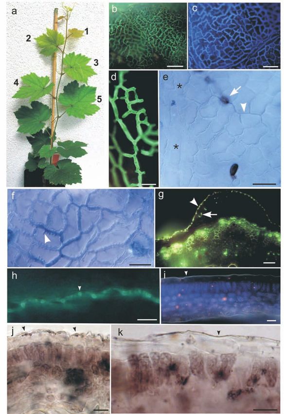
Fig. 3. (a,b) Spore types of G.
bidwellii. (a) Ascospores (b) pycn-
idiospores, (a inset) spermatia,
(d) non-germinating spermatia
(arrowheads) on leaf surface (7
dpi). c,e-g) Dependence of germ
tube length of germinating pycn-
idiospores (arrowhead) and as-
cospores (arrow) on leaf age: (c,e)
short germ tubes on younger
87
(position 2 and 3) leaves (1-3 dpi).
(f,g) Long germ tubes on older
(position 4) leaves (1 dpi). (h-m)
Dependence of hyphal growth on
leaf age: (h,i) position 3 and 5
leaves (2 dpi); arrowheads point
to developing hyphal nets; (j,k)
position 1 and 5 leaves (3 dpi);
(l,m) position 1 and 5 leaves (4
dpi). (n,o) Variability of hyphal
growth within the same leaf (po-
sition 4); (n) dense subcuticular
hyphal net; (o) germinated
spores and appressoria with sub-
cuticular hyphae still at initial
stage of development (6 dpi).
Numbers in brackets: (leaf num-
ber) and [dpi]. Bars = 20 µm
(a-c,a,h-j,l); 50 µm (d-g,n); 100
µm (k,m-p).
sition 1 to 3) leaves (2 dpi: Fig. 3h; 3 dpi: Fig. 3c, j; 4 dpi: in the season, the surface of fruit mummies is completely
Fig. 3l) than in older (position 5) leaves (Fig. 3i, k, m). covered with pseudothecia. (Fig. 4h). On fruit mummies
Whereas in young leaves the subcuticular hyphae had which remain on the plants (Fig. 4g, h) or on the soil sur-
vigorously spread and formed hyphal nets, in older face, asci differentiate (Fig. 4i-k) and release ascospores
leaves only very short, unbranched hyphae were present that infect the new crop. From overwintering fruit mum-
that had reached only short distances from the infection mies collected from within the trellis more than 105 as-
site. In addition, on older expanded leaves we found ar- cospores per mummy were obtained (HOFFMAN et al.,
eas with strong growth of subcuticular hyphae (Fig. 3n) 2004). In our study, ascospores were rapidly and vigor-
and on the same leaf areas with inhibited spread ously released when fruit mummies stored at -20°C were
(Fig. 3o). brought in contact with water after being conditioned by
incubation at +20°C in humid Perlite for 14 d (Fig. 4l, m).
Development of pycnidia
At about 12 to 14 dpi the hyphal nets became increasingly
dense, while the surrounding tissue started to become Discussion
necrotic. Thick hyphal aggregates developed into pycnid-
ia (Fig. 4a-e). In spring, the primary infections of the grape foliage are
initiated by ascospores released from fruit mummies, but
Development of asci conidia formed in pycnidia on overwintering, diseased
In the vineyard, lesions with pycnidia occur on all green canes are also suspected to play a role. During the season
parts of the plant (Fig. 4e, f). Infected berries become de- the disease is spread by pycnidiospores. The size and
hydrated and develop into fruit mummies (Fig. 4g). Later shape of pycnidiospores (formed on agar plates) and as-
Journal für Kulturflanzen 61. 2009CORNELIA I. ULLRICH et al., Biology of the black rot pathogen, Guignardia bidwellii…
Originalarbeit
88
Fig. 4. (a-d) Development of
pycnidia within a lesion, 14 dpi;
aniline blue staining. (a) Net of
thick hyphae; (b) aggregating hy-
phae; (c) detail of hyphal aggre-
gate; (d) lesion area at low
magnification with numerous
pycnidial initials (arrows). (e-g)
Symptoms of black rot; (e,f) le-
sions on leaves and berries; (g)
fruit mummies. (h-m) Develop-
ment of asci. (h) Cross section (45
µm thickness) showing the den-
sity of pseudothecia on mummi-
fied berries (arrows). (i)
Pseudothecium with asci, DIC; (j)
pseudothecium with asci, aniline
blue staining. (k) Mature
pseudothecium; (l, m) bursting
pseudothecia and asci ejecting
ascospores. Bars = 20 µm (a,c,
j-m); 50 µm (b,i); 200 µm (d,k).
cospores (isolated from fruit mummies) observed in the spermatia formed on autoclaved grape leaves in Petri
present study is in good agreement with the measure- dishes, was as described by SIVANESAN and HOLLIDAY
ments given by SIVANESAN and HOLLIDAY (1981) and KUO (1981), but their size deviated from the measurements
and HOCH (1996a). However, the appendages of the pyc- given by these authors by a factor of about 2. Following
nidiospores, as depicted by SIVANESAN and HOLLIDAY inoculation onto leaves the spermatia did not germinate,
(1981) and also observed by scanning electron microsco- which is in agreement with previous reports (ALEXOPOU-
py by KUO and HOCH (1996a), were not seen in our study. LOS, 1966).
In the differential interference contrast of the light micro- Interestingly, spore germination, formation of mela-
scope numerous spherical inclusion bodies were ob- nized appressoria and infection of the leaf was identical
served. By transmission electron microscopy the pycnid- for pycnidiospores and ascospores. Spore germination
iospores were shown to contain lipid bodies, mitochon- was not affected by leaf age. Unequivocal effects of leaf
dria and two nuclei (SHAW et al., 1998). In the scanning position on germ tube lengths were not observed either,
electron microscope the pycnidiospore surface appears although in many cases germ tubes were longer on older,
smooth (KUO and HOCH, 1996a). fully expanded leaves than on young ones. Overall, the
In nature, spermagonia containing hyaline, one-celled pre-penetration events observed were in agreement with
spermatia (Leptodothiorella sp.) are produced toward the the results reported by KUO and HOCH (1996b). In sam-
end of the growing season in berry mummies in associa- ples viewed two days after inoculation, hyphae emerging
tion with ascogonial stromata (PEARSON and GOHEEN, from appressoria started to colonize the leaf. Within
1988). It is assumed that they act as male cells in sperma- three to four days the subcuticular hyphal nets were fully
tization (ALEXOPOULOS, 1966). In our study, the shape of developed.
Journal für Kulturflanzen 61. 2009CORNELIA I. ULLRICH et al., Biology of the black rot pathogen, Guignardia bidwellii…
The hyphae of G. bidwellii grew primarily between the trobin that has good curative activity against blackrot (D.
cuticle and the anticlinal cell walls and preferentially MOLITOR, personal communication). Pyraclostrobin has
Originalarbeit
close to the vascular bundles. Only in later stages (8 dpi) translaminar activity but is neither xylem systemic nor
did they occasionally overgrow the periclinal walls of the phloem mobile (BARTLETT et al., 2002).
epidermal cells. When the cuticle of infected leaves was It is known since long that the susceptibility to black
enzymatically separated from the leaf tissues, the hyphae rot depends on leaf age (SCRIBNER and VIALA, 1888; PRU-
remained attached to it, confirming that they did not pro- NET, 1898). This was also confirmed in the present study,
trude into the epidermis or mesophyll tissue. The hyphal where young leaves (pos. 2 and 3) showed the typical dis-
nets were found on petioles, canes, and twines as well. ease symptoms, whereas very young (pos. 1) and older
Subcuticular growth of fungal hyphae following pene- leaves (pos. 5 and older) were not or only scarcely infect-
tration of the cuticle is common among plant pathogenic ed. The weaker disease on very young leaves is explain-
fungi. However, in many cases it is only transient, before able by the fact that after growing less infection sites are
growth becomes inter- or intracellular (for example present per unit area due to the greater expansion of
DIÉGUEZ-URIBEONDO et al., 2005). Among the fungi grow- these leaves, which, as judged by the large lesion size, are
ing below the cuticle throughout the asexual life cycle, highly susceptible (KUO and HOCH, 1996b). However,
the apple scab fungus Venturia inaequalis is probably the with increasing maturity the leaves become increasingly
best studied example. However, different from G. bidwell- resistant to the pathogen. In our study this ontogenetic
ii, V. inaequalis does not strictly grow above epidermal resistance was reflected by the fungal development ob-
anticlinal walls. Instead, a stroma is formed that grows in served in whole leaf mounts. In mature leaves the fungus 89
the apoplast between the cuticle and the epidermal cells penetrated the cuticle, but further development was lim-
(MACHARDY, 1996). ited to a few subcuticular hyphae that failed to branch
G. bidwellii is able to produce its dense hyphal nets and to form the typical subcuticular hyphal nets seen in
without forming haustoria or penetrating or visually young tissue. In this respect our results confirm those of
damaging the epidermal or mesophyll cells. The question KUO and HOCH (1996b), who observed that in mature
concerning the mechanism to retrieve nutrients is in- leaves the hyphae seldom expanded more than 30 µm
triguing. What might be the signals for this specialized from the site of penetration. Similar to the situation with
growth pattern, assimilation products such as sugars and black rot, ontogenetic resistance in apple leaves against
amino acids? Preferred hyphal spreading on and around V. inaequalis is expressed by formation of a stroma only
the vascular bundles may indicate an easier availability about one-third in thickness and poorly developed com-
of nutrients and water at these sites compared to other pared to the stroma in young leaves. Interestingly,
parts of the leaf. On cuticle fragments isolated from growth of the fungus is similarly impaired in genetically
leaves by enzymatic digestion hyphal imprints were resistant leaves (MACHARDY, 1996) and in leaves that had
found, indicating specific biochemical processes involved been protected with a chemical inducer of plant resis-
in softening the cuticle. Concurrent with the develop- tance (ORTEGA et al., 1998). The physiological cause for
ment of hyphal nets the leaf surface gets slightly wavy or limited hyphal growth in mature tissues is not known.
wrinkled. Probably, also permeabilizing agents are in- Factors like thickness of the cuticle may affect the success
volved, which lead to gradual nutrient efflux from the of penetration, but for limitation of hyphal growth in the
epidermal cytoplasm into the cell wall apoplast. space between the cuticle and the epidermal cells obvi-
Usually, G. bidwellii grows on the upper, adaxial leaf ously other defence mechanisms must play a role. In the
surface, but inoculation of the lower, abaxial surface was present study we observed paramural-like bodies with
also successful. In this case, the fungal hyphae did not numerous vesicles in epidermal cells in the vicinity of ap-
penetrate into the mesophyll through the open stomata pressoria and subcuticular hyphae of G. bidwellii, as de-
but remained on the anticlinal cell walls around the sto- scribed for barley leaves infected with powdery mildew
mata, where nutrient sources obviously are easier to (AN et al., 2007). The possible role of such secretory ves-
reach. By this strategy the fungus may even avoid recog- icles has been discussed to serve the exchange of materi-
nition by the host plant and stimulation of plant defence als between the invading fungus and the host cell (BRACK-
mechanisms. Subcuticular growth of hyphae and forma- ER and LITTLEFIELD, 1973). Although it is not known if this
tion of pycnidia and pycnidiospores was identical on both is part of a defence reaction, it at least indicates a re-
leaf surfaces. Preliminary observations indicate that sponse of the host plant to the presence of the parasite.
symptom development is even more pronounced when Correspondingly, the subcuticular hyphae were sur-
the lower leaf surface is inoculated compared to inocula- rounded by distinct electron-lucent fungal walls. The na-
tion of the upper leaf surface (unpublished). This would ture and possible function of such structures in suscepti-
mean that in viticulture a protection not only of the upper ble host plants has been assumed to serve the prevention
but also the lower leaf surface is necessary. On the other of any direct physical contact between the protoplast of
hand, the location of the fungus directly beneath the cu- the plant and the pathogen (POLITIS and WHEELER, 1973).
ticle suggests that a curative control of this pathogen In the present study a certain variability of the extent
should be possible also with fungicides that are not com- of fungal growth in expanding leaves (pos. 4) was noted.
pletely systemic, provided they are able to penetrate the In some of the samples, all of which had been taken ran-
cuticle. An example is the strobilurin fungicide pyraclos- domly from different leaf areas, fully developed hyphal
Journal für Kulturflanzen 61. 2009CORNELIA I. ULLRICH et al., Biology of the black rot pathogen, Guignardia bidwellii…
nets were present, whereas in others only initial hyphal BARTLETT, D.W., J.M. CLOUGH, J.R. GODWIN, A.A. HALL, M. HAMER, B.
branching was observed. Growing, still expanding leaves PARR-DOBRZANSKI, 2002: Review: the strobilurin fungicides, Pest
Originalarbeit
Manag. Sci. 58, 649-662.
of dicots are not homogeneous with respect to ontogenic BESSELAT, B., J. BOUCHET, 1984: Black-rot: a cause for concern in some
age of their different parts. The tissues at the base of vineyards. Phytoma 356, 33-35.
BMELV, 2004: Bericht des Bundesministeriums für Verbrauchers-
many leaf blades are more differentiated and thus more chutz, Ernährung und Landwirtschaft zur Schwarzfäule in den
mature than those of the central and distal areas deutschen Weinbaugebieten, September 2004. Internet. Online.
(SCHMUNDT et al., 1998). We therefore strongly suppose http://www.bmelv.de
BRACKER, C.E., L.J. LITTLEFIELD, 1973: Structural concepts of
that the observed differences in fungal growth in differ- host-pathogen interfaces. In: R. J. W. BYRDE, C. V. CUTTING (eds.):
ent samples reflect differences in ontogenetic maturity. Fungal pathogenicity and the plant’s response. London, New York,
Academic Press, 159-318.
The location of G. bidwellii below the cuticle allows to BRUNDRETT, M.C., Y. PICHÉ, R.L. PETERSON, 1984: A new method for
visualize the subcuticular hyphae even by scanning elec- observing the morphology of vesicular-arbuscular mycorrhizae.
Can. J. Bot. 62, 2128-2134.
tron microscopy (KUO and HOCH, 1996b). However, for DIÉGUEZ-URIBEONDO, J., H. FÖRSTER, A. SOTO-ESTRATA, J.E. ADASKAVEG,
routine use, for example for studying the development of 2005: Subcuticular-intracellular hemibiotrophic and intercellular
the fungus in varieties differing in susceptibility (MOLD- necrotrophic development of Colletotrichum acutatum on al-
mond. Phytopathology 95, 751-768.
ENHAUER et al., 2006), or after application of fungicides HOFFMAN, L.E., W.Y. WILCOX, D.M. GADOURY, R.C. SEEM, D.G. RIEGEL,
(WATKINS et al., 1977), or resistance inducing agents (OR- 2004: Integrated control of grape black rot: influence of host phe-
nology, inoculum availability, sanitation, and spray timing. Phy-
TEGA et al., 1998), fast and less elaborate methods, like topathology 94, 641-650.
the microscopy of whole leaf mounts are needed. HOLZ, B., C. HOFFMANN, G. NACHTIGALL, 2005: Schwarzfäule der Rebe
In our study, trypan blue, Blankophor, Calcofluor and (Black Rot). Informationsblatt der BBA, 1-2.
90 JOCHEM, P., 1986: Protonentransportierende ATPase am Tonoplasten
aniline blue were all suitable to stain spores, germ tubes von Blattzellen der CAM-Pflanze Kalanchoë daigremontiana. PhD
and appressoria of G. bidwellii in whole leaf mounts. Due thesis, TH Darmstadt.
KUO, K.C., H.C. HOCH, 1995: Visualization of the extracellular matrix
to the rapidity of the staining procedure, Blankophor and surrounding pycnidiospores, germlings, and appressoria of Phyl-
Calcofluor can be particularly recommended. Both losticta ampelicida. Mycologia 87, 759-771.
stains, however, are inadequate to visualize the subcutic- KUO, K.C., H.C. HOCH, 1996a: Germination of Phyllosticta ampelicida
pycnidiospores: Prerequisite of adhesion to the substratum and
ular hyphae. The latter can be rapidly stained with trypan the relationship of substratum wettability. Fung. Genet. Bio. 20,
blue, and the samples viewed with a normal microscope. 18-29.
KUO, K.C., H.C. HOCH, 1996b: The parasitic relationship between
If a fluorescence microscope is available, the method of Phyllosticta ampelicida and Vitis vinifera. Mycologia 88, 626-634.
choice is staining with aniline blue. In samples treated MACHARDY, W.E., 1996: Pathogen development and host tissue re-
action. In: Apple scab: biology, epidemiology and management.
with this dye the subcuticular hyphae show a bright yel- W. E. MACHARDY (ed.). American Phytopathological Society, St.
low-greenish fluorescence, contrasting well with the sur- Paul, 117-151.
rounding plant tissue. MAURI, G., U. KOBEL, 1988: Black rot – a new severe grape disease in
Ticino. Schweiz. Z. Obst- Weinbau, Wädenswil 124, 473-475.
In conclusion, black rot of grapevine caused by Guig- MOLDENHAUER, J., B.M. MOERSCHBACHER, A.J. VAN DER WESTHUIZEN,
nardia bidwellii is a challenging disease to basic research 2006: Histological investigation of stripe rust (Puccinia striiformis
f. sp. tritici) development in resistant and susceptible wheat cul-
and to the development of control methods for organic tivars. Plant Pathol. 55, 469-474.
viticulture. The microscopical methods described here ORTEGA, F., U. STEINER, H.W. DEHNE, 1998: Induced resistance to apple
can be used as a tool to study the infection process and to scab: microscopic studies on the infection cycle of Venturia
inaequalis (Cke.). J. Phytopathol. 146, 399-405.
characterize the growth of the parasite in grapevine PEARSON, R.G., A.C. GOHEEN, (eds.) 1988: Compendium of grape dis-
leaves prior to the development of visible disease symp- eases. American Phytopathological Society, 93 pp.
POLITIS, D.J., H. WHEELER, 1973: Ultrastructural study of penetration
toms. of maize leaves by Colletotrichum gramicidola. Physiol. Plant
Pathol. 3, 465-471.
PRUNET, A., 1898: Observations et expériences sur le black rot II. Con-
ditions internes du développement du black rot. Rev. Vit. 9,
Acknowledgements 601-603.
REDDICK, D., 1911: The black rot disease of grapes. Cornell Univ. Agri.
Exp. Stat. Bull. 293, 289-364.
We thank Helga RADKE, Birgit WEIHRAUCH and Petra ZINK SCHMUNDT, D., M. STITT, U. SCHURR, 1998: Quantitative analysis of the
for their valuable technical assistance. This research was local rates of growth of dicot leaves at a high temporal and spatial
resolution, using image sequence analysis. Plant J. 16, 505-514.
financially supported by „Bundesprogramm Ökologi- SCRIBNER, F.L., P. VIALA, 1888: Black rot (Laestadia bidwellii). US Dept.
scher Landbau“ („The Federal Organic Farming Agr. Botan. Div. Sec. Vegetable Pathol. Bull. 7, 1-29.
Scheme“), supported by the German Federal Ministry of SHAW, B.D., K.C. KUO, H.C. HOCH, 1998: Germination and appresso-
rium development of Phyllosticta ampelicida pycnidiospores. My-
Food, Agriculture and Consumer Protection (BMELV). cologia 90, 258-268.
SIVANESAN, A., P. HOLLIDAY, 1981: Guignardia bidwellii, IMI Descrip-
tions of Fungi and Bacteria 71, Sheet 710.
STRUCK, C., M. HAHN, K. MENDGEN, 1998: Infection structures of plant
References pathogenic fungi – potential targets for plant disease control. Z.
Pflanzenkr. Pflanzensch. 105, 581-589.
WATKINS, J.E., L.J. LITTLEFIELD, G.D. STATLER, 1977: Effect of the sys-
AN, Q., A.J.E. VAN BEL, R. HÜCKELHOVEN, 2007: Do plant cells secrete temic fungicide 4-n-butyl-1,2,4-triazole on the development of
exosomes derived from multivesicular bodies? Plant Signal. Be- Puccinia recondita f. sp. tritici in wheat. Phytopathology 67,
hav. 2, 4-7. 985-989.
ALEXOPOULOS, C.J., 1966: Einführung in die Mykologie. Stuttgart, G. WILCOX, W.F., 2003: Black rot. Cornell Cooperative Extension. 3 pp.
Fischer Verl., 495 S. Publication No 102 GSFG-D4.
Journal für Kulturflanzen 61. 2009You can also read