Degradation of the Acyl Side Chain of the Steroid Compound Cholate in Pseudomonas sp. Strain Chol1 Proceeds via an Aldehyde Intermediate
←
→
Page content transcription
If your browser does not render page correctly, please read the page content below
Erschienen in: Journal of bacteriology ; 195 (2013), 3. - S. 585-595

Degradation of the Acyl Side Chain of the Steroid Compound Cholate
in Pseudomonas sp. Strain Chol1 Proceeds via an Aldehyde
Intermediate
Johannes Holert,a Žarko Kulić,b Onur Yücel,a Vemparthan Suvekbala,c Marc J.-F. Suter,d Heiko M. Möller,b Bodo Philippa
University of Münster, Institute of Molecular Microbiology and Biotechnology, Münster, Germanya; Departments of Chemistryb and Biology,c University of Konstanz,
Konstanz, Germany; Eawag, Swiss Federal Institute of Aquatic Science and Technology, Duebendorf, Switzerlandd

Bacterial degradation of steroids is widespread, but the metabolic pathways have rarely been explored. Previous studies with
Pseudomonas sp. strain Chol1 and the C24 steroid cholate have shown that cholate degradation proceeds via oxidation of the A
ring, followed by cleavage of the C5 acyl side chain attached to C-17, with 7a,12b-dihydroxy-androsta-1,4-diene-3,17-dione
(12b-DHADD) as the product. In this study, the pathway for degradation of the acyl side chain of cholate was investigated in
vitro with cell extracts of strain Chol1. For this, intermediates of cholate degradation were produced with mutants of strain
Chol1 and submitted to enzymatic assays containing coenzyme A (CoA), ATP, and NAD1 as cosubstrates. When the C24 steroid
(22E)-7a,12a-dihydroxy-3-oxochola-1,4,22-triene-24-oate (DHOCTO) was used as the substrate, it was completely transformed
to 12a-DHADD and 7a-hydroxy-androsta-1,4-diene-3,12,17-trione (HADT) as end products, indicating complete removal of
the acyl side chain. The same products were formed with the C22 steroid 7a,12a-dihydroxy-3-oxopregna-1,4-diene-20-carboxy-
late (DHOPDC) as the substrate. The 12-keto compound HADT was transformed into 12b-DHADD in an NADPH-dependent
reaction. When NAD1 was omitted from assays with DHOCTO, a new product, identified as 7a,12a-dihydroxy-3-oxopregna-
1,4-diene-20S-carbaldehyde (DHOPDCA), was formed. This aldehyde was transformed to DHOPDC and DHOPDC-CoA in the
presence of NAD1, CoA, and ATP. These results revealed that degradation of the C5 acyl side chain of cholate does not proceed
via classical b-oxidation but via a free aldehyde that is oxidized to the corresponding acid. The reaction leading to the aldehyde is
presumably catalyzed by an aldolase encoded by the gene skt, which was previously predicted to be a b-ketothiolase.

S   teroids constitute a large and diverse class of natural com-
    pounds. While steroids serve various functions in eukaryotic
organisms, such as membrane constituent and hormonal func-
                                                                                     (13). Recently, it was shown that the degradation of rings A and B
                                                                                     can also precede side chain degradation in Rhodococcus jostii (14).
                                                                                         While the reactions leading to D1,4-3-keto structures and the
tions, they occur only rarely in prokaryotic organisms. Neverthe-                    breakdown of ADDs have been well studied, the removal of the
less, many bacteria are capable of transforming steroids, and this                   steroid side chain has not yet been explored in great detail. We
property is used for the biotechnological production of steroid                      investigated this process by studying the degradation of the C5 acyl
drugs (1–3). In addition, bacteria from diverse phylogenetic                         side chain of the bile salt cholate (compound I in Fig. 1) by Pseu-
groups can degrade naturally occurring steroids completely and                       domonas sp. strain Chol1 (15). Bile salts are surface-active steroid
use them as sources of carbon and energy. Bacteria are also able to                  compounds that aid the digestion of lipophilic nutrients in the
degrade synthetic steroids, which is relevant for public health,                     digestive tracts of vertebrates and enter the environment in signif-
because the occurrence of steroid drugs in the environment is                        icant amounts by excretion (16, 17).
believed to influence the fertility of animals and humans (4).                           The degradation of the C5 acyl side chain is believed to proceed
Compared to the degradation of other natural compounds, such                         via the stepwise removal of an acetyl and a propionyl residue from
as aromatic compounds, relatively little is known about bacterial                    the steroid skeleton. This pathway is supported by the analysis of
                                                                                     two transposon mutants of strain Chol1 that have defects in the
degradation of steroids. To date, only one bacterial metabolic
                                                                                     degradation of the acyl side chain of cholate.
pathway for aerobic degradation of steroids, the 9,10-seco path-
                                                                                         The first mutant, strain G12, is defective in the gene skt, which
way, has been described in detail (5–9).
                                                                                     encodes a putative thiolase (18). When this mutant is incubated
    The 9,10-seco pathway starts with oxidative reactions at the A
                                                                                     with cholate, two steroid compounds with a complete C5 acyl side
ring of the steroid that lead to the formation of D1,4-3-keto struc-
                                                                                     chain accumulate in culture supernatants, namely, (22E)-7a,12a-
tures. Subsequently, the side chain attached to the D ring at C-17                   dihydroxy-3-oxochola-1,4,22-triene-24-oate (DHOCTO, XX) and
is removed in a stepwise fashion, leading to the production of
androsta-D1,4-diene-3,17-diones (ADDs), which are the central
intermediates of the 9,10-seco pathway (8). ADDs are further de-
graded by a monooxygenase-catalyzed hydroxylation at C-9,
which causes the opening of the B ring concomitantly with the
aromatization of the A ring (10). The resulting 9,10-seco steroids
are further degraded by the breakdown of the aromatic A ring (11,
12). The remaining C and D rings form acidic perhydroindane
derivatives that are further degraded by as-yet-unknown reactions

                                                                                                                                                                   585

                                                         Konstanzer Online-Publikations-System (KOPS)
                                                      URL: http://nbn-resolving.de/urn:nbn:de:bsz:352-282520
FIG 1 Section of the proposed pathway of cholate (compound I) degradation in Pseudomonas sp. strain Chol1. The following compounds have been identified:
II, 3-ketocholate; III, D1/4-3-ketocholate (the position of the double bond has not been identified yet; for simplicity, only the more probable D4 isomer was
chosen); IV, D1,4-3-ketocholate; V, cholyl-CoA; VI, 3-ketocholyl-CoA; VII, D1/4-3-ketocholyl-CoA; VIII, D1,4-3-ketocholyl-CoA; IX, CoA ester of
(22E)7a,12a-dihydroxy-3-oxochola-1,4-triene-24-oate (DHOCTO); XI, 7a,12a-dihydroxy-3-oxopregna-1,4-diene-20S-carbaldehyde (DHOPDCA);
XII, 7a,12a-dihydroxy-3-oxopregna-1,4-diene-20-carboxylate (DHOPDC); XIII, CoA ester of DHOPDC; XVI, 7a,12a-dihydroxy-androsta-1,4-diene-
3,17-dione (12a-DHADD); XVII, 7a-hydroxy-androsta-1,4-diene-3,12,17-trione (HADT); XVIII, 12b-DHADD; XIX, 3,7,12-trihydroxy-9,10-secoan-
drosta-1,3,5(10)triene-9,17-dione (THSATD); XX, DHOCTO; and XXI, 7a,12a22-trihydroxy-3-oxochola-1,4-diene-24-oate (THOCDO). The com-
pounds shown in brackets are plausible intermediates that have not been detected yet.

7a,12a,22-trihydroxy-3-oxochola-1,4-diene-24-oate (THOCDO,                      the last two reactions of the removal of the C3 acyl side chain have
XXI). The second mutant, strain R1, is defective in the gene acad,              also not yet been verified.
which encodes a putative acyl coenzyme A (CoA) dehydrogenase                        Thus, the goal of our study was to elucidate the pathway for
(19). When this mutant is incubated with cholate, a steroid com-                degradation of the C5 acyl side chain of cholate in vitro by using
pound with a C3 acyl side chain, namely, 7a,12a-dihydroxy-3-                    cell extracts of strain Chol1 and intermediates of cholate degrada-
oxopregna-1,4-diene-20-carboxylate (DHOPDC, XII), accumu-                       tion (such as DHOCTO and DHOPDC) as substrates. In addition,
lates in culture supernatants as a dead-end product.                            we wanted to elucidate the fate of the acetyl-CoA and propionyl-
    The phenotypes and genotypes of these mutants strongly sug-                 CoA residues, which are released in the course of the side chain
gest that the first part of side chain degradation proceeds via a               degradation of cholate.
classical b-oxidation, in which the putative thiolase Skt would
catalyze the thiolytic cleavage of acetyl-CoA from the side chain               MATERIALS AND METHODS
with the CoA ester of DHOPDC as a product (8). For this, a b-                   Cultivation of bacteria. Pseudomonas sp. strain Chol1 and the transposon
keto-acyl-CoA substrate for Skt originating from oxidation of the               mutant strains G12 (defective in skt) and R1 (defective in acad) were
C-22 hydroxyl group of THOCDO-CoA (X) has to be postulated.                     grown in the phosphate-buffered mineral medium MMChol as described
DHOPDC-CoA would then be the substrate for Acad that would                      previously (15, 18, 19). Strain Chol1 was grown with cholate (2 mM), and
be responsible for introducing a double bond into the acyl side                 strains G12 and R1 were grown with mixtures of cholate (2 mM) and
chain by an oxidative reaction. After hydration of the double                   succinate (12 mM) in the presence of kanamycin (10 mg ml21). To induce
                                                                                strain G12 for the transformation of cholate, precultures were grown with
bond, the C3 acyl side chain is believed to be cleaved from the
                                                                                12b-DHADD (XVIII) (ca. 2 mM) as described previously (18). For the
steroid skeleton by an aldolase reaction (8).                                   production of 12b-DHADD, strain Chol1 was grown with cholate under
    While this pathway for degradation of the C5 acyl side chain                anoxic conditions with nitrate as the electron acceptor, as described pre-
appears biochemically plausible, it has never been confirmed in                 viously (15).
vitro and, thus, is still hypothetical. In particular, the putative                 Preparation of cell suspensions and cell extracts. Cells of strains
b-keto-acyl-CoA substrate for Skt has never been detected, and                  Chol1, R1, and G12 were harvested in the late-exponential growth phase

586
by centrifugation at 9,000 3 g for 10 min at 4°C. For cell suspension ing to 70% eluent B within 9 min, and returning to 20% eluent B within 1
experiments, cells were washed with MMChol medium without a carbon min, followed by an equilibration of 6 min. For the detection and purifi-
source and finally resuspended in MMChol with cholate (2 mM) to an cation of D1/4- and D1,4-3-ketocholate and DHOCTO, a gradient method
optical density at 600 nm (OD600) of 1. For the preparation of cell extracts, was used, starting with 20% eluent B for 2 min, increasing to 34% eluent
cells were washed in 50 mM K-Na-phosphate buffer (pH 7.0), resus- B within 9 min, and returning to 20% eluent B within 1 min, followed by
pended in a small volume of the buffer used for the respective enzyme an equilibration of 6 min. For analysis of the CoA activation assays and
assay, and broken by three passages through a cooled French press purification of P5 and P7, a gradient method was used, starting with 10%
(Aminco) at 138 MPa. Homogenates were centrifuged at 17,900 3 g for 30 eluent B for 2 min, increasing to 56% eluent B within 23 min, and return-
min at 4°C to separate the cell extracts from cell debris. To remove mol- ing to 10% eluent B within 1 min, followed by an equilibration of 6 min.
ecules with molecular masses of ,5,000 Da, cell extracts were subjected to Purification of steroid compounds. Steroid compounds from culture
a desalting step using a Sephadex G-25 matrix (PD-10; GE Healthcare). supernatants and from the CoA activation assays were purified by organic
Protein concentration was determined by the BCA assay (Pierce, USA) extraction with ethylacetate followed by semipreparative HPLC as de-
with bovine serum albumin as the standard. All cell extracts were imme- scribed previously (19) using the HPLC gradient methods described
diately used for enzyme assays or stored at 220°C. above and by solid-phase extraction as described below. The purity of the
Enzyme assays. All enzyme assays were performed at 30°C. Enzyme steroid compounds was assessed by HPLC analysis. For some steroid
assays for acyl-CoA ligase activities contained 50 mM morpholinepro- compounds, the purity was also assessed by LC-MS analysis and nuclear
panesulfonic acid (MOPS) buffer (pH 7.8), 1 mM CoA, 1 mM ATP, 2.5 magnetic resonance (NMR) spectroscopy.
mM MgCl2, and cell extracts (0.6 to 0.8 mg protein ml21) and were started For the purification of DHOCTO (XX), DHOPDC (XII), and D1/4-
by adding 1 mM cholate or 100 to 200 mM D1/4- and D1,4-3-ketocholate, and D1,4-3-ketocholate, the mutant strains G12 and R1 were cultivated as
DHOCTO, or DHOPDC. Electron acceptors were added in final concen- described above. In the supernatants of these cultures, either DHOCTO
trations of 1 mM [NAD1, K3Fe(CN)6] or 25 mM phenazine methosulfate (strain G12) or DHOPDC (strain R1) accumulated along with D1/4- and
(PMS). Samples were withdrawn immediately after the reaction was D1,4-3-ketocholate (III and IV, respectively) in both cases (18, 19). Those
started (t0 5 0 min) and at defined time intervals thereafter and subse- supernatants were used for the purification of D1/4- and D1,4-3-keto-
quently analyzed by high-performance liquid chromatography (HPLC) cholate by semipreparative HPLC. For the further purification of
or liquid chromatography-mass spectrometry (LC-MS). To hydrolyze DHOCTO and DHOPDC, cultures of the respective mutant strains in the
CoA-ester bonds, samples were incubated with 500 mM NaOH at 30°C late-exponential growth phase were diluted 1:10 (vol/vol) with fresh
for at least 30 min. MMChol containing succinate (12 mM) and kanamycin (10 mg ml21) for
The activity of citrate synthase (EC 2.3.3.1) was measured by monitor- a second growth passage. Under these conditions, the residual D1/4- and
ing the acetyl-CoA-, oxaloacetate-, and 5,5=-dithiobis-2-nitrobenzoic D1,4-3-ketocholate were completely transformed into DHOCTO (strain
acid (DTNB)-dependent formation of thionitrobenzoate in a spectropho- G12) or DHOPDC (strain R1), which remained in the culture superna-
tometer at 412 nm (ε 5 13.6 mM21 cm21). Assays contained HEPES tants as dead-end products. After organic extraction, DHOCTO- and
buffer (pH 8.0), 1 mM DTNB, 0.1 mM acetyl-CoA, and cell extracts (0.5 to DHOPDC-containing extracts were submitted to semipreparative HPLC
0.6 mg protein ml21) and were started by adding 2 mM oxaloacetate. The or to solid-phase extraction using Chromabond C18 ec columns (Mach-
activity of 2-methylcitrate synthase (EC 2.3.3.5) was measured in the same erey-Nagel). The columns were washed and equilibrated with 1 column
way as for citrate synthase, except propionyl-CoA was used instead of volume of methanol followed by 1 volume of MOPS buffer (50 mM, pH
acetyl-CoA. 7.8) before DHOCTO- or DHOPDC-containing extracts were loaded on
The activity of isocitrate dehydrogenase (EC 1.1.1.42) was measured the columns. After washing the columns with 3 volumes of MOPS buffer,
by monitoring the D-isocitrate-dependent formation of NADPH in a DHOCTO or DHOPDC was eluted with absolute methanol, which was
spectrophotometer at 365 nm (ε 5 3.4 mM21 cm21). Assays contained evaporated under low pressure (Eppendorf concentrator 5301). Both
100 mM Tris-HCl buffer (pH 8.0), 10 mM MgCl2, 2.5 mM NADP1, and compounds were resuspended in MOPS buffer, and the purity was con-
cell extracts (0.5 to 0.6 mg protein ml21) and were started by adding 1 mM trolled by HPLC analysis. Product patterns of CoA activation assays
D-isocitrate. showed that there was no difference between the DHOCTO and
The activity of isocitrate lyase (EC 4.1.3.1) was measured in a coupled DHOPDC purifications that were generated by semipreparative HPLC or
assay by determining the isocitrate-dependent oxidation of NADH in a spec- by solid-phase extraction. The concentrations of DHOCTO and
trophotometer at 340 nm (ε 5 6.2 mM21 cm21). The assay is based on the DHOPDC in the resulting solutions were estimated by measuring their
NADH-dependent reduction of glyoxylate to glycolate by lactate dehydroge- absorbance at 245 nm, which is a characteristic maximum for steroid
nase. The assays contained 50 mM HEPES buffer (pH 7.0), 2.5 mM MgCl2, compounds with a D1/4- and D1,4-3-keto structure (21) (Fig. 2A). Accord-
0.3 mM NADH, 1 U lactate dehydrogenase, and cell extracts (0.4 to 0.5 mg ing to calculations based on an averaged molar extinction coefficient of
protein ml21) and were started by adding 5 mM D-isocitrate. ε245 nm of 14.7 cm21 mM21, the final concentrations were in a range of 3 to
The activity of pyruvate dehydrogenase (EC 1.2.4.1) was measured 5 mM.
by the pyruvate- and CoA-dependent formation of NADH in a spectro- P5 and P6 were purified from the respective enzyme assay mixtures by
photometer at 365 nm (ε 5 3.4 mM21 cm21). The assays contained 50 solid-phase extraction and subsequent semipreparative HPLC as de-
mM Tris-HCl buffer (pH 7.8), 2 mM MgCl2, 2.5 mM dithiothreitol, 0.4 scribed above before using them as the substrates for the enzymatic assays.
mM thiamine pyrophosphate, 0.1 mM CoA, 5 mM NAD1, and cell ex- For NMR analysis, steroid compounds were purified from the respec-
tracts (0.4 to 0.5 mg protein ml21) and were started by adding 5 mM tive in vitro assays (P4, P5, P7) or from culture supernatants (12b-
pyruvate. DHADD) using organic extraction, solid-phase extraction, and semi-
HPLC. All steroid compounds were analyzed with a reversed-phase preparative HPLC purification as described previously (19) but without
HPLC system equipped with a UV-visible (UV-Vis) light diode array de- acidification of the aqueous phase for the second organic extraction step
tector as described previously (15, 20). K-Na-phosphate buffer (10 mM, after HPLC purification. Residues were resuspended in deuterated sol-
pH 7.1) (eluent A) and acetonitrile (eluent B) were used as eluents with a vents as indicated in Table 1.
total flow rate of 0.8 ml min21. For purification of steroid compounds, a UV-Vis spectroscopy. UV-Vis spectra of steroid compounds were re-
semipreparative reversed-phase column was used (250 by 8 mm, Euro- corded with a diode array detector during HPLC analyses and were used for a
sphere II, 100-5 C18 H [Knauer]), with a flow rate of 2 ml min21. For the first classification of the configuration of the A ring as well as for determina-
detection and purification of P4, P6, 12b-DHADD, and DHOPDC, a tion of CoA ester formation. Degradation products of cholate with a D1/4- or
gradient method was used, starting with 20% eluent B for 2 min, increas- D1,4-3-keto structure of the A ring exhibit the aforementioned distinct ab-
587t:o (B) XII
IV
!.. /
.Q
/m
"'g!
~
~
2 10 220 230 2•0 250 2&0 270 280 290 300 G 7 8 9 10 II 12 !3 14
Wavelength (nm] Retention time (min]
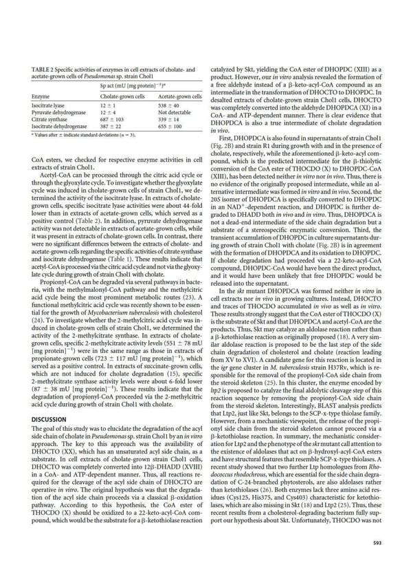
FIG 2 UV spectroscopic and chromatographic properties of the intermediates of cholate degradation. (A) UV spectra of a 1·4-3-ketocholate (N in Fig. I) (black
line), a•·•-3-ketocholyi-CoA (Vl!I in Fig. 1) (dotted black line), and cholyi-CoA (V in Fig. I) (gray line) as characteristic examples for steroid compounds with
a a 114 -or a 1•4-3-ketostructure of the A ring (absorption maximum at 245 nm) and for their acyi-CoAesters (absorption maximum at 250 nm), respectively. (B)
HPLC chromatogram of a culture supernatant of Pseudomonas sp. strain Choll growing with cholate after 8 h of incubation. The analysis wavelength was 245 nm.
sorption maxima around 245 om, while the CoA moieties of CoA esters increase to 90% eluent Bin 30 min. The gradient composition was kept at
exhibit maxima around 260 nm (Fig. 2A). If an intermediate has both struc- 90% eluent B for 4 min and then returned to initial conditions in 1 min,
tural elements, the absorption of the a 114 - or /l 1..-3-keto structure of the A followed by 3 min ofreequilibration, giving a total run time of 40 min. The
ring is added to the absorption of the CoA moiety, resulting in an absorption injection volume was 50 111. The scan range was 300 to 1,200 Da at a mass
maxirnun1 that is shifted to 250 nm (Fig. 2A). resolution of 100,000 at mlz 400. The needle voltage was set to 5,000 V, the
Mass spectrometry; Mass spectra were obtained on an LTQ Orbitrap capillary voltage to 42 V, the tube lens to 135 V, and the capillary temper-
XL LC-MS/MS instrument (Thermo Scientific) using electrospray in the ature to 275°C. The normalized collision energy was set to 35 V for the
positive-ion mode. Chromatographic separation was performed using an MS/MS experiments, with an isolation width of 1 Da. Mass accuracies
HTS PAL autosampler together with a Rheos 2200 HPLC and a Macherey- determined with the calibration mixture from Thermo were better than
Nagel AG 125/2 Nucleosill20.3 C 18 column. Acetic acid {O.So/oo [vollvol] 1.7 ppm {0.2 ppm at 195 Da, 0.5 ppm at 524 Da, 1.2 ppm at 1,222 Da, 1.4
in H2 0 ) was added postcolumn at 20 J.LI min - 1 using a Bischoff2200 ppm at 1,422 Da, and 1.6 ppm at 1,622 Da).
HPLC pump. The following gradient system was used: 10 mM ammo- For measurements in the negative-ion mode, mass spectra were ob-
nium acetate (pH 7) (eluent A) and acetonitrile (eluent B) at a flow rate of tained on a LXQ LC-MS/MS instrument (Thermo Scientific) using elec-
250111 min- •,starting with 5% eluentB for2 min and followed by a linear trospray in the negative mode. Chromatographic separation was per-
TABLE I 1H and 13C chemical shifts of 12a-DHADD, 1213-DHADD, 12a-20S-DHOPDCA, and 12a-20R-DHOPDCA"
12a-DHADD (XVI) in MeOD 12~-DHADD (XVIII) in 0,0 12a-20S-DHOPDCA (XI) in MeOD 12a-20R-DHOPDCA in MeOD
Atom
no. SC(ppm) SH (ppm), 2"AJ H H b SC (ppm) SH (ppm), 2 "3 "4ltformed using a Dionex Ultimate 3000 HPLC system equipped with a
Dionex Acclaim 120 reversed-phase column (C18, 5 mm, 300/4.6). The
following gradient system was used: 50 mM ammonium acetate (pH 5)
(eluent A) and methanol (eluent B) at a flow rate of 600 ml min21, starting
with 10% eluent B for 2 min and followed by a linear increase to 90%
eluent B in 20 min. The gradient composition was kept at 90% eluent B for
30 min and then returned to initial conditions in 1 min. The injection volume
was 50 ml. The scan range was from 50 to 1,300 Da. The needle voltage was set
to 4,000 V, the capillary voltage to 27 V, the tube lens to 25.1 V, and the
capillary temperature to 300°C. The normalized collision energy was set to 35
V for MS/MS experiments, with an isolation width of 1 Da.
NMR spectroscopy. NMR spectra were acquired at 300K on a Bruker
Avance III 600 MHz spectrometer equipped with a 5-mm TCI-H/C/N
triple-resonance cryoprobe with an actively shielded Z gradient. HPLC-
purified samples were dissolved in D2O or methanol-d4. The proton one-
dimensional (1D) spectra were acquired with 32,768 data points and a
spectral width of 16 ppm. Solvent signals were suppressed using the exci- FIG 3 HPLC chromatograms of a CoA activation assay with cell extracts of
tation-sculpting sequence (22). The 2D correlated spectroscopy (COSY) Pseudomonas sp. strain Chol1 containing a mixture of D1/4- and D1,4-3-keto-
cholate (III and IV in Fig. 1) as the substrates incubated for 0 min (A) and 160
experiments were recorded with 256 increments and 2,000 detected com-
min (B). P1 and P2 represent the CoA esters of D1/4- and D1,4-3-ketocholate
plex points. The heteronuclear single quantum correlation (HSQC) and (VII and VIII in Fig. 1), respectively, which are hydrolyzed completely after
heteronuclear multiple-bond correlation (HMBC) spectra were recorded treatment with NaOH (C). The analysis wavelengths were 245 nm (black) and
with 256 increments and 2,000 and 4,000 detected complex points and 260 nm (gray).
with spectral widths of 200 ppm and 250 ppm in the indirect dimension,
respectively. The spectra were processed by applying zero filling to double
the number of real points in both dimensions and using a squared sine bell
function with a sine bell shift of p/4 and p/3 in F2 and F1, respectively. tants of the skt mutant strain G12 and the acad mutant strain R1,
Chemical shifts of the 13C nuclei were assigned indirectly from the HSQC respectively, and submitted to in vitro assays for CoA activation.
and HMBC spectra. Acquisition, processing, and analyses of all spectra HPLC analyses of assays containing nondesalted cell extracts
were performed with the Bruker Topspin (v3.0) software. In general, di- and DHOCTO as the substrate revealed cell extract-, ATP-, and
astereotopic protons were not assigned stereospecifically, since high spec-
CoA-dependent consumption of DHOCTO, the transient forma-
tral overlap prevented a sufficient resolution of some of the decisive cou-
tion of a new product, P3, with a UV spectrum typical for CoA
pling patterns.
esters, and the formation of an accumulating new product, P4
(Fig. 4B) with a lmax around 245 nm. LC-MS analyses of these
RESULTS products revealed ions with m/z [M1H]1 of 1,124.2 and 317.1 for
Transformation of D1/4- and D1,4-3-ketocholate using nonde- P3 and P4, respectively. The mass of the P3 ion indicated a molec-
salted cell extracts. To test whether D1/4- and D1,4-3-ketocholate ular mass of 1,123 Da (C43H64N7O20P3S), which exactly matches
(Fig. 1, III and IV, respectively) can be activated with CoA and the mass of the CoA ester of DHOPDC (19) (Fig. 1, XIII). In
subsequently be transformed into other intermediates, these com- agreement with that, the same product was also formed in identi-
pounds were purified from culture supernatants of the mutant cal assays with DHOPDC as the substrate (Fig. 5B). These results
strains G12 and R1 and submitted to in vitro assays for CoA acti- indicate that P3 was DHOPDC-CoA. Interestingly, DHOPDC-
vation using nondesalted cell extracts of strain Chol1. HPLC anal- CoA was not hydrolyzed after treatment with NaOH. P4 also ac-
yses of these assays revealed cell extract-, ATP-, and CoA-depen- cumulated in CoA activation assays with DHOPDC as the sub-
dent consumption of D1/4- and D1,4-3-ketocholate and the strate (Fig. 5B). The mass of the P4 ion indicated a molecular mass
concomitant formation of two products, P1 and P2 (Fig. 3A and of 316 Da (C19H24O4), which exactly matches the mass of
B), which showed typical UV absorption spectra of CoA esters DHADD (XVIII), the end product of anaerobic nitrate-depen-
with a lmax around 250 nm (Fig. 2A). After alkaline treatment of dent cholate degradation by strain Chol1 (15). However, chro-
the respective assay mixtures, these products disappeared, while matographic comparisons of DHADD showed that P4 and
D1/4- and D1,4-3-ketocholate reappeared (Fig. 3C). These results DHADD had different retention times. When cell extracts of suc-
indicate that the products P1 and P2 were the CoA esters of D1/4- cinate-grown cells were used, which are not induced for cholate
and D1,4-3-ketocholate (VII and VIII), respectively. No further degradation (15), DHOCTO and DHOPDC were not converted at
steroid compounds with UV spectra characteristic for a D1/4- or all (data not shown).
D1,4-3-keto structure of the A ring (lmax around 245 nm) (Fig. 2A) Identification of P4 as 12a-DHADD. To elucidate the struc-
were detected in these assays. Further steroid compounds were ture of P4 (Fig. 1, XVI) and to compare it to DHADD (XVIII),
also not detected when the assays were supplied with K3Fe(CN)6 both compounds were purified from assay mixtures and culture
as an electron acceptor, indicating that the respective CoA esters supernatants of anaerobically grown cells, respectively, and sub-
could not be further degraded by cell extracts of strain Chol1 un- mitted to NMR analysis. Both compounds were fully character-
der the applied conditions. ized by 1D and 2D NMR spectroscopy, and all proton and carbon
Transformation of DHOCTO and DHOPDC using nonde- resonances could be assigned (Table 1).
salted cell extracts. In the next step, we investigated whether While the chemical shifts of rings A, B, and D were very similar
DHOCTO (XX) and DHOPDC (XII) could be activated with CoA for both compounds, ring C and its direct vicinity showed signif-
and subsequently transformed into other intermediates. For this, icantly different chemical shifts. Analysis employing HSQC,
DHOCTO and DHOPDC were purified from culture superna- HMBC, and COSY spectra revealed the exact same chemical
589FIG 4 HPLC chromatograms of CoA activation assays with cell extracts of Pseudomonas sp. strain Chol1 containing DHOCTO (XX in Fig. 1) as the substrate. (A) Representative t0 for all assays. (B) Assay with nondesalted cell extracts incubated for 60 and 120 min. (C) Assay with desalted cell extracts incubated for 60 min. (D) Assay with desalted cell extracts in the presence of NAD1 incubated for 60 and 120 min. P3 was identified as the CoA ester of DHOPDC (XIII), P4 was identified as 12a-DHADD (XVI), P5 was identified as DHOPDCA (XI), and P6 was identified as HADT (XVII). The analysis wavelengths were 245 nm (black) and 260 nm (gray). connectivities for both compounds, corresponding to DHADD. formation of the OH group at C-12, which is in agreement with The differences in the chemical shifts turned out to arise from the their identical molecular masses. P4 was therefore identified as configuration of the stereogenic center at C-12. In DHADD, 12a-DHADD (XVI), while the end product of anaerobic cholate the carbon-bound H12 exhibits a double doublet with 3JHH degradation by strain Chol1 was identified as 12b-DHADD couplings of 11.2 Hz and 4.7 Hz. The two coupling constants (XVIII). This difference in stereochemistry must be the reason for correspond to an axial-axial and an axial-equatorial coupling, the different retention times of 12a-DHADD and 12b-DHADD respectively. This coupling pattern is in agreement with an during HPLC analysis. equatorial (b) position of the C-12 OH group. In addition, the Transformation of DHOCTO and DHOPDC using desalted upfield-shifted resonances of the axial ring protons H9 and cell extracts. To further dissect the reaction sequence of the side H14 at 1.54 ppm and 1.53 ppm, respectively, are characteristic chain degradation leading from DHOCTO to 12a-DHADD, we of a distant b-OH group at C-12. used desalted cell extracts in the DHOCTO and DHOPDC activa- In contrast, P4 exhibits resonances around 2 ppm for protons tion assays. HPLC analyses of assay mixtures containing desalted H9 and H14, characteristic of a proximate axial (a) 12-OH group, cell extracts and DHOCTO as the substrate revealed cell extract-, leading to deshielding of the axial protons H9 and H14. In the case CoA-, and ATP-dependent consumption of DHOCTO and the of P4, the coupling constants of protons H11 and H12 could not formation and accumulation of a new product, P5, with a higher be resolved due to the overlapping signals of impurities. retention time (Fig. 4C). None of the products found with non- Thus, the only difference between DHADD and P4 is the con- desalted cell extracts were formed in those assays. P5 had a UV FIG 5 HPLC chromatograms of CoA activation assays with cell extracts of Pseudomonas sp. strain Chol1 containing DHOPDC (XII in Fig. 1) as the substrate. (A) Representative t0 for all assays. (B) Assay with nondesalted cell extracts incubated for 60 and 120 min. (C) Assay with desalted cell extracts incubated for 120 min. (D) Assay with desalted cell extracts in the presence of NAD1 incubated for 120 min. (E) Assay with desalted cell extracts in the presence of NAD1 and PMS incubated for 120 min. P3 was identified as the CoA ester of DHOPDC (XIII), P4 was identified as 12a-DHADD (XVI), and P6 was identified as HADT (XVII). The analysis wavelengths were 245 nm (black) and 260 nm (gray). 590
spectrum with a lmax around 245 nm, and LC-MS analysis re-
vealed an ion with m/z [M1H]1 of 359.2. Based on the fact that
steroid compounds containing a D1,4-3-keto structure exhibit
similar molar extinction coefficients at 245 nm (21), the concen-
tration of accumulated P5 estimated from its peak area is in the
same range as the initial substrate concentration, indicating a
complete conversion of DHOCTO into P5.
    When the assay mixtures were supplied with NAD1 as an elec-
tron acceptor, P5 was not produced, while transient formation of
DHOPDC-CoA and 12a-DHADD as observed with nondesalted
cell extracts was restored. In addition, a new product, P6, accumu-
lated and had a lmax around 245 nm (Fig. 4D). According to the
peak area at 245 nm, DHOCTO was completely converted
into P6.
    This result suggested that further degradation of P5 was NAD1
dependent. To test this possibility, assay mixtures for DHOCTO
activation were supplied with NAD1 after P5 had been formed. In
these assays, P5 was consumed completely, while DHOPDC-CoA
and 12a-DHADD were formed transiently, and P6 accumulated
as the end product (data not shown). When NADP1 (1 mM) was
added as an electron acceptor, P5 was also transformed, but the
reaction time was significantly lower, and only DHOPDC-CoA
and 12a-DHADD were formed.
    When desalted cell extracts were used in the DHOPDC activa-
tion assays, the conversion of DHOPDC into DHOPDC-CoA and                FIG 6 (A) Transformation of DHOPDCA (squares) into DHOPDC (circles)
12a-DHADD was significantly slower and incomplete (Fig. 5C).             with a desalted cell extract of Pseudomonas sp. strain Chol1 in the presence of
Supplementation of those assays with NAD1 (Fig. 5D) or with              NAD1 (black lines). Controls without cell extract (dashed gray lines) or
                                                                         without NAD1 (solid gray line) showed no transformation of DHOPDCA
PMS alone (data not shown) did not restore the original reaction         and no formation of DHOPDC. (B) Chemical and biochemical reactions of
kinetics. The addition of NAD1 and PMS simultaneously led to             DHOPDCA: NAD1-dependent oxidation of 20S-DHOPDCA to DHOPDC
the formation and accumulation of P6 (Fig. 5E). According to the         and chemical transformation of 20S-DHOPDCA to 20R-DHOPDCA un-
peak area at 245 nm, DHOPDC was completely converted into P6.            der alkaline conditions; the 20R isomer was not oxidized by cell extracts of
Notably, formation of P5 was not observed in any of the assays           strain Chol1.
with DHOPDC as the substrate.
    Identification of P5 as 7a,12a-dihydroxy-3-oxopregna-1,4-
diene-20S-carbaldehyde (20S-DHOPDCA). To elucidate the                   enolate and subsequent nonstereospecific reprotonation at C-20,
structure of P5, it was purified and submitted to NMR analysis.          yielding a 1:1 mixture of the two diastereomers.
The side chain of P5 was identified by the distinct resonance of the         In supernatants of strain Chol1 growing with cholate, a prod-
aldehyde group and its vicinal branch H20, which exhibits corre-         uct which coeluted with and had the same UV spectrum as 20S-
lations to the ring proton H17, the methyl group H21, and the            DHOPDCA accumulated transiently (Fig. 2B). The same product
aldehyde proton H22 in the 2D COSY spectrum. CH/CH2 groups               was also detected in culture supernatants of strain R1 during
11 and 12 could not be assigned due to high spectral overlap and         growth with succinate in the presence of cholate. These results
the presence of impurities. Nevertheless, the configuration of the       indicate that the aldehyde intermediate 20S-DHOPDCA is also
12-OH group could be assigned to be in the a position according          formed in vivo during the degradation of cholate.
to the characteristic resonance patterns of CH groups 9 and 14.              Oxidation of 20S-DHOPDCA to DHOPDC in desalted cell
The proton resonances around 2 ppm and carbon resonances                 extracts. To further characterize 20S-DHOPDCA, it was purified
around 40 ppm of these groups would be shifted to ca. 1.5 ppm            from the respective DHOCTO activation assays and was used as
and 44 ppm, respectively, in the case of a b-OH group at C-12            the substrate for enzymatic tests containing desalted cell extracts
(Table 1). The downfield-shifted proton resonances arise from the        of strain Chol1 and NAD1. Under these conditions, 20S-DHOP-
axial 12-OH group, which has an influence on the axial protons           DCA was immediately transformed into DHOPDC (Fig. 6) as the
H9 and H14.                                                              only product. This reaction was cell extract and NAD1 dependent.
    When the pH values of solutions containing P5 were raised to         The identity of DHOPDC was confirmed by coelution experi-
10 to 12 with NaOH, a second product, P7, was formed chemically          ments with purified DHOPDC (data not shown).
from P5 with a very similar UV spectrum but a higher retention               When a mixture of 20S- and 20R-DHOPDCA was used as the
time. P7 exhibits the same chemical connectivities, with slightly        substrate, only the 20S stereoisomer reacted, while the 20R isomer
shifted NMR resonances in the proximity of the stereogenic center at     remained unchanged (data not shown), indicating that further
C-20, like those of P5 (Table 1). Therefore, P5 was identified as        enzymatic transformation of DHOPDCA is stereospecific regard-
7a,12a-dihydroxy-3-oxopregna-1,4-diene-20S-carbaldehyde (20S-            ing the configuration at C-20.
DHOPDCA) (Fig. 1, XI), and P7 was identified as the respective 20R           Transformation of DHOCTO and DHOPDC in cell extracts
diastereomer. P7 could be formed from P5 by abstraction of the           of the mutant strains G12 and R1. The same CoA activation assays
acidic proton H20 under alkaline conditions, leading to the respective   as those carried out with the wild-type strain Chol1 were used to

                                                                                                                                                    591
the addition of NAD1 alone or with PMS (data not shown). This
result indicated that strain G12 was obviously not induced for
further degradation of DHOPDC-CoA.
When DHOCTO was used as the substrate for CoA activation
with desalted cell extracts of the acad mutant strain R1, HPLC
analysis showed the consumption of DHOCTO and the formation
of DHOPDCA (XI) as an end product. When NAD1 was supplied,
DHOPDC (XII) and DHOPDC-CoA (XIII) were formed. When
DHOPDC was used as the substrate for CoA activation with a
desalted or nondesalted cell extract of strain R1, DHOPDC-CoA
(XIII) was the only product. Neither 12a-DHADD nor P6 was
formed in assays with cell extracts of strain R1 (data not shown).
Transformation of 12a-DHADD to 12b-DHADD in de-
salted cell extracts and identification of P6 as 7a-hydroxy-
FIG 7 HPLC chromatograms of a CoA activation assay with nondesalted cell androsta-1,4-diene-3,12,17-trione (HADT). To analyze the reac-
extracts of the skt mutant Pseudomonas sp. strain G12 containing DHOCTO tions involved in the stereoinversion of the 12-hydroxyl group,
(XX in Fig. 1) as the substrate incubated for 0 min (A) and 160 min (B). The 12a-DHADD was purified from the respective CoA activation
analysis wavelengths were 245 nm (gray) and 260 nm (black). P8 was identified
as the CoA ester of DHOCTO (IX) and was hydrolyzed completely after treat- assays and used as the substrate for enzymatic tests with desalted
ment with NaOH (C). cell extracts of strain Chol1. When assay mixtures were supplied
with NAD1, 12a-DHADD was transformed into a product that
coeluted with, and had the same UV spectrum as, P6. Transfor-
investigate the fate of DHOCTO and DHOPDC in CoA activation mation of 12a-DHADD into P6 was NAD1 dependent and could
assays with cell extracts of the mutant strains G12 and R1, both of be accelerated by the addition of PMS (data not shown).
which have defects in the degradation of the acyl side chain of cholate. To further characterize P6, it was purified from DHOPDC ac-
When DHOCTO was used as the substrate for CoA activation tivation assays (see above) and used as the substrate in assays con-
with desalted or nondesalted cell extracts of the skt mutant strain taining desalted cell extracts of strain Chol1. When assay mixtures
G12, HPLC analysis showed the formation of only one product were supplied with 1 mM NADH, P6 was transformed into a com-
(P8) with a UV spectrum characteristic for a CoA ester (lmax pound that coeluted with, and had the same UV spectrum as,
around 250 nm), which disappeared after alkaline treatment, 12a-DHADD (Fig. 8B). When the assay mixtures were supplied
while DHOCTO remained (Fig. 7). LC-MS analyses revealed an with 1 mM NADPH, P6 was transformed into a compound that
ion with m/z [M1H]1 of 1,150.2, indicating a molecular mass of coeluted with, and had the same UV spectrum as, 12b-DHADD
1,149 Da (C43H64N7O20P3S). These results show that P8 was the (Fig. 8C). Without the addition of an electron donor, P6 did not
CoA ester of DHOCTO (Fig. 1, IX). Furthermore, the LC-MS react at all.
analyses revealed an ion with m/z [M2H]2 of 417.15. This ion MS analysis of purified P6 revealed an ion with m/z [M1H]1
indicates a molecular mass of 418 Da (C24H34O6), which exactly of 315.1, indicating a molecular mass of 314 Da and a molecular
matches the mass of THOCDO (XXI). Formation of DHOPDCA formula of C19H22O4. This molecular mass and the redox reac-
was not observed in those assays. These results indicate that fur- tions leading to the different isomers of DHADD indicate that P6
ther degradation of THOCDO is prevented in the absence of the is 7a-hydroxy-androsta-1,4-diene-3,12,17-trione (HADT) (Fig.
enzyme encoded by the skt gene. 1, XVII). Obviously, the stereoinversion of the 12-hydroxyl group
When DHOPDC was used as the substrate for CoA activation of DHADD proceeds via the 12-keto intermediate HADT.
with desalted or nondesalted cell extracts of strain G12, HPLC Degradation of acetyl-CoA and propionyl-CoA. During deg-
analysis showed the formation of DHOPDC-CoA (XIII), while radation of the acyl side chain of cholate, one acetyl-CoA and one
other products, such as 12a-DHADD, were not formed, even after propionyl-CoA are released. To investigate the fate of these two
FIG 8 Chromatograms of enzyme assays with desalted cell extracts of Pseudomonas sp. strain Chol1 containing P6 as the substrate. (A) Representative t0 for all
assays. (B) Assay with NADH incubated for 30 min. (C) Assay with NADPH incubated for 30 min. P6 was identified as HADT (XVII in Fig. 1). The analysis
wavelength was 245 nm. (D) Isomerization of 12a-DHADD to 12b-DHADD via HADT. The oxidation to HADT is NAD1 dependent, and the back reaction
could be measured in vitro; the reduction of HADT to 12b-DHADD is NADPH dependent; 12b-DHADD is further degraded to THSATD.
592TABLE 2 Specific activities of enzymes in cell extracts of cholate- and catalyzed by Skt, yielding the CoA ester ofDHOPDC (XIII) as a
acetate-grown cells of Pseudomonas sp. strain Choll product. However, our in vitro analysis revealed the formation of
Sp act (mU [mg protein)- ' )" a free aldehyde instead of a 13-keto-acyl-CoA compound as an
intermediate in the transformation ofDHOCTO to DHOPDC. In
Enzyme Cholate-grown cells Acetate-grown cells
desalted extracts of cholate-grown strain Choll cells, DHOCTO
Jsocitrate lyase 12 ± 1 538 :!: 40 was completely converted into the aldehyde DHOPDCA (XI) in a
Pyruvate dehydrogenase 12 ± 4 Not detectable CoA- and ATP-dependent manner. There is clear evidence that
Citrate synthase 687 ± 103 339 :!: 14
lsocitrate dehydrogenase 387 ± 22 655 ± 100
DHOPDCA is also a true intermediate of cholate degradation
in vivo.
• Values after ± indicate standard deviations (t1 = 3).
First, DHOPDCA is also found in supernatants of strain Choll
(Fig. 2B) and strain R1 during growth with and in the presence of
cholate, respectively, while the aforementioned 13-keto-acyl com-
CoA esters, we checked for respective enzyme activities in cell pound, which is the predicted intermediate for the 13-thiolytic
extracts of strain Choll. conversion of the CoA ester ofTHOCDO (X) to DHOPDC-CoA
Acetyl-CoA can be processed through the citric add cycle or (XIII), has been detected neither in vitro nor in vivo. Thus, there is
through the glyoxylate cycle. To investigate whether the glyoxylate no evidence of the originally proposed intermediate, while an al-
cycle was in duced in cholate-grown cells of strain Choll, we de- ternative intermediate was formed in vitro and in vivo. Second, the
termined the activity of the isocitrate lyase. In extracts of cholate- 20S isomer ofDHOPDCA is specifically converted to DHOPDC
grown cells, specific isocitrate lyase activities were about 44-fold in an NAD +-dependent reaction, and DHOPDC is further de-
lower than in extracts of acetate-grown cells, which served as a graded to DHADD both in vivo and in vitro. Thus, DHOPDCA is
positive control (Table 2). In addition, pyruvate dehydrogenase not a dead-end intermediate of the side chain degradation but a
activity was not detectable in extracts of acetate-grown cells, while substrate of a stereospecific enzymatic conversion. Third, the
it was present in extracts of cholate-grown cells. In contrast, there transient accumulation ofDHOPDC in culture supernatants dur-
were no significant differences between the extracts of cholate- and ing growth of strain Choll with cholate (Fig. 2B) is in agreement
acetate-grown cells regarding the specific activities of citrate synthase with the formation ofDHOPDCA and its oxidation to DHOPDC.
and isocitrate dehydrogenase (Table 1). These results indicate that If cholate degradation had proceeded via a 22-keto-acyl-CoA
acetyi-CoA is processed via the citric add cycle and not via the glyoxy- compotmd, DHOPDC-CoA would have been the direct product,
late cycle during growth ofstrain Choll with cholate. and it would have been unlikely that free DHOPDC would be
Propionyl-CoA can be degraded via several pathways in bacte- released into the supernatant.
ria, with the methylmalonyl-CoA pathway and the methylcitric In the skt mutant DHOPDCA was formed neither in vitro in
acid cycle being the most prominent metabolic routes (23). A cell extracts nor in vivo in growing cultures. Instead, DHOCTO
functional methylcitric acid cycle was recently shown to be essen- and traces ofTHOCDO accumulated in vivo as well as in vitro.
tial for the growth of Mycobacterium tuberculosis with cholesterol These results strongly suggest that the CoA ester ofTHOCDO (X)
(24). To investigate whether the 2-methylcitric acid cycle was in- is the substrate ofSkt and that DHOPDCA and acetyl-CoAare the
duced in cholate-grown cells of strain Choll, we determined the products. Thus, Skt may catalyze an aldolase reaction rather than
activity of the 2-methylcitrate synthase. In extracts of cholate- a 13-ketothiolase reaction as originally proposed (18). A very siln-
grown cells, specific 2-methylcitrate activity levels (551 :t 78 mU ilar aldolase reaction is proposed to be the last step of the side
[mg proteinr 1) were in the same range as those in extracts of chain degradation of cholesterol and cholate (reaction leading
propionate-grown cells (723 :t 117 mU [mg protein]- 1), which from XV to XVI). A candidate gene for tllis reaction is located in
served as a positive control. In extracts of succinate-grown cells, the igr gene cluster in M. tuberculosis strain H37Rv, which is re-
which are not induced for cholate degradation (15), specific sponsible for the removal of the propionyl-CoA side chain from
2-methylcitrate synthase activity levels were about 6 -fold lower the steroid skeleton (25). In this cluster, the enzyme encoded by
(87 :t 38 mU [mg p rotein] - 1). These results indicate that the ltp2 is proposed to catalyze the final aldolytic cleavage step of this
degradation of propionyl-CoA proceeded via the 2-methylcitric reaction sequence by removing the propionyl-CoA side chain
acid cycle during growth of strain Choll with cholate. from the steroid skeleton. Interestingly, BLAST analysis predicts
that Ltp2, just like Skt, belongs to the SCP-x-type thiolase family.
DISCUSSION However, from a mechanistic viewpoint, the release of the propi-
The goal of this study was to elucidate the degradation of the acyl onyl side chain from the steroid skeleton cannot proceed via a
side chain of cholate in Pseudomonas sp. strain Choll by an in vitro 13-ketothiolase reaction.Jn summary, the mechanistic consider-
approach. The key to this approach was the availability of ation for Ltp2 and the phenotype of the skt mutant call attention to
DHOCTO (XX), which has an unsaturated acyl side chain, as a the existence of aldolases that act on 13-hydroxyl-acyl-CoA esters
substrate. In cell extracts of cholate-grown strain Choll cells, and have structural features that resemble SCP-x-type thiolases. A
DHOCTO was completely converted into 1213-DHADD (XVIII) recent study showed that two further Ltp homologues from Rho-
in a CoA- and ATP-dependent manner. Thus, all reactions re- dococcus rhodochrous, which are essential for the side chain degra-
quired for the cleavage of the acyl side chain of DHOCTO are dation of C-24-branched phytosterols, are also aldolases rather
operative in vitro. The original hypothesis was that the degrada- than ketothiolases (26). Both enzymes lack three amino acid res-
tion of the acyl side chain proceeds via a classical 13-oxidation idues (Cys125, His375, and Cys403) characteristic for ketothio-
pathway. According to this hypothesis, the CoA ester of 1ases, which are also missing in Skt (18) and Ltp2 (25). Thus, these
THOCDO (X) should be oxidized to a 22-keto-acyl-CoA com- recent results from a cholesterol-degrading bacterium fully sup-
pound, which would be the substrate for a 13-ketothiolase reaction port our hypothesis about Skt. Unfortunately, THOCDO was not
59 3available as a substrate for further studies on the function of Skt, for electron acceptors and donors involved in this inversion. In the
because it was produced in only very small amounts by the skt presence of NAD1, 12a-DHADD is oxidized to HADT (XVII).
mutant strain G12. With NADH, HADT reacted back to 12a-DHADD, while in the
Our in vitro studies indicate that the aldehyde DHOPDCA is presence of NADPH the 12-keto group is reduced to the stereoin-
transformed to DHOPDC-CoA by an NAD1-dependent alde- verted 12b-hydroxyl group, yielding 12b-DHADD. The function
hyde dehydrogenase and a DHOPDC acyl-CoA ligase. Thus, this of this specificity for the reduction of HADT is not known. In C.
pathway leads to the same product as the previously proposed testosteroni, the formation of 12b-DHADD is necessary for the
b-oxidation. A clear disadvantage of this pathway in comparison subsequent hydroxylation at C-9 and the formation of THSATD
to b-oxidation is the additional expenditure of ATP for CoA acti- (32). The same might be true for strain Chol1, because 12b-
vation of DHOPDC. A possible advantage, however, could be that DHADD, the end product of anaerobic cholate degradation, is
the formation of the b-keto acid is avoided, which tends to decar- immediately transformed into THSATD by cell suspensions upon
boxylate spontaneously, thereby leading to a potential dead-end aeration (15). With respect to this, it can be assumed that high
metabolite. Such an alternative strategy for the degradation of an intracellular levels of NADPH pull the reaction sequence toward
acyl side chain via a free aldehyde intermediate has been reported the degradation of the steroid skeleton.
so far only for the degradation of ferulic acid by Delftia acidovorans Our study revealed that all acidic intermediates of the side
(27) and two Pseudomonas strains (28, 29). chain degradation of cholate available to us, namely, D1,4-3-keto-
With wild-type cell extracts, DHOPDC-CoA (XIII) was further cholate (IV) and the respective monoene (III), DHOCTO (XX)
transformed into 12a-DHADD (XVI). The respective reaction and DHOPDC (XII), could be activated with CoA in vitro. Never-
sequence should comprise a dehydrogenation of the side chain theless, it is still unclear whether substrate-specific CoA ligases act
followed by a hydration of the double bond and an aldolytic cleav- on the free acids or whether only one or two ligases with broad
age of propionyl-CoA from the steroid skeleton. As no reaction substrate specificities are involved. Recently, two CoA ligases,
intermediates between DHOPDC-CoA and 12a-DHADD accu- CasG and CasI, specific for the CoA activation of cholate and of
mulated in our in vitro assays, the respective reaction steps obvi- DHOPDC-like steroids, respectively, were identified in R. jostii
ously occur at a very high rate. The fact that DHOPDC-CoA was (33). As CoA activation is an energy-consuming process, it is also
not further degraded in assays with cell extracts of strain R1 sup- reasonable that CoA transferases could be involved, reducing the
ports the idea that acad encodes the acyl-CoA dehydrogenase re-
demand for ATP during the side chain degradation of steroid
quired for initiating the further conversion of DHOPDC-CoA
compounds. It is also still unclear whether the activated cholyl-
and, thus, confirmed the phenotype of the mutant strain R1 in
CoA molecule or only the free acidic molecule can be the substrate
vitro. Within the aforementioned igr gene cluster of M. tuberculo-
for A ring-oxidizing enzymes. The resistance of the CoA ester of
sis, the proteins encoded by fadE28 and fadE29 were shown to
DHOPDC toward hydrolysis at alkaline pH (11 to 12) might be
catalyze the dehydrogenation of the C3 side chain (25). The en-
caused by steric hindrance of the branched side chain and the D
zyme encoded by acad therefore has a function analogous to that
ring, as observed for other b-branched thioesters in native chem-
of the heterodimeric complex of enzymes FadE28 to FadE29 and
ical ligation reactions (34).
shows a similarity of 49% to FadE29 but only 12% to FadE28. In
proximity to acad is a gene (30) (ORF_11377) that encodes a pro- Our in vitro study has also revealed the fate of the acetyl-CoA
tein with high similarity to the aforementioned Ltp2 (63% identity and the propionyl-CoA residues that are released from the steroid
on an amino acid level), suggesting that ORF_11377 is responsible skeleton during degradation of the acyl side chain. The acetyl res-
for the reaction leading from compound XV to 12a-DHADD and idue was channeled into the energy metabolism via the tricarbox-
propionyl-CoA. ylic acid (TCA) cycle and not into the assimilatory metabolism via
While the dehydrogenation of DHOPDC-CoA was possible the glyoxylate cycle. This could be the reason for the fact that strain
with cell extracts of strain Chol1, the dehydrogenation of D1,4-3- R1 is not able to grow with cholate as the sole carbon source,
ketocholyl-CoA (VIII) was not. Possible reasons for this difference despite its ability to release one acetyl-CoA residue from the sub-
could be that the respective acyl-CoA dehydrogenases require dif- strate (19). The propionyl residue was further degraded via the
ferent electron acceptors or that the electron acceptor for the de- methylcitric acid cycle, which is supported by the fact that all genes
hydrogenation of DHOPDC-CoA was not limiting in cell extracts for this pathway are present in the genome of strain Chol1, while
of strain Chol1. The electron acceptors for both reactions are not those for the methylmalonyl-CoA pathway are not (32).
known, but according to analyses of other acyl-CoA dehydroge- To our knowledge, our study is the first detailed in vitro recon-
nases, it appears that a flavine cofactor is likely to be involved (31). stitution of the complete side chain degradation of a steroid com-
The dehydrogenation of D1,4-3-ketocholyl-CoA may also be a bot- pound in bacteria. In earlier studies, degradation of the steroid
tleneck of the whole reaction sequence in vivo, because D1,4-3- side chain was shown with cell extracts of different Mycobacterium
ketocholate is always transiently accumulating in large amounts in strains leading from 3-oxo-24-ethylcholest-4-en-26-oic acid and
the wild-type culture supernatants. (Fig. 2B). lithocholic acid to the respective 17-keto products (35, 36). How-
The isomerization of 12a-DHADD (XVI) to 12b-DHADD ever, there were no CoA esters or other intermediates detected in
(XVIII) via HADT (XVII) has also been observed during cholate those earlier studies. Based on this in vitro study and on the ge-
degradation by Comamonas testosteroni strain TA441 and is cata- nome information, molecular and biochemical studies for the full
lyzed by the consecutive action of a dehydrogenase (SteA) and a elucidation of the cholate degradation pathway in strain Chol1 are
reductase (SteB) (32). Genes (ORF_11442 and ORF_11447) en- on the way in our laboratory. Given the high similarity of Acad
coding predicted proteins with high similarities to SteA (58%) and and Skt to predicted proteins in Comamonas testosteroni, it is likely
SteB (70%), respectively, are also found in the genome of strain that the same pathway is present in this well-characterized steroid-
Chol1 (30). Our in vitro studies revealed an interesting specificity degrading proteobacterium as well.
594ACKNOWLEDGMENTS pound cholate in Pseudomonas sp. strain Chol1. FEMS Microbiol. Lett.
318:123–130.
Initial parts of this work were performed at the University of Konstanz 19. Birkenmaier A, Holert J, Erdbrink H, Möller HM, Friemel A, Schoe-
(Germany) in the group of Bernhard Schink, who is acknowledged for his nenberger R, Suter MJ, Klebensberger J, Philipp B. 2007. Biochemical
support. We thank Karin Niermann for technical assistance. Mass spec- and genetic investigation of initial reactions in aerobic degradation of the
trometric measurements by Marc Schürmann (Münster) are highly ac- bile acid cholate in Pseudomonas sp. strain Chol1. J. Bacteriol. 189:7165–
knowledged. 7173.
H.M.M. was responsible for the NMR analysis. 20. Jagmann N, Brachvogel H, Philipp B. 2010. Parasitic growth of Pseu-
This work was funded by a grant of the Deutsche Forschungsgemein- domonas aeruginosa in co-culture with the chitinolytic bacterium Aeromo-
schaft (DFG, PH71/3-1) (to B.P.) and by a grant of the Deutscher Akade- nas hydrophila. Environ. Microbiol. 12:1787–1802.
21. Tenneson M, Baty J, Bilton R, Mason A. 1979. The degradation of cholic
mischer Austauschdienst (DAAD) (to V.S.). acid by Pseudomonas sp. NCIB 10590. Biochem. J. 184:613– 618.
22. Hwang T, Shaka A. 1995. Water suppression that works. Excitation
REFERENCES sculpting using arbitrary wave-forms and pulsed-field gradients. J. Magn.
Reson. 112:275–279.
1. Bortolini O, Medici A, Poli S. 1997. Biotransformations on steroid 23. Textor S, Wendisch V, Graaf A, Müller U, Linder M, Linder D, Buckel
nucleus of bile acids. Steroids 62:564 –577. W. 1997. Propionate oxidation in Escherichia coli: evidence for operation
2. Donova MV, Egorova OV. 2012. Microbial steroid transformations: of a methylcitrate cycle in bacteria. Arch. Microbiol. 168:428 – 436.
current state and prospects. Appl. Microbiol. Biotechnol. 94:1423–1447. 24. Griffin JE, Pandey AK, Gilmore SA, Mizrahi V, McKinney JD, Bertozzi
3. Mahato SB, Garai S. 1997. Advances in microbial steroid biotransforma- CR, Sassetti CM. 2012. Cholesterol catabolism by Mycobacterium tuber-
tion. Steroids 62:332–345. culosis requires transcriptional and metabolic adaptations. Chem. Biol.
4. Silva CP, Otero M, Esteves V. 2012. Processes for the elimination of 19:218 –227.
estrogenic steroid hormones from water: a review. Environ. Pollut. 165: 25. Thomas ST, VanderVen BC, Sherman DR, Russell DG, Sampson NS.
38 –58. 2011. Pathway profiling in Mycobacterium tuberculosis: elucidation of cho-
5. Hayakawa S. 1982. Microbial transformation of bile acids. A unified lesterol-derived catabolite and enzymes that catalyze its metabolism. J.
scheme for bile acid degradation and hydroxylation of bile acids. Z Allg. Biol. Chem. 286:43668 – 43678.
Mikrobiol. 22:309 –326. 26. Wilbrink MH, Van Der Geize R, Dijkhuizen L. 2012. Molecular char-
6. Horinouchi M, Hayashi T, Kudo T. 2012. Steroid degradation in Coma- acterization of ltp3 and ltp4, essential for C24-branched chain sterol side
monas testosteroni. J. Steroid Biochem. Mol. Biol. 129:4 –14. chain degradation in Rhodococcus rhodochrous DSM43269. Microbiology
7. Kieslich K. 1985. Microbial side-chain degradation of sterols. J. Basic doi:10.1099/mic.0.059501-0.
Microbiol. 25:461– 474. 27. Plaggenborg R, Steinbüchel A, Priefert H. 2001. The coenzyme A-de-
8. Philipp B. 2011. Bacterial degradation of bile salts. Appl. Microbiol. Bio- pendent, non-beta-oxidation pathway and not direct deacetylation is the
technol. 89:903–915. major route for ferulic acid degradation in Delftia acidovorans. FEMS Mi-
9. Yam KC, Okamoto S, Roberts JN, Eltis LD. 2011. Adventures in Rho- crobiol. Lett. 205:9 –16.
dococcus—from steroids to explosives. Can. J. Microbiol. 57:155–168. 28. Narbad A, Gasson MJ. 1998. Metabolism of ferulic acid via vanillin using
10. Petrusma M, Hessels G, Dijkhuizen L, Van Der Geize R. 2011. Multi- a novel CoA-dependent pathway in a newly-isolated strain of Pseudomo-
plicity of 3-ketosteroid-9a-hydroxylase enzymes in Rhodococcus rhodo- nas fluorescens. Microbiology 144:1397–1405.
chrous DSM43269 for specific degradation of different classes of steroids. J. 29. Overhage J, Priefert H, Steinbüchel A. 1999. Biochemical and genetic
Bacteriol. 193:3931–3940. analyses of ferulic acid catabolism in Pseudomonas sp. strain HR199. Appl.
11. Dresen C, Lin L, D’Angelo I, Tocheva E, Strynadka N, Eltis L. 2010. A Environ. Microbiol. 65:4837– 4847.
flavin-dependent monooxygenase from Mycobacterium tuberculosis in- 30. Holert J, Alam I, Larsen M, Antunes A, Bajic V, Stingl U, Philipp B.
volved in cholesterol catabolism. J. Biol. Chem. 285:22264 –22275. 2013. Genome sequence of Pseudomonas sp. strain Chol1, a model organ-
12. Yam K, D’Angelo I, Kalscheuer R, Zhu H, Wang J, Snieckus V, Ly L, ism for the degradation of bile salts and other steroid compounds. Ge-
Converse P, Jacobs W, Jr, Strynadka N. 2009. Studies of a ring-cleaving nome Announc. doi:10.1128/genomeA.00014-12.
dioxygenase illuminate the role of cholesterol metabolism in the patho- 31. Ghisla S, Thorpe C. 2004. Acyl-CoA dehydrogenases. Eur. J. Biochem.
genesis of Mycobacterium tuberculosis. PLoS Pathog. 5:e1000344. doi:10 271:494 –508.
.1371/journal.ppat.1000344. 32. Horinouchi M, Hayashi T, Koshino H, Malon M, Yamamoto T, Kudo
13. Lack NA, Yam KC, Lowe ED, Horsman GP, Owen RL, Sim E, Eltis LD. T. 2008. Identification of genes involved in inversion of stereochemistry of
2010. Characterization of a carbon-carbon hydrolase from Mycobacterium a C-12 hydroxyl group in the catabolism of cholic acid by Comamonas
tuberculosis involved in cholesterol metabolism. J. Biol. Chem. 285:434 – testosteroni TA441. J. Bacteriol. 190:5545–5554.
443. 33. Mohn WW, Wilbrink MH, Casabon I, Stewart GR, Liu J, van der Geize
14. Swain K, Casabon I, Eltis LD, Mohn WW. 28 September 2012. Two R, Eltis LD. 28 September 2012. A gene cluster encoding cholate catabo-
transporters essential for the reassimilation of novel cholate metabolites lism in Rhodococcus spp. J. Bacteriol. doi:10.1128/JB.01169-12.
by Rhodococcus jostii RHA1. J. Bacteriol. doi:10.1128/JB.01167-12. 34. Pollock SB, Kent SBH. 2011. An investigation into the origin of the
15. Philipp B, Erdbrink H, Suter MJ, Schink B. 2006. Degradation of and dramatically reduced reactivity of peptide-prolyl-thioesters in native
sensitivity to cholate in Pseudomonas sp. strain Chol1. Arch. Microbiol. chemical ligation. Chem. Commun. (Camb.) 47:2342–2344.
185:192–201. 35. Fujimoto Y, Chen C, Szeleczky Z, Ditullio D, Sih C. 1982. Microbial
16. Hofmann AF, Mysels KJ. 1987. Bile salts as biological surfactants. Col- degradation of the phytosterol side chain. I. Enzymic conversion of 3-oxo-
loids Surf. 30:145–173. 24-ethylcholest-4-en-26-oic acid into 3-oxochol-4-en-24-oic acid and
17. Ridlon J, Kang D, Hylemon P. 2006. Bile salt biotransformations by androst-4-ene-3,17-dione. J. Am. Chem. Soc. 104:4718 – 4720.
human intestinal bacteria. J. Lipid Res. 47:241–259. 36. Lee K, Park H. 1991. The conversion of lithocholic acid into 5b-
18. Birkenmaier A, Möller H, Philipp B. 2011. Identification of a thiolase androstan-3,17-dione in the cell-free system of Mycobacterium sp. NRRL
gene essential for b-oxidation of the acyl side chain of the steroid com- B-3805. Arch. Pharm. Res. 14:261–265.
595You can also read