Stem Cell Reports Ar ticle
←
→
Page content transcription
If your browser does not render page correctly, please read the page content below
Stem Cell Reports
Ar ticle

CRISPR-Mediated Genomic Deletion of Sox2 in the Axolotl Shows a
Requirement in Spinal Cord Neural Stem Cell Amplification during Tail
Regeneration
Ji-Feng Fei,1 Maritta Schuez,1 Akira Tazaki,1 Yuka Taniguchi,1 Kathleen Roensch,1 and Elly M. Tanaka1,*
1DFG Center for Regenerative Therapies, Dresden (CRTD), Technische Universität Dresden, Dresden 01307, Germany

*Correspondence: elly.tanaka@crt-dresden.de
 http://dx.doi.org/10.1016/j.stemcr.2014.06.018
This is an open access article under the CC BY-NC-ND license (http://creativecommons.org/licenses/by-nc-nd/3.0/).

SUMMARY

The salamander is the only tetrapod that functionally regenerates all cell types of the limb and spinal cord (SC) and thus represents an
important regeneration model, but the lack of gene-knockout technology has limited molecular analysis. We compared transcriptional
activator-like effector nucleases (TALENs) and clustered regularly interspaced short palindromic repeats (CRISPRs) in the knockout of
three loci in the axolotl and find that CRISPRs show highly penetrant knockout with less toxic effects compared to TALENs. Deletion
of Sox2 in up to 100% of cells yielded viable F0 larvae with normal SC organization and ependymoglial cell marker expression such as
GFAP and ZO-1. However, upon tail amputation, neural stem cell proliferation was inhibited, resulting in spinal-cord-specific regenera-
tion failure. In contrast, the mesodermal blastema formed normally. Sox3 expression during development, but not regeneration, most
likely allowed embryonic survival and the regeneration-specific phenotype. This analysis represents the first tissue-specific regeneration
phenotype from the genomic deletion of a gene in the axolotl.

INTRODUCTION                                                               Tanaka, 2005; Kawakami et al., 2006; Khattak et al., 2013;
                                                                           Mercader et al., 2005; Roy et al., 2000; Whited et al.,
The molecular study of regeneration in animals such as the                 2013). However, the lack of stable genomic knockout in
salamander serves as an important basis for understanding                  the axolotl had limited molecular studies. Recently,
the limited regenerative abilities of other animals. The sal-              exciting progress has been made on genome editing using
amander is the only tetrapod where full cellular reconstitu-               zinc-finger nucleases, transcriptional activator-like effector
tion of a lesioned spinal cord occurs and where the adult                  nucleases (TALENs), and clustered regularly interspaced
limb functionally regenerates all constituent tissues, and                 short palindromic repeat (CRISPR) systems (for review,
it therefore plays particular significance in understanding                see Gaj et al., 2013). With these methods, highly efficient
regenerative ability and how it may be improved (Kragl                     gene knockout or homologous gene knockin has been
et al., 2008, 2009; Simon and Tanaka, 2013; Tanaka and                     achieved in a variety of species including vertebrates such
Ferretti, 2009). The relevance of salamander regeneration                  as Xenopus, zebrafish, and rodents. Studies implementing
to mammals was highlighted in an analysis of mouse digit                   TALENs in Xenopus reported that targeted deletions reach
tip regeneration, which showed that the molecular factors                  efficiencies up to 100% of alleles (Lei et al., 2012; Suzuki
mediating tissue interactions between nerve and skin were                  et al., 2013). The effectiveness of TALENs was also recently
similar to those first identified during salamander limb                   demonstrated in the ribbed Iberian newt, Pleurodeles waltl
regeneration (Takeo et al., 2013). Furthermore, the exten-                 (Hayashi et al., 2014). These results indicate that TALENs
sive skin-regenerative capacities of the spiny mouse were                  are a highly versatile system for targeting deletions and
shown to utilize an extracellular matrix (ECM) environ-                    gene insertions into vertebrate genomes.
ment similar to those defined for salamander limb re-                         Recently, an alternative method of genome editing was
generation, suggesting that the implementation of certain                  described, based on nucleases of the bacterial antiphage
ECM-expression programs is associated with deep regener-                   ‘‘immune’’ system, CRISPR (for review, see Gaj et al.,
ative ability (Seifert et al., 2012). Therefore, deepening our             2013; Mali et al., 2013a). Several groups engineered a
understanding of the molecular programs underlying the                     two-component system for genome editing in which a sin-
regenerative response in salamanders is an important                       gle guide RNA (gRNA) was coexpressed with the Cas9 gene
endeavor.                                                                  (Cong et al., 2013; Jinek et al., 2012; Mali et al., 2013b).
   Targeted overexpression of genes via electroporation,                   This two-component system resulted in highly efficient
viral transduction, or transgenesis as well as knockdown                   generation of indels at the target site. In human embryonic
of protein expression via electroporation morpholinos,                     stem cells, depending on the target, 1%–34% of expressing
respectively, has been an important means of molecularly                   cells showed a deletion of at least one targeted allele. The
analyzing regeneration in the axolotl (Echeverri and                       method has also been used to demonstrate the possibility

444 Stem Cell Reports j Vol. 3 j 444–459 j September 9, 2014 j ª2014 The Authors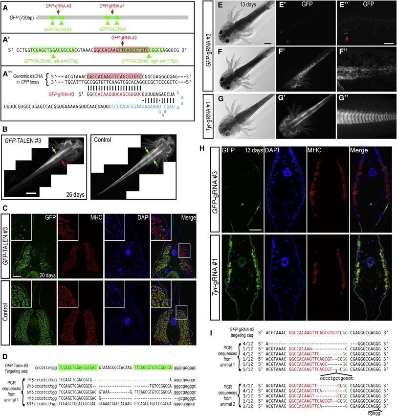
Stem Cell Reports
CRISPR-Mediated Sox2 Deletion in Axolotl

of generating genomic deletions in several model systems,          GFP-axolotl strain (Sobkow et al., 2006) to have an easy
including the axolotl (Blitz et al., 2013; Flowers et al., 2014;   readout for successful gene knockout. Two pairs of GFP-
Hwang et al., 2013; Nakayama et al., 2013).                        TALENs and two pairs of CRISPR GFP-gRNAs in correspond-
  In most vertebrate models, mRNAs encoding TALENs                 ing regions were designed according to published protocols
and CRISPRs have been injected into the egg to generate            (Cermak et al., 2011; Cong et al., 2013; Mali et al., 2013b)
genomic deletions from the beginning of development.               (Figures 1A–1A00 ; see the Experimental Procedures). We in-
Since regeneration is a late event, a question is whether          jected the TALEN mRNAs (mixture of left- and right-arm
this means of knocking out genes can be applied to study-          mRNAs) or CRISPR RNAs (mixture of Cas9 mRNA and
ing specific cell types during regeneration or whether more        GFP-gRNA) into freshly laid axolotl embryos with the aim
sophisticated methods to induce knockouts later in devel-          of eliciting knockout of the genomic GFP as early as possible.
opment will be necessary. Here, we investigated the utility        In the egg injection experiments, we titrated the amount of
of the CRISPR system to knock out a key neural stem gene,          injected TALEN (Table S1 available online) and CRISPR
Sox2, with the aim to study its role in the regenerating spi-      (Table S2) RNAs and found that the CRISPR RNAs were
nal cord. SOX2 is an SRY-related high-mobility group box           significantly less toxic to embryos (evaluated at 10 days
transcription factor that supports embryonic pluripotency          postinjection) (Tables S1 and S2). We next assessed the effi-
(Avilion et al., 2003), and it later plays a key role in           ciency of genomic modification. Injections of GFP-TALEN
the development, maintenance, and expansion of neural              pair #1 or #3 mRNA, but not control TALENs (#1 left-arm
stem/progenitor cells (Bylund et al., 2003; Favaro et al.,         plus #3 right-arm mRNAs), into embryos resulted in 24%–
2009; Takemoto et al., 2011). In addition, Sox2 is required        34% of the larvae showing a visible loss or reduction of
to maintain the proliferative progenitor pool in various           GFP expression in at least some tissue/body cells in the
adult epithelia (Arnold et al., 2011; Que et al., 2009). In        resulting larvae (Figures 1B and C; Table S3). GFP expres-
the axolotl, SOX2 is expressed in the mature spinal cord           sion was markedly reduced in the normally strongly ex-
and in cultured spinal cord neurospheres that harbor mul-          pressing muscle, as confirmed on histological cross-sections
tipotent neural stem cells that reconstitute the different         (Figure 1C). The genomic generation of indels was con-
spinal cord cell types (McHedlishvili et al., 2012; Tapia          firmed by genomic PCR (Figure 1D). Animals modified by
et al., 2012). The molecular factors that support the rapid        GFP-TALENs #1 and #3 were grown to adulthood, and
expansion and self-renewal of neural stem cells during spi-        both showed germline transmission of the GFP deletion
nal cord regeneration after tail amputation were unknown.          with a representative transmission frequency of 56%.
Here, we show by the CRISPR-mediated genomic deletion                 In comparison to TALENs, injection of CRISPR GFP-gRNA
of Sox2 that axolotls harboring up to 100% Sox2 deleted            #3 generated GFP deletions at a higher penetrance and effi-
cells survive embryonic development to generate larvae             ciency. A total of 55% of injected animals showed at least
with a normal spinal cord cell complement and neuronal             some loss of GFP expression. Of the animals showing modi-
number. Morpholino knockdown of SOX2 in the embryo                 fication, 60% showed greater than 50% of cells lacking GFP
confirms lack of embryonic lethality. Upon tail amputa-            expression (Table S3; Figures 1E–1F00 ). Control CRISPR
tion, however, the spinal cord cells do not increase their         Tyrosinase(Tyr)-gRNA did not yield knockdown of GFP
proliferation, resulting in defective expansion and an over-       expression (Figures 1G–1G00 ). Cryosections confirmed the
all lack of spinal cord in the regenerating tail. Expression       loss of GFP expression in GFP-gRNA#3-injected, but not
analysis of Sox3 shows overlapping expression with Sox2            control Tyr-gRNA#1-injected, animals at the cellular level
during neural tube development, but not regeneration,              (Figure 1H). Finally, PCR cloning and sequencing of
rationalizing the spinal cord regeneration-specific pheno-         genomic DNA isolated from individual injected animals
type. Despite severe defects in the regenerating spinal            showed a large proportion of clones (cells) with deletions
cord, the regenerative epidermis and the mesenchymal               at the target GFP sequence (Figure 1I). Control-injected em-
blastema form normally in Sox2-CRISPR animals.                     bryos showed no modification of the GFP locus. Germline
                                                                   transmission of CRISPRs is still to be determined. In sum-
                                                                   mary, our results showed that GFP-TALENs and GFP-
RESULTS                                                            CRISPRs can successfully modify the GFP-transgene, with
                                                                   CRISPR mediating more efficient, less toxic effects.
CRISPRs Mediate More Efficient, Less Toxic Gene                    Efficient Knockout of the Endogenous Tyrosinase Gene
Knockout Than TALENs in Axolotl                                    To assess the effectiveness of TALENs and CRISPRs on an
Germline-Transmitted Knockout of a GFP-Transgene                   endogenous gene, we first chose the axolotl Tyrosinase
To determine if TALENs and CRISPRs can mediate efficient           (Tyr) gene as it is a nonessential gene whose knockout gives
gene deletions, we initially chose to knock out a genomi-          a clear, pigmentation defect. To knock out Tyr, two pairs
cally inserted GFP-transgene in our germline-transgenic            of Tyr-TALENs (#3 and #4) and three CRISPR gRNAs

                                                          Stem Cell Reports j Vol. 3 j 444–459 j September 9, 2014 j ª2014 The Authors 445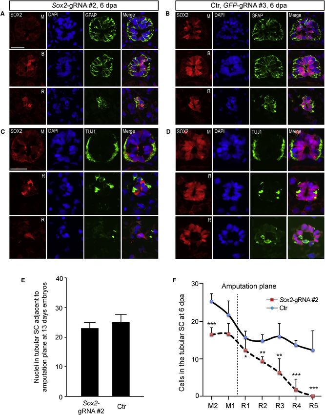
Stem Cell Reports
                                                                                         CRISPR-Mediated Sox2 Deletion in Axolotl

Figure 1. Deletions in a GFP-Transgene in the Axolotl Genome via TALEN or CRISPR Injections
(A–A00 ) TALENs (green) and CRISPR gRNAs (red) were designed to target the GFP sequence and injected into one-cell-stage eggs.
(B) Representative axolotl larvae that received GFP-TALEN #3 in which half of the body (red arrow compared to green arrow) has lost GFP
expression. Control (GFP-TALEN #1 left-arm plus #3 right-arm mRNAs) injection showed bilaterally symmetric GFP expression (green
arrows). Scale bar, 2 mm.
(C) Tail cross-sections from GFP-TALEN #3 knockdown versus control knockdown animals counterstained for DAPI and muscle-specific
myosin heavy chain (MHC) immunostaining. GFP fluorescence signal is missing in muscle cells of the GFP-TALEN section, but not in a
comparable region of the control animal. Region of interest is shown and zoomed by the white box. Scale bar, 200 mm.
                                                                                                      (legend continued on next page)

446 Stem Cell Reports j Vol. 3 j 444–459 j September 9, 2014 j ª2014 The Authors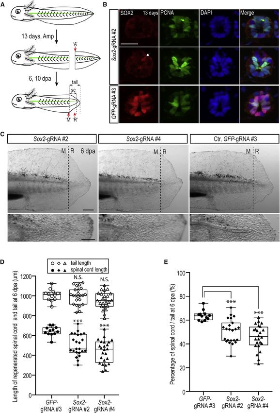
Stem Cell Reports
CRISPR-Mediated Sox2 Deletion in Axolotl
(Figure 2A) were assessed by injection of RNAs into em- the phenotype and the other four Sox2-gRNAs gave no
bryos and examined 13 days postinjection (Table S4). A to- apparent phenotype. Of 487 eggs injected with Sox2-
tal of 60% of animals injected with Tyr-TALEN #4 mRNA gRNA#2, 403 survived, hatched, and grew to normal sizes.
showed at least some loss of pigment in the body (Figures Upon closer visual inspection of behavior, 274 (68%)
2B and 2B0 ). In comparison, the most efficient CRISPR showed a curved body (Figure 3C) when swimming, and
Tyr-gRNA#1 yielded 99% of animals showing loss in many appeared to have excess blood in the olfactory bulb
pigment, with 86% showing apparently complete loss of area (Figure 3C0 , red arrow). The same phenotype was
pigment (Table S4; Figures 2E and 2E0 ). Animals injected observed from Sox2-gRNA#4-injected animals (Figures 3E
with control TALEN (left arm of Tyr-TALEN #4) and GFP- and 3E0 ). This phenotype was never observed in the GFP-
gRNA#3 showed no loss in pigmentation (Figures 2C, gRNA#1-injected animals (Figures 3D, 3D0 , 3F, and 3F0 ).
2C0 , 2F, and 2F0 ). Again, PCR amplification, cloning, and Immunohistological examination of the olfactory bulb
sequencing of the target Tyr sequence showed a higher confirmed a severe reduction in SOX2-positive cells at the
rate of deletions from Tyr-CRISPR (Figure 2G) when lumen of the olfactory bulb and a general loss of cell
compared to Tyr-TALEN (Figure 2D). Our results show number (Figure S1A). The observation of identical pheno-
that for two independent gene-knockout trials, CRISPRs types from two different Sox2-gRNAs, combined with the
showed a higher frequency and penetrance of gene knock- absence of the phenotype in all animals injected with
down with less animal lethality compared to TALENs. GFP and Tyrosinase gRNAs, indicates that the phenotypes
are specific to Sox2 deletion and not due to off-target
Axolotls Harboring CRISPR-Mediated Deletions in effects.
Sox2 Show Normal Size and Organization of the Spinal The genetic knockout of Sox2 in mice results in preim-
Cord during Development plantation lethality (Avilion et al., 2003), and there-
Given the higher success rate with CRISPRs, we sought to fore, the survival through embryonic development was
determine the role of Sox2 in axolotl spinal cord develop- surprising, although knockdown of Sox2 by morpho-
ment and regeneration by the generation of CRISPR-medi- lino in amphibian or fish has not been reported. We
ated gene deletions. In axolotls, SOX2 expression was asso- therefore analyzed the penetrance of CRISPR-mediated
ciated with neural stem cells that have the ability to Sox2 deletions within individual animals at two develop-
clonally reconstitute spinal cord cell types during tail mental stages: embryonic stage 15 (neural plate stage)
regeneration (McHedlishvili et al., 2012). In order to effec- and day 13 larvae. At stage 15, when there was no
tively disrupt the protein-coding sequence of Sox2, seven scorable phenotype, injected animals were randomly
CRISPR targeting sites were selected as close as possible to selected and genotyped by PCR. Among this cohort,
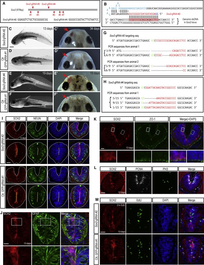
the translational start codon (Figures 3A and 3B). Sox2- 4 out of 11 animals yielded 100% of cloned inserts with
CRISPR RNAs were injected into fertilized eggs at the sin- deletions in Sox2, indicating that animals can survive to
gle-cell stage and analyzed 13 days postinjection. Of the stage 15 with what appears to be complete penetrance
seven Sox2-gRNAs designed, two of them, Sox2-gRNA#2 of Sox2 deletion (Figure S1B). Day 13 larvae could be
targeting the N-terminal region and Sox2-gRNA#4 target- phenotypically sorted based on the curved-body pheno-
ing the middle of the sequence, gave identical, penetrant type (68% of total). Genotyping of the curved-body
phenotypes described below, while a smaller proportion animals showed 8 out of 11 animals with 100% of
of Sox2-gRNA#5-injected animals gave a milder version of clones containing deletions in Sox2 (Figures 3G and
(D) Analysis of genomic target site in two individuals injected with GFP-TALEN #3 by PCR amplification and sequencing of clones. A total
of 9 out of 15 clones showed deletions or deletions plus insertions in the genomic sequence, reflecting a high rate of genome modification
in the cells.
(E) Representative axolotl larva that received CRISPR GFP-gRNA#3 in which >50% of cells have lost GFP expression.
(E0 ) GFP expression is reduced throughout the body.
(E00 ) High-magnification view of tail tissue. Only a few remaining GFP cells are visible (red arrow).
(F–F00 ) Another representative axolotl larva with extensive knockout in 50% of the body.
(G–G00 ) Injection of CRISPR gRNAs targeting the axolotl Tyrosinase (Tyr) gene as a control shows no knockdown of the GFP-transgene.
Scale bars represent 1 mm in (E) and 500 mm in (E00 ).
(H) Tail cross-sections from CRISPR GFP knockdown versus control Tyrosinase knockdown animals counterstained for DAPI and MHC
immunostaining. GFP fluorescence signal is missing in most cells of the GFP-gRNA#3 section. Scale bars, 100 mm.
(I) Analysis of genomic target site in two individuals injected with GFP-gRNA#3 by PCR amplification and sequencing of clones. In both
samples, all 12 clones showed deletions or deletions plus insertions in the genomic sequence, reflecting a high rate of genome modifi-
cation in the cells.
See also Tables S1–S3.
Stem Cell Reports j Vol. 3 j 444–459 j September 9, 2014 j ª2014 The Authors 447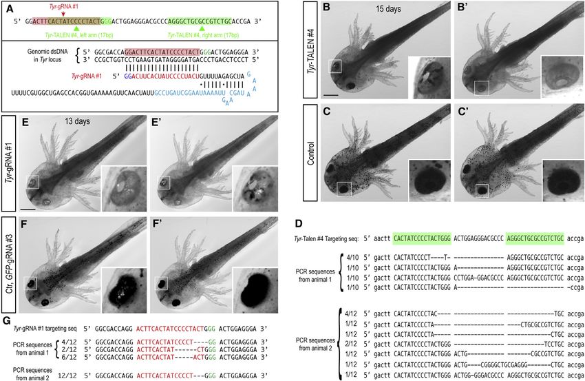
Stem Cell Reports
                                                                                             CRISPR-Mediated Sox2 Deletion in Axolotl

Figure 2. Deletions in the Tyr Gene via TALEN or CRISPR Injections
(A) TALEN and CRISPR gRNAs were designed to target the Tyr sequence and RNAs injected into one-cell-stage eggs.
(B and B0 ) Two representative axolotl larva that had been injected with Tyr-TALEN #4 mRNAs in which >50% of cells have lost pigmentation.
Scale bar, 1 mm.
(C and C0 ) Two representative axolotl larvae injected with left-arm-only TALEN mRNAs show no loss of pigmentation.
(D) Analysis of the genomic target site in two individuals injected with Tyr-TALEN #4 by PCR amplification and sequencing of clones. In
animal 1, seven out of ten clones showed deletions in the genomic sequence, while in animal 2, 11 out of 12 samples showed genomic
modification, suggesting a high rate of modification in this animal.
(E and E0 ) Two representative axolotl larva that had been injected with Tyr-gRNA#1 in which nearly all (left panel) or >50% (right panel) of
cells have lost pigmentation. Scale bar, 1 mm.
(F and F0 ) Two representative axolotl larvae injected with control GFP-gRNA#3 show no loss of pigmentation.
(G) Analysis of the genomic target site in two individuals injected with Tyr-gRNA#1 by PCR amplification and sequencing of clones. In
sample 1, all (12 out of 12) clones showed deletions in the genomic sequence, while in animal 2, all (12 out of 12) samples showed the
same genomic modification, suggesting that the deletion was made in the early egg/embryo.
See also Table S4.

S1C). Analysis of animals injected with Sox2-gRNA#4                        To confirm our conclusions from the CRISPR-mediated
also showed a high-penetrance Sox2 genomic locus                         deletions, we investigated the embryonic phenotype of
modification (Figure 3H). In contrast, genotyping of                     knocking down SOX2 protein via injection of fluorescein
Sox2-gRNA#2-injected animals that did not show the                       isothiocyanate-coupled morpholinos against the Sox2
curved-body/olfactory bulb phenotype showed only a                       translational start site into one-cell-stage embryos. Sox2
very low frequency of gene modification (Figure S1D).                    morpholino or control morpholinos were injected at
These results show that animals harboring a very high                    three different concentrations (Figure S2A) and yielded an
penetrance of CRISPR-mediated Sox2 deletions in the ge-                  indistinguishable proportion of viable embryos when
nomes of their somatic cells can survive embryonic devel-                screened at embryonic stage 37 (Figures S2A–S2C). Immu-
opment to day 13 larvae.                                                 nostaining for SOX2 on cross-sections of stage 16/17

448 Stem Cell Reports j Vol. 3 j 444–459 j September 9, 2014 j ª2014 The Authors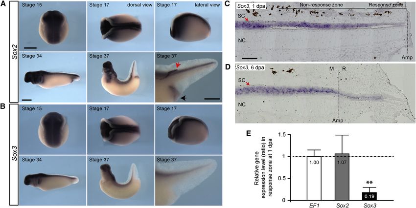
Stem Cell Reports
CRISPR-Mediated Sox2 Deletion in Axolotl

animals confirmed downregulation of SOX2 expression in           reflect significant G1 and S phases of the slow cell cycle
Sox2 morpholino-injected embryos (Figures S2D and S2E).          found in these neural stem cells under homeostatic condi-
The independent knockdown of SOX2 via morpholinos                tions (A. Rodrigo-Albors, personal communication). We
also revealed no detectable embryonic phenotype, con-            then examined whether the overall length of the regener-
firming the results from CRISPR-mediated Sox2 deletion.          ated spinal cord and tail was affected by following live
   To initially characterize the effects of SOX2 deletion        animals at 6 and 10 days after tail amputation under obli-
in the uninjured spinal cord by immunohistochemistry,            que transillumination conditions. Animals injected with
we used animals that showed approximately half-body              Sox2-gRNA#2 or with Sox2-gRNA#4 showed reduced or
knockout of SOX2 to have an internal control. Based on           lack of spinal cord in the regenerated portion of the tail.
our experience with GFP-CRISPR knockouts, animals can            (Figure 4C). In contrast, control GFP-gRNA#1-injected ani-
often show deletion of the gene in one-half of their             mals showed clear outgrowth of the spinal cord into the
body, presumably due to action of the CRISPR in one of           blastema (Figure 4C). We quantitated this phenotype by
the first two blastomeres during development. When we            measuring the length of the overall tail regenerate versus
analyzed sections of the anterior spinal cord of such ani-       the length of the visible spinal cord (Figure 4A). The overall
mals, at least one-half of the spinal cord was lacking           tail regenerate length was unaffected in Sox2-CRISPR ani-
SOX2 expression in the cells lining the spinal cord lumen        mals, while the spinal cord showed a significantly reduced
(Figure 3I). Surprisingly, in such animals, the left and right   length resulting in a shifted ratio of spinal cord/tail regen-
side of the spinal cord were similar in size and organiza-       erate length (Figures 4D and 4E). We further correlated the
tion, showing NEUN+ neuronal cells at the outer circum-          extent of Sox2 deletion found in the removed portion of
ference (Figure 3I). We also observed normal expression          the tail (see above) with the spinal cord regeneration
of other markers associated with axolotl neural stem cells       phenotype at 6 days and found that those specimens
in the Sox2-deleted cells, including normal expression of        showing complete knockout of SOX2 immunofluores-
GFAP and the tight junction protein ZO-1, as well as the         cence at the amputation plane displayed the strongest spi-
proliferative markers proliferating cell nuclear antigen         nal cord growth inhibition (Figures S1E and S1F). Analysis
(PCNA) and phosphohistone H3 (PH3) (Figures 3J–3L).              of the live samples at 10 days postamputation showed a
We further examined the profile of proliferation markers         continued defect in spinal cord outgrowth into the blas-
by injection of 5-ethynyl-20 -deoxyuridine (EdU). In the         tema (Figures S3A–S3C). By this later time point, a mild
uninjured spinal cord, the incorporation of EdU was              effect on blastema length was observed in some Sox2-
similar in the SOX2+ and the SOX2 side of the spinal            deleted animals.
cord (Figure 3M).                                                  To characterize the spinal cord regeneration defect in
                                                                 more detail, we examined immunohistological sections of
Defective Spinal Cord Regeneration due to Lack of                the spinal cord at different distances behind the amputa-
Enhanced Proliferation after Tail Amputation in                  tion plane and in the regenerate at 6 and 10 days postampu-
Sox2-CRISPR Animals                                              tation. Behind the amputation plane, in the region of the
Since the uninjured spinal cord showed normal organiza-          spinal cord that acts as the source zone for the regenerate
tion under conditions of Sox2 deletion, we went on to            (McHedlishvili et al., 2007), the spinal cord showed com-
determine if there is a phenotype during tail regeneration.      parable morphology and marker expression to control
We amputated the tails of Sox2-CRISPR animals that               animals including robust GFAP expression as well as
showed the curved-body phenotype in order to select for          neuron-specific tubulin, TUJ1 expression at the outer
animals that had a high penetrance of deletions (Figure 4A).     circumference (Figures 5A–5D). However, in contrast to
We characterized the spinal cords in the removed portion         the Sox2-CRISPR spinal cords that prior to amputation
of the tail next to the amputation plane to determine the        showed normal cell numbers (Figure 5E), in the 6- and 10-
penetrance of SOX2 deletion and the organization of the          day regenerates, the number of cells per cross-section in
spinal cord at the starting point of the regeneration exper-     the source zone behind the amputation plane was reduced
iment. As expected, most animals showed massive or com-          compared to control regenerates (M1 and M2; Figures 5F
plete loss of the SOX2 spinal cord immunofluorescence            and S3D). This suggests that cells from the source zone
signal at the amputation plane in the removed portion of         contribute to the regenerate but are not replenished. The re-
the tail (Figures 4A, 4B, and S1E). Consistent with our          generating region of the spinal cord (R1–R5; Figures 5F and
analysis above, we found that the spinal cord size, organi-      S3D) showed a starkly reduced number of GFAP+ spinal cord
zation, and PCNA expression was normal in spinal cords           cells, especially in more distal regions of the regenerate, sug-
lacking SOX2+ cells (Figure 4B). PCNA expression in the          gesting a defect in self-renewal and also a disorganization of
uninjured ependymal cells of both control and Sox2-              TUJ1+ cells (Figures 5A, 5C, and S3E). Interestingly, despite
CRISPR animals showed differential intensity levels that         the majority of the Sox2-gRNA#2-injected animals showing

                                                        Stem Cell Reports j Vol. 3 j 444–459 j September 9, 2014 j ª2014 The Authors 449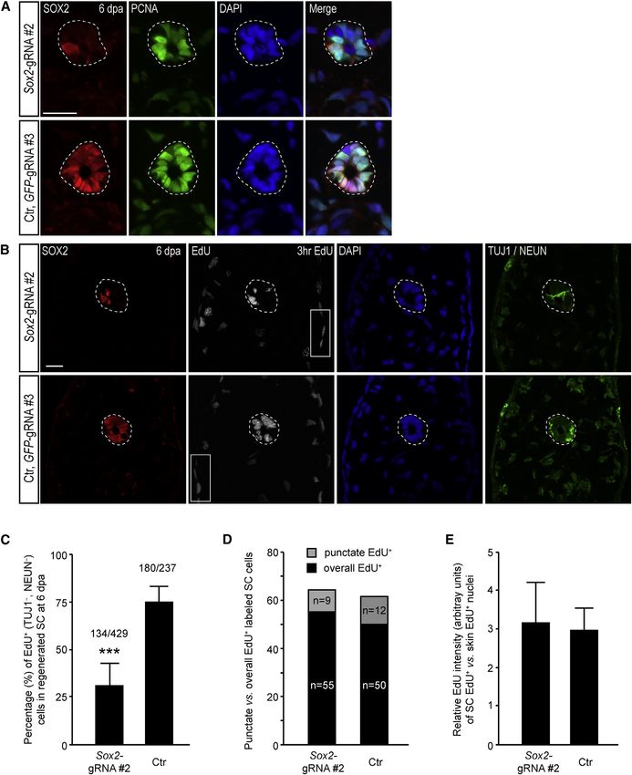
Stem Cell Reports
                                                                                   CRISPR-Mediated Sox2 Deletion in Axolotl

                                                                                                       (legend on next page)

450 Stem Cell Reports j Vol. 3 j 444–459 j September 9, 2014 j ª2014 The Authors
Stem Cell Reports
CRISPR-Mediated Sox2 Deletion in Axolotl
complete or nearly complete loss of SOX2 expression at the proliferative activity of the stem/progenitor cells. The
amputation plane (Figures 4B, S1C, and S1E), the regenerate timing or duration of the cell cycle in neural progenitors
routinely contained some cells that were immunopositive can be modulated at various phases of the cell cycle,
for SOX2. As described in Sox2 heterozygous knockout including the Gap phases as well as S phase (Arai et al.,
mice, we observed specific, cytoplasmic rather than nuclear 2011). We examined whether S phase may have slowed
staining in some of the regenerating samples in which the down in the Sox2-depleted cycling cells but found no evi-
few remaining SOX2+ cells seem to have contributed to dence for a change in average EdU fluorescence intensity
the regenerated spinal cord (Avilion et al., 2003) (Figures (Figure 6D and 6E), suggesting that the S phase properties
5C and S3E). were not altered but rather a higher percentage of cells re-
The above phenotype suggested that deletion of Sox2 in mained in G1 or G2/M in the Sox2-CRISPR animals. These
neural stem cells results in a defect in proliferative expan- results indicate that lack of SOX2 results in a hampered pro-
sion of neural stem cells specifically after tail amputation. liferation and expansion of the neural stem/progenitor cell
To gain further insight into these proliferative properties, pool without changing a number of key markers of radial
we first examined PCNA expression in the 6-day regener- glial cell identity.
ated spinal cords. While all cells in the regenerating spinal
cord of Sox2-CRISPR animals were positive for PCNA (Fig- Downregulation of Sox3 during Spinal Cord
ure 6A), they showed the varying signal intensities that Regeneration
were more similar to the slow-cycling, nonregenerating, Considering the survival of Sox2-CRISPR animals through
control spinal cord cells (Figure 4B) compared to the embryonic development but the strong regeneration-spe-
more homogeneous staining typical of fast-cycling neural cific phenotype, we turned to Sox3 as a potential family
stem cells in the control regenerates (Figure 6A). This sug- member whose expression may rationalize the Sox2 depen-
gested that in Sox2-CRISPR animals, cells do not accelerate dence of regeneration, but not development. In frog and
their cell cycle efficiently and possess a significant G1 fish embryos, Sox3, whose overexpression caused expan-
phase. To corroborate this hypothesis, we performed pulse sion of the CNS and whose knockdown caused reduction
labeling with the S phase marker EdU on regeneration day of CNS tissue, was shown to control Sox2 expression, but
6 for 3 hr prior to harvesting the tail for immunohisto- not vice versa (Dee et al., 2008; Rogers et al., 2009). No
chemical processing. In the Sox2-CRISPR animals, we phenotype for Sox2 knockdown in these models was
observed a marked reduction in the percentage (from described. Sox2 deletion in the mouse CNS showed a mild
75% to 31%) of TUJ1NEUN spinal cord cells that incor- phenotype, with a compensatory upregulation of Sox3
porated EdU (Figures 6B and 6C), indicating a decreased (Miyagi et al., 2008). We therefore examined expression
Figure 3. Normal Spinal Cord Organization in Animals with CRISPR-Mediated Deletions in the Sox2 Locus
(A) Schema depicting the locations of seven and sequence of two CRISPR gRNAs designed against the Sox2 coding sequence.
(B) Schema illustrating the binding between Sox2-gRNA#2 and the Sox2 target sequence.
(C–F00 ) Representative axolotl larvae that received Sox2-gRNA#2 (C and C0 ) and Sox2-gRNA#4 (E and E0 ) show curved body and/or olfactory
bulb defects (C0 and E0 , red arrows). Animals receiving control (Ctr) GFP-gRNA#1 show no such defects (D, D0 , F, and F0 ). Scale bars represent
1 mm in (C) and (E) and 0.5 mm in (C0 ) and (E0 ).
(G and H) Analysis of the genomic target site in two (G) or one (H) individuals injected with Sox2-gRNA#2 or Sox2-gRNA#4, respectively, by
PCR amplification and sequencing of clones. Note: these individuals show a very high proportion of clones with modification.
(I) Sox2-CRISPR animals maintain normal organization of neurons. Immunofluorescence staining for SOX2 (red) and NEUN (green)
combined with DAPI (blue) on spinal cord cross-sections from animals injected with Sox2-gRNA#2 (upper panels) or control (Ctr) GFP-
gRNA#1. Two individual spinal cord sections along the anterior-posterior axis are shown. A significant number of Sox2-gRNA#2-injected
animals are lacking SOX2 protein expression in at least half of the spinal cord indicating biallelic loss in one-half of the body, suggesting
modification at the two-cell stage. Both SOX2+ and SOX2 sides of the spinal cord have NEUN+ neurons at the outer circumference. Dashed
lines indicate spinal cord area; yellow lines indicate apical surface. Scale bars, 50 mm.
(J and K) Sox2-deleted cells maintain GFAP and ZO-1 expression. Immunofluorescence for SOX2 (red) and GFAP (green, J) or ZO-1 (green, K)
combined with DAPI (blue) from Sox2-gRNA#2-injected animals. Both the side with SOX2+ cells and the side without SOX2+ cells express
GFAP. Square boxes denote insets shown at higher magnification below. Scale bars, 50 mm.
(L) Normal expression of proliferative markers in Sox2-deleted cells. Immunofluorescence for SOX2 (red), PCNA (white), and phospho-
histone H3 (green) combined with DAPI (blue) on cross-sections. Scale bar, 50 mm.
(M) Incorporation of EdU in Sox2-deleted cells in the uninjured spinal cord. Immunofluorescence for SOX2 (red) combined with EdU
(green) and DAPI (blue) on spinal cord cross-sections from animals injected with Sox2-gRNA#2, with 3 hr EdU labeling before harvest.
Scale bar, 50 mm.
See also Figures S1 and S2.
Stem Cell Reports j Vol. 3 j 444–459 j September 9, 2014 j ª2014 The Authors 451Stem Cell Reports
CRISPR-Mediated Sox2 Deletion in Axolotl
(legend on next page)
452 Stem Cell Reports j Vol. 3 j 444–459 j September 9, 2014 j ª2014 The AuthorsStem Cell Reports
CRISPR-Mediated Sox2 Deletion in Axolotl
of the axolotl Sox3 gene during development and tail gene deletions and other genome modifications in the
regeneration. Whole-mount in situ hybridization of axolotl for regeneration studies.
axolotl embryos at stages 15, 17, 34, and 37 showed indis- Our work examining CRISPR-mediated Sox2 deletion
tinguishable Sox2 and Sox3 expression in the CNS, showed mild phenotypes during development but a strong
including the spinal cord all along the body axis (Figures inhibition of spinal cord regeneration after tail amputa-
7A and 7B). This finding most likely explains the lack of tion. The lack of a developmental phenotype due to
embryonic phenotype in animals with reduced SOX2 SOX2 knockdown was corroborated by the injection of
either by CRISPR- or morpholino-mediated knockdown. anti-Sox2 morpholinos that knocked down SOX2 immuno-
When we checked Sox3 expression during spinal cord staining in the embryo but yielded the same frequency of
regeneration by quantitative PCR and by in situ hybridiza- normally developing embryos as control-injected cohorts.
tion of longitudinal sections at day1 and day 6, we It is interesting that no knockdown experiments for Sox2
observed a specific downregulation of Sox3 in the regener- have been reported in amphibian and zebrafish embryos,
ating portion of the spinal cord (Figures 7C–7E). These re- raising the possibility that in these vertebrates, Sox2 is
sults would correspond to a heightened sensitivity of the either not solely responsible for maintenance of pluripo-
regenerating spinal cord to Sox2 deletion compared to the tent cells or that maternal stores allow survival through
developing spinal cord. early stages. Indeed, when we examined Sox3, its distribu-
tion overlapped with Sox2 expression in the embryo, sug-
gesting it can compensate for lack of Sox2, especially since
DISCUSSION Sox3 has been shown to be upstream of Sox2 in Xenopus em-
bryos (Rogers et al., 2009). During regeneration, axolotl
This work represents a number of important advances for Sox3 was downregulated in the spinal cord, which would
the study of axolotl regeneration. First, we have shown be consistent with an enhanced sensitivity of the regen-
for several genomic loci that TALENs and CRISPRs can erate to Sox2 knockdown.
induce targeted deletions in a large proportion of cells Our analysis of tail regeneration in the Sox2-CRISPR ani-
from injected animals. In all cases, CRISPRs yielded higher mals showed that despite maintaining a number of radial
viability of injected animals and higher penetrance of the glial cell markers, Sox2-deleted spinal cord shows defective
phenotype, reflecting deletion of both alleles within often proliferation of GFAP-positive cells and therefore, reduced
a large majority of the cells. The Tyr and Sox2 experiments outgrowth of the spinal cord at 6 and 10 days postamputa-
show strong cellular phenotypes associated with homozy- tion. In other work, we have defined the cell-cycle charac-
gous gene deletion. In addition, for the GFP-transgene, teristics of SOX2+PCNA+-positive cells before and after
we have shown germline transmission of the deletion after regeneration and found that in the uninjured state, resi-
TALEN-mediated gene deletion. We therefore conclude dent cells cycle slowly, on average once every 14 days.
that CRISPRs will be an important mode of generating Upon tail amputation, the cell cycle accelerates to a 4-day
Figure 4. Deficient Spinal Cord Regeneration in Sox2-CRISPR Animals
(A) Cartoon illustrates regeneration experimental scheme. Amp, amputation; dpa, day post amputation; ‘A’, amputated spinal cord site;
‘M’, mature spinal cord area; ‘R’, regenerated area. Dashed line indicates the amputation plane, the tip of the spinal cord, and tip of the tail.
(B) Immunofluorescence for SOX2 (red) and PCNA (green) combined with DAPI (blue) on cross-sections from the removed piece of tail just
at the amputation plane (‘A’ in A) from animals injected with Sox2-gRNA#2 or GFP-gRNA#3. Upper and middle panels are two adjacent
sections from the same animal; one shows complete loss of the SOX2-positive signal, but the other shows the presence of a single Sox2-
positive cell (arrow). Scale bar, 50 mm.
(C) Images of live, 6-day regenerates. Left: Sox2-gRNA#2; middle: Sox2-gRNA#4; right: control (Ctr), GFP-gRNA#3. Lower panels show
larger-magnification images of regenerating spinal cord area. In animals injected with Sox2-gRNA#2 (left lower panels) and Sox2-gRNA#4
(middle lower panels), a clear spinal cord tube that extends into the blastema is not visible compared to the control sample (GFP-gRNA#3).
‘M’, mature spinal cord area; ‘R’, regenerated area. Dashed lines represent amputation planes. Scale bars represent 500 mm in the upper
panel and 200 mm in the lower panel.
(D) Quantitation of spinal cord and tail length in CRISPR Sox2-gRNAs versus control GFP-gRNA-injected animals at 6 days of regeneration.
Regenerated tail length was indistinguishable between the Sox2-gRNA#2- (n = 23), Sox2-gRNA#4- (n = 24), and GFP-gRNA#3-injected
(n = 12) animals. In contrast, Sox2-gRNAs-injected animals showed a significantly shorter spinal cord outgrowth. Each bullet represents
one individual animal. Error bars, SD; ***p < 0.001.
(E) Spinal cord length to tail length ratio in 6-day regenerates. The spinal cord represents a smaller fraction of the total tail length in Sox2-
gRNA-injected animals compared to controls. The same data set from (D) was used for data plotting. Each bullet represents one individual
animal. Error bars, SD; ***p < 0.001.
See also Figure S3.
Stem Cell Reports j Vol. 3 j 444–459 j September 9, 2014 j ª2014 The Authors 453Stem Cell Reports
CRISPR-Mediated Sox2 Deletion in Axolotl
(legend on next page)
454 Stem Cell Reports j Vol. 3 j 444–459 j September 9, 2014 j ª2014 The AuthorsStem Cell Reports
CRISPR-Mediated Sox2 Deletion in Axolotl
cycle (A. Rodrigo-Albors, personal communication). Given zygous GFP-transgenic and white axolotl animals were used for in-
that the SOX2TUJ1NEUN cells at the amputation jection. For other experiments, eggs derived from either wild-type
plane maintained expression of PCNA and normal levels or white animals were used for injection. Axolotl larvae were kept
of EdU incorporation before regeneration, we interpret individually in plastic cups with a change of fresh tap water every
second day and fed Artemia daily. Axolotl larvae were anaesthe-
this result to mean that the SOX2+ cells that would nor-
tized within 0.01% ethyl-p-aminobenzoate (benzocaine; Sigma)
mally accelerate their cell cycle are, in the absence of
prior to imaging or amputation.
SOX2, unable to accelerate their cell cycle in response to
injury cues. The role of SOX2 in promoting proliferation
of neural stem/progenitor cells has previously been docu- DNA Extraction, Genotyping, and Sequencing
Genomic DNA extraction and PCR were carried out using
mented in the brain and in CNS tumors (Favaro et al.,
REDExtract-N-Amp Tissue PCR Kit (Sigma) according to the manu-
2009, 2014), and our work would support a role for SOX2
facturer’s instructions. Each genomic locus was PCR amplified
in injury mediated rapid cell cycles required to expand with gene-specific primer pairs (see the Supplemental Experi-
the pool of neural stem cells to reconstitute the missing mental Procedures). The resulting PCR products were cloned into
spinal cord. pGEMT vector (Promega). Individual clones were sequenced with
T7 primer.
EXPERIMENTAL PROCEDURES
Microscopy, Quantifications, and Statistics
For further details, see the Supplemental Experimental Procedures. Fluorescence images were acquired with a Zeiss Observer or
All other methods were performed according to standard confocal microscope. Bright-field or color images were acquired
protocols. with an Olympus dissecting microscope. The length of the regen-
erated spinal cords and tails was measured with CellSens Standard
CRISPR Design and RNA Synthesis software. Spinal cord cells were counted using Photoshop soft-
For better comparison, relevant TALENs and CRISPRs were de- ware. EdU+TUJ1NEUN cells, within 300 mm of amputation
signed against the identical sequence of each targeted gene locus. plane, in the regenerated spinal cord were used for quantifica-
The design and RNA synthesis were carried out according to the tion in Figure 4C. EdU fluorescence intensity (in Figure 4E)
published protocols (Bedell et al., 2012; Cermak et al., 2011; was measured with Software Image J, restricted to the spinal
Hwang et al., 2013). cells expressing overall EdU (TUJ1, NEUN) versus epidermal
cells expressing overall EdU. The punctate EdU-labeled cells
Axolotl Care and Egg Injection were most likely excluded because they were at either the begin-
Animal experiments were carried out according to German animal ning or the end of S phase during EdU pulse. After subtracting
welfare legislation. Axolotl egg injection was performed accord- the background value, the relative EdU intensity was calculated
ing to previously published protocols (Khattak et al., 2014). by dividing the average EdU intensity value of each individual
Briefly, 125–500 pg TALEN mRNAs, 500–1,000 pg CRISPR RNAs, spinal cord cell with the average EdU intensity value, minimally
or 5–20 pmol of morpholinos was injected into freshly laid derived from five epidermal cells of the same section. Data plot-
single-cell-stage embryos. For TALEN- and CRISPR-mediated GFP ting was carried out using Microsoft Excel and Prism. Student’s
knockout experiments, eggs derived from the breeding of hetero- t test was used for the p value calculation.
Figure 5. Cellular Analysis of Spinal Cord Regeneration defect in Sox2-CRISPR Animals
The defect in the regenerating spinal cord was investigated by immunofluorescence staining of cross-sections at different locations along
the length of the regenerating spinal cord starting behind the amputation plane.
(A and B) Immunostaining for SOX2 (red) and GFAP (green) with DAPI shows that while the spinal cord in the mature region (M) shows
similar morphology between the Sox2-gRNA#2- and GFP-gRNA#3-injected animals, in the middle region of the regeneration blastema (R),
there are fewer cells in the spinal cord of the Sox2-gRNA#2-injected animals. Interestingly, some cells showing SOX2 staining are evident
in the regenerating portion of the spinal cord. B, boundary between mature and regenerate. Scale bars, 50 mm.
(C and D) Immunostaining for SOX2 (red) and TUJ1 (green) with DAPI (blue) shows the comparable organization of neurons and axons in
the mature portion of the Sox2-gRNA#2- and GFP-gRNA#3-injected animals. In contrast, the distribution of TUJ1+ cells is scattered and
disorganized in the mid portion of the Sox2-gRNA#2-injected animals.
(E) Quantification of the number of nuclei in the intact spinal cord shows that Sox2-gRNA#2-injected animals have similar numbers of
spinal cord cells to control (Ctr), GFP-gRNA#3-injected animals. Data are the mean value of determinants derived from four control and
seven Sox2-gRNA#2 animals (each determinant is the average value of at least three adjacent sections close to amputation plane). Error
bars, SD.
(F) Quantification of the number of nuclei in the regenerating spinal cord at different points along its length shows that cell number
decreases markedly along the length of the regenerate in Sox2-gRNA#2-injected animals while it stays relatively constant in control GFP-
gRNA#3-injected animals. Data are the mean value of determinants derived from four control and seven Sox2-gRNA#2 animals (each
determinant is the average value of three sections, with 50 mm distance). Each bar on the x axis stands for 150 mm. Error bars, SD.
Stem Cell Reports j Vol. 3 j 444–459 j September 9, 2014 j ª2014 The Authors 455Stem Cell Reports
CRISPR-Mediated Sox2 Deletion in Axolotl
Figure 6. Reduced Proliferative Capacity of Regenerating Spinal Cord Cells in Sox2-CRISPR Animals
(A) Immunostaining for SOX2 (red) and PCNA (green) with DAPI (blue) shows that essentially all cells in the regenerating portion of Sox2-
CRISPR animals at 6 dpa show PCNA staining (dotted lines). However, the heterogeneity in PCNA intensity levels more closely resembles
the profile seen in an unamputated spinal cord (see Figure 4B) compared to the more uniform staining observed in a control regenerating
spinal cord (Ctr). Scale bar, 50 mm.
(B) EdU incorporation was assayed to interrogate passage through the cell cycle. After a 3 hr EdU pulse, cells in the regenerating portion
of the Sox2-gRNA#2-injected animals showed fewer EdU+ cells in the spinal cord (dotted lines) compared to GFP-gRNA#3-injected
control (Ctr) animals. In contrast, the amount of EdU incorporation in epidermis (box) was similar between the two types of animals.
Scale bar, 50 mm.
(legend continued on next page)
456 Stem Cell Reports j Vol. 3 j 444–459 j September 9, 2014 j ª2014 The AuthorsStem Cell Reports
CRISPR-Mediated Sox2 Deletion in Axolotl
Figure 7. Sox3 Expression Overlaps with Sox2 during Development, but Not Regeneration
(A and B) Sox2 (A) and Sox3 (B) whole-mount in situ hybridization of axolotl embryos at stage 15, 17, 34, and 37. Note the indistin-
guishable Sox2 and Sox3 expression in the CNS, including the spinal cord, at all stages analyzed. Sox2, but not Sox3, is also expressed in the
lateral line (red arrow) and cloaca (black arrow) at stage 37. Scale bars represent 1 mm in (A), left panels and 0.5 mm in (A), lower right
panel.
(C) Sox3 in situ hybridization of a 1-day regenerating tail, longitudinal section. Sox3 expression is reduced in the 500 mm zone next to the
amputation plane that will be the source of the regenerating spinal cord.
(D) Sox3 in situ hybridization of a 6-day regenerating tail, longitudinal section. Sox3 expression is low all along the regenerating portion
of the spinal cord. SC, spinal cord; NC, nodal cord; M, mature; R, regenerate. Dashed lines represent amputation planes. Scale bar, 200 mm.
(E) Results of quantitative RT-PCR of Sox2 and Sox3 expression in the 500 mm response zone next to the amputation plane at 1 dpa as shown
in (C) versus the 500 mm developing tail portion adjacent to the amputation plane at the day of amputation (0 dpa). Data are derived from
three individual experiments and expressed as ratio of gene expression level in the response/nonresponse zone, with the ratio obtained in
the case of control EF1 being set as1 and the ratios obtained in Sox2 and Sox3 being expressed relative to it. Error bars, SD; **p < 0.01.
ACCESSION NUMBERS with this article online at http://dx.doi.org/10.1016/j.stemcr.
2014.06.018.
The GenBank accession numbers for the Sox2 and Sox3 sequences
reported in this paper are KJ999995 and KJ999996, respectively. AUTHOR CONTRIBUTIONS
J.F.F. designed, performed, and analyzed experiments and wrote
SUPPLEMENTAL INFORMATION
the manuscript. M.S., A.T., Y.T., and K.R. performed experiments.
Supplemental Information includes Supplemental Experimental E.M.T. provided funding, designed and analyzed experiments,
Procedures, three figures, and four tables and can be found and wrote the manuscript.
(C) Quantification of EdU+ (TUJ, NEUN) spinal cord cells as shown in (B) in the regenerated spinal cord within 300 mm of the amputation
plane. Cells from Sox2-gRNA-injected animals showed a 31% incorporation rate compared to 75% in GFP-gRNA#3-injected control animals.
In total, 429 and 237 spinal cord cells, from ten Sox2-gRNA#2- and four GFP-gRNA#3-injected control animals, respectively, were used for
data plotting. Error bars, SD.
(D) Quantification of the distribution of EdU nuclear staining patterns shows that both types of animals have the same relative fraction
‘‘punctate’’ versus ‘‘overall’’ EdU label. Data derived from three Sox2-gRNA#2- and two GFP-gRNA#3-injected control animals were used for
plotting.
(E) Quantification of the EdU intensity in spinal cord cells shows similar levels of incorporation, suggesting that in Sox2-gRNA#2-injected
animals, S phase still progresses at a similar rate to controls, but more cells spend a longer time in G0, G1, or G2 phases. The ‘‘overall’’ EdU+
cells shown in (D), derived from three Sox2-gRNA#2- and two GFP-gRNA#3-injected control animals, were used for plotting. Error bars, SD.
Stem Cell Reports j Vol. 3 j 444–459 j September 9, 2014 j ª2014 The Authors 457Stem Cell Reports
CRISPR-Mediated Sox2 Deletion in Axolotl
ACKNOWLEDGMENTS tenance require Sox2-dependent regulation of Shh. Nat. Neurosci.
12, 1248–1256.
The authors are grateful to Beate Gruhl, Sabine Mögel, Mark
Armisted, and Anja Wagner for outstanding animal care. The Favaro, R., Appolloni, I., Pellegatta, S., Sanga, A.B., Pagella, P., Gam-
work was supported by an HFSP program grant, DFG 274/3-2, bini, E., Pisati, F., Ottolenghi, S., Foti, M., Finocchiaro, G., et al.
DFG-274/2-3/SFB655 from Cells into Tissues, and DFG 274/5-2/ (2014). Sox2 is required to maintain cancer stem cells in a mouse
SPP1356 from Pluripotency, a CRTD seed grant on zinc finger nu- model of high-grade oligodendroglioma. Cancer Res. 74, 1833–
cleases, and from central funds from the DFG Research Center, 1844.
CRTD. Flowers, G.P., Timberlake, A.T., McLean, K.C., Monaghan, J.R., and
Crews, C.M. (2014). Highly efficient targeted mutagenesis in
Received: May 2, 2014 axolotl using Cas9 RNA-guided nuclease. Development 141,
Revised: June 28, 2014 2165–2171.
Accepted: June 30, 2014
Gaj, T., Gersbach, C.A., and Barbas, C.F., 3rd. (2013). ZFN, TALEN,
Published: August 7, 2014
and CRISPR/Cas-based methods for genome engineering. Trends
Biotechnol. 31, 397–405.
REFERENCES Hayashi, T., Sakamoto, K., Sakuma, T., Yokotani, N., Inoue, T., Ka-
Arai, Y., Pulvers, J.N., Haffner, C., Schilling, B., Nüsslein, I., Cale- waguchi, E., Agata, K., Yamamoto, T., and Takeuchi, T. (2014).
gari, F., and Huttner, W.B. (2011). Neural stem and progenitor Transcription activator-like effector nucleases efficiently disrupt
cells shorten S-phase on commitment to neuron production. the target gene in Iberian ribbed newts (Pleurodeles waltl), an
Nat. Commun. 2, 154. experimental model animal for regeneration. Dev. Growth Differ.
56, 115–121.
Arnold, K., Sarkar, A., Yram, M.A., Polo, J.M., Bronson, R., Sen-
gupta, S., Seandel, M., Geijsen, N., and Hochedlinger, K. (2011). Hwang, W.Y., Fu, Y., Reyon, D., Maeder, M.L., Tsai, S.Q., Sander,
Sox2(+) adult stem and progenitor cells are important for tissue J.D., Peterson, R.T., Yeh, J.R., and Joung, J.K. (2013). Efficient
regeneration and survival of mice. Cell Stem Cell 9, 317–329. genome editing in zebrafish using a CRISPR-Cas system. Nat.
Biotechnol. 31, 227–229.
Avilion, A.A., Nicolis, S.K., Pevny, L.H., Perez, L., Vivian, N., and
Lovell-Badge, R. (2003). Multipotent cell lineages in early Jinek, M., Chylinski, K., Fonfara, I., Hauer, M., Doudna, J.A., and
mouse development depend on SOX2 function. Genes Dev. 17, Charpentier, E. (2012). A programmable dual-RNA-guided DNA
126–140. endonuclease in adaptive bacterial immunity. Science 337,
816–821.
Bedell, V.M., Wang, Y., Campbell, J.M., Poshusta, T.L., Starker,
C.G., Krug, R.G., 2nd, Tan, W., Penheiter, S.G., Ma, A.C., Leung, Kawakami, Y., Rodriguez Esteban, C., Raya, M., Kawakami, H.,
A.Y., et al. (2012). In vivo genome editing using a high-efficiency Martı́, M., Dubova, I., and Izpisúa Belmonte, J.C. (2006). Wnt/
TALEN system. Nature 491, 114–118. beta-catenin signaling regulates vertebrate limb regeneration.
Blitz, I.L., Biesinger, J., Xie, X., and Cho, K.W. (2013). Biallelic Genes Dev. 20, 3232–3237.
genome modification in F(0) Xenopus tropicalis embryos using Khattak, S., Schuez, M., Richter, T., Knapp, D., Haigo, S.L., San-
the CRISPR/Cas system. Genesis 51, 827–834. doval-Guzmán, T., Hradlikova, K., Duemmler, A., Kerney, R., and
Bylund, M., Andersson, E., Novitch, B.G., and Muhr, J. (2003). Tanaka, E.M. (2013). germline transgenic methods for tracking
Vertebrate neurogenesis is counteracted by Sox1-3 activity. Nat. cells and testing gene function during regeneration in the axolotl.
Neurosci. 6, 1162–1168. Stem Cell Rep. 1, 90–103.
Cermak, T., Doyle, E.L., Christian, M., Wang, L., Zhang, Y., Khattak, S., Murawala, P., Andreas, H., Kappert, V., Schuez, M., San-
Schmidt, C., Baller, J.A., Somia, N.V., Bogdanove, A.J., and Voytas, doval-Guzmán, T., Crawford, K., and Tanaka, E.M. (2014). Opti-
D.F. (2011). Efficient design and assembly of custom TALEN and mized axolotl (Ambystoma mexicanum) husbandry, breeding,
other TAL effector-based constructs for DNA targeting. Nucleic metamorphosis, transgenesis and tamoxifen-mediated recombina-
Acids Res. 39, e82. tion. Nat. Protoc. 9, 529–540.
Cong, L., Ran, F.A., Cox, D., Lin, S., Barretto, R., Habib, N., Hsu, Kragl, M., Knapp, D., Nacu, E., Khattak, S., Schnapp, E., Epperlein,
P.D., Wu, X., Jiang, W., Marraffini, L.A., and Zhang, F. (2013). H.H., and Tanaka, E.M. (2008). Novel insights into the flexibility of
Multiplex genome engineering using CRISPR/Cas systems. Science cell and positional identity during urodele limb regeneration. Cold
339, 819–823. Spring Harb. Symp. Quant. Biol. 73, 583–592.
Dee, C.T., Hirst, C.S., Shih, Y.H., Tripathi, V.B., Patient, R.K., and Kragl, M., Knapp, D., Nacu, E., Khattak, S., Maden, M., Epperlein,
Scotting, P.J. (2008). Sox3 regulates both neural fate and differenti- H.H., and Tanaka, E.M. (2009). Cells keep a memory of their tissue
ation in the zebrafish ectoderm. Dev. Biol. 320, 289–301. origin during axolotl limb regeneration. Nature 460, 60–65.
Echeverri, K., and Tanaka, E.M. (2005). Proximodistal patterning Lei, Y., Guo, X., Liu, Y., Cao, Y., Deng, Y., Chen, X., Cheng, C.H.,
during limb regeneration. Dev. Biol. 279, 391–401. Dawid, I.B., Chen, Y., and Zhao, H. (2012). Efficient targeted
Favaro, R., Valotta, M., Ferri, A.L., Latorre, E., Mariani, J., Giachino, gene disruption in Xenopus embryos using engineered transcrip-
C., Lancini, C., Tosetti, V., Ottolenghi, S., Taylor, V., and Nicolis, tion activator-like effector nucleases (TALENs). Proc. Natl. Acad.
S.K. (2009). Hippocampal development and neural stem cell main- Sci. USA 109, 17484–17489.
458 Stem Cell Reports j Vol. 3 j 444–459 j September 9, 2014 j ª2014 The AuthorsStem Cell Reports
CRISPR-Mediated Sox2 Deletion in Axolotl
Mali, P., Esvelt, K.M., and Church, G.M. (2013a). Cas9 as a versatile Seifert, A.W., Kiama, S.G., Seifert, M.G., Goheen, J.R., Palmer, T.M.,
tool for engineering biology. Nat. Methods 10, 957–963. and Maden, M. (2012). Skin shedding and tissue regeneration in
Mali, P., Yang, L., Esvelt, K.M., Aach, J., Guell, M., DiCarlo, J.E., African spiny mice (Acomys). Nature 489, 561–565.
Norville, J.E., and Church, G.M. (2013b). RNA-guided human Simon, A., and Tanaka, E.M. (2013). Limb regeneration. Wiley
genome engineering via Cas9. Science 339, 823–826. Interdiscip. Rev. Dev. Biol. 2, 291–300.
McHedlishvili, L., Epperlein, H.H., Telzerow, A., and Tanaka, E.M.
Sobkow, L., Epperlein, H.H., Herklotz, S., Straube, W.L., and Ta-
(2007). A clonal analysis of neural progenitors during axolotl spi-
naka, E.M. (2006). A germline GFP transgenic axolotl and its use
nal cord regeneration reveals evidence for both spatially restricted
to track cell fate: dual origin of the fin mesenchyme during devel-
and multipotent progenitors. Development 134, 2083–2093.
opment and the fate of blood cells during regeneration. Dev. Biol.
McHedlishvili, L., Mazurov, V., Grassme, K.S., Goehler, K., Robl, B., 290, 386–397.
Tazaki, A., Roensch, K., Duemmler, A., and Tanaka, E.M. (2012).
Reconstitution of the central and peripheral nervous system dur- Suzuki, K.T., Isoyama, Y., Kashiwagi, K., Sakuma, T., Ochiai, H.,
ing salamander tail regeneration. Proc. Natl. Acad. Sci. USA 109, Sakamoto, N., Furuno, N., Kashiwagi, A., and Yamamoto, T.
E2258–E2266. (2013). High efficiency TALENs enable F0 functional analysis by
targeted gene disruption in Xenopus laevis embryos. Biol. Open
Mercader, N., Tanaka, E.M., and Torres, M. (2005). Proximodistal
identity during vertebrate limb regeneration is regulated by Meis 2, 448–452.
homeodomain proteins. Development 132, 4131–4142. Takemoto, T., Uchikawa, M., Yoshida, M., Bell, D.M., Lovell-Badge,
Miyagi, S., Masui, S., Niwa, H., Saito, T., Shimazaki, T., Okano, H., R., Papaioannou, V.E., and Kondoh, H. (2011). Tbx6-dependent
Nishimoto, M., Muramatsu, M., Iwama, A., and Okuda, A. (2008). Sox2 regulation determines neural or mesodermal fate in axial
Consequence of the loss of Sox2 in the developing brain of the stem cells. Nature 470, 394–398.
mouse. FEBS Lett. 582, 2811–2815.
Takeo, M., Chou, W.C., Sun, Q., Lee, W., Rabbani, P., Loomis, C.,
Nakayama, T., Fish, M.B., Fisher, M., Oomen-Hajagos, J., Thomsen, Taketo, M.M., and Ito, M. (2013). Wnt activation in nail epithe-
G.H., and Grainger, R.M. (2013). Simple and efficient CRISPR/ lium couples nail growth to digit regeneration. Nature 499,
Cas9-mediated targeted mutagenesis in Xenopus tropicalis. Gene- 228–232.
sis 51, 835–843.
Tanaka, E.M., and Ferretti, P. (2009). Considering the evolution of
Que, J., Luo, X., Schwartz, R.J., and Hogan, B.L. (2009). Multiple
regeneration in the central nervous system. Nat. Rev. Neurosci. 10,
roles for Sox2 in the developing and adult mouse trachea. Develop-
713–723.
ment 136, 1899–1907.
Rogers, C.D., Harafuji, N., Archer, T., Cunningham, D.D., and Tapia, N., Reinhardt, P., Duemmler, A., Wu, G., Araúzo-Bravo, M.J.,
Casey, E.S. (2009). Xenopus Sox3 activates sox2 and geminin Esch, D., Greber, B., Cojocaru, V., Rascon, C.A., Tazaki, A., et al.
and indirectly represses Xvent2 expression to induce neural pro- (2012). Reprogramming to pluripotency is an ancient trait of verte-
genitor formation at the expense of non-neural ectodermal deriv- brate Oct4 and Pou2 proteins. Nat. Commun. 3, 1279.
atives. Mech. Dev. 126, 42–55. Whited, J.L., Tsai, S.L., Beier, K.T., White, J.N., Piekarski, N.,
Roy, S., Gardiner, D.M., and Bryant, S.V. (2000). Vaccinia as a tool Hanken, J., Cepko, C.L., and Tabin, C.J. (2013). Pseudotyped retro-
for functional analysis in regenerating limbs: ectopic expression of viruses for infecting axolotl in vivo and in vitro. Development 140,
Shh. Dev. Biol. 218, 199–205. 1137–1146.
Stem Cell Reports j Vol. 3 j 444–459 j September 9, 2014 j ª2014 The Authors 459You can also read


