Is Senescence-Associated β-Galactosidase a Reliable in vivo Marker of Cellular Senescence During Embryonic Development?
←
→
Page content transcription
If your browser does not render page correctly, please read the page content below
REVIEW
                                                                                                                                       published: 28 January 2021
                                                                                                                                   doi: 10.3389/fcell.2021.623175

                                            Is Senescence-Associated
                                            β-Galactosidase a Reliable in vivo
                                            Marker of Cellular Senescence
                                            During Embryonic Development?
                                            José Antonio de Mera-Rodríguez 1 , Guadalupe Álvarez-Hernán 1 , Yolanda Gañán 2 ,
                                            Gervasio Martín-Partido 1 , Joaquín Rodríguez-León 2* and Javier Francisco-Morcillo 1*
                                            1
                                             Área de Biología Celular, Departamento de Anatomía, Biología Celular y Zoología, Facultad de Ciencias, Universidad
                                            de Extremadura, Badajoz, Spain, 2 Área de Anatomía y Embriología Humana, Departamento de Anatomía, Biología Celular y
                          Edited by:        Zoología, Facultad de Medicina, Universidad de Extremadura, Badajoz, Spain
                     Wolfgang Knabe,
        Universität Münster, Germany
                       Reviewed by:
                                            During vertebrate embryonic development, cellular senescence occurs at multiple
                    Luis Covarrubias,       locations. To date, it has been accepted that when there has been induction of
     National Autonomous University         senescence in an embryonic tissue, β-galactosidase activity is detectable at a pH as high
                   of Mexico, Mexico
                 Valery Krizhanovsky,       as 6.0, and this has been extensively used as a marker of cellular senescence in vivo
 Weizmann Institute of Science, Israel      in both whole-mount and cryosections. Such senescence-associated β-galactosidase
                Vassilis G. Gorgoulis,
 National and Kapodistrian University
                                            (SA-β-GAL) labeling appears enhanced in degenerating regions of the vertebrate embryo
                   of Athens, Greece        that are also affected by programmed cell death. In this sense, there is a strong SA-
                  *Correspondence:          β-GAL signal which overlaps with the pattern of cell death in the interdigital tissue of the
             Joaquín Rodríguez-León         developing limbs, and indeed, many of the labeled cells detected go on to subsequently
                       jrleon@unex.es
             Javier Francisco-Morcillo      undergo apoptosis. However, it has been reported that β-GAL activity at pH 6.0 is
                     morcillo@unex.es       also enhanced in healthy neurons, and some retinal neurons are strongly labeled with
                                            this histochemical technique when they begin to differentiate during early embryonic
                    Specialty section:
         This article was submitted to      development. These labeled early post-mitotic neurons also express other senescence
               Cell Death and Survival,     markers such as p21. Therefore, the reliability of this histochemical technique in studying
                a section of the journal
  Frontiers in Cell and Developmental
                                            senescence in cells such as neurons that undergo prolonged and irreversible cell-
                                Biology     cycle arrest is questionable because it is also expressed in healthy post-mitotic cells.
        Received: 29 October 2020           The identification of new biomarkers of cellular senescence would, in combination
        Accepted: 05 January 2021
                                            with established markers, increase the specificity and efficiency of detecting cellular
        Published: 28 January 2021
                                            senescence in embryonic and healthy mature tissues.
                             Citation:
               de Mera-Rodríguez JA,        Keywords: cell death, cell senescence, retina, development, histochemistry, limb
         Álvarez-Hernán G, Gañán Y,
 Martín-Partido G, Rodríguez-León J
   and Francisco-Morcillo J (2021) Is
             Senescence-Associated
                                           CELLULAR SENESCENCE
   β-Galactosidase a Reliable in vivo
Marker of Cellular Senescence During
                                           The study of cellular senescence was initiated by Hayflick and Moorhead (1961). Those authors
           Embryonic Development?          reported that human fibroblasts isolated from embryonic tissues cease to proliferate after a
     Front. Cell Dev. Biol. 9:623175.      limited number of cell divisions. We now know that the loss of proliferative activity is the
     doi: 10.3389/fcell.2021.623175        consequence of progressive shortening of telomeres in each replicative round (for a review, see

Frontiers in Cell and Developmental Biology | www.frontiersin.org                1                                        January 2021 | Volume 9 | Article 623175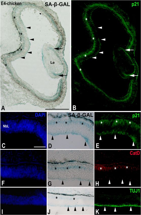
de Mera-Rodríguez et al.                                                                               SA-β-GAL Histochemistry in Embryonic Tissues

Bernadotte et al., 2016). Currently, we have learnt a lot about the        SA-β-GAL HISTOCHEMISTRY
stimuli that trigger cellular senescence and about the intracellular
effector pathways that execute this process. Cellular senescence           Lysosomal β-GAL cleaves β-D-galactose residues in β-D-
is characterized by a prolonged and irreversible cell-cycle arrest         galactosides. Detectable β-GAL activity is the most extensively
with secretory features, macromolecular damage, and altered                used marker for senescent or aging cells whether in culture or in
metabolism (Campisi et al., 2011; Rodier and Campisi, 2011;                mammalian tissues (Dimri et al., 1995; Debacq-Chainiaux et al.,
Childs et al., 2015; Hernandez-Segura et al., 2018; Gorgoulis              2009). Specifically, at pH 6.0, the β-GAL enzyme hydrolyses
et al., 2019). The cells that exhibit these features of senescence         5-bromo-4-chloro-3-indoyl-β-d-galactopyranoside (X-gal), a
normally accumulate in aging tissues, further linking this cellular        colorless, soluble compound consisting of galactose linked to an
state with the aging process in general (Dimri et al., 1995).              indole. This reaction releases a deep blue, insoluble product on
Cellular senescence is now also considered to be a suppressive             the cell culture or in the tissue. This histochemical technique
mechanism against oncogenesis (Lee and Lee, 2019), acting to               is distinct from the acidic β-GAL activity, present in lysosomes
block proliferation in cells with oncogenic mutations (Campisi             of all non-senescent cells and detectable at pH 4.0 (Kuilman
et al., 2011). Recent studies also describe beneficial effects of          et al., 2010). However, it has been demonstrated (Lee et al.,
cellular senescence during embryonic development, tissue repair            2006) that lysosomal β-GAL is the origin of SA-β-GAL activity,
and regeneration, and cellular reprogramming (Czarkwiani and               with the increased SA-β-GAL activity detected in senescent cells
Yun, 2018; Rhinn et al., 2019). Senescence during embryonic                clearly being a result of the increased expression of GLB1, the
development is involved in tissue remodeling and is usually                gene encoding the lysosomal enzyme. Furthermore, increased
linked to areas of cell death that arise in degenerating structures        lysosomal biogenesis has been described in senescent cells (Kurz
(Muñoz-Espín and Serrano, 2014). Therefore, senescent cells                et al., 2000; Severino et al., 2000; Hernandez-Segura et al., 2018).
can be detected from embryogenesis (when they contribute                       Nonetheless, some researchers have clearly shown that in
to tissue development) to adulthood (when they prevent the                 some cases β-GAL activity is not indicative of senescence.
propagation of damaged cells and contribute to tissue repair and           For instance, it has been described that β-GAL activity at pH
tumor suppression).                                                        6.0 might reflect the activity of autophagy due to the intense
    Senescent cells are metabolically active and possess some              biogenesis of lysosomes that occurs during this process (Young
features in vitro and in vivo which are known biomarkers of                et al., 2009). Intense β-GAL histochemical signals have also
cellular senescence (Bernadotte et al., 2016; Matjusaitis et al.,          been reported in the visceral endoderm at early stages of
2016; Hernandez-Segura et al., 2018; Wang and Dreesen, 2018).              mouse embryo development (Huang and Rivera-Pérez, 2014)
One of the most used methods to assess cellular senescence is              and in the luminal cells of the duodenum (Going et al., 2002).
the detection of β-galactosidase (β-GAL) activity at pH 6.0, a             Furthermore, endogenous β-GAL activity at pH 6.0 is detected
histochemical assay that is called “senescence associated β-GAL”           cytochemically in immortalized cultured cells and macrophage-
(SA-β-GAL), because it labels senescent cells, both in vivo and            like cells (Yegorov et al., 1998), in macrophages and osteoclasts
in vitro (Dimri et al., 1995). However, intense β-GAL labelling            in mature tissues (Bursuker et al., 1982; Kopp et al., 2007; Hall
at pH 6.0 could also reflect an alteration in lysosomal number             et al., 2017), and in confluent non-transformed fibroblast cultures
or activity in non-proliferating cells (Yegorov et al., 1998) or           (Severino et al., 2000). A recent study has shown that Purkinje,
in terminally differentiated cells such as neurons (Piechota               choroid plexus, heart muscle, intestinal, and pancreatic cells in
et al., 2016, inter alia). It has been demonstrated that the so-           mammalian tissues are strongly positive for SA-β-GAL activity
called SA-β-GAL increases with aging in subsets of neurons                 (Raffaele et al., 2020). Also, serum starvation and confluent
in different brain areas, but this enzymatic activity is also              culture increase SA-β-GAL activity (Yang and Hu, 2005). In the
present in specific populations of neurons in very young mice              case of the nervous system, it has been proposed that β-GAL
(for a review, see Walton and Andersen, 2019). In this sense,              activity at pH 6.0 might be detected in differentiated neurons
it is known that neurons become post-mitotic very early in                 (Piechota et al., 2016, see below), and even at early stages of
development, and a recent study performed in our laboratory                development (de Mera-Rodríguez et al., 2019, see below). All
clearly demonstrated that β-GAL activity at pH 6.0 is intense              these findings indicate that the reliability of the “so-called” SA-
in recently differentiated ganglion cells in the developing avian          β-GAL assays is questionable because, since the enzyme is not
retina (de Mera-Rodríguez et al., 2019).                                   always specific for cell-aging, this histochemical technique is
    In the present communication, we discuss the reliability of            insufficient to characterize cellular senescence.
SA-β-GAL activity in identifying cellular senescence in vitro                  Senescent cells also show additional signature features that
and in vivo. We first focus on the description of the cell-                could be used in combination with SA-β-GAL to identify the
type-specific labeling of SA-β-GAL in embryonic structures of              state of senescence. Morphologically, cultured senescent cells
different vertebrates. Then, we consider the possible relationship         become flat, large, multi-nucleated, and vacuolated (Denoyelle
between areas of intense SA-β-GAL-staining and areas affected              et al., 2006). Recently, it has been shown that senescent cells
by massive cell death in different embryonic structures.                   in tissues of aged mice are larger than non-senescent cells
Finally, we compare the staining pattern of SA-β-GAL activity              (Biran et al., 2017) and that senescent alveolar progenitors show
with the distribution of other markers of cell senescence,                 abnormal elongated morphology (Kobayashi et al., 2020). In
cell death, and cell differentiation in the developing visual              contrast, in embryonic living tissues, senescent cells usually
system of vertebrates.                                                     have a normal morphology (Muñoz-Espín and Serrano, 2014).

Frontiers in Cell and Developmental Biology | www.frontiersin.org      2                                    January 2021 | Volume 9 | Article 623175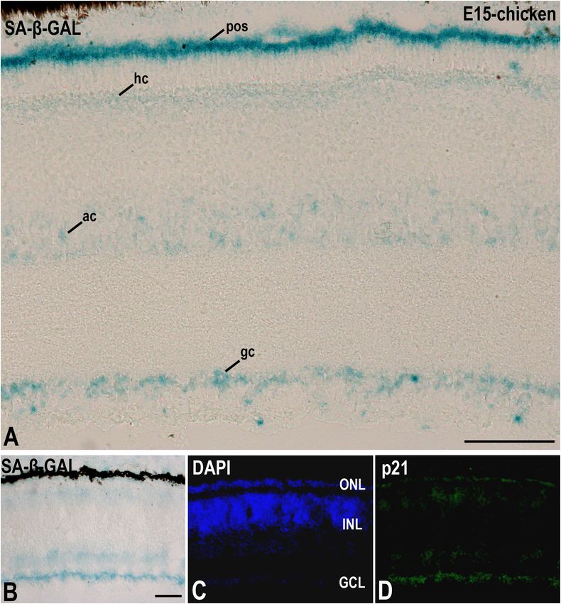
de Mera-Rodríguez et al.                                                                                  SA-β-GAL Histochemistry in Embryonic Tissues

Senescence-associated heterochromatic foci (SAHF) contain                   correlates with SA-β-GAL staining (Lorda-Díez et al., 2019), as
trimethylation at Lys9 of histone 3 (H3K9me3), heterochromatin              do areas of cell death in the somites, tail bud, and CNS (Muñoz-
protein 1 homologue-γ (HP1 γ), and macroH2A (Sharpless                      Espín et al., 2013; Storer et al., 2013). In the case of the developing
and Sherr, 2015; Bernadotte et al., 2016; Wang and Dreesen,                 otic vesicle in the chicken, SA-β-GAL labeled cells are associated
2018; Rhinn et al., 2019). Senescent cells undergo long-term exit           with areas of increased apoptosis (Figures 1A–C) (Gibaja et al.,
from the cell cycle, lacking markers of cell proliferation such as          2019; Magariños et al., 2020). In mice, it has been found that
Ki67 or PCNA (Rhinn et al., 2019). They also activate tumor                 senescence in the endolymphatic sac has a morphogenetic role
suppressor networks, including p16INK4A and p19ARF which                    analogous to that of apoptosis (Muñoz-Espín et al., 2013).
function by activating the Rb protein and the p53 transcription                 The chronotopographical coincidence of cell senescence and
factor, respectively (Lowe and Sherr, 2003).                                apoptosis in the embryo is even more evident in the developing
    These are among the most commonly used markers of                       limb. Since the scope of this article is discussion of the reliability
cell senescence in cultured cells and in aged or pathological               of β-galactosidase staining as an in vivo marker for senescence
tissues. However, the analysis of senescence signatures in some             detection, we would encourage the reader to refer to the detailed
populations of cells in the developing embryo shows enhanced                review of limb apoptosis and senescence in another article
expression of the cell cycle inhibitor p21, but not that of typical         included in this issue authored by Montero et al. (2020). At
markers of senescence in aging including p53, p16, or p19                   early stages of limb development, limb outgrowth is controlled
(Muñoz-Espín et al., 2013; Storer et al., 2013).                            by an epithelial thickening at the distal tip of the structure –
                                                                            the apical ectodermal ridge (AER) (Figures 1D,H,L). The cells
                                                                            of this signaling center proliferate together with the other tissues
SA-β-GAL STAINING IN THE                                                    in the limb bud but undergo apoptosis from soon after the bud’s
DEVELOPING EMBRYO                                                           formation until its disappearance when all the phalanxes have
                                                                            been formed (Rodríguez-León et al., 2013). Concomitant with
SA-β-GAL Staining in Whole Mount                                            the apoptotic process (Figures 1H,L), AER cells also exhibit SA-
Embryos                                                                     β-GAL activity (Figure 1D) and express different markers of
                                                                            cellular senescence such as p21 (Storer et al., 2013; Muñoz-Espín
Programmed cellular senescence is an essential process during
                                                                            et al., 2013; Lorda-Díez et al., 2015; Li et al., 2018). In mice, a lack
vertebrate embryonic development (for reviews, see Muñoz-
                                                                            of p21 activity results in senescence defects and AER structural
Espín and Serrano, 2014; Czarkwiani and Yun, 2018; Rhinn
                                                                            deficiencies, thereby impairing normal limb development (Storer
et al., 2019; Da Silva-Álvarez et al., 2019; Sacco et al., 2021). SA-
                                                                            et al., 2013). Indeed, during limb development in the mouse,
β-GAL labeled cells are found stereotypically in well-defined time
                                                                            strong SA-β-GAL activity is detected in the AER at early
windows during embryonic development (Muñoz-Espín et al.,
                                                                            stages but almost disappears at more advanced stages (Li et al.,
2013; Storer et al., 2013). SA-β-GAL staining in whole-mount
                                                                            2018). These last authors suggest that some senescent cells in
mammalian embryos clearly shows that abundant labeled cells
                                                                            this region of the developing mouse limb undergo apoptosis
accumulates in the developing limbs, nails, focal areas of the
                                                                            and are removed by phagocytosis. However, a sub-population
skin, the tip of the tail, heart, eye tissues, inner ear, olfactory
                                                                            of these embryonic cells positive for ß-GAL activity and p21
epithelium, and the closing neural tube (Muñoz-Espín et al.,
                                                                            expression remains in the limb tissues after birth, and re-
2013; Storer et al., 2013; Zhao et al., 2018), with a staining
                                                                            enter the cell cycle, proliferating in situ (Li et al., 2018). These
pattern similar to that observed in whole-mount chicken embryos
                                                                            results could suggest that some cells in the population are non-
(Figures 1A–C) (cf. Storer et al., 2013; Lorda-Díez et al., 2015,
                                                                            senescent cells or a degree of plasticity in the process of cellular
2019; Gibaja et al., 2019). More recently (Da Silva-Álvarez et al.,
                                                                            senescence during development. Future works are needed to
2020), putative senescent cells during zebrafish development have
                                                                            clarify if cellular senescence during development is a process
been characterized in detail by using SA-β-GAL staining in whole
                                                                            that have some unique features that distinguish it from the adult
mounts, with the strong activity being detected in the yolk,
                                                                            cellular senescence.
cloaca, central nervous system (CNS), intestine, liver, pronephric
                                                                                But this is not the only structure that undergoes apoptosis
ducts, and lens.
                                                                            during limb development. The anterior and posterior margins
                                                                            of the limb bud, as well as the interdigital areas, enter the
SA-β-GAL Staining in Degenerating                                           apoptosis program to sculpt the final shape of the organ (reviewed
Embryo Areas: The Case of the                                               by Montero et al., 2020). Moreover, the interdigital regression
Developing Limb                                                             during digit formation has been regarded as an excellent model
The distribution of SA-β-GAL positive areas in the whole embryo             for the study of how cellular senescence and programmed cell
strongly correlates with the distribution of apoptotic cells. Thus,         death are related (Lorda-Díez et al., 2015; Sanchez-Fernandez
intense SA-β-GAL staining is detected in degenerating structures            et al., 2019). Apoptotic interdigital areas can be observed for
such as the pronephros of fish (Villiard et al., 2017) and                  approximately 48 h in the developing chicken limb – from 6.5
amphibians (Davaapil et al., 2017; Villiard et al., 2017), and in           to 8.5 days of incubation (Montero and Hurlé, 2010; Lorda-Díez
the mesonephros of birds (Nacher et al., 2006) and mammals                  et al., 2015). The classical way to detect the interdigital apoptotic
(Muñoz-Espín et al., 2013; Da Silva-Álvarez et al., 2018). The              pattern is to use vital dyes such as Neutral Red (Figures 1H–K).
pattern of cell death in the developing avian heart also strongly           Staining with this dye allows visualization of tissue removal

Frontiers in Cell and Developmental Biology | www.frontiersin.org       3                                      January 2021 | Volume 9 | Article 623175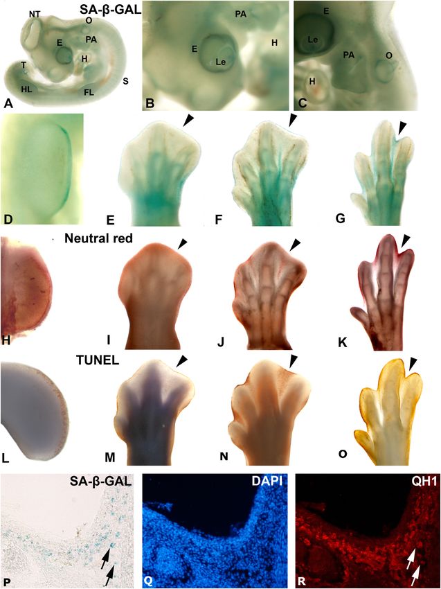
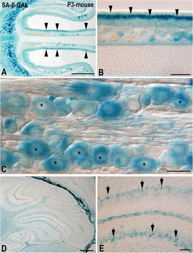
de Mera-Rodríguez et al.                                                                                                  SA-β-GAL Histochemistry in Embryonic Tissues

  FIGURE 1 | Areas segmenting SA-β-GAL activity and apoptosis during avian embryonic development. Detection of SA-β-GAL activity in embryonic day 3.5 (A,B)
  and E4 (C) chicken embryos, and E3.5 (D), E6 (E), E7 (F), and E8 (G) hindlimbs. Neutral red staining for cell death detection in E3.5 (H), E6 (I), E7 (J), and E8 (K)
  hindlimbs. TUNEL assay for apoptosis detection in E4 (L), E6 (M), E7 (N), and E8 (O) hindlimbs. Labeling of AER can be noted in (D,H, and L). Arrowheads in
  (E–G), (I–K), and (M–O) point to the third interdigital space during the establishment of cell senescence and the progression of interdigital programmed cell death.
  SA-β-GAL histochemistry (P) and QH1 immunostaining (R) label macrophages in the interdigital mesenchyme of quail at stage 36. DAPI staining (Q) shows the
  structure of the interdigital space. E, eye; Fl, forelimb; H, heart, HL, hindlimb, Le, lens; NT, neural tube, O, otic vesicle; PA, pharyngeal arches; T, tail bud.

in the AER (Figure 1H) and the progression of interdigital                               developing tissue) from necrosis (the passive death of cells due,
cell death (Figures 1K,L). But it cannot distinguish apoptosis                           for instance, to toxicity or cellular damage). For this reason, since
(the active and programmed process of death in the cells of a                            the decade of the 1990s, the TUNEL assay has been used to

Frontiers in Cell and Developmental Biology | www.frontiersin.org                    4                                           January 2021 | Volume 9 | Article 623175
de Mera-Rodríguez et al.                                                                               SA-β-GAL Histochemistry in Embryonic Tissues

ensure the detection of apoptotic areas during limb development           Piechota et al., 2016) and in prolonged-cultured hippocampal
(Figures 1L–O). The population of apoptotic cells detected by             neurons (Dong et al., 2011; Xu et al., 2019). Furthermore, this
the two techniques in the interdigital areas coincides with the           enzymatic activity is greater in hippocampal neurons after an
pattern of SA-β-GAL labeling in these regions (Figures 1E–G),             injury (Tominaga et al., 2019) and in the Purkinje cells of adult
evidence for the correlation of the two processes during limb             mice (Jurk et al., 2012). Neurodegenerative diseases also cause
development. Indeed, different studies have shown that cellular           increased SA-β-GAL staining in astrocytes, oligodendrocytes,
senescence and apoptosis coincide during limb development and             and microglial cells (Kritsilis et al., 2018). In sum therefore, SA-
are essential for proper interdigital regression (Muñoz-Espín             β-GAL activity increases in mammalian neurons and glial cells
et al., 2013; Lorda-Díez et al., 2015; Montero et al., 2016).             during the aging process and under pathological conditions.
    Besides the activation of SA-β-GAL activity, another essential            Intense SA-β-GAL activity has also been detected in several
feature of cell senescence is cell cycle arrest. In the case of           types of neurons in very young mice (1–3 months old) in vivo
limb interdigital regression, this is connected to up-regulation          and in vitro (Jurk et al., 2012; Piechota et al., 2016; Bussian et al.,
of genes such as p21, p63, and p73 which block cell cycle                 2018; Musi et al., 2018; Raffaele et al., 2020). In this sense, intense
progression (Lorda-Díez et al., 2015; Sanchez-Fernandez et al.,           SA-β-GAL staining is detected in several populations of neurons
2020). Also, various members of the Btg/Tob tumor suppressor              in horizontal cryosections of the head of a 3-day-old mouse.
gene family are expressed in the interdigital space during the            Strong SA-β-GAL signal is detected in the intermediate layer of
progression of programmed cell death, and overexpression of               the olfactory epithelium, the region where the olfactory sensory
Btg2 in the early limb mesenchyme results in an anti-proliferative        neurons are located (Figures 2A,B), but also in neurons of the
and pro-apoptotic effect on the tissue, leading to the formation          trigeminal ganglion (Figure 2C) and in the cerebellar Purkinje
of shortened limbs (Lorda-Díez et al., 2015). Interestingly, the          cells (Figures 2D,E). Therefore, the detection of this enzymatic
expression of these tumor suppressor genes in chicken and mouse           activity in the CNS at relatively early stages of the postnatal life
embryos, species with free digits, is up-regulated during the             suggests that it cannot be attributed solely to cell senescence
course of programmed cell death in the interdigital spaces, but is        either in vivo or in vitro. Given this scenario, it is quite possible
down-regulated, or maintained at stable levels, in the interdigital       that β-GAL activity at pH 6.0 could be detected in neurons even
areas of the duck which maintains webbed digits in adulthood              in embryonic tissues.
(Lorda-Díez et al., 2015).                                                    The vertebrate retina contains six well-known types of
    Another important feature of cell senescence is the                   neurons – ganglion, amacrine, horizontal, bipolar, cone, and
up-regulation of different components of the senescence-                  rod cells. Much is known about the intrinsic programs (mainly
associated secretory phenotype (SASP), namely, different matrix           coded by transcription factors, but also by extrinsic factors
metalloproteinases, IgfBP5, TNF signaling pathway members,                such as growth factors) which are involved in retinogenesis
and interleukin 8 (Rhinn et al., 2019). Several of these SASP             and in the maintenance of neuronal phenotypes (Álvarez-
members are up-regulated in the interdigital areas (Lorda-Díez            Hernán et al., 2013, 2018, 2019, 2020; Xiang, 2013; Bejarano-
et al., 2015) and in the AER (Storer et al., 2013) when cells are         Escobar et al., 2014, 2015). Furthermore, several phases of cell
dying by apoptosis.                                                       death have been reported to occur during development of the
    All this evidence, namely detection of SA-ß-GAL activity and          vertebrate retina (Cook et al., 1998; Knabe et al., 2000; Péquignot
upregulation of cell cycle inhibitors as well as SASP components,         et al., 2003; Francisco-Morcillo et al., 2004, 2014; Rodríguez-
supports that SA-β-GAL staining is detected in senescent cells            Gallardo et al., 2005; Chavarría et al., 2007, 2013; Valenciano
during development in apoptotic areas like those detected in the          et al., 2009; Bejarano-Escobar et al., 2011, 2013). Therefore,
tetrapod limb. Nevertheless, other populations of non-senescent           the embryonic retina constitutes an excellent model with which
cells with a high lysosomal mass and/or increased β-GAL                   to study whether SA-β-GAL activity is linked with neuronal
lysosomal activity, such as QH1-immunoreactive macrophages                differentiation processes and/or with cell death areas.
in the quail interdigital area, also show strong SA-β-GAL                     Previous studies have shown that SA-β-GAL histochemistry
labeling (Figures 1P–R), coinciding with previous studies in mice         assay shows staining of the retinal pigment epithelium in the
(Hall et al., 2017).                                                      developing avian retina (de Mera-Rodríguez et al., 2019) and
                                                                          in the mature retina of rats (Lamoke et al., 2015) and primates
                                                                          (Mishima et al., 1999), including humans (Hjelmeland et al., 1999;
SA-β-GAL Staining in the Nervous                                          Matsunaga et al., 1999). In this sense, some retinal pathologies are
System: The Case of the Developing and                                    linked to cellular senescence that occurs in the retinal pigment
Mature Retina                                                             epithelium (Kozlowski, 2012). Specific SA-β-GAL enzymatic
The analysis of the SA-β-GAL labeling in the developing and               activity is observed in human retinal blood vessels, mainly
mature CNS is a topic of controversy in the field of neuronal             in the endothelial and smooth muscle cells (López-Luppo
senescence. SA-β-GAL activity has been used to detect putative            et al., 2017), and intense SA-β-GAL activity is observed in the
senescent cells in the aging brain of mice (Ori et al., 2015), in         microvasculature of diabetic rats (Lamoke et al., 2015). With
cultures of primary cortical neurons (Chernova et al., 2006), and         regard to SA-β-GAL activity in neural retinal tissue, in the mouse
in cerebellar granule neurons (Bhanu et al., 2010). SA-β-GAL              at early postnatal stages, SA-β-GAL staining is enhanced in
is detected cytochemically in neurons of the hippocampus,                 retinal ganglion cells and subpopulations of neurons dispersed
and its activity increases in old animals (Geng et al., 2010;             throughout the inner nuclear layer (INL) (Oubaha et al., 2016).

Frontiers in Cell and Developmental Biology | www.frontiersin.org     5                                      January 2021 | Volume 9 | Article 623175
de Mera-Rodríguez et al. SA-β-GAL Histochemistry in Embryonic Tissues FIGURE 2 | The presence of SA-β-GAL activity in the postnatal day P3 mouse head tissue. Horizontal (A,B) and sagittal (C–E) cryosections were treated with SA-β-GAL histochemistry. (A,B) Intense SA-β-GAL signal is found in the intermediate layer of the olfactory epithelium (arrowheads). (C) Strong SA-β-GAL staining is detected in sensory neurons in the trigeminal ganglion (asterisks). (D,E) SA-β-GAL activity is detected in the cerebellum, mainly in the Purkinje cell layer (arrows). Scale bars: 200 µm (A,D), 50 µm (B,E), and 20 µm (C). It has also been reported that, in the adult mouse, SA- proliferative diabetic retinopathy, also lead to cell senescence of β-GAL activity in retinal ganglion cells might be increased by several types of retinal neurons (Sapieha and Mallette, 2018). acute intraocular pressure induced ischæmic injury (Li et al., A recent study performed in our laboratory (de Mera- 2017). Other diseases, such as retinopathy of prematurity and Rodríguez et al., 2019) has shown that, in the laminated Frontiers in Cell and Developmental Biology | www.frontiersin.org 6 January 2021 | Volume 9 | Article 623175
de Mera-Rodríguez et al.                                                                                                     SA-β-GAL Histochemistry in Embryonic Tissues

avian retina, even at embryonic stages, SA-β-GAL labeling is                              recently differentiated and mature retinal neurons. We also
intense in subpopulations of neurons located in the ganglion                              found that SA-β-GAL labeling strongly correlates with p21
cell layer (GCL) and in subpopulations of interneurons mainly                             immunoreactivity in both the laminated (Figures 3B–D) and the
located in the amacrine cell layer and the horizontal cell layer                          undifferentiated (Figures 4A–E) retina, even in the lens tissue
(Figures 3A,B). Therefore, SA-β-GAL activity is intense in                                (Figures 4A,B) (de Mera-Rodríguez et al., 2019). SA-β-GAL

  FIGURE 3 | The presence of SA-β-GAL activity in the embryonic day E15 chicken retina. Cryosections of retinas were treated with SA-β-GAL histochemistry (A,B)
  and antibodies against p21 (B–D). DAPI staining shows the laminated structure of the retina (C). SA-β-GAL staining is found in the photoreceptor outer segments
  and in subpopulations of amacrine and ganglion cells (A,B). The horizontal cell layer appears faintly labeled (A,B). p21 immunostaining strongly correlates with the
  SA-β-GAL labeling pattern. ac, amacrine cells; gc, ganglion cells; GCL, ganglion cell layer; hc, horizontal cells; INL, inner nuclear layer; ONL, outer nuclear layer; pos,
  photoreceptor outer segments. Scale bars: 50 µm (A and B–D).

Frontiers in Cell and Developmental Biology | www.frontiersin.org                     7                                             January 2021 | Volume 9 | Article 623175
de Mera-Rodríguez et al.                                                                                                  SA-β-GAL Histochemistry in Embryonic Tissues

  FIGURE 4 | The presence of SA-β-GAL activity in the embryonic day E4 chicken retina. Cryosections of retinas were treated with SA-β-GAL histochemistry and
  antibodies against p21 (A–E), CatD (F–H), and TUJ1 (I–K). DAPI staining shows that the neural retina consists of a NbL (C,F,I). SA-β-GAL staining is detected in the
  scleral (asterisks in A,D,G,J) and vitreal (arrowheads in A,D,G,J) regions of the retina. p21 immunostaining correlates with the SA-β-GAL staining pattern in the
  undifferentiated retina (arrowheads and asterisks in B,E) and lens (arrows in B). CatD immunoreactivity (arrowheads and asterisks in H) is strongly coincident with the
  SA-β-GAL histochemistry signal (arrowheads and asterisks in G). TUJ1 immunoreactivity is intense in the vitreal surface of the NbL (arrowheads in K), coinciding with
  the vitreal SA-β-GAL histochemistry signal detected in the same region (arrowheads in J). Le, lens; NbL, neuroblastic layer. Scale bars: 150 µm (A,B), 50 µm (C–K).

Frontiers in Cell and Developmental Biology | www.frontiersin.org                   8                                            January 2021 | Volume 9 | Article 623175
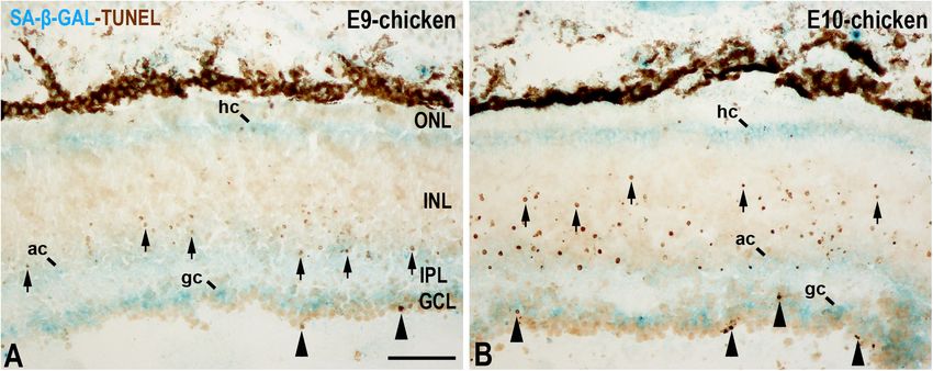
de Mera-Rodríguez et al.                                                                                                        SA-β-GAL Histochemistry in Embryonic Tissues

  FIGURE 5 | SA-β-GAL activity and cell death in the embryonic day E9 (A) and E10 (B) chicken retina. Cryosections were doubly stained with SA-β-GAL
  histochemistry and TUNEL technique. TUNEL-positive nuclei are mainly detected in the GCL (arrowheads) and in the middle region of the INL (arrows). SA-β-GAL
  activity is observed in the GCL, amacrine cell layer, and horizontal cell layer. ac, amacrine cells; gc, ganglion cells; GCL, ganglion cell layer; hc, horizontal cells; INL,
  inner nuclear layer; IPL, inner plexiform layer; ONL, outer nuclear layer; pos, photoreceptor outer segments. Scale bar: 50 µm.

staining in the non-laminated retina is mainly restricted to                                CONCLUSION
the vitreal and scleral surfaces of the neuroblastic layer (NbL)
(Figures 4A,D,G,J), strongly correlating with cathepsin D                                   SA-β-GAL activity increases with aging in neurons of the
immunoreactivity (Figures 4F–H) (de Mera-Rodríguez et al.,                                  mammalian brain (for a review, see Walton and Andersen,
2019), a marker for increased lysosome number or activity (Kurz                             2019). In the developing limb, this SA-β-GAL activity correlates
et al., 2000; Wassélius et al., 2003; Ahuja et al., 2008; Bejarano-                         well with cellular senescence in the areas of programmed
Escobar et al., 2011; Lorda-Díez et al., 2019). The SA-β-GAL                                cell death that occur physiologically during the development
activity located in the vitreal surface of the retina is detected in                        process. Consequently, a proper balance between senescence
TUJ1-positive newborn ganglion cell neuroblasts (Figures 4I–K).                             and apoptosis is needed for accurate formation of the limbs
Therefore, SA-β-GAL labeling in the developing visual system                                (Lorda-Díez et al., 2015). However, intense SA-β-GAL is also
correlates strongly with the location of the lysosomal mass, but                            detected in cells that have intrinsically high lysosomal β-GAL
also with other senescence markers, and seems to be linked to                               activity, such as macrophages, but also in a wide range of post-
neuronal differentiation.                                                                   mitotic cells, including neurons, even at early stages of embryonic
   But is SA-β-GAL activity also linked to apoptotic cells                                  development. Other senescence-associated markers, such as
in the developing retina? In the undifferentiated vertebrate                                p21, are also activated in parallel with SA-β-GAL activity in
retina, apoptotic cells are either dispersed throughout the                                 recently differentiated retinal neurons (de Mera-Rodríguez et al.,
NbL or concentrated in areas that surround the optic nerve                                  2019). It is likely that there are several common mechanisms
head (Mayordomo et al., 2003; Francisco-Morcillo et al., 2004;                              involved in both the acquisition of the senescent phenotype
Rodríguez-Gallardo et al., 2005; Valenciano et al., 2009; Bejarano-                         and the maintenance of long-term non-dividing cells’ non-
Escobar et al., 2011, 2013). However, SA-β-GAL activity is                                  proliferating status. Therefore, it is important to discriminate
mainly concentrated in the vitreal and scleral surfaces of the                              between senescent cells and post-mitotic cells in studies about
avian NbL (de Mera-Rodríguez et al., 2019). Later, in the                                   aging of the CNS because some accepted markers of senescence
laminated retina, apoptosis follows spatiotemporal patterns that                            (SA-β-GAL, p21 expression) are less specific than originally
are analogs of the cell differentiation pattern (Figure 5) (Cook                            was expected. The identification of new candidate biomarkers
et al., 1998; Marín-Teva et al., 1999). In the E9 chicken retina,                           of cellular senescence would, in combination with established
cell death is mainly concentrated in the GCL and in the                                     markers, increase the specificity and efficiency of detecting
amacrine cell layer (Figure 5A) (Cook et al., 1998), and, at                                senescence in vivo and in vitro. A future requirement will be
E10, TUNEL-positive bodies spread vitreally to the bipolar                                  to have markers for senescence-like phenotypes in long-term
cell layer (Figure 5B). By these stages, SA-β-GAL staining is                               non-proliferating cells, such as neurons.
detected homogeneously in the GCL, amacrine cell layer, and
horizontal cell layer (Figure 5) (de Mera-Rodríguez et al., 2019).
There is therefore no correlation between SA-β-GAL activity                                 AUTHOR CONTRIBUTIONS
and the chronotopographical distribution of dying cells in the
developing avian retina. In this case, β-GAL activity at pH 6.0                             GÁ-H, JM-R, YG, GM-P, JR-L, and JF-M wrote and
seems to be related to terminal cell differentiation rather than to                         critically reviewed the manuscript. All authors approved
cellular senescence.                                                                        the final manuscript.

Frontiers in Cell and Developmental Biology | www.frontiersin.org                       9                                              January 2021 | Volume 9 | Article 623175
de Mera-Rodríguez et al.                                                                                                          SA-β-GAL Histochemistry in Embryonic Tissues

FUNDING                                                                                       Ministerio de Economía y Competitividad (CGL2015-64650P),
                                                                                              Dirección General de Investigación del Ministerio de Educación y
GÁ-H was a recipient of a Fellowship from the Universidad                                     Ciencia (BFU2017-85547-P), and Junta de Extremadura, Fondo
de Extremadura. This work was supported by grants from the                                    Europeo de Desarrollo Regional, “Una manera de hacer Europa”
Spanish Ministerio de Ciencia y Tecnología (BFU2007-67540),                                   (GR15158, GR18114, and IB18113).

REFERENCES                                                                                    Chavarría, T., Baleriola, J., Mayordomo, R., De Pablo, F., and De La Rosa, E. J.
                                                                                                 (2013). Early neural cell death is an extensive, dynamic process in the embryonic
Ahuja, S., Ahuja-Jensen, P., Johnson, L. E., Caffé, A. R., Abrahamson, M., Ekström,              chick and mouse retina. Sci. World J. 2013:627240.
   P. A. R., et al. (2008). Rd1 mouse retina shows an imbalance in the activity of            Chavarría, T., Valenciano, A. I., Mayordomo, R., Egea, J., Comella, J. X., Hallböök,
   cysteine protease cathepsins and their endogenous inhibitor cystatin. Investig.               F., et al. (2007). Differential, age-dependent MEK-ERK and PI3K-Akt activation
   Ophthalmol. Vis. Sci. 49, 1089–1096. doi: 10.1167/iovs.07-0549                                by insulin acting as a survival factor during embryonic retinal development.
Álvarez-Hernán, G., Andrade, J. P., Escarabajal-Blázquez, L., Blasco, M., Solana-                Dev. Neurobiol. 67, 1777–1788. doi: 10.1002/dneu.20554
   Fajardo, J., Martín-Partido, G., et al. (2019). Retinal differentiation in                 Chernova, T., Nicotera, P., and Smith, A. G. (2006). Heme deficiency is associated
   syngnathids: comparison in the developmental rate and acquisition of retinal                  with senescence and causes suppression of N-Methyl-D-Aspartate receptor
   structures in altricial and precocial fish species. Zoomorphology 138, 371–385.               subunits expression in primary cortical neurons. Mol. Pharmacol. 69, 697–705.
   doi: 10.1007/s00435-019-00447-3                                                               doi: 10.1124/mol.105.016675
Álvarez-Hernán, G., Bejarano-Escobar, R., Morona, R., González, A., Martín-                   Childs, B. G., Durik, M., Baker, D. J., and Van Deursen, J. M. (2015). Cellular
   Partido, G., and Francisco-Morcillo, J. (2013). Islet-1 immunoreactivity in the               senescence in aging and age-related disease: from mechanisms to therapy. Nat.
   developing retina of Xenopus laevis. Sci. World J. 2013:740420.                               Med. 21, 1424–1435. doi: 10.1038/nm.4000
Álvarez-Hernán, G., Hernández-Núñez, I., Rico-Leo, E. M., Marzal, A., de Mera-                Cook, B., Portera-Cailliau, C., and Adler, R. (1998). Developmental neuronal
   Rodríguez, J. A., Rodríguez-León, J., et al. (2020). Retinal differentiation in an            death is not a universal phenomenon among cell types in the chick embryo
   altricial bird species, Taeniopygia guttata: an immunohistochemical study. Exp.               retina. J. Comp. Neurol. 396, 12–19. doi: 10.1002/(sici)1096-9861(19980622)
   Eye Res. 190:107869. doi: 10.1016/j.exer.2019.107869                                          396:13.0.co;2-l
Álvarez-Hernán, G., Sánchez-Resino, E., Hernández-Núñez, I., Marzal, A.,                      Czarkwiani, A., and Yun, M. H. (2018). Out with the Old. In with the new:
   Rodríguez-León, J., Martín-Partido, G., et al. (2018). Retinal histogenesis in                senescence in development. Curr. Opin. Cell Biol. 55, 74–80. doi: 10.1016/j.
   an Altricial Avian Species, the Zebra Finch (Taeniopygia guttata. Vieillot 1817).             ceb.2018.05.014
   J. Anat. 233, 106–120. doi: 10.1111/joa.12809                                              Da Silva-Álvarez, S., Guerra-Varela, J., Sobrido-Cameán, D., Quelle, A., Barreiro-
Bejarano-Escobar, R., Álvarez-Hernán, G., Morona, R., González, A., Martín-                      Iglesias, A., Sánchez, L., et al. (2020). Developmentally-programmed cellular
   Partido, G., and Francisco-Morcillo, J. (2015). Expression and function of                    senescence is conserved and widespread in zebrafish. Aging 12, 17895–17901.
   the LIM-Homeodomain transcription factor Islet-1 in the developing and                        doi: 10.18632/aging.103968
   mature vertebrate retina. Exp. Eye Res. 138, 22–31. doi: 10.1016/j.exer.2015.              Da Silva-Álvarez, S., Lamas-González, O., Ferreirós, A., González, P., Gómez, M.,
   06.021                                                                                        García-Caballero, T., et al. (2018). Pkd2 deletion during embryo development
Bejarano-Escobar, R., Blasco, M., Durán, A. C., Martín-Partido, G., and Francisco-               does not alter mesonephric programmed cell senescence. Int. J. Dev. Biol. 62,
   Morcillo, J. (2013). Chronotopographical distribution patterns of cell death                  637–640. doi: 10.1387/ijdb.180078mc
   and of lectin-positive Macrophages/Microglial cells during the visual system               Da Silva-Álvarez, S., Picallos-Rabina, P., Antelo-Iglesias, L., Triana-Martínez,
   ontogeny of the small-spotted catshark Scyliorhinus canicula. J. Anat. 223,                   F., Barreiro-Iglesias, A., Sánchez, L., et al. (2019). The development of cell
   171–184. doi: 10.1111/joa.12071                                                               senescence. Exp. Gerontol. 128:110742.
Bejarano-Escobar, R., Blasco, M., Martín-Partido, G., and Francisco-Morcillo, J.              Davaapil, H., Brockes, J. P., and Yun, M. H. (2017). Conserved and novel
   (2014). Molecular characterization of cell types in the developing, mature, and               functions of programmed cellular senescence during vertebrate development.
   regenerating fish retina. Rev. Fish Biol. Fish. 24, 127–158. doi: 10.1007/s11160-             Development 144, 106–114. doi: 10.1242/dev.138222
   013-9320-z                                                                                 de Mera-Rodríguez, J. A., Álvarez-Hernán, G., Gañán, Y., Martín-Partido, G.,
Bejarano-Escobar, R., Holguín-Arévalo, M. S., Montero, J. A., Francisco-Morcillo,                Rodríguez-León, J., and Francisco-Morcillo, J. (2019). Senescence-Associated
   J., and Martín-Partido, G. (2011). Macrophage and microglia ontogeny in the                   β-Galactosidase activity in the developing avian retina. Dev. Dyn. 248, 850–865.
   mouse visual system can be traced by the expression of Cathepsins B and D.                    doi: 10.1002/dvdy.74
   Dev. Dyn. 240, 1841–1855. doi: 10.1002/dvdy.22673                                          Debacq-Chainiaux, F., Erusalimsky, J. D., Campisi, J., and Toussaint, O. (2009).
Bernadotte, A., Mikhelson, V. M., and Spivak, I. M. (2016). Markers of cellular                  Protocols to detect senescence-associated beta-galactosidase (SA-Bgal) activity,
   senescence telomere shortening as a marker of cellular senescence. Aging 8,                   a biomarker of senescent cells in culture and in vivo. Nat. Protoc. 4, 1798–1806.
   3–11. doi: 10.18632/aging.100871                                                              doi: 10.1038/nprot.2009.191
Bhanu, U. M., Mandraju, R. K., Bhaskar, C., and Kondapi, A. K. (2010). Cultured               Denoyelle, C., Abou-Rjaily, G., Bezrookove, V., Verhaegen, M., Johnson, T. M.,
   cerebellar granule neurons as an in vitro aging model: topoisomerase IIβ as an                Fullen, D. R., et al. (2006). Anti-oncogenic role of the endoplasmic reticulum
   additional biomarker in DNA repair and aging. Toxicol. Vitr. 24, 1935–1945.                   differentially activated by mutations in the MAPK pathway. Nat. Cell Biol. 8,
   doi: 10.1016/j.tiv.2010.08.003                                                                1053–1063. doi: 10.1038/ncb1471
Biran, A., Zada, L., Karan, P. A., Vadai, E., Roitman, L., Ovadya, Y., et al. (2017).         Dimri, G. P., Lee, X., Basile, G., Acosta, M., Scott, G., Roskelley, C., et al. (1995).
   Quantitative identification of senescent cells in aging and disease. Aging Cell 16,           A biomarker that identifies senescent human cells in culture and in aging skin
   661–671. doi: 10.1111/acel.12592                                                              in vivo. Proc. Natl. Acad. Sci. U.S.A. 92, 9363–9367. doi: 10.1073/pnas.92.20.
Bursuker, I., Rhodes, J. M., and Goldman, R. (1982). B-Galactosidase—an indicator                9363
   of the maturational stage of mouse and human mononuclear phagocytes. J. Cell               Dong, W., Cheng, S., Huang, F., Fan, W., Chen, Y., Shi, H., et al. (2011).
   Physiol. 112, 385–390. doi: 10.1002/jcp.1041120312                                            Mitochondrial dysfunction in long-term neuronal cultures mimics changes
Bussian, T. J., Aziz, A., Meyer, C. F., Swenson, B. L., van Deursen, J. M., and Baker,           with aging. Med. Sci. Monit. 17, 91–96.
   D. J. (2018). Clearance of senescent glial cells prevents tau-dependent pathology          Francisco-Morcillo, J., Bejarano-Escobar, R., Rodríguez-León, J., Navascués, J., and
   and cognitive decline. Nature 562, 578–582. doi: 10.1038/s41586-018-                          Martín-Partido, G. (2014). Ontogenetic cell death and phagocytosis in the visual
   0543-y                                                                                        system of vertebrates. Dev. Dyn. 243, 1203–1225. doi: 10.1002/dvdy.24174
Campisi, J., Andersen, J. K., Kapahi, P., and Melov, S. (2011). Cellular senescence:          Francisco-Morcillo, J., Hidalgo-Sánchez, M., and Martín-Partido, G. (2004). Spatial
   a link between cancer and age-related degenerative disease? Semin. Cancer Biol.               and temporal patterns of apoptosis during differentiation of the retina in the
   21, 354–359.                                                                                  turtle. Anat. Embryol. 208, 289–299.

Frontiers in Cell and Developmental Biology | www.frontiersin.org                        10                                              January 2021 | Volume 9 | Article 623175
de Mera-Rodríguez et al. SA-β-GAL Histochemistry in Embryonic Tissues
Geng, Y. Q., Guan, J. T., Xu, X. H., and Fu, Y. C. (2010). Senescence-associated microaneurysm formation during aging. Investig. Ophthalmol. Vis. Sci. 58,
beta-galactosidase activity expression in aging hippocampal neurons. Biochem. 2832–2842. doi: 10.1167/iovs.16-20312
Biophys. Res. Commun. 396, 866–869. doi: 10.1016/j.bbrc.2010.05.011 Lorda-Díez, C. I., García-Riart, B., Montero, J. A., Rodríguez-León, J., García-
Gibaja, A., Aburto, M. R., Pulido, S., Collado, M., Hurle, J. M., Varela-Nieto, I., Porrero, J. A., and Hurlé, J. M. (2015). Apoptosis during embryonic tissue
et al. (2019). TGFβ2-induced senescence during early inner ear development. remodeling is accompanied by cell senescence. Aging 7, 974–985. doi: 10.18632/
Sci. Rep. 9, 1–13. doi: 10.1016/b978-0-12-408088-1.00001-4 aging.100844
Going, J. J., Stuart, R. C., Downie, M., Fletcher-Monaghan, A. J., and Nicol Lorda-Díez, C. I., Solis-Mancilla, M. E., Sanchez-Fernandez, C., Garcia-Porrero,
Keith, W. (2002). “Senescence-Associated” β-Galactosidase activity in the upper J. A., Hurle, J. M., and Montero, J. A. (2019). Cell senescence, apoptosis and
gastrointestinal tract. J. Pathol. 196, 394–400. doi: 10.1002/path.1059 DNA damage cooperate in the remodeling processes accounting for heart
Gorgoulis, V., Adams, P. D., Alimonti, A., Bennett, D. C., Bischof, O., Bishop, C., morphogenesis. J. Anat. 234, 815–829. doi: 10.1111/joa.12972
et al. (2019). Cellular senescence: defining a path forward. Cell 179, 813–827. Lowe, S. W., and Sherr, C. J. (2003). Tumor suppression by Ink4a-Arf: progress and
doi: 10.1016/j.cell.2019.10.005 puzzles. Curr. Opin. Genet. Dev. 13, 77–83. doi: 10.1016/s0959-437x(02)00013-8
Hall, B. M., Balan, V., Gleiberman, A. S., Strom, E., Krasnov, P., Virtuoso, L. P., et al. Magariños, M., Barajas-Azpeleta, R., Varela-Nieto, I., and Aburto, M. R. (2020).
(2017). P16(Ink4a) and senescence-associated β-Galactosidase can be induced Otic neurogenesis is regulated by TGFβ in a senescence-independent manner.
in macrophages as part of a reversible response to physiological stimuli. Aging Front. Cell. Neurosci. 14:217. doi: 10.3389/fncel.2020.00217
9, 1867–1884. doi: 10.18632/aging.101268 Marín-Teva, J. L., Almendros, A., Calvente, R., Cuadros, M. A., and Navascués, J.
Hayflick, L., and Moorhead, P. S. (1961). The serial cultivation of human (1999). Proliferation of actively migrating ameboid microglia in the developing
diploid cell strains. Exp. Cell Res. 25, 585–621. doi: 10.1016/0014-4827(61)901 quail retina. Anat. Embryol. 200, 289–300. doi: 10.1007/s004290050280
92-6 Matjusaitis, M., Chin, G., Sarnoski, E. A., and Stolzing, A. (2016). Biomarkers to
Hernandez-Segura, A., Nehme, J., and Demaria, M. (2018). Hallmarks of cellular identify and isolate senescent cells. Ageing Res. Rev. 29, 1–12. doi: 10.1016/j.arr.
senescence. Trends Cell Biol. 28, 436–453. doi: 10.1016/j.tcb.2018.02.001 2016.05.003
Hjelmeland, L. M., Cristofalo, V. J., Funk, W., Rakoczy, E., and Katz, M. L. (1999). Matsunaga, H., Handa, J. T., Aotaki-Keen, A., Sherwood, S. W., West, M. D., and
Senescence of the retinal pigment epithelium. Mol. Vis. 5, 33. Hjelmeland, L. M. (1999). β-Galactosidase histochemistry and telomere loss
Huang, T., and Rivera-Pérez, J. A. (2014). Senescence-associated β-Galactosidase in senescent retinal pigment epithelial cells. Investig. Ophthalmol. Vis. Sci. 40,
activity marks the visceral endoderm of mouse embryos but is not indicative of 197–202.
senescence. Genesis 52, 300–308. doi: 10.1002/dvg.22761 Mayordomo, R., Valenciano, A. I., De La Rosa, E. J., and Hallböök, F. (2003).
Jurk, D., Wang, C., Miwa, S., Maddick, M., Korolchuk, V., Tsolou, A., et al. (2012). Generation of retinal ganglion cells is modulated by caspase-dependent
Postmitotic neurons develop a P21-Dependent senescence-like phenotype programmed cell death. Eur. J. Neurosci. 18, 1744–1750. doi: 10.1046/j.1460-
driven by a DNA damage response. Aging Cell 11, 996–1004. doi: 10.1111/j. 9568.2003.02891.x
1474-9726.2012.00870.x Mishima, K., Handa, J. T., Aotaki-Keen, A., Lutty, G. A., Morse, L. S.,
Knabe, W., Süss, M., and Kuhn, H. J. (2000). The patterns of cell death and of and Hjelmeland, L. M. (1999). Senescence-Associated β-Galactosidase histo-
macrophages in the developing forebrain of the tree shrew tupaia belangeri. chemistry for the primate eye. Investig. Ophthalmol. Vis. Sci. 40, 1590–1593.
Anat. Embryol. 201, 157–168. doi: 10.1007/pl00008237 Montero, J. A., and Hurlé, J. M. (2010). Sculpturing digit shape by cell death.
Kobayashi, Y., Tata, A., Konkimalla, A., Katsura, H., Lee, R. F., Ou, J., et al. (2020). Apoptosis 15, 365–375. doi: 10.1007/s10495-009-0444-5
Persistence of a regeneration-associated, transitional alveolar epithelial cell state Montero, J. A., Lorda-Diez, C. I., and Hurle, J. M. (2020). Confluence of cellular
in pulmonary fibrosis. Nat. Cell Biol. 22, 934–946. doi: 10.1038/s41556-020- degradation pathways during interdigital tissue remodeling in embryonic
0542-8 tetrapods. Front. Cell Dev. Biol. 8:593761. doi: 10.3389/fcell.2020.593761
Kopp, H. G., Hooper, A. T., Shmelkov, S. V., and Rafii, S. (2007). β-Galactosidase Montero, J. A., Sanchez-Fernandez, C., Lorda-Diez, C. I., Garcia-Porrero, J. A., and
staining on bone marrow. The osteoclast pitfall. Histol. Histopathol. 22, Hurle, J. M. (2016). DNA damage precedes apoptosis during the regression of
971–976. the interdigital tissue in vertebrate embryos. Sci. Rep. 6, 1–12.
Kozlowski, M. R. (2012). RPE cell senescence: a key contributor to age-related Muñoz-Espín, D., Cañamero, M., Maraver, A., Gómez-López, G., Contreras,
macular degeneration. Med. Hypotheses 78, 505–510. doi: 10.1016/j.mehy.2012. J., Murillo-Cuesta, S., et al. (2013). Programmed cell senescence during
01.018 mammalian embryonic development. Cell 155, 1104–1118. doi: 10.1016/j.cell.
Kritsilis, M., Rizou, S. V., Koutsoudaki, P. N., Evangelou, K., Gorgoulis, V. G., and 2013.10.019
Papadopoulos, D. (2018). Ageing, cellular senescence and neurodegenerative Muñoz-Espín, D., and Serrano, M. (2014). Cellular senescence: from physiology to
disease. Int. J. Mol. Sci. 19:2937. doi: 10.3390/ijms19102937 pathology. Nat. Rev. Mol. Cell Biol. 15, 482–496. doi: 10.1038/nrm3823
Kuilman, T., Michaloglou, C., Mooi, W. J., and Peepers, D. S. (2010). The essence Musi, N., Valentine, J. M., Sickora, K. R., Baeuerle, E., Thompson, C. S., Shen, Q.,
of senescence. Genes Dev. 24, 2463–2479. doi: 10.1101/gad.1971610 et al. (2018). Tau protein aggregation is associated with cellular senescence in
Kurz, D. J., Decary, S., Hong, Y., and Erusalimsky, J. D. (2000). Senescence- the brain. Aging Cell 17:e12840. doi: 10.1111/acel.12840
Associated β-galactosidase reflects an increase in lysosomal mass during Nacher, V., Carretero, A., Navarro, M., Armengol, C., Llombart, C., Rodríguez, A.,
replicative ageing of human endothelial cells. J. Cell Sci. 113, 3613–3622. et al. (2006). The quail mesonephros: a new model for renal senescence? J. Vasc.
Lamoke, F., Shaw, S., Yuan, J., Ananth, S., Duncan, M., Martin, P., et al. Res. 43, 581–586. doi: 10.1159/000096076
(2015). Increased oxidative and nitrative stress accelerates aging of the retinal Ori, A., Toyama, B. H., Harris, M. S., Bock, T., Iskar, M., Bork, P., et al.
vasculature in the diabetic retina. PLoS One 10:e0139664. doi: 10.1371/journal. (2015). Integrated transcriptome and proteome analyses reveal organ-specific
pone.0139664 proteome deterioration in old rats. Cell Syst. 1, 224–237. doi: 10.1016/j.cels.
Lee, B. Y., Han, J. A., Im, J. S., Morrone, A., Johung, K., Goodwin, E. C., et al. (2006). 2015.08.012
Senescence-Associated β-Galactosidase is lysosomal β-Galactosidase. Aging Cell Oubaha, M., Miloudi, K., Dejda, A., Guber, V., Mawambo, G., Germain, M. A., et al.
5, 187–195. (2016). Senescence-associated secretory phenotype contributes to pathological
Lee, S., and Lee, J. S. (2019). Cellular senescence: a promising strategy for cancer angiogenesis in retinopathy. Sci. Transl. Med. 8:362ra144. doi: 10.1126/
therapy. BMB Rep. 52, 35–41. doi: 10.5483/bmbrep.2019.52.1.294 scitranslmed.aaf9440
Li, L. U., Zhao, Y., and Zhang, H. (2017). P16INK4a upregulation mediated by Péquignot, M. O., Provost, A. C., Sallé, S., Taupin, P., Sainton, K. M., Marchant,
TBK1 induces retinal ganglion cell senescence in ischemic injury. Cell Death D., et al. (2003). Major Role of BAX in apoptosis during retinal development
Dis. 8, 1–12. and in establishment of a functional postnatal retina. Dev. Dyn. 228, 231–238.
Li, Y., Zhao, H., Huang, X., Tang, J., Zhang, S., Li Yan, et al. (2018). Embryonic doi: 10.1002/dvdy.10376
senescent cells re-enter cell cycle and contribute to tissues after birth. Cell Res. Piechota, M., Sunderland, P., Wysocka, A., Nalberczak, M., Sliwinska, M. A.,
28, 775–778. doi: 10.1038/s41422-018-0050-6 Radwanska, K., et al. (2016). Is Senescence-Associated β-Galactosidase a marker
López-Luppo, M., Catita, J., Ramos, D., Navarro, M., Carretero, A., Mendes- of neuronal senescence? Oncotarget 7, 81099–81109. doi: 10.18632/oncotarget.
Jorge, L., et al. (2017). Cellular senescence is associated with human retinal 12752
Frontiers in Cell and Developmental Biology | www.frontiersin.org 11 January 2021 | Volume 9 | Article 623175de Mera-Rodríguez et al. SA-β-GAL Histochemistry in Embryonic Tissues Raffaele, M., Kovacovicova, K., Bonomini, F., Rezzani, R., Frohlich, J., and Villiard, É, Denis, J.-F., Hashemi, F. S., Igelmann, S., Ferbeyre, G., and Roy, Vinciguerra, M. (2020). Senescence-like phenotype in post-mitotic cells of mice S. (2017). Senescence gives insights into the morphogenetic evolution of entering middle age. Aging 12, 13979–13990. doi: 10.18632/aging.103637 anamniotes. Biol. Open 6, 891–896. doi: 10.1242/bio.025809 Rodríguez-León, J., Tomas, A. R., Johnson, A., and Kawakami, Y. (2013). Recent Walton, C. C., and Andersen, J. K. (2019). Unknown fates of (Brain) oxidation or advances in the study of limb development: the emergence and function of the UFO: close encounters with neuronal senescence. Free Radic. Biol. Med. 134, apical ectodermal ridge. J. Stem Cells 8, 79–98. 695–701. doi: 10.1016/j.freeradbiomed.2019.01.012 Rhinn, M., Ritschka, B., and Keyes, W. M. (2019). Cellular senescence in Wang, A. S., and Dreesen, O. (2018). Biomarkers of cellular senescence and skin development, regeneration and disease. Development 146:dev151837. doi: 10. aging. Front. Genet. 9:247. doi: 10.3389/fgene.2018.00247 1242/dev.151837 Wassélius, J., Wallin, H., Abrahamson, M., and Ehinger, B. (2003). Cathepsin B in Rodier, F., and Campisi, J. (2011). Four faces of cellular senescence. J. Cell Biol. 192, the rat eye. Graefe’s Arch. Clin. Exp. Ophthalmol. 241, 934–942. doi: 10.1007/ 547–556. doi: 10.1083/jcb.201009094 s00417-003-0782-x Rodríguez-Gallardo, L., Lineros-Domínguez, M. D. C., Francisco-Morcillo, J., Xiang, M. (2013). Intrinsic control of mammalian retinogenesis. Cell Mol. Life Sci. and Martín-Partido, G. (2005). Macrophages during retina and optic nerve 70, 2519–2532. doi: 10.1007/s00018-012-1183-2 development in the mouse embryo: relationship to cell death and optic fibres. Xu, T., Sun, L., Shen, X., Chen, Y., Yin, Y., Zhang, J., et al. (2019). NADPH Anat. Embryol. 210, 303–316. doi: 10.1007/s00429-005-0051-3 Oxidase 2-Mediated NLRP1 inflammasome activation involves in neuronal Sacco, A., Belloni, L., and Latella, L. (2021). From development to aging: the path senescence in hippocampal neurons in vitro. Int. Immunopharmacol. 69, 60–70. to cellular senescence. Antioxid Redox Signal. doi: 10.1089/ars.2020.8071 [Epub doi: 10.1016/j.intimp.2019.01.025 ahead of print]. Yang, N. C., and Hu, M. L. (2005). The limitations and validities of senescence Sanchez-Fernandez, C., Lorda-Díez, C. I., García-Porrero, J. A., Montero, J. A., associated-β-galactosidase activity as an aging marker for human foreskin and Hurlé, J. M. (2019). UHRF genes regulate programmed interdigital tissue fibroblast Hs68 cells. Exp. Gerontol. 40, 813–819. doi: 10.1016/j.exger.2005. regression and chondrogenesis in the embryonic limb. Cell Death Dis. 10:347. 07.011 doi: 10.1038/s41419-019-1575-4 Yegorov, Y. E., Akimov, S. S., Hass, R., Zelenin, A. V., and Prudovsky, I. A. (1998). Sanchez-Fernandez, C., Lorda-Diez, C. I., Hurlé, J. M., and Montero, J. A. (2020). Endogenous β-Galactosidase activity in continuously nonproliferating cells. The methylation status of the embryonic limb skeletal progenitors determines Exp. Cell Res. 243, 207–211. doi: 10.1006/excr.1998.4169 their cell fate in chicken. Commun. Biol. 3, 1–12. Young, A. R. J., Narita, M., Ferreira, M., Kirschner, K., Sadaie, M., Darot, J. F. J., Sapieha, P., and Mallette, F. A. (2018). Cellular senescence in postmitotic cells: et al. (2009). Autophagy mediates the mitotic senescence transition. Genes Dev. beyond growth arrest. Trends Cell Biol. 28, 595–607. doi: 10.1016/j.tcb.2018. 23, 798–803. doi: 10.1101/gad.519709 03.003 Zhao, Y., Tyshkovskiy, A., Muñoz-Espín, D., Tian, X., Serrano, M., De Magalhaes, Severino, J., Allen, R. G., Balin, S., Balin, A., and Cristofalo, V. J. (2000). Is J. P., et al. (2018). Naked mole rats can undergo developmental, oncogene- β-Galactosidase staining a marker of senescence in vitro and in vivo? Exp. Cell induced and dna damage-induced cellular senescence. Proc. Natl. Acad. Sci. Res. 257, 162–171. doi: 10.1006/excr.2000.4875 U.S.A. 115, 1801–1806. doi: 10.1073/pnas.1721160115 Sharpless, N. E., and Sherr, C. J. (2015). Forging a signature of in vivo senescence. Nat. Rev. Cancer 15, 397–408. doi: 10.1038/nrc3960 Conflict of Interest: The authors declare that the research was conducted in the Storer, M., Mas, A., Robert-Moreno, A., Pecoraro, M., Ortells, M. C., Di Giacomo, absence of any commercial or financial relationships that could be construed as a V., et al. (2013). Senescence is a developmental mechanism that contributes potential conflict of interest. to embryonic growth and patterning. Cell 155, 1119–1130. doi: 10.1016/j.cell. 2013.10.041 Copyright © 2021 de Mera-Rodríguez, Álvarez-Hernán, Gañán, Martín-Partido, Tominaga, T., Shimada, R., Okada, Y., Kawamata, T., and Kibayashi, K. Rodríguez-León and Francisco-Morcillo. This is an open-access article distributed (2019). Senescence-Associated-β-Galactosidase staining following traumatic under the terms of the Creative Commons Attribution License (CC BY). The brain injury in the mouse cerebrum. PLoS One 14:e0213673. doi: 10.1371/ use, distribution or reproduction in other forums is permitted, provided the journal.pone.0213673 original author(s) and the copyright owner(s) are credited and that the original Valenciano, A. I., Boya, P., and De La Rosa, E. J. (2009). Early neural cell death: publication in this journal is cited, in accordance with accepted academic practice. numbers and cues from the developing neuroretina. Int. J. Dev. Biol. 53, No use, distribution or reproduction is permitted which does not comply with 1515–1528. doi: 10.1387/ijdb.072446av these terms. Frontiers in Cell and Developmental Biology | www.frontiersin.org 12 January 2021 | Volume 9 | Article 623175
You can also read


