Small molecule photocatalysis enables drug target identifi-cation via energy transfer
←
→
Page content transcription
If your browser does not render page correctly, please read the page content below
bioRxiv preprint doi: https://doi.org/10.1101/2021.08.02.454797; this version posted August 2, 2021. The copyright holder for this preprint
(which was not certified by peer review) is the author/funder, who has granted bioRxiv a license to display the preprint in perpetuity. It is made
                                            available under aCC-BY-NC-ND 4.0 International license.

Small molecule photocatalysis enables drug target identifi-
cation via energy transfer
Aaron D. Trowbridge1*, Ciaran P. Seath1*, Frances P. Rodriguez-Rivera2*†, Beryl X. Li1, Barbara E. Dul3, Adam
G. Schwaid4, Jacob B. Geri1, James V. Oakley1, Olugbeminiyi O. Fadeyi5, Rob C. Oslund5, Keun Ah Ryu5, Cory
White5, Tamara Reyes-Robles5, Paul Tawa6, Dann L. Parker, Jr.2†, David W. C. MacMillan1†

Abstract:
The identification of cellular targets that can be exploited for therapeutic benefit, broadly known as target ID, re-
mains a fundamental goal in drug discovery. In recent years, the application of new chemical and biological tech-
nologies that accelerate target ID has become commonplace within drug discovery programs, as a complete under-
standing of how molecules react in a cellular environment can lead to increased binding selectivity, improved safety
profiles, and clinical efficacy. Established approaches using photoaffinity labelling (PAL) are often costly and time-
consuming due to poor signal-to-noise coupled with extensive probe optimization. Such challenges are exacerbated
when dealing with low abundance membrane proteins or multiple protein target engagement, typically rendering
target ID unfeasible. Herein, we describe a general platform for photocatalytic small molecule target ID, which
hinges upon the generation of high-energy carbene intermediates via visible light-mediated Dexter energy transfer.
By decoupling the reactive warhead from the drug, catalytic signal amplification results in multiple labelling events
per drug, leading to unprecedented levels of target enrichment. Through the development of cell permeable photo-
catalyst conjugates, this method has enabled the quantitative target and off target identification of several drugs
including (+)-JQ1, paclitaxel, and dasatinib. Moreover, this methodology has led to the target ID of two GPCRs –
ADORA2A and GPR40 – a class of drug target seldom successfully uncovered in small molecule PAL campaigns.

Main text:
The identification of biological targets and understanding of their interactions at the molecular level (target ID) is
essential for the successful design of new therapeutic candidates and their progression into the clinic1,2. In recent
years however, the intrinsic challenges associated with fully characterizing drug targets has manifested in low suc-
cess rates and lengthy timelines, resulting in an industry-wide bottleneck within the developmental pipeline3,4.
Therefore, the development of new methods to elucidate small molecule targets has the potential to significantly
increase the success of therapeutic target selections, which should in turn lead to a reduction in clinical attrition and
ultimately patient morbidity (Scheme 1a)1,5,6.
       Over the last two decades, technological advancements in the fields of mass spectrometry7, chemical genet-
ics8, and bioinformatics9 have transformed drug target identification leading to improvements in our understanding
of biological pathways and cellular signalling2,10. However, while this information has provided a more focused
route to the complex process of drug discovery, there remains a demand for target identification technologies for
proteins without a well-described mechanism-of-action11. To address this need, affinity-based approaches12, and
photoaffinity labelling (PAL) in particular, have now become routinely used tools in drug discovery (Scheme 1a)13.
PAL works by the incorporation of a stoichiometric photoactivatable group, such as a diazirine, and an affinity
handle, such as biotin, into the small-molecule architecture14. Following UV-activation and affinity-based enrich-
ment, immunoblotting and proteomic analysis can be used to gather information regarding the identity of the target
protein15.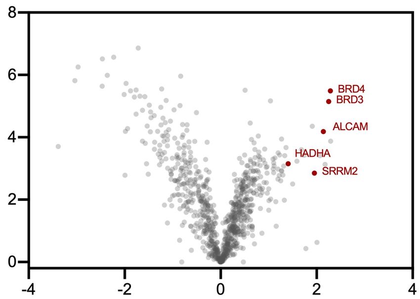
bioRxiv preprint doi: https://doi.org/10.1101/2021.08.02.454797; this version posted August 2, 2021. The copyright holder for this preprint
(which was not certified by peer review) is the author/funder, who has granted bioRxiv a license to display the preprint in perpetuity. It is made
available under aCC-BY-NC-ND 4.0 International license.
a. Small molecule target ID in phenotypic screeing-based drug discovery b. Photocatalytic target ID enabled by µMapping
Small molecule Phenotype State-of-the-art PAL
cell death, carbene N N
drug candidate
proliferation etc. UV light
F3C t1/2 ~ 4 ns F3C
tBuO N Dexter
N energy
O = affinity transfer
handle
N N Unknown protein F3C
Cl gene F
N interaction O
N
Stoichiometric
Me Me diazirine N
S N
probe H N
Me Ir F
N F
>99% insertion into water
Me N
poor signal-to-noise
F
F3C
time consuming
and costly µMap target ID
Target Identification multiple signal
Photoaffinity One labelling event
leads to higher success possible per probe labelling
labelling (PAL) amplification
rate in clinic events
blue light
c. Development of intracellular µMap catalyst suitable for small molecule target ID d. µMap photocatalyst cell permeability
Geri et al. Science (2020) This work: Halotag cell permeability assay (HEK293T)
Carboxylic acid
CO2
conjugation handle
M G2-
SO
F3C CF3
M
Ir-G1-hexyl-Cl Ir-G2-hexyl-Cl
e
Ir-
D
F F CO2H
Me Me Cell permeable
N N
Probe (µM) – 10 5 10 5 10
MeO TAMRA-Cl (µM) 5 5 5 5 5 5
N
F F
N Commercially
Ir Ir
Me N F F N available TOM20-
Me halotag
N Photocatalyst N Me
O SAR development No SAR required anti-TAMRA
F F PF6
F3C CF3 GAPDH
CO2H Multiple derivatives
anti-GAPDH
prepared (Ir-PEG3-
Gen 1 photocatalyst Gen 2 photocatalyst NH2, Ir-DBCO, Ir-
Halotag chaser assay reveals only Ir-G2
extracellular µMap intracellular µMap PEG3-CO2H) catalyst is cell permeable
antibody labelling small-molecule target ID
Scheme 1. Photoaffinity labelling comprises a critical component of small molecule target ID. a, Target ID campaigns are critical for the development
of successful drugs, though often rely on challenging photoaffinity labelling campaigns that employ the stoichiometric activation of diazirine small-
molecule conjugates with UV light. b, Our approach separates the warhead from the small molecule probe, instead employing the photocatalytic activation
of diazirines using visible light, giving rise to significant signal enhancement. c, Development of cell-penetrating, generation 2 photocatalyst suitable for
small-molecule conjugation and target ID. d, Cell permeability of Ir-photocatalysts determined by halotag chaser assay; photocatalyst PEG-hexyl chloride
conjugate and TAMRA hexyl chloride incubated with HEK293T cells expressing TOM20-halotag. Western blot analysis and immunostaining TOM20
with anti-TAMRA reveals off-compete only in the presence of Ir-G2 catalyst.
While these methods have been extremely empowering for a number of protein classes16–18, practically they
remain capricious and typically struggle to determine the complete interactome due to low receptor and protein
abundance and short half-lives, leading to low cross-linking yields and high background15,19. The use of diazirine-
based probes in particular has been challenging in this context as >99% of the carbenes generated upon UV irradi-
ation react with water and not the target20. These spent probes serve to further block the binding of unreacted mol-
ecules, further hampering labelling efficiency. As a result, costly and time-consuming structural optimization cam-
paigns are often required to overcome these shortfalls.
Indeed, the inherent difficulties associated with PAL have inspired the development of several elegant methods
that hinge upon the use of stoichiometric activated electrophiles12,21–24, single-electron transfer events25, or specific
oxidizable residues26 to identify target proteins. However, many of these technologies are limited to a single label-
ling event per drug molecule, often require extensive structural optimization (linker length and composition), and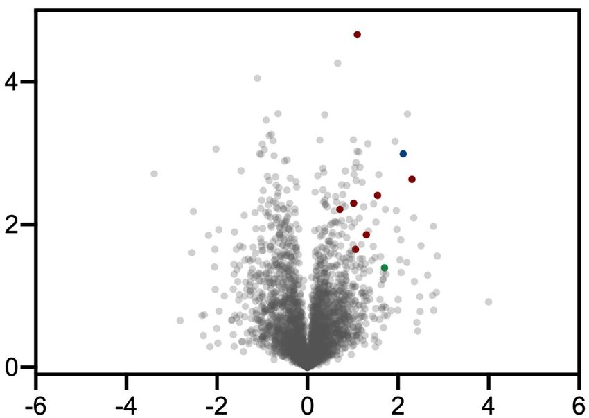
bioRxiv preprint doi: https://doi.org/10.1101/2021.08.02.454797; this version posted August 2, 2021. The copyright holder for this preprint
(which was not certified by peer review) is the author/funder, who has granted bioRxiv a license to display the preprint in perpetuity. It is made
available under aCC-BY-NC-ND 4.0 International license.
(+)-JQ1-Gen 2-Ir (1) Labelling of recombinant CA & BRD4 Intracellular labelling in HeLa cells Scheme 2. Development of photo-
Me targets BDR4 Biotin-PEG3-Dz
catalytic target ID platform for in-
a Biotin-PEG3-Dz
Me (250 µM)
450 nm (15 min) b (250 µM)
450 nm (15 min)
teractome mapping of (+)-JQ1 in
S
Me Cl Ir-NHBoc (1.3 µM) + – – HeLa cells. Structure of JQ1-based
N (+)-JQ1-G1 (5 µM) – + –
N F3C
(+)-JQ1-G2 (1.3 µM) – + – photocatalyst conjugate and state-of-
N F PF6 (+)-JQ1-G2 (5 µM) + – –
N (–)-JQ1-G2 (1.3 µM) – – + the-art PAL probe. a, Labelling of
O Me
N Steptavidin
BRD4 recombinant BRD4 protein vs. spec-
(pull down)
NH N
F
tator protein carbonic anhydrase us-
3 Ir
F Total protein
H3 ing free iridium-, (+)-JQ1-, and (–)-
(inputs)
O
H
N JQ1-probe by immunostaining with
N N
streptavidin. b, Comparing permea-
No labelling with inactive (–)-JQ1 enantiomer No labelling with (+)-JQ1-G1-Ir catalyst
O F3C
F bility of G1 and G2-based (+)-JQ1
probes following irradiation in HeLa
Time dependent labelling of BRD4 in HeLa cells (+)-JQ1-G2-Ir (1) vs. Ir-NHBoc (free-Ir) cells, streptavidin bead enrichment,
HeLa cells
c
Biotin-PEG3-Dz 450 nm 450 nm 450 nm d and immunostaining with anti-
(250 µM) (2 min) (5 min) (15 min)
BRD4. c, BRD4 labelling increases
over time (2 min, 5 min, and 15 min
–Log10 (p-value)
(+)-JQ1-G2 (5 µM) + – + – + – –
Ir-NHBoc (5 µM) – + – + – + – irradiation) through photocatalytic
signal amplification using (+)-JQ1-
BRD4
(pull down) G2 probe in HeLa cells following
streptavidin bead enrichment and
H3
(inputs) immunostaining with anti-BRD4. d,
TMT-based quantitative chemopro-
teomic analysis of JQ1-labelling in
Labelling increases with time due to signal amplification
HeLa cells comparing intracellular
Conclusive target ID using µMap by chemoproteomics
labelling by (+)-JQ1-G2 catalysts
Log2 (fold change) and unconjugated iridium catalyst
(control) reveals BRD proteins as
(+)-JQ1-G2-Ir (1) vs. Ir-NHBoc + (+)-JQ1 (+)-JQ1-G2 (1) vs. (–)-JQ1-G2 (inactive) highly enriched in addition to known
e HeLa cells HeLa cells
f JQ1 off-targets. e, TMT-based quan-
titative chemoproteomic analysis of
–Log10 (p-value)
JQ1-labelling in HeLa cells compar-
–Log10 (p-value)
BRD2 ing intracellular labelling by (+)-
BRD3 JQ1-G2 catalysts and unconjugated
iridium catalyst + (+)-JQ1 (control).
f, TMT-based quantitative chemo-
proteomic analysis of JQ1-labelling
in HeLa cells comparing intracellu-
lar labelling between active (+)-JQ1-
G2 and inactive (–)-JQ1-G2 cata-
lysts reveals only (+)-isomer labels
Log2 (fold change) BRD proteins. g, State-of-the-art
Log2 (fold change)
PAL employing active (+)-JQ1- and
State-of-the-art UV photoaffinity labelling using JQ1-Dz-alkyne (+)-JQ1-Dz (2) vs. (–)-JQ1-Dz (inactive) inactive (–)-JQ1-Dz-alkyne probes
State-of-the-art PAL in HeLa cells h
HeLa cells reveals no selective enrichment of
Me
UV (20 min)
BRD4 by western blotting despite
S Me broad biotinylation visible by im-
–Log10 (p-value)
Me (+)-JQ1-Dz-alkyne (µM)
Cl 5 – munostaining with streptavidin. h,
N
N N
(–)-JQ1-Dz-alkyne (µM) – 5 TMT-based quantitative chemopro-
N
g
BRD4
(pull down)
teomic analysis in HeLa cells com-
O
paring state-of-the-art PAL employ-
HN ing active (+)-JQ1- and inactive (–)-
N N JQ1-Dz-alkyne probes reveals no
Steptavidin
(pull down)
enrichment of BRD proteins.
(+)-JQ1-Dz-alkyne (2) BRD4
State-of-the-art
photoaffinity probe
Log2 (fold change)
No conclusive target ID by quantitative chemoproteomics using state-of-the-art photoaffinity labelling
require low-yielding downstream ‘click’ processing19. Moreover, intracellular labelling technologies are often ham-
pered by low cell-permeability leading to high background signal5. We therefore reasoned that the development of
a catalytic target ID technology that separated the drug molecule from the reactive warhead could overcome these
challenges through multiple labelling events leading to signal amplification (Scheme 1b).
We recently disclosed a novel antibody-based proximity labelling platform for cell surface microenvironment
elucidation, termed µMap27. This method relies upon the activation of diazirine molecules in close proximity to a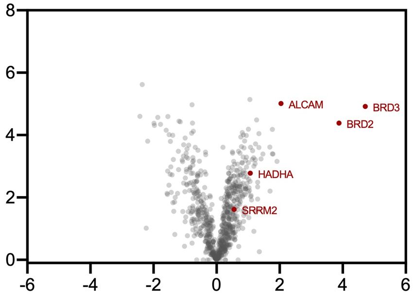
bioRxiv preprint doi: https://doi.org/10.1101/2021.08.02.454797; this version posted August 2, 2021. The copyright holder for this preprint
(which was not certified by peer review) is the author/funder, who has granted bioRxiv a license to display the preprint in perpetuity. It is made
                                            available under aCC-BY-NC-ND 4.0 International license.

set of photocatalysts appended to an antibody via Dexter energy transfer. Inspired by this unique activation mode,
we questioned whether such a tactic could be leveraged for small molecule target ID through the incorporation of a
photocatalyst onto a bioactive small molecule. However, at the outset of the investigation, we were cognizant of
several challenges inherent in developing such a technology, such as catalyst cell permeability and biocompatibility,
ease of chemical manipulation, retention of biological activity, and labelling efficiency (given each antibody con-
tained an average of 6–8 photocatalysts). However, we reasoned that by ‘switching on’ catalysis through visible
light activation, labelling could be controlled both spatially and temporally, bypassing intrinsic reactivity problems
and enabling the identification of novel targets across numerous drug discovery programs.
    We began by investigating cell permeability: employing a halotag-based chaser assay off-competing a TAMRA
dye in HEK293T cells, we identified that our previous catalyst design (Gen 1) was impermeable by virtue of its
neutral net charge and two carboxylic acid residues (Scheme 1d). Through screening different photocatalyst struc-
tures, we realized that Ir-catalysts containing both the dFCF3-phenyl pyridine moiety and 4,4-dialkyl bpy ligand
were crucial in achieving the necessary triplet energy27. Pleasingly, by removing the carboxylic acid groups, the
cationic photocatalyst (Gen 2) was rendered cell permeable (Scheme 1d). With this in mind, we evaluated conjuga-
tion handles based around the 4,4-dMebpy ligand, opting for a distal carboxylic acid to enable facile amide coupling.
Importantly, our G2-iridium catalyst can be accessed on gram-scale and be readily conjugated to a range of linkers
and complex small molecules (vide infra).
    Confident in our ability to access almost any Ir-drug conjugate, we initiated our target ID campaign with the
validated epigenetic tool compound (+)-JQ128. A potent inhibitor of the BET family of bromodomain proteins
(BRD2/3/4), several JQ1 structural analogues are in clinical trials for a variety of cancers including NUT midline
carcinoma29. We prepared the corresponding (+)-JQ1-G2 conjugate (1) (Scheme 2) and validated target engagement
in vitro with recombinant BRD4 in a competition assay vs. bovine carbonic anhydrase (CA). An equimolar amount
of CA and BRD4 was treated with (+)-JQ1-G2 probe (1) and an excess of diazirine-PEG3-biotin prior to irradiation
at 450 nm. Labelling intensity was measured by western blotting with a streptavidin stain. Pleasingly, these prelim-
inary experiments revealed a 20-fold increase in labelling for BRD4 vs. CA compared to the unconjugated (free)
photocatalyst (Scheme 2a). Importantly, the (–)-JQ1-G2 conjugate, which is known to not bind BRD428, showed
significantly reduced labelling, demonstrating that labelling is as a result of a ligand/protein binding event (Scheme
2c, A). In addition, we were able to confirm this through microscale thermophoresis (MST), where the addition of
the Ir-catalyst made only a minor impact on the binding constant (Figure S1).
    Based on these results, we sought to apply this method to live cells. We treated HeLa cells with 5 µM (+)-JQ1-
Gen 2 (1) for 3h before the addition of 250 µM Dz-PEG3-Biotin and subsequent 15 min irradiation (450 nm).
Following lysis and streptavidin-bead enrichment, western blot analysis with anti-BRD4 showed a clear labelling
of the target protein compared to DMSO control (Figure S2). In line with previous findings, the corresponding (+)-
JQ1-Gen 1 catalyst, while demonstrating similar in vitro labelling capability, showed no such enrichment of the
target protein in cells (Scheme 2b). Consistent with our hypothesis, the intensity of labelling was found to be linearly
related to irradiation time, demonstrating the photocatalytic signal amplification and temporal control offered by
the µMap platform (Scheme 2c). This was also observable by confocal microscopy, wherein the degree of biotinyl-
ation imparted by increased significantly over time (Figure S3). Encouraged by our western blot validation data,
we moved to TMT-based quantitative chemoproteomics in order to more completely assess the interactome of (+)-
JQ1. To our delight, by comparing the labelling by (+)-JQ1-Gen 2 (1) vs. unconjugated (free) photocatalyst in HeLa
cells, we observed several BRD proteins as the most enriched, although the precise identity of which however
remains difficult to ascertain due to structural homology (Scheme 2d). We also identified two previously annotated
(+)-JQ1 off-targets, HADHA30 and SRRM231. ALCAM (CD166), a transmembrane glycoprotein, was also identi-
fied as being significantly enriched, but currently has no reported interaction with (+)-JQ1. CD166 exerts a pro-
bioRxiv preprint doi: https://doi.org/10.1101/2021.08.02.454797; this version posted August 2, 2021. The copyright holder for this preprint
(which was not certified by peer review) is the author/funder, who has granted bioRxiv a license to display the preprint in perpetuity. It is made
available under aCC-BY-NC-ND 4.0 International license.
carcinogenic role via the inhibition of transcription factors along the FOXO/AKT axis and is considered a novel
therapeutic target for liver cancer32. Interestingly, BET inhibition by (+)-JQ1 has been shown to upregulate expres-
sion of FOXO1, although the mechanism remains unclear33. In order to evaluate whether protein enrichment was
as a direct result of labelling or up-regulation by virtue of the presence of (+)-JQ1, we repeated the experiment with
Kinase µMap target ID using dasatanib-G2-iridium conjugate
desHEP-Dasatinib-PEG5-Gen 2-Ir (3)
Intracellular labelling of p38 using desHEP-Das-PEG5-G2 (3) vs. Ir-G2-NHEt
F3C PF6
Src/Abl tyrosine kinase inhibitor F desHEP-Das-PEG5-G2 in THP1 cells THP1 cells MYLK Kinase
Me
O
Me
N
b Lysosomal
H 5000
S N N N Off target
N O F a
H Ir
N
–Log10 (p-value)
Cl N N F
N 4000 ABCC1
average densitometry
Me 5
HN N p38α
SRPK1
Dasatinib-Gen 2-Ir (4) Src
3000 LYN
O F
full length conjugate F3C MARK3
Me
FN3KRP
2000 CTSS
Cl N N N
H
N
S N N Biological
1000
O N activity retained
Me
CF3 PF6
F O Conclusive 0
Me µMap target ID das-G2 das-G2 + free Ir
N
(10 µM) das (10X) (20 µM)
N O Log2 (fold change)
F O
Ir
F
N Dasatinib-G2 (4) ± Dasatinib (off-compete) Dasatinib-Dz (5) ± Dasatinib (off-compete)
N NH K562 cells Me K562 cells
d Kinase
F Lysosomal e N N N Me Dasatinib-Dz-
O H
CF3 alkyne (5),
Off target N
N N S PAL probe
H
–Log10 (p-value)
N O
–Log10 (p-value)
Dasatinib-G2 (4) functional assay Cl
CSK
FECH Myt1 O
c 18 h DMSO Das Das-G2
p38α N N
incubation LAT3 CTSD O N
probe (µM) H
5 5 5
MAPK1
BTK BTK
phospho-p38
CTSD
CSK
phospho-Abl
GAPDH
Log2 (fold change) Log2 (fold change)
µMap target ID of microtubules using paclitaxel-G2-iridium conjugate
Paclitaxel-G2 vs Ir(dFCF3)(dMebpy)+
Paclitaxel-Gen 2-Iridium conjugate (6) Intracellular labelling in MCF7 cells
MCF7 cells
targets tubulin F3C
F PF6 f Biotin-PEG3-Dz 450 nm irradiation g
(250 µM) (20 min)
Me
O N
–Log10 (p-value)
N paclitax.-G2 (20 µm) + – –
O Me O F
Bz Me H Ir Ir-NHBoc (20 µM) – + –
NH O O N O F
MeO 3
N
Me
Ph O N α-tubulin
Me (pull down)
OH
F
HO F3C
BzO O O α-tubulin
(inputs)
Me O
µMap labelling in MCF7 cells by paclitaxel-G2 catalyst reveals enriched α- and β-tubulin isoforms
Log2 (fold change)
Scheme 3. Intracellular photocatalytic target ID and interactome mapping of dasatinib and paclitaxel. a, Enrichment of p38 by western blot for labelling using
desHEP-dasatinib-PEG5-G2 labelling in THP1 cells. b, Label free proteomic analysis in THP1 cells comparing intracellular labelling by desHEP-dasatinib-PEG5-G2
catalyst vs. Ir-G2-NHEt reveals enrichment of several kinases (red), as well as lysosomal proteins (green) and off-targets (blue). c, Kinase activity assays reveals
dasatinib-G2 retains inhibition activity against Abl and p38, as well as general tyrosine phosphorylation, in K562 cells. d, TMT-based quantitative chemoproteomic
analysis in K562 cells comparing intracellular labelling by dasatinib-G2 catalyst vs. dasatinib-G2 + dasatinib (off-compete control) reveals enrichment of several
kinases (red), as well as lysosomal proteins (green) and established off-targets (blue). e, TMT-based quantitative chemoproteomic analysis in K562 cells comparing
intracellular labelling by dasatinib-Dz-alkyne (PAL probe) vs. off-compete control does not reveal enrichment of kinases suitable for conclusive target ID. f, Initial
western blot studies for paclitaxel-G2 labelling in MCF7 cells following irradiation and streptavidin bead enrichment reveals significant enrichment of a-tubulin by
immunostaining compared to unconjugated iridium and DMSO controls. g, TMT-based quantitative chemoproteomic analysis in MCF7 cells comparing intracellular
labelling by paclitaxel-G2 catalyst and unconjugated iridium catalyst (control) reveals enrichment of several tubulin isoforms.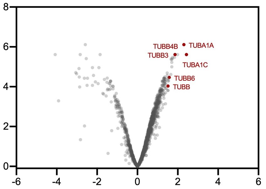
bioRxiv preprint doi: https://doi.org/10.1101/2021.08.02.454797; this version posted August 2, 2021. The copyright holder for this preprint
(which was not certified by peer review) is the author/funder, who has granted bioRxiv a license to display the preprint in perpetuity. It is made
                                            available under aCC-BY-NC-ND 4.0 International license.

an equivalent of (+)-JQ1 in the free iridium control (Scheme 2e). Upon chemoproteomic analysis we found that
CD166 was similarly enriched, indicating that it may be a putative off-target binder of (+)-JQ1, although further
biological validation is required. We further compared the interactomes of the enantiomers of JQ1-G2 and found
the active (+) enantiomer, (1), delivered BRD2/3/4 as top hits, and while CD166 was detected it was not enriched,
indicating that binding may not be affected by the stereogenic center (Scheme 2e). In contrast to these data, the
same analysis using classical UV-based PAL employing (+)-JQ-Dz-alkyne (2)34, in our hands, did not lead to en-
richment of BRD proteins by western blot (Scheme 2g) or chemoproteomic analysis (Scheme 2h).
    The dual Src/Abl tryrosine kinase inhibitor dasatinib displays significant antileukemic effects against various
imatinib-resistant mutants35. However, despite well-documented BCR/ABL inhibition, its precise downstream cel-
lular MOA remains to be fully understood. While the dasatinib interactome has been previously characterized16,
most methods have been performed with recombinant protein or in cell-lysate; live cell data is typically restricted
to kinase-based assays that measure downstream phosphorylation or residence at engineered kinase constructs,
which can be challenging to deconvolute and fail to identify non-kinase based off targets36–39.
    As previous studies have demonstrated difficulties in maintaining potency and cell-permeability using dasatinib-
derived probes16, we started by synthesizing three truncated (desHydroxyEthylPiperazinyl)-dasatinib iridium con-
jugates using our cell-permeable Ir-G2 catalyst with varying PEG linker lengths (n = 3–5) (3) (Scheme 3, top).
Gratifyingly, upon subjection of the desHEP-dasatinib-G2 conjugates (3) (5 µM) to our standard µMap protocol,
all of the conjugates revealed enrichment of p38 (MAP kinase) by western blot analysis compared to off-compete
(4X dasatinib) controls in THP1 cells (Figure S4). As the corresponding PEG5-G2 conjugate showed the greatest
enrichment (3.5X enrichment vs. off-compete and 9.5X enrichment vs. free-Ir) (Scheme 3a), we undertook label
free proteomic analysis of these reactions revealing significant enrichment of p38a (Figure S5), which has been
shown to play a critical role in its antileukemic properties40, as well as several other established kinase interactors
including Src and Lyn (Scheme 3b)41. Furthermore, we identified multidrug resistance transporter ABCC1 amongst
the most enriched proteins – an important off target; understanding the interaction between drug molecules and
efflux transporters is an important consideration in many drug discovery efforts42. Lysosomal sequestration of da-
satinib43, due to its lipophilic and weakly basic properties, was evident by the presence of cathepsin S (CTSS)
amongst the most enriched proteins. Encouraged by these initial results, we turned our attention to the underex-
plored full dasatinib-PEG3-G2 catalyst (4), which retains the 2-hydroxyethylpiperazine tail. Importantly, we found
a similar kinase inhibition profile against p38, in addition to Abl, by evaluation of downstream phosphorylation in
Ph+ K562 cells, compared to the parent drug, again highlighting the compatibility of the iridium photocatalyst
towards maintaining biological function and cell permeability (Scheme 3c) (Figure S6). Gratifyingly, subjection of
our µMap labelling to TMT-based chemoproteomics revealed extensive enrichment of p38a as well as Myt1 and
CSK kinases, both well-established binders of dasatinib (Scheme 3d)39. Moreover, known kinase off target fer-
rochelatase (FECH)44 was also significantly enriched, alongside large amino acid transporter (LAT3)39. Similarly,
lysosomal protein cathepsin D (CTSD) was amongst the most enriched proteins. Notably, in our hands, state-of-
the-art photoaffinity labelling, employing dasatinib-diazirine-alkyne (5), revealed only trace enrichment of CSK
and the kinases BTK and MAPK1 (BTK was found to be similarly enriched by µMap) (Scheme 3e).
    The anti-cancer properties of the natural product paclitaxel (Taxol) have been proposed to be derived from bind-
ing to microtubules, leading to stabilization and mitotic arrest; however, the full extent of its mechanism remains
unclear45. Based on its widespread use and intriguing mechanism, we prepared the corresponding paclitaxel-Gen 2-
iridium conjugate (6) (Scheme 3, bottom) and assessed its cellular activity. Through a series of cell proliferation
assays we found that our paclitaxel-G2 conjugate displayed similar anti-proliferative properties as the native com-
pound, suggesting that the pendent Ir-catalyst did not disrupt the native function of paclitaxel (Figure S7). Encour-
aged by this, we proceeded to study the efficiency of labelling in the breast cancer cell line MCF7. Following our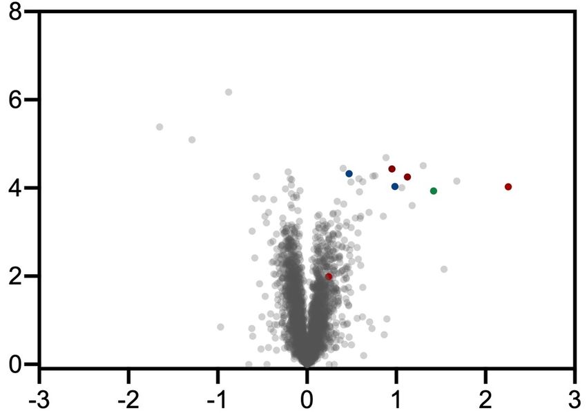
bioRxiv preprint doi: https://doi.org/10.1101/2021.08.02.454797; this version posted August 2, 2021. The copyright holder for this preprint
(which was not certified by peer review) is the author/funder, who has granted bioRxiv a license to display the preprint in perpetuity. It is made
available under aCC-BY-NC-ND 4.0 International license.
standard µMap protocol with 20 µM paclitaxel-G2 conjugate (6) for 3h, western blot analysis with anti-a-tubulin
showed clear labelling of the target protein compared both the free iridium and DMSO controls (Scheme 3f). Sub-
jection of our µMap labelling to TMT-based chemoproteomics revealed extensive labelling of tubulin isotypes aIa,
bIII, bIVb, and aIc (Scheme 3g), which is in good agreement with previous photoaffinity labelling studies on ex-
tracted tubulin46.
Having established the efficacy of µMap target ID for intracellular proteins, we turned our attention to the cell
surface. The exceedingly low abundance, lack of exposed residues, and aggregation-prone hydrophobic domains
oftentimes confounds the detection and manipulation of membrane proteins, rendering target ID unfeasible19,47,48.
These challenges are exacerbated when combined with the high background labelling, poor sensitivity, and low
cross-linking yields systemic in PAL campaigns. We therefore felt that our µMap target ID platform was ideally
placed to tackle these challenges by virtue of our catalytic signal amplification. We chose the adenosine receptor
A2a (ADORA2A) as an exemplar membrane target. This GPCR has become an important target for immunother-
apy49, but critically, has never been identified through live cell chemoproteomics50,51. Using a reported ligand for
ADORA2A that binds from the extracellular face, SCH5826152, we prepared both a tethered diazirine-conjugate,
SCH58261-Dz (7), and an Ir-conjugate based on the more hydrophilic G1 catalyst, SCH58261-G1 (8); the low cell
Cell surface µMap target ID of GPCR ADORA2A using SCH58261-G1-iridium conjugate
SCH58261-Dz-alkyne (7) SCH58261-Dz (7) ± SCH58261 (off-compete) State-of-
State-of-the-art photoaffinity probe b A2a-expressing HEK293T cells the-art
N
N
N PAL
O N N
N N H
N N
–Log10 (p-value)
No conclusive
H 2N O
target ID of
ADORA2A using
SCH58261-Gen 1-Iridum conjugate (8) CO2 diazirine probe
targets A2A (ADORA2A) F3C (over-expressed)
F ADORA2A
Me Me
N N
NH N O
N
N Ir F
Me N F
O
Me
O N
N O OMe
F
N F3C Log2 (fold change)
O
CO2H
N
N SCH58261-G1 (8) ± SCH58261 (off-compete)
N
N hydrophilic catalyst for PC-12 cells Unequivocal
H 2N O cell-surface labelling c
µMap-target ID
of GPCR at
Membrane labelling in HEK293T cells ADORA2A
–Log10 (p-value)
native
Biotin-PEG3-Dz expression
SO
SO
SCH58261 SCH58261 SCH58261 Ir-alkyne
(250 µM)
M
M
-Dz-alkyne -Dz-biotin -G1-Iridium (free Ir)
D
D
probe (µM) – 1 1 1 1 – 1 1 1 1 Catalytic signal
SCH58261 (µM) – – 100 – 100 – – 100 – 100 amplification
kDa 49 overcomes low
concentration
38
a
cat
28
Streptavidin/anti-ADORA2A
Log2 (fold change)
Scheme 4. Extracellular photocatalytic target ID of GPCR ADORA2A using SCH58261 at native expression. a, Structure of SCH58261-based Dz-
alkyne probe for PAL labelling and iridium conjugate based on hydrophilic G1 photocatalyst. b, Initial western blot studies for SCH58261-G1 labelling in
A2a-expressing HEK293T cells following irradiation and streptavidin bead enrichment reveals significant biotinylation by immunostaining compared to un-
conjugated iridium and DMSO controls, in addition to PAL labelling. c, TMT-based quantitative chemoproteomic analysis in A2a-expressing HEK293T cells
comparing extracellular labelling by SCH58261-Dz-alkyne vs. SCH58261-Dz-alkyne + SCH58261 (off-compete control) reveals inconclusive target ID of
ADORA2A. d, TMT-based quantitative chemoproteomic analysis in PC-12 cells comparing extracellular labelling by SCH58261-G1 catalyst vs. SCH58261-
G1 + SCH58261 (off-compete control) reveals significant enrichment of ADORA2A.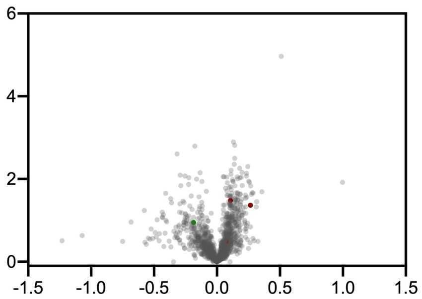
bioRxiv preprint doi: https://doi.org/10.1101/2021.08.02.454797; this version posted August 2, 2021. The copyright holder for this preprint
(which was not certified by peer review) is the author/funder, who has granted bioRxiv a license to display the preprint in perpetuity. It is made
                                            available under aCC-BY-NC-ND 4.0 International license.

permeability affording a higher effective concentration of photocatalyst probe on the cell surface (Scheme 4a).
Photocatalytic labelling applied to A2a-expressing HEK293T cells, followed by western blot visualization, revealed
a stark difference in labelling between the SCH58261-G1 (8) and the corresponding off-compete controls (Scheme
4b). Tandem mass tag (TMT)-based chemoproteomic analysis of these reactions confirmed our initial result, with
our photocatalytic-labelling method using SCH58261-G1 (8) showing a 10-fold enrichment for ADORA2A with
respect to off-competing with the parent SCH58261 ligand, and >20-fold enrichment versus free-Ir photocatalyst
(Figure S8a). In contrast, PAL using SCH58261-Dz (7), showed poor enrichment of ADORA2A by quantitative
chemoproteomics (Scheme 4c), in line with western blot data (Figure S8b). Based on the degree of enrichment in
A2a-expressing HEK293T cells, we were keen to ascertain how the µMap target ID platform performed at native
levels of membrane protein concentration, wherein classical PAL remains extremely challenging. Remarkably, pho-
tocatalytic labelling using SCH58261-G1 (8) in PC-12 cells, which have previously been validated to natively ex-
press A2a53, revealed similarly high levels of enrichment for the target protein ADORA2A – highlighting the signal
amplification conferred by the µMap platform (Scheme 4d). Finally, we further validated this labelling technology
by identifying the long chain fatty acid receptor GPR40, an important anti-diabetic therapeutic target, using the
small molecule probe MK-8666, further expanding the repertoire of µMap membrane target ID (Figure S9–S12)54.
     In conclusion, we describe a general platform for photocatalytic target ID that utilizes cell-penetrating iridium
conjugated-small molecules, which can bind protein targets, to locally activate proximal diazirines via Dexter en-
ergy transfer. The catalytic signal amplification conferred by µMap target ID has allowed for the identification of
multiple protein targets and off targets across multiple drug classes and cellular compartments where established
PAL have not been successful. As such, we envision that µMap target ID will find immediate use in providing a
deeper biological understanding of efficacy target networks, quickly revealing off-target pharmacology, and ulti-
mately driving pharmacotherapy forward against novel targets within drug discovery programmes in both academic
and industrial settings.

Acknowledgments.
Research reported in this publication was supported by the NIH National Institute of General Medical Sciences
(R01- GM103558-03) and gifts from Merck & Co., Inc., Kenilworth, New Jersey, USA. ADT would like to thank
the European Union’s Horizon 2020 research and innovation programme under Marie Sklodowska-Curie Grant
Agreement No.891458. JBG acknowledges the NIH for a postdoctoral fellowship (F32-GM133133-01). JVO
acknowledges the National Science Foundation Graduate Research Fellowship Programme under Grant No. (DGE-
1656466). Any opinions, findings, and conclusions or recommendations expressed in this material are those of the
authors and do not necessarily reflect the views of the National Science Foundation. The authors thank Saw Kyin
and Henry H. Shwe at the Princeton Proteomics Facility. The authors thank Brande Thomas-Fowlkes and Xiaoping
Zhang (MRL, Merck & Co., Inc, Kenilworth, NJ, USA), for providing GPR40-HEK cells and running IP-1 assay,
respectively. HEK-hA2aR cell line was received as a gift from Jeremy Presland (MRL, Merck & Co., Inc, Boston,
MA, USA). We acknowledge the use of Princeton’s Imaging and Analysis Center, which is partially supported by
the Princeton Center for Complex Materials, a National Science Foundation/Materials Research Science and Engi-
neering Centers programme (DMR-1420541). We also acknowledge V. G. Vendavasi and the use of Princeton’s
Biophysics Core Facility. We thank Antony Burton for assistance in performing confocal microscopy. We thank T.
W. Muir and members of the Muir Laboratory for their advice and analytical support.

Author Affiliations and Contributions
1
  Merck Center for Catalysis at Princeton University, Princeton, NJ 08544, USA.
2
  Discovery Chemistry, Merck & Co., Inc, Kenilworth NJ 07033, USA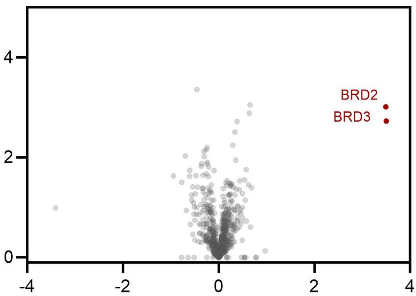
bioRxiv preprint doi: https://doi.org/10.1101/2021.08.02.454797; this version posted August 2, 2021. The copyright holder for this preprint
(which was not certified by peer review) is the author/funder, who has granted bioRxiv a license to display the preprint in perpetuity. It is made
                                            available under aCC-BY-NC-ND 4.0 International license.

3
  Department of Chemistry, Princeton University, Princeton, NJ 08544, USA
4
  Discovery Chemistry, Merck & Co., Inc., Boston, MA 02115, USA.
5
  Merck Exploratory Science Center, Merck & Co., Inc., Cambridge, MA 02141, USA.
6
  Pharmacology, Merck & Co., Inc., Kenilworth, NJ 07033, USA.

Author contributions: ADT, CPS, FPR-R, DLP, DWCM conceived the work. FPR-R, DLP, CPS, ADT, BXL,
BD, JBG, JVO, AGS, OOF, RCO designed and executed the experiments. PT, TR-R, KAR provided insight into
experimental design. ADT, CPS, FPR-R, DLP, DWCM prepared this manuscript. *These authors contributed
equally

Competing interest: A provisional U.S. patent has been filed by DWCM, ADT, CPS based in part on this work,
62/982,366; 63/076,658. International Application No. PCT/US2021/019959. DWCM declares an ownership inter-
est, and ADT and CPS declare an affiliation interest, in the company Dexterity Pharma LLC, which has commer-
cialized materials used in this work. DWCM declares an ownership interest in Penn PhD, which has commercialized
materials used in this work.

Correspondence: †Corresponding authors. Correspondence and requests for materials should be addressed to
D.W.C.M     (dmacmill@princeton.edu),    FPR-R     (frances.rodriguez-rivera@merck.com),    and     DLP
(dann_parker@merck.com).

References

(1)      Chan, J. N. Y.; Nislow, C.; Emili, A. Recent Advances and Method Development for Drug Target
         Identification. Trends Pharmacol. Sci. 2010, 31 (2), 82–88.
         https://doi.org/https://doi.org/10.1016/j.tips.2009.11.002.
(2)      Schenone, M.; Dančík, V.; Wagner, B. K.; Clemons, P. A. Target Identification and Mechanism of Action
         in Chemical Biology and Drug Discovery. Nat. Chem. Biol. 2013, 9 (4), 232–240.
         https://doi.org/10.1038/nchembio.1199.
(3)      Hart, C. P. Finding the Target after Screening the Phenotype. Drug Discov. Today 2005, 10 (7), 513–519.
         https://doi.org/https://doi.org/10.1016/S1359-6446(05)03415-X.
(4)      Williams, M. Target Validation. Curr. Opin. Pharmacol. 2003, 3 (5), 571–577.
         https://doi.org/https://doi.org/10.1016/j.coph.2003.06.001.
(5)      Ziegler, S.; Pries, V.; Hedberg, C.; Waldmann, H. Target Identification for Small Bioactive Molecules:
         Finding the Needle in the Haystack. Angew. Chemie Int. Ed. 2013, 52 (10), 2744–2792.
         https://doi.org/10.1002/anie.201208749.
(6)      Morgan, P.; Brown, D. G.; Lennard, S.; Anderton, M. J.; Barrett, J. C.; Eriksson, U.; Fidock, M.; Hamrén,
         B.; Johnson, A.; March, R. E.; Matcham, J.; Mettetal, J.; Nicholls, D. J.; Platz, S.; Rees, S.; Snowden, M.
         A.; Pangalos, M. N. Impact of a Five-Dimensional Framework on R&D Productivity at AstraZeneca. Nat.
         Rev. Drug Discov. 2018, 17 (3), 167–181. https://doi.org/10.1038/nrd.2017.244.
(7)      Schirle, M.; Bantscheff, M.; Kuster, B. Mass Spectrometry-Based Proteomics in Preclinical Drug
         Discovery. Chem. Biol. 2012, 19 (1), 72–84.
         https://doi.org/https://doi.org/10.1016/j.chembiol.2012.01.002.
(8)      Wagner, B. K.; Clemons, P. A. Connecting Synthetic Chemistry Decisions to Cell and Genome Biology
         Using Small-Molecule Phenotypic Profiling. Curr. Opin. Chem. Biol. 2009, 13 (5), 539–548.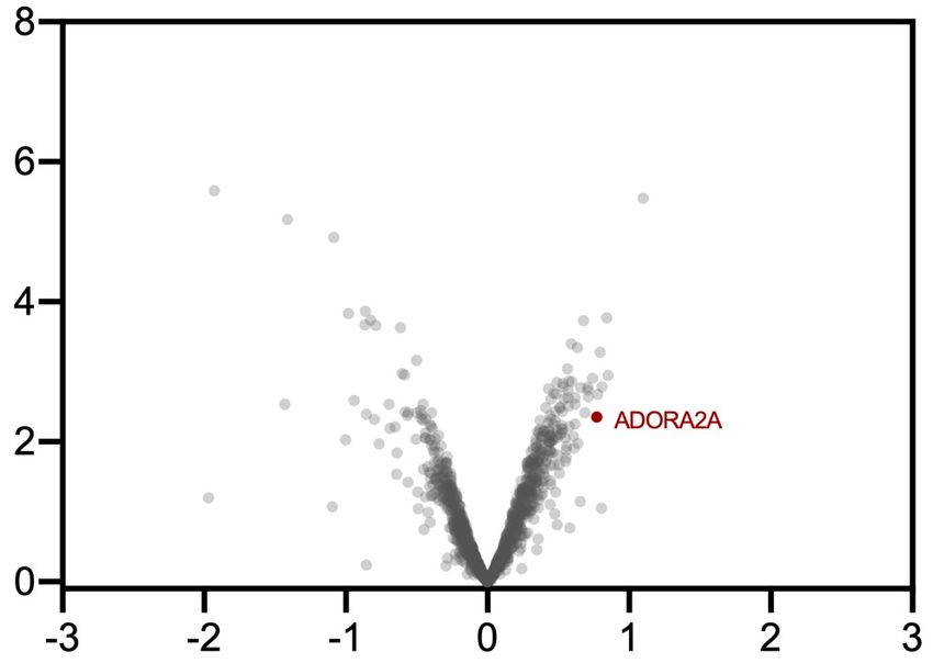
bioRxiv preprint doi: https://doi.org/10.1101/2021.08.02.454797; this version posted August 2, 2021. The copyright holder for this preprint
(which was not certified by peer review) is the author/funder, who has granted bioRxiv a license to display the preprint in perpetuity. It is made
                                            available under aCC-BY-NC-ND 4.0 International license.

         https://doi.org/https://doi.org/10.1016/j.cbpa.2009.09.018.
(9)      Butcher, E. C.; Berg, E. L.; Kunkel, E. J. Systems Biology in Drug Discovery. Nat. Biotechnol. 2004, 22
         (10), 1253–1259. https://doi.org/10.1038/nbt1017.
(10)     Schürmann, M.; Janning, P.; Ziegler, S.; Waldmann, H. Small-Molecule Target Engagement in Cells. Cell
         Chem. Biol. 2016, 23 (4), 435–441. https://doi.org/https://doi.org/10.1016/j.chembiol.2016.03.008.
(11)     Simon, G. M.; Niphakis, M. J.; Cravatt, B. F. Determining Target Engagement in Living Systems. Nat.
         Chem. Biol. 2013, 9 (4), 200–205. https://doi.org/10.1038/nchembio.1211.
(12)     Kawatani, M.; Osada, H. Affinity-Based Target Identification for Bioactive Small Molecules.
         Medchemcomm 2014, 5 (3), 277–287. https://doi.org/10.1039/C3MD00276D.
(13)     Hill, J. R.; Robertson, A. A. B. Fishing for Drug Targets: A Focus on Diazirine Photoaffinity Probe
         Synthesis. J. Med. Chem. 2018, 61 (16), 6945–6963. https://doi.org/10.1021/acs.jmedchem.7b01561.
(14)     Ge, S.-S.; Chen, B.; Wu, Y.-Y.; Long, Q.-S.; Zhao, Y.-L.; Wang, P.-Y.; Yang, S. Current Advances of
         Carbene-Mediated Photoaffinity Labeling in Medicinal Chemistry. RSC Adv. 2018, 8 (51), 29428–29454.
         https://doi.org/10.1039/C8RA03538E.
(15)     Smith, E.; Collins, I. Photoaffinity Labeling in Target- and Binding-Site Identification. Future Med. Chem.
         2015, 7 (2), 159–183. https://doi.org/10.4155/fmc.14.152.
(16)     Shi, H.; Zhang, C.-J.; Chen, G. Y. J.; Yao, S. Q. Cell-Based Proteome Profiling of Potential Dasatinib
         Targets by Use of Affinity-Based Probes. J. Am. Chem. Soc. 2012, 134 (6), 3001–3014.
         https://doi.org/10.1021/ja208518u.
(17)     Zuhl, A. M.; Nolan, C. E.; Brodney, M. A.; Niessen, S.; Atchison, K.; Houle, C.; Karanian, D. A.;
         Ambroise, C.; Brulet, J. W.; Beck, E. M.; Doran, S. D.; O’Neill, B. T.; am Ende, C. W.; Chang, C.;
         Geoghegan, K. F.; West, G. M.; Judkins, J. C.; Hou, X.; Riddell, D. R.; Johnson, D. S. Chemoproteomic
         Profiling Reveals That Cathepsin D Off-Target Activity Drives Ocular Toxicity of β-Secretase Inhibitors.
         Nat. Commun. 2016, 7 (1), 13042. https://doi.org/10.1038/ncomms13042.
(18)     Ito, T.; Ando, H.; Suzuki, T.; Ogura, T.; Hotta, K.; Imamura, Y.; Yamaguchi, Y.; Handa, H. Identification
         of a Primary Target of Thalidomide Teratogenicity. Science 2010, 327 (5971), 1345 LP – 1350.
         https://doi.org/10.1126/science.1177319.
(19)     Wright, M. H.; Sieber, S. A. Chemical Proteomics Approaches for Identifying the Cellular Targets of
         Natural Products. Nat. Prod. Rep. 2016, 33 (5), 681–708. https://doi.org/10.1039/C6NP00001K.
(20)     Park, J.; Koh, M.; Koo, J. Y.; Lee, S.; Park, S. B. Investigation of Specific Binding Proteins to
         Photoaffinity Linkers for Efficient Deconvolution of Target Protein. ACS Chem. Biol. 2016, 11 (1), 44–52.
         https://doi.org/10.1021/acschembio.5b00671.
(21)     Hayashi, T.; Hamachi, I. Traceless Affinity Labeling of Endogenous Proteins for Functional Analysis in
         Living Cells. Acc. Chem. Res. 2012, 45 (9), 1460–1469. https://doi.org/10.1021/ar200334r.
(22)     Yamaura, K.; Kiyonaka, S.; Numata, T.; Inoue, R.; Hamachi, I. Discovery of Allosteric Modulators for
         GABAA Receptors by Ligand-Directed Chemistry. Nat. Chem. Biol. 2016, 12 (10), 822–830.
         https://doi.org/10.1038/nchembio.2150.
(23)     Fujishima, S.; Yasui, R.; Miki, T.; Ojida, A.; Hamachi, I. Ligand-Directed Acyl Imidazole Chemistry for
         Labeling of Membrane-Bound Proteins on Live Cells. J. Am. Chem. Soc. 2012, 134 (9), 3961–3964.
         https://doi.org/10.1021/ja2108855.
(24)     Herner, A.; Marjanovic, J.; Lewandowski, T. M.; Marin, V.; Patterson, M.; Miesbauer, L.; Ready, D.;
         Williams, J.; Vasudevan, A.; Lin, Q. 2-Aryl-5-Carboxytetrazole as a New Photoaffinity Label for Drug
         Target Identification. J. Am. Chem. Soc. 2016, 138 (44), 14609–14615.
         https://doi.org/10.1021/jacs.6b06645.
bioRxiv preprint doi: https://doi.org/10.1101/2021.08.02.454797; this version posted August 2, 2021. The copyright holder for this preprint
(which was not certified by peer review) is the author/funder, who has granted bioRxiv a license to display the preprint in perpetuity. It is made
available under aCC-BY-NC-ND 4.0 International license.
(25) Sato, S.; Nakamura, H. Ligand-Directed Selective Protein Modification Based on Local Single-Electron-
Transfer Catalysis. Angew. Chemie Int. Ed. 2013, 52 (33), 8681–8684.
https://doi.org/10.1002/anie.201303831.
(26) Sato, S.; Tsushima, M.; Nakamura, H. Target-Protein-Selective Inactivation and Labelling Using an
Oxidative Catalyst. Org. Biomol. Chem. 2018, 16 (34), 6168–6179. https://doi.org/10.1039/C8OB01484A.
(27) Geri, J. B.; Oakley, J. V.; Reyes-Robles, T.; Wang, T.; McCarver, S. J.; White, C. H.; Rodriguez-Rivera,
F. P.; Parker, D. L.; Hett, E. C.; Fadeyi, O. O.; Oslund, R. C.; MacMillan, D. W. C. Microenvironment
Mapping via Dexter Energy Transfer on Immune Cells. Science 2020, 367 (6482), 1091–1097.
https://doi.org/10.1126/science.aaz5074.
(28) Filippakopoulos, P.; Qi, J.; Picaud, S.; Shen, Y.; Smith, W. B.; Fedorov, O.; Morse, E. M.; Keates, T.;
Hickman, T. T.; Felletar, I.; Philpott, M.; Munro, S.; McKeown, M. R.; Wang, Y.; Christie, A. L.; West,
N.; Cameron, M. J.; Schwartz, B.; Heightman, T. D.; La Thangue, N.; French, C. A.; Wiest, O.; Kung, A.
L.; Knapp, S.; Bradner, J. E. Selective Inhibition of BET Bromodomains. Nature 2010, 468 (7327), 1067–
1073. https://doi.org/10.1038/nature09504.
(29) Shorstova, T.; Foulkes, W. D.; Witcher, M. Achieving Clinical Success with BET Inhibitors as Anti-
Cancer Agents. Br. J. Cancer 2021, 124 (9), 1478–1490. https://doi.org/10.1038/s41416-021-01321-0.
(30) Kurzawa, N.; Becher, I.; Sridharan, S.; Franken, H.; Mateus, A.; Anders, S.; Bantscheff, M.; Huber, W.;
Savitski, M. M. A Computational Method for Detection of Ligand-Binding Proteins from Dose Range
Thermal Proteome Profiles. Nat. Commun. 2020, 11 (1), 5783. https://doi.org/10.1038/s41467-020-19529-
8.
(31) Tyler, D. S.; Vappiani, J.; Cañeque, T.; Lam, E. Y. N.; Ward, A.; Gilan, O.; Chan, Y.-C.; Hienzsch, A.;
Rutkowska, A.; Werner, T.; Wagner, A. J.; Lugo, D.; Gregory, R.; Ramirez Molina, C.; Garton, N.;
Wellaway, C. R.; Jackson, S.; MacPherson, L.; Figueiredo, M.; Stolzenburg, S.; Bell, C. C.; House, C.;
Dawson, S.-J.; Hawkins, E. D.; Drewes, G.; Prinjha, R. K.; Rodriguez, R.; Grandi, P.; Dawson, M. A.
Click Chemistry Enables Preclinical Evaluation of Targeted Epigenetic Therapies. Science 2017, eaal2066.
https://doi.org/10.1126/science.aal2066.
(32) Yu, W.; Wang, J.; Ma, L.; Tang, X.; Qiao, Y.; Pan, Q.; Yu, Y.; Sun, F. CD166 Plays a Pro-Carcinogenic
Role in Liver Cancer Cells via Inhibition of FOXO Proteins through AKT. Oncol Rep 2014, 32 (2), 677–
683. https://doi.org/10.3892/or.2014.3226.
(33) Tan, Y.; Wang, L.; Du, Y.; Liu, X.; Chen, Z.; Weng, X.; Guo, J.; Chen, H.; Wang, M.; Wang, X.
Inhibition of BRD4 Suppresses Tumor Growth in Prostate Cancer via the Enhancement of FOXO1
Expression. Int J Oncol 2018, 53 (6), 2503–2517. https://doi.org/10.3892/ijo.2018.4577.
(34) Li, Z.; Wang, D.; Li, L.; Pan, S.; Na, Z.; Tan, C. Y. J.; Yao, S. Q. “Minimalist” Cyclopropene-Containing
Photo-Cross-Linkers Suitable for Live-Cell Imaging and Affinity-Based Protein Labeling. J. Am. Chem.
Soc. 2014, 136 (28), 9990–9998. https://doi.org/10.1021/ja502780z.
(35) Shah, N. P.; Tran, C.; Lee, F. Y.; Chen, P.; Norris, D.; Sawyers, C. L. Overriding Imatinib Resistance with
a Novel ABL Kinase Inhibitor. Science 2004, 305 (5682), 399 LP – 401.
https://doi.org/10.1126/science.1099480.
(36) Karaman, M. W.; Herrgard, S.; Treiber, D. K.; Gallant, P.; Atteridge, C. E.; Campbell, B. T.; Chan, K. W.;
Ciceri, P.; Davis, M. I.; Edeen, P. T.; Faraoni, R.; Floyd, M.; Hunt, J. P.; Lockhart, D. J.; Milanov, Z. V;
Morrison, M. J.; Pallares, G.; Patel, H. K.; Pritchard, S.; Wodicka, L. M.; Zarrinkar, P. P. A Quantitative
Analysis of Kinase Inhibitor Selectivity. Nat. Biotechnol. 2008, 26 (1), 127–132.
https://doi.org/10.1038/nbt1358.
(37) Hantschel, O.; Rix, U.; Schmidt, U.; Bürckstümmer, T.; Kneidinger, M.; Schütze, G.; Colinge, J.; Bennett,
K. L.; Ellmeier, W.; Valent, P.; Superti-Furga, G. The Btk Tyrosine Kinase Is a Major Target of the Bcr-
Abl Inhibitor Dasatinib. Proc. Natl. Acad. Sci. 2007, 104 (33), 13283–13288.bioRxiv preprint doi: https://doi.org/10.1101/2021.08.02.454797; this version posted August 2, 2021. The copyright holder for this preprint
(which was not certified by peer review) is the author/funder, who has granted bioRxiv a license to display the preprint in perpetuity. It is made
available under aCC-BY-NC-ND 4.0 International license.
https://doi.org/10.1073/pnas.0702654104.
(38) Vasta, J. D.; Corona, C. R.; Wilkinson, J.; Zimprich, C. A.; Hartnett, J. R.; Ingold, M. R.; Zimmerman, K.;
Machleidt, T.; Kirkland, T. A.; Huwiler, K. G.; Ohana, R. F.; Slater, M.; Otto, P.; Cong, M.; Wells, C. I.;
Berger, B.-T.; Hanke, T.; Glas, C.; Ding, K.; Drewry, D. H.; Huber, K. V. M.; Willson, T. M.; Knapp, S.;
Müller, S.; Meisenheimer, P. L.; Fan, F.; Wood, K. V; Robers, M. B. Quantitative, Wide-Spectrum Kinase
Profiling in Live Cells for Assessing the Effect of Cellular ATP on Target Engagement. Cell Chem. Biol.
2018, 25 (2), 206-214.e11. https://doi.org/10.1016/j.chembiol.2017.10.010.
(39) Friedman Ohana, R.; Levin, S.; Hurst, R.; Rosenblatt, M. M.; Zimmerman, K.; Machleidt, T.; Wood, K. V;
Kirkland, T. A. Streamlined Target Deconvolution Approach Utilizing a Single Photoreactive
Chloroalkane Capture Tag. ACS Chem. Biol. 2021, 16 (2), 404–413.
https://doi.org/10.1021/acschembio.0c00987.
(40) Dumka, D.; Puri, P.; Carayol, N.; Lumby, C.; Balachandran, H.; Schuster, K.; Verma, A. K.; Terada, L. S.;
Platanias, L. C.; Parmar, S. Activation of the P38 Map Kinase Pathway Is Essential for the Antileukemic
Effects of Dasatinib. Leuk. Lymphoma 2009, 50 (12), 2017–2029.
https://doi.org/10.3109/10428190903147637.
(41) Williams, N. K.; Lucet, I. S.; Klinken, S. P.; Ingley, E.; Rossjohn, J. Crystal Structures of the Lyn Protein
Tyrosine Kinase Domain in Its Apo- and Inhibitor-Bound State. J. Biol. Chem. 2009, 284 (1), 284–291.
https://doi.org/https://doi.org/10.1074/jbc.M807850200.
(42) Gottesman, M. M.; Fojo, T.; Bates, S. E. Multidrug Resistance in Cancer: Role of ATP–Dependent
Transporters. Nat. Rev. Cancer 2002, 2 (1), 48–58. https://doi.org/10.1038/nrc706.
(43) Ruzickova, E.; Skoupa, N.; Dolezel, P.; Smith, D. A.; Mlejnek, P. The Lysosomal Sequestration of
Tyrosine Kinase Inhibitors and Drug Resistance. Biomolecules 2019, 9 (11), 675
https://doi.org/10.3390/biom9110675.
(44) Klaeger, S.; Gohlke, B.; Perrin, J.; Gupta, V.; Heinzlmeir, S.; Helm, D.; Qiao, H.; Bergamini, G.; Handa,
H.; Savitski, M. M.; Bantscheff, M.; Médard, G.; Preissner, R.; Kuster, B. Chemical Proteomics Reveals
Ferrochelatase as a Common Off-Target of Kinase Inhibitors. ACS Chem. Biol. 2016, 11 (5), 1245–1254.
https://doi.org/10.1021/acschembio.5b01063.
(45) Wang, T.-H.; Wang, H.-S.; Soong, Y.-K. Paclitaxel-Induced Cell Death. Cancer 2000, 88 (11), 2619–
2628. https://doi.org/10.1002/1097-0142(20000601)88:113.0.CO;2-J.
(46) Yang, C.-P. H.; Yap, E.-H.; Xiao, H.; Fiser, A.; Horwitz, S. B. 2-(m-Azidobenzoyl)Taxol Binds
Differentially to Distinct β-Tubulin Isotypes. Proc. Natl. Acad. Sci. 2016, 113 (40), 11294–11299.
https://doi.org/10.1073/pnas.1613286113.
(47) Comess, K. M.; McLoughlin, S. M.; Oyer, J. A.; Richardson, P. L.; Stöckmann, H.; Vasudevan, A.;
Warder, S. E. Emerging Approaches for the Identification of Protein Targets of Small Molecules - A
Practitioners’ Perspective. J. Med. Chem. 2018, 61 (19), 8504–8535.
https://doi.org/10.1021/acs.jmedchem.7b01921.
(48) Helbig, A. O.; Heck, A. J. R.; Slijper, M. Exploring the Membrane Proteome—Challenges and Analytical
Strategies. J. Proteomics 2010, 73 (5), 868–878. https://doi.org/https://doi.org/10.1016/j.jprot.2010.01.005.
(49) Cekic, C.; Linden, J. Adenosine A2A Receptors Intrinsically Regulate CD8+ T Cells in the Tumor
Microenvironment. Cancer Res. 2014, 74 (24), 7239–7249. https://doi.org/10.1158/0008-5472.CAN-13-
3581.
(50) Muranaka, H.; Momose, T.; Handa, C.; Ozawa, T. Photoaffinity Labeling of the Human A2A Adenosine
Receptor and Cross-Link Position Analysis by Mass Spectrometry. ACS Med. Chem. Lett. 2017, 8 (6),
660–665. https://doi.org/10.1021/acsmedchemlett.7b00138.
(51) Yang, X.; Michiels, T. J. M.; de Jong, C.; Soethoudt, M.; Dekker, N.; Gordon, E.; van der Stelt, M.;bioRxiv preprint doi: https://doi.org/10.1101/2021.08.02.454797; this version posted August 2, 2021. The copyright holder for this preprint
(which was not certified by peer review) is the author/funder, who has granted bioRxiv a license to display the preprint in perpetuity. It is made
available under aCC-BY-NC-ND 4.0 International license.
Heitman, L. H.; van der Es, D.; IJzerman, A. P. An Affinity-Based Probe for the Human Adenosine A2A
Receptor. J. Med. Chem. 2018, 61 (17), 7892–7901. https://doi.org/10.1021/acs.jmedchem.8b00860.
(52) Zocchi, C.; Ongini, E.; Conti, A.; Monopoli, A.; Negretti, A.; Baraldi, P. G.; Dionisotti, S. The Non-
Xanthine Heterocyclic Compound SCH 58261 Is a New Potent and Selective A2a Adenosine Receptor
Antagonist. J. Pharmacol. Exp. Ther. 1996, 276 (2), 398–404.
(53) Kudlacek, O.; Just, H.; Korkhov, V. M.; Vartian, N.; Klinger, M.; Pankevych, H.; Yang, Q.; Nanoff, C.;
Freissmuth, M.; Boehm, S. The Human D2 Dopamine Receptor Synergizes with the A2A Adenosine
Receptor to Stimulate Adenylyl Cyclase in PC12 Cells. Neuropsychopharmacology 2003, 28 (7), 1317–
1327. https://doi.org/10.1038/sj.npp.1300181.
(54) Defossa, E.; Wagner, M. Recent Developments in the Discovery of FFA1 Receptor Agonists as Novel
Oral Treatment for Type 2 Diabetes Mellitus. Bioorg. Med. Chem. Lett. 2014, 24 (14), 2991–3000.
https://doi.org/https://doi.org/10.1016/j.bmcl.2014.05.019.You can also read