MiRNAome expression profiles in the gonads of adult Melopsittacus undulatus - PeerJ
←
→
Page content transcription
If your browser does not render page correctly, please read the page content below
miRNAome expression profiles in the
                                          gonads of adult Melopsittacus undulatus
                                          Lan Jiang1 ,2 , Qingqing Wang2 ,3 , Jue Yu4 , Vinita Gowda5 , Gabriel Johnson6 ,
                                          Jianke Yang7 , Xianzhao Kan2 ,3 and Xiaojun Yang1
                                      1
                                        State Key Laboratory of Genetic Resources and Evolution, Kunming Institute of Zoology, Chinese Academy
                                        of Sciences, Kunming, China
                                      2
                                        The Institute of Bioinformatics, College of Life Sciences, Anhui Normal University, Wuhu, China
                                      3
                                        The Provincial Key Laboratory of the Conservation and Exploitation Research of Biological Resources
                                        in Anhui, Wuhu, China
                                      4
                                        College of Foreign Studies, Anhui Normal University, Wuhu, China
                                      5
                                        Department of Biological Sciences, Indian Institute of Science Education and Research, Bhopal,
                                        Madhya Pradesh, India
                                      6
                                        Department of Botany, National Museum of Natural History, Smithsonian Institution, Washington,
                                        District of Columbia, USA
                                      7
                                        School of Basic Medicine, Wannan Medical College, Wuhu, China

                                              ABSTRACT
                                              The budgerigar (Melopsittacus undulatus) is one of the most widely studied parrot
                                              species, serving as an excellent animal model for behavior and neuroscience research.
                                              Until recently, it was unknown how sexual differences in the behavior, physiol-
                                              ogy, and development of organisms are regulated by differential gene expression.
                                              MicroRNAs (miRNAs) are endogenous short non-coding RNA molecules that can
                                              post-transcriptionally regulate gene expression and play a critical role in gonadal
                                              differentiation as well as early development of animals. However, very little is known
                                              about the role gonadal miRNAs play in the early development of birds. Research on
                                              the sex-biased expression of miRNAs in avian gonads are limited, and little is known
                                              about M. undulatus. In the current study, we sequenced two small non-coding RNA
                                              libraries made from the gonads of adult male and female budgerigars using Illumina
Submitted 1 November 2017                     paired-end sequencing technology. We obtained 254 known and 141 novel miRNAs,
Accepted 22 March 2018                        and randomly validated five miRNAs. Of these, three miRNAs were differentially
Published 9 April 2018                        expressed miRNAs and 18 miRNAs involved in sexual differentiation as determined
Corresponding authors                         by functional analysis with GO annotation and KEGG pathway analysis. In conclusion,
Xianzhao Kan,                                 this work is the first report of sex-biased miRNAs expression in the budgerigar, and
xianzhao@ahnu.edu.cn
Xiaojun Yang, yangxj@mail.kiz.ac.cn           provides additional sequences to the avian miRNAome database which will foster
                                              further functional genomic research.
Academic editor
Marc Robinson-Rechavi
Additional Information and                Subjects Biochemistry, Bioinformatics, Genomics, Molecular Biology, Zoology
Declarations can be found on
page 13                                   Keywords Gonads, Sex-biased, miRNA, Adult, Melopsittacus undulatus

DOI 10.7717/peerj.4615

    Copyright                             INTRODUCTION
2018 Jiang et al.
                                          MicroRNAs (miRNAs) are all non-coding RNAs that play vital role in post-transcriptional
Distributed under                         regulation of various animals and plants (Bartel, 2009). Almost thirty thousand entries
Creative Commons CC-BY 4.0
                                          have been released in the latest miRBase database (v21) (Kozomara & Griffiths-Jones, 2014)
OPEN ACCESS                               where each miRNA can target hundreds of messenger RNAs (mRNAs) in diverse binding

                                          How to cite this article Jiang et al. (2018), miRNAome expression profiles in the gonads of adult Melopsittacus undulatus. PeerJ 6:e4615;
                                          DOI 10.7717/peerj.4615
Table 1 Previous studies on the avian miRNAs.
No. miRNAs samples Reference
1 Identified 84 miRNAs in 0.5–5-day-old chicken embryos Darnell et al. (2006)
2 Discovered 449 new miRNAs in chicken embryos Glazov et al. (2008)
3 Detected 160 miRNAs (14 novel) in the embryonic chicken Hicks, Tembhurne & Liu (2008)
4 Found 663 miRNAs in breeder cock testes Wu et al. (2016)
5 Suggested that the MIR202* of chicken might play a critical Bannister et al. (2009)
role in regulating testicular development
6 Observed that miR-101, miR-31 and miR-202-5p of Cutting et al. (2012)
chicken had roles in testicular and ovarian development
7 Found 55 differentially expressed miRNAs between the Yu et al. (2013)
ovaries of laying and non-laying ducks
8 Detected 353 differentially expressed miRNAs between the Xu et al. (2014)
ovaries of laying and broody geese
9 Identified 93 differentially expressed miRNAs between the Kang et al. (2013)
ovaries of mature and immature chickens
sites, resulting in the enhancement or suppression of gene expression (Lim et al., 2005). The
importance of miRNA is evident from their evolutionary conservation and by the various
biological processes in which they are involved, including development and physiology
(Friedman & Burge, 2014; Skalsky et al., 2014). Animal miRNA are involved in neuronal
cell fate, cell proliferation and differentiation, metabolism, aging, apoptosis and organ
morphogenesis, suggest that miRNAs are particularly critical in the development, health,
and aging of animals (Ameres & Zamore, 2013).
Sexual dimorphism is a universal trait among animals where morphological and
behavioral differences between genders play an important role in their sexual selection.
Gonads are the principal reproductive organs that are involved in sexual differentiation
wherein they are involved in the production of sex hormones and gametes. Although
sexual dimorphism is most visual in birds (example peacock and peahen, hummingbirds,
songbirds, paradise fly-catcher), the miRNAs involved in gender differentiation among
Psittaciformes are not known. Most recently, few miRNAs that are involved in gender
differentiation have been reported in many non-avian species, including human (Ali et al.,
2016), fruit flies (Marco, 2014), sheep (Torley et al., 2011), pigs (Mai et al., 2016), marine
bivalves (Rosani, Pallavicini & Venier, 2016), Schistosoma mansoni (Marco et al., 2013) and
the yellow catfish (Jing et al., 2014). In birds, previous miRNA studies have mainly focused
on miRNAs that regulate germ cells in various stages of chicken embryo development
and breeder cock testes (Table 1). While the latest release (v21) of the miRBase database
contains a total of 1328 mature miRNAs in birds, these sequences are based exclusively on
domesticated poultry species and do not represent the vast diversity of Class Aves.
Parrots, comprising the parakeets, cockatiels, macaws and cockatoos, are members of
the Psittaciformes order. Currently, within the order Psittaciformes at least 397 species
are recognized across 94 genera (Gill & Donsker, 2017). This order is classified into four
families: Strigopidae (New Zealand parrots), Cacatuidae (Cockatoos), Psittacidae (African
& New world parrots), and Psittaculidae (Old world parrots). Parrots are generally
Jiang et al. (2018), PeerJ, DOI 10.7717/peerj.4615 2/19recognized as the most remarkable intelligent animals along with corvids, chimps,
dolphins, and humans. The budgerigar (Melopsittacus undulatus) is a small parrot from
Australia and a popular domestic pet throughout the world (Del Hoyo et al., 2015). It
is also the most widely studied parrot species, frequently used as a model organism for
behavioral studies, feather pigmentation and neuroscience, specifically in the fields of
behavioral and neurosciences due to their easy availability, small size and easy breeding in
controlled conditions. Budgerigars were also well-fitted for genetic mapping experiments,
the polyketide synthase was abolished by the R644W substitution, which blocked the
synthesis of yellow pigmentation in the budgerigars (Cooke et al., 2017). Several studies
have shown that vocal learning in adult budgerigars is gender-biased. In males, right-sided
dominance of molecular neuronal activation was found in answer to mate calls in the
CMM male (Eda-Fujiwara et al., 2016), several studies found that vocal learning in adult
budgerigars is sex-biased (Striedter et al., 2003). These differences in learning have been
well documented at various developmental stages in males and females as well (Hoeschele
& Bowling, 2016). Nevertheless, the biological functions of gonadal miRNAs in budgerigars
are largely unknown and the sexual differential expression (DE) profile of gonadal miRNAs
in budgerigars has not been reported.
In the current study, miRNA expression profiles were collected in two groups of
budgerigars (male testes and female ovaries) using Illumina paired-end sequencing
technology. Based on the new data generated for M. undulatus and the existing information
from miRBase, we achieved the following goals: (1) identification of known miRNAs
and novel miRNAs in M. undulatus gonads; (2) nucleotide bias in these miRNAs; (3)
identification of those that are differentially expressed.
METHODS
Ethics statement
Animals in the current study were authorized by the Ethics Committee of Anhui Normal
University (Anhui, China) with authorization number #20150612.
Tissue collection, RNA preparation and sequencing
Six adult 1.5-year-old budgerigars (three males and three females) were obtained from
Wuhu (31◦ 330 N, 118◦ 370 E, southeast of China) in 2015. Total RNAs from six gonadal
samples were extracted using TRIzol reagent (Invitrogen, Carlsbad, CA, USA). The quality
and integrity of the RNAs were examined using an ND-8000 spectrophotometer (Nanodrop
Technologies, Wilmington, DE, USA) and a 2100-Bioanalyzer (Agilent Technologies, Santa
Clara, CA, USA), which having a RNA integrity number >7.0 (Fig. S1). To reduce the
individual differences between samples, two RNA pools for deep sequencing were prepared
using equal amounts of the extracted RNA from three ovaries and three testes. The Truseq
Small RNA Sample Preparation Kit (Illumina, San Diego, CA, USA) was used to isolate small
RNA (sRNA) to construct a miRNA library according to manufacturer’s specifications. The
resulting cDNA products were sequenced by the Illumina Hiseq 2500 sequencer (Illumina
Inc, San Diego, CA, USA). The resulting sequence data have been submitted to the Short
Read Archive at NCBI and are available through accession SRR5664259 and SRR5664260.
Jiang et al. (2018), PeerJ, DOI 10.7717/peerj.4615 3/19Analyses of sequencing data
The low-quality reads and adaptor contamination were identified by FastQC v0.11.5
(https://www.bioinformatics.babraham.ac.uk/projects/fastqc/) using the PHRED
algorithm and removed using cutadapt v1.14 (Martin, 2011), resulting in fragments
corresponding to RNAs of 14-41 nt in length. Subsequently, analyses of the length
distribution and clustering of sRNA reads revealed the characteristics of the sRNAs. Next,
the resulting sRNA sequences were aligned against expressed sequence tags (ESTs) stored in
NCBI (https://blast.ncbi.nlm.nih.gov), Rfam 11.0 (Burge et al., 2012), and RepBase (Jurka
et al., 2005). The aligned sequences were removed, and the small RNAs were annotated into
different categories, such as rRNA, tRNA, small nuclear RNA (snRNA), and small nucleolus
RNA (snoRNA), and reads mapped to those were removed as well. The Bowtie v1.2.1.1
(Langmead et al., 2009) and the SOAP2 (Li et al., 2009b) software were used, allowing for
≤ 1 mismatch, to map with the budgerigar genome (v6.3) (Accession: NW_004848282.1)
(Ganapathy et al., 2014). Conserved miRNAs were identified in the budgerigar by matching
the unannotated data to precursor and mature miRNA sequences in miRBase v21. Venn
diagram drawn using BioVenn (Hulsen, Vlieg & Alkema, 2008).
Prediction of potential miRNAs and analysis of miRNA function
miRDeep2 software was used to discover known and novel miRNAs from sequence data
(Friedländer et al., 2012). The Dicer-binding sites and the free energies were combined in
evaluating these candidate miRNAs to assign them score numbers that correspond to the
reliability if the miRDeep2 predictions (Gong et al., 2017), then, the miRDeep2 predictions
were used to map the remaining results with the genome (mismatch < 1). The criteria we
used to distinguish miRNAs from other classes of small RNAs amounted to a score of total
reads >5, and a true positive prediction ≥ 90%. Mfold program based on the free energy
minimization, was used to predict their propensity to form hairpin loops as potential
pre-miRNAs (Zuker, 2003).
To investigate differentially expressed miRNAs, each library was normalized to
transcripts per million (TPM) by DESeq (Anders & Huber, 2010). Three miRNAs that
were differentially expressed between ovaries and testes were determined to be statistically
significant using p-value ≤ 0.05 (q < 0.01) and a minimum fold change of 2 (Benjamini
& Hochberg, 1995). Samtools (Li et al., 2009a) were used to extract 3UTR sequence. Using
3UTR regions of budgerigar for targeting. The potential target genes for the seed sequence
of three significantly differentially expressed miRNAs and eighteen miRNAs were predicted
using miRanda (score ≥ 150, MFE (minimum free energy)Table 2 Raw reads and clean reads in M. undulatus gonads.
Category Total reads
Ovaries Testes
Raw reads 9,191,836 100% 6,383,927 100%
Clean reads 7,540,057 82.03% 5,389,781 84.43%
reliability of the predicted miRNAs. Primers to detect miRNAs were designed using
miRprimer2, as previously described (Busk, 2014). The cDNA was synthesized using
Rayscript cDNA Synthesis KIT (GENEray, GK8030). Based on the protocol of the AceQ
TM Q-PCR Probe Master Mix (Vazyme, Q112-02), the PCR reaction and temperature
conditions were performed in a two-step Q-PCR method using the ABI 7500 qPCR
instruments (Applied Biosystems Inc., Foster City, CA, USA). The housekeeping gene
5S ribosomal RNA was used as an internal normalization control. The Q-PCR reaction
mixture (20 µL) contained 5 µL cDNA product, 0.5 µL primer forward and 0.5 µL primer
reverse, 10 µL Taqman Mix with 0.4 µL ROX, and 3.6 µL ddH2 O. PCR cycles were as
follows: 2 min at 95 ◦ C, followed by 40 cycles of 10 s at 95 ◦ C, and 49s at 60 ◦ C. All reactions
were duplicated three times, and after amplification, melting curves were analyzed for all
reactions. We calculated relative quantification expression results by the 2−11CT method
(Livak & Schmittgen, 2001). Analyses were performed in R (http://www.R-project.org/)
using the t -test function with a p-value ≤ 0.05, which was used to detect the significantly
differentially expressed miRNAs between ovaries and testes.
RESULTS
Small RNA sequence profile
Two small RNA libraries were sequenced using Illumina Hiseq 2500 in a single lane.
A total of 9,191,836 and 6,383,927 raw reads were obtained from the ovarian and the
testicular samples, respectively. After filtering for adaptors and low quality reads, 82.03%
and 84.43% of the total reads were recovered (Table 2). The proportion of clean and
unique reads that matched to the M. undulatus genome were 6,927,475 (91.88%) / 448,234
(78.34%) and 4,646,370 (86.21%) / 2,007,263 (84.68%) in the ovarian and testicular
libraries, respectively. Fewer ovarian reads mapped to CDS, intron, and 3UTR upstream
and downstream, compared to those of the testis; however, more ovarian reads mapped
to 5UTR sequences (Fig. 1). We also mapped unique reads to the Rfam, RepBase, EST
database and miRBase (v21). The sequences that mapped perfectly to the pre-miRNAs
and mature miRNAs in miRBase (v21), were considered to be mappable reads. To avoid
influencing miRNA identification, the unmappable reads were removed by searching
against noncoding RNAs (tRNA, rRNA, snoRNA, and snRNA) deposited in the RepBase
and Rfam databases (Fig. 2). Finally, 7,540,057 cleaned sequences of ovaries representing
572,201 unique reads and 5,389,781 cleaned sequences of testes representing 2,370,340
unique reads were used for subsequent analysis.
Jiang et al. (2018), PeerJ, DOI 10.7717/peerj.4615 5/19Figure 1 Unique reads mapped to the genome.
Full-size DOI: 10.7717/peerj.4615/fig-1
Figure 2 Mappable unique reads about small RNA classification.
Full-size DOI: 10.7717/peerj.4615/fig-2
Identification of new members of known miRNAs and novel miRNAs
The unique reads from the two deep-sequencing libraries were mapped to the budgerigar
genome. The secondary stable hairpin structures were predicted for identification of new
miRNAs (see Fig. S2). The abundances of known miRNAs that mapped to the budgerigar
genome and miRBase v21. Predicted pre-miRNAs were analyzed and represent 101 miRNA
Jiang et al. (2018), PeerJ, DOI 10.7717/peerj.4615 6/19Figure 3 Budgerigar miRNAs compared with zebra finch and chicken in miRBase v21.
Full-size DOI: 10.7717/peerj.4615/fig-3
families, including 254 miRNAs previously identified in Aves (see Table S1). Differences
in expression frequencies among miRNAs reads were detected.
MiRNA family represents sequences that evolved from a common ancestor. The let-7
family is a conserved miRNA family, both in sequence and function and plays a vital role
in animal development. To date, more than thirteen types of let-7 family miRNAs have
been identified in animals, and the seven detected in this study, mun-let-7, mun-let-7a,
mun-let-7c, mun-let-7e, mun-let-7f, mun-let-7g, mun-let-7i have not been previously
described. These members of let-7 family in budgerigar are highly differentially expressed.
We identified several putative novel miRNAs using the reads that did not map to known
miRNAs. A total of 141 novel miRNA candidates were predicted (Table S2). In total,
395 miRNAs were identified including 254 known and 141 novel miRNAs from the two
libraries. Furthermore, 282 of these were co-expressed in male and female gonads, while
113 were gender-specific: 88 female and 25 male (Table S3). Compared with the Aves
database (zebra finch and chicken) in miRBase v21, we used Venn diagrams to compare
three species (zebra finch, chicken and budgerigar), 135 miRNAs were found co-expressed
in the three species, 76 miRNAs were found in zebra finch and in budgerigar, and 43
miRNAs were found in chicken and in budgerigar (Fig. 3).
miRNA nucleotide bias
The first 50 end nucleotide of the budgerigar miRNAs, of any length, was not frequently
uridine, as seen in 42.8% of gonadal samples, various nucleotides are detected at the 50 end
of miRNA sequences. In general, U is the predominant nucleotide at 50 end of all miRNAs of
Jiang et al. (2018), PeerJ, DOI 10.7717/peerj.4615 7/19Figure 4 The percentage distribution of base composition at each position of M. undulatus miRNAs in
all combined tissues.
Full-size DOI: 10.7717/peerj.4615/fig-4
all lengths. Adenine accounts for 23.8%, Cytosine accounts for 23.3%, Guanidine accounts
for 10.1% across all miRNA nucleotide positions. Averaged across both gonad samples,
66.6% of the nucleotides consisted of A+U in the first bias (Fig. 4). In the seed region of
the miRNAs, i.e., the second to eighth nucleotide positions, A-U was the most frequently
observed pair.
Differentially expressed and sex-biased miRNAs identification
Notably, three of the miRNAs (mun-215-5p, mun-novel24-5p and mun-novel10-3p)
were expressed differentially between the gonadal samples with statistical significance,
differentially expressed miRNAs were detected using both the absolute value of fold change
>2 and q-valueTable 3 Three significant expression miRNAs and eighteen miRNAs in M. undulatus gonads.
miRNA name Ovaries Testes Ovaries_TPM Testes_TPM FoldChange_Log2 Pvalue Qvalue
mun-miR-215-5p 61,621 81 18,361.36013 230.9738828 −6.312799391 1.05E–06 0.000494547
mun-novel24-3p 0 943 0 2,688.992241 Inf 1.99E–06 0.000494547
mun-novel10-3p 0 489 0 1,394.397885 Inf 3.83E–05 0.006351634
mun-novel15-3p 0 152 0 433.4324715 Inf 0.001679481 0.129097846
mun-novel68-3p 0 97 0 276.5983535 Inf 0.003671809 0.202765425
mun-novel31-3p 0 27 0 76.99129428 Inf 0.005862203 0.224116531
mun-novel45-3p 0 27 0 76.99129428 Inf 0.005862203 0.224116531
mun-novel72-3p 0 30 0 85.54588253 Inf 0.005261171 0.224116531
mun-novel29-3p 0 30 0 85.54588253 Inf 0.005261171 0.224116531
mun-novel46-3p 0 23 0 65.58517661 Inf 0.007334974 0.260391575
mun-miR-203-5p 0 16 0 45.62447068 Inf 0.014360624 0.419837066
mun-novel36-3p 0 12 0 34.21835301 Inf 0.02575755 0.609595344
mun-novel72-5p 0 10 0 28.51529418 Inf 0.037043893 0.836750495
mun-novel66-3p 0 9 0 25.66376476 Inf 0.045293686 0.865806236
mun-novel7-3p 0 9 0 25.66376476 Inf 0.045293686 0.865806236
mun-novel102-3p 1 194 0.297972446 553.1967071 10.85839792 0.002078034 0.129097846
mun-novel8-3p 2 59 0.595944893 168.2402357 8.14112813 0.015983738 0.427715185
mun-novel101-3p 9 202 2.681752018 576.0089424 7.746771562 0.013714368 0.419837066
mun-miR-2954-3p 218 803 64.95799333 2,289.778122 5.139556933 0.040127921 0.836750495
mun-miR-375-3p 2,228 11 663.8826108 31.3668236 −4.403616818 0.040406463 0.836750495
mun-miR-194-5p 2,669 5 795.2884597 14.25764709 −5.801670415 0.009804135 0.324843664
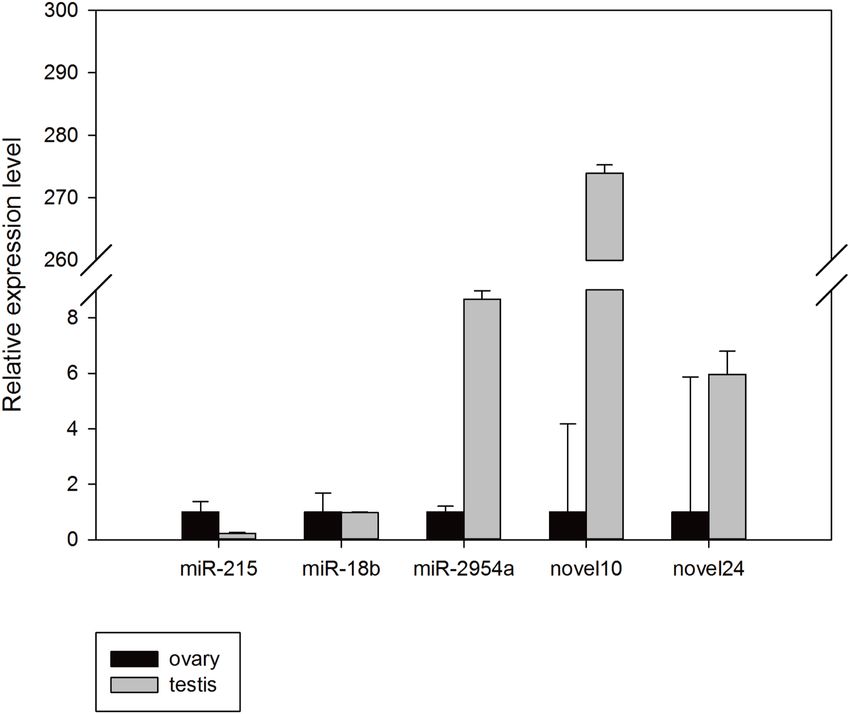
results were concordant with their relative expression trends for our miRNA-seq analyses
basically, and confirmed that these miRNAs exist in the budgerigar. The expression level of
mun-miR-18b-5p, mun-miR-2954-3p, mun-novel10-5p, and mun-novel24-3p in the male
samples were higher than in the female samples, whereas mun-miR-215-5p expression
was higher in the ovaries. There were a minor differences between the miRNA-seq analysis
and Q-PCR assays results. For example, mun-novel10-5p and mun-novel24-3p expression
were not detected in the sRNA sequence data of the ovaries (Table 3), but weak expression
was detected in the validation experiments (Fig. 5).
Putative target prediction for known and novel miRNAs
The putative target genes for these 21 miRNAs were predicted by miRanda, seedVicious
and Targetscan. One thousand, five hundred and sixty-seven genes were determined to be
feasible targets and had complete complementarity to the seed sequence of 21 miRNAs.
These predictions suggest that a single miRNA might target more than one mRNA, such as
mun-miR-2954-3p, which is predicted to target 129 budgerigar genes (Table S4). Similarly,
one gene can be controlled by one or more miRNAs. For instance, mun-novel102-
3p could target FOXG1, and FOXP1 has three miRNAs target sites (mun-novel45-3p,
mun-novel102-3p and mun-miR-2954-3p), and mun-novel7-3p, mun-novel36-3p, mun-
novel102-3p and mun-miR-203-3p could target FOXP2 (Table S4). The miRNAs identified
in this study can target multiple transcription factor genes, such as GATA4, GTF2E1,
Jiang et al. (2018), PeerJ, DOI 10.7717/peerj.4615 9/19Figure 5 Random validation of miRNAs with expression using Q-PCR.
Full-size DOI: 10.7717/peerj.4615/fig-5
GTF3C3, JUN, LZTFL1, MBTPS1, MTF1, NFYA, NFYC, PBX3, RFX4, and SOX10.
Moreover, computational analysis of the 21 miRNA sequences with known, functional
miRNA–mRNA regulatory modules suggested these miRNAs may target genes encoding
KPNA3, PKP4, CHN1, SMG7, FAM53A, and calmodulin, which are included in a series of
important physiological processes and metabolic networks.
GO annotation and KEGG pathway analyses
The genes found to be potentially regulated by miRNAs from this study were annotated
using GO annotation and KEGG pathway analyses. Gene Ontogeny annotations were
classified as cellular component, biological process and molecular function, using GO
rank 2 with p-value ≤ 0.05 (FDR < 0.05). We found that many of the miRNAs detected
in this study were involved in the organogenesis. Seven subcategories within ‘‘cellular
components’’ were found, with ‘‘cell part’’ and ‘‘membrane-bounded organelle’’ being
most represented. Sixteen subcategories of ‘‘biological processes’’ were also identified,
with ‘‘single-organism cellular process’’ being most abundant. Furthermore, many genes
were assigned to five subcategories in ‘‘molecular function,’’ with the largest proportion in
‘‘protein binding’’ (Fig. 6). Notably, mun-miR-215-5p, which demonstrated significantly
biased expression in the ovaries, is likely a primary modulator of a protein complex
involved in signaling and/or catalytic enzyme activity, like RABGAP1, PRR5, FGF13,
SDCBP, TIAL1, PRDX4 (Table S4). From the GO term, we found nine GO annotations
Jiang et al. (2018), PeerJ, DOI 10.7717/peerj.4615 10/19Figure 6 Gene ontology classification annotated by gene2go for target genes of differentially expressed
miRNAs. The figure shows partial GO enrichment for the predicted target genes in ontologies of molecu-
lar function, cellular component, biological processes.
Full-size DOI: 10.7717/peerj.4615/fig-6
were related to female, which the detected miRNAs could regulated some genes, like
FKBP4, C1QBP, VMP1, AKT1, BMPR1B, ADAMTS1, PHB2, IF2B2, MED1, DACH2,
CHD7, AK8, TBP, TAF4, DACH1, LHX9, TYRO3, KIT, EIF2B2, DACH2 and PDGFRA,
thirteen GO terms were found related to male and six GO annotations wete detected
as gonadal function as well (Table S5). In addition, FOXP1 (mun-miR-2954-3p, mun-
novel102-3p and mun-novel45-3p) and FOXP2 (mun-miR-203-5p, mun-novel102-3p,
mun-novel36-3p and mun-novel7-3p) were detected to regulate vocal learning, moreover,
VDAC1, CNTN2, GRIN1, FOXP1, FOXP2, HIF1A, VDAC3, CAMK4, PRKAR1B, ITGA8,
GRIA1, PLK2,HMGCR, KCNAB1, CHST10, RELN, FGF13, ABI1, APP, SRF, LIS1, AK8,
CTNND2 and ATAD1 were illustrated to regulate learning ability (Table S5). The KEGG
pathway analysis demonstrated that the target genes were related to significantly expressed
miRNAs. According to the KEGG pathway analysis (FDR < 0.05), three pathways were
significantly enriched, such as the cell communication (FDR = 0.00317745), excretory
system (FDR = 0.00317745) and signal transduction (FDR = 0.00446146) (Table S6).
Jiang et al. (2018), PeerJ, DOI 10.7717/peerj.4615 11/19DISCUSSION
MiRNA research is needed in more phylogenetically disparate avian species to obtain a
more accurate concept of the miRNAome. Furthermore, some important avian miRNAs
involved in regulating gonadal sex differentiation and development were demonstrated
(Table 1). However, avian studies of sex-biased miRNA expression between ovaries and
testes are limited. The role of non-coding RNAs in the gonads is an area of active research.
Here we used Illumina sequencing to investigate the differentially expressed miRNAs in
the male and female gonads of budgerigars. We observed numerous miRNA families in
our data that may potentially work as key regulators of gene expression. For example, the
let-7 family has been shown to function as a heterochronic switch, and loss of these could
cause periods of cell fate reiteration in adults. In contrast, increasing the gene dosage would
led to premature expression in adult fates (Reinhart et al., 2000). Consequently, the let-7
family miRNAs are considered highly conserved in Animalia (Hertel et al., 2012). Let-7 and
miR-125 are associated with polycistronic transcripts and work as two key regulators of
development in Bilateria.
MiR2954, which is known in chicken (Garcia-Riart et al., 2017; Liu et al., 2017) and
zebra finch (Lin, Balakrishnan & Clayton, 2014), is an bird-specific gene (absent in the
mammalian lineage) and is encoded on the Z chromosome which is known to result in
its higher expression in males than females (Lin, Balakrishnan & Clayton, 2014; Luo et al.,
2012). It has been proposed that this might affect the neurogenomic mechanisms that lead
to sexually dimorphic bird song habituation (Lin, Balakrishnan & Clayton, 2014). Based
on bioinformatical and experimental analysis from chicken and zebra finch, miR-2954-3p
is male-biased and Z-linked miRNA, which targets across a range of bird species, could
help the study field about the evolutionary dynamics of partial dosage compensation and
the genetic architecture underlying gonadal characteristics (Warnefors et al., 2017). The
bioinformatical prediction and the qPCR validation in the present study has confirmed
that there is a male-biased expression of miR2954 in M. undulatus, further corroborating
its involvement in sexual dimorphism. Putative genes targeted by miR2954 include a
TLE4 transcription factor family, which might be associated with nervous system function,
including Ca2+ / calmodulin-dependent protein kinase IV (CaMKIV), SCAMP1, and
SMARCA2. Mun-miR-2954 is also related to development, environmental adaptation,
the nervous system, signaling molecules and interaction, and substance dependence as
indicated in the KEGG analysis. Previous study in zebra finch has elaborated miR2954
could target FOXP2 to regulate vocal learning and detected higher male expression in many
tissues (Fu et al., 2014), however, we haven’t detected mun-miR2954-3p target FOXP2,
whereas mun-miR-203-3p, mun-novel102-3p, mun-novel36-3p and mun-novel7-3p
might regulate FOXP2, mun-miR-2954-3p, mun-novel102-3p and mun-novel45-3p might
regulate FOXP1, which related to vocal learning in the budgerigars and we also measured
higher male expression in gonads.
Further investigations of GO analysis, miRNA in current study might regulate several
sex-related genes. For instance, FKBP4 was considered to be markers of hypospermatogenic
testis (D’Aurora et al., 2017), we found higher male expression of mun-novel7-3p and
Jiang et al. (2018), PeerJ, DOI 10.7717/peerj.4615 12/19mun-novel72-3p could target FKBP4. mun-miR-194-5p and mun-novel31-3p might
regulate SALL1 which the transcriptional regulators of adipose-specific sex-different genes
(Karastergiou & Fried, 2017). LHX9 is needed for ovarian function (Workman, 2017), mun-
novel72-3p and mun-novel8-3p could regulate it. Mun-miR-215-5p which significantly
expressed in ovaries could target PRDX4 (sex-linked gene) (Tippabathani et al., 2017). In
mice gonadal development, miR-181a suppressed granulosa cell proliferation by targeting
ACVR2A, we detected that mun-novel7-3p could regulated ACVR2A as well (Zhang et
al., 2013). Currently, mun-novel7-3p could also target AKT1 (with prior report for sex
differences (Seney et al., 2013)). And mun-novel31-3p could target RNF2 (associated with
regulation of genetic imprinting (Li, Zhang & Wu, 2017)). mun-novel68-3p might regulate
HSF2 which related to sex-determining (Literman et al., 2018), mun-miR-2954-3p and
mun-novel72-3p could target KITLG (sex development) (Hersmus et al., 2017) (Table S4).
Of all the miRNAs tested, we found three gonad-enriched miRNAs (mun-miR-215-5p,
mun-novel10-5p and mun-novel24-3p). In the present study, the presence of these miRNAs
in the gonads suggests that they might serve a similar function in the budgerigar. These
miRNAs may produce sex-specific responses to potential biological mechanisms that have
not yet been described. Although the physiological and biochemical functions of mun-
novel10-5p and mun-novel24-3p remain unclear, their differentially expressed patterns
indicate that they might play important roles in sexual differentiation and development.
CONCLUSIONS
In this study, the whole gonadal miRNAome of budgerigars was sequenced, consisting
of a total of 12,929,838 clean reads and 2,942,541 unique reads. Moreover, differential
expression of 254 known miRNAs and 141 novel miRNAs were analyzed in the gonadal
tissues of budgerigars. The majority of these miRNAs were evolutionarily conserved within
chordates while some of them were budgerigar- or avian-specific. In conclusion, this work
describes the characteristics of sex-biased miRNAs of M. undulatus and adds new sequences
to the avian miRNAome database to facilitate further functional genomic research.
ADDITIONAL INFORMATION AND DECLARATIONS
Funding
This study was supported by the Major Program of Natural Science Foundation of the
Higher Education Institutions (No. KJ2016SD22), the Promotion Plan and Teaching
Reform in Scientific Research Foundation of the Higher Education Institutions of Anhui
Province, China (No. 2015zdjy035) and the Key Program of Natural Science Foundation of
the Anhui Higher Education Institutions (KJ2016A735). The funders had no role in study
design, data collection and analysis, decision to publish, or preparation of the manuscript.
Grant Disclosures
The following grant information was disclosed by the authors:
Major Program of Natural Science Foundation of the Higher Education Institutions:
KJ2016SD22.
Jiang et al. (2018), PeerJ, DOI 10.7717/peerj.4615 13/19Higher Education Institutions of Anhui Province, China: 2015zdjy035.
The Key Program of Natural Science Foundation of the Anhui Higher Education
Institutions: KJ2016A735.
Competing Interests
The authors declare there are no competing interests.
Author Contributions
• Lan Jiang performed the experiments, analyzed the data, prepared figures and/or tables,
authored or reviewed drafts of the paper, approved the final draft.
• Qingqing Wang, Jue Yu, Vinita Gowda, Gabriel Johnson and Jianke Yang performed the
experiments, analyzed the data, authored or reviewed drafts of the paper, approved the
final draft.
• Xianzhao Kan and Xiaojun Yang conceived and designed the experiments, contributed
reagents/materials/analysis tools, authored or reviewed drafts of the paper, approved the
final draft.
Animal Ethics
The following information was supplied relating to ethical approvals (i.e., approving body
and any reference numbers):
Animals in the current study were authorized by the Ethics Committee of Anhui Normal
University (Anhui, China) with authorization number #20150612.
Data Availability
The following information was supplied regarding data availability:
SRA database accession number SRR5664259 and SRR5664260.
Jiang, Lan; Kan, Xianzhao (2018): budgerigar_mirnaomes_rawdata. figshare. https:
//doi.org/10.6084/m9.figshare.5187874.v1.
Supplemental Information
Supplemental information for this article can be found online at http://dx.doi.org/10.7717/
peerj.4615#supplemental-information.
REFERENCES
Ali NM, Boo L, Yeap SK, Ky H, Satharasinghe DA, Liew WC, Ong HK, Cheong
SK, Kamarul T. 2016. Probable impact of age and hypoxia on proliferation and
microRNA expression profile of bone marrow-derived human mesenchymal stem
cells. PeerJ 4:e1536 DOI 10.7717/peerj.1536.
Ameres SL, Zamore PD. 2013. Diversifying microRNA sequence and function. Nature
Reviews Molecular Cell Biology 14:475–488 DOI 10.1038/nrm3611.
Anders S, Huber W. 2010. Differential expression analysis for sequence count data.
Genome Biology 11:Article R106 DOI 10.1186/gb-2010-11-10-r106.
Jiang et al. (2018), PeerJ, DOI 10.7717/peerj.4615 14/19Ashburner M, Ball CA, Blake JA, Botstein D, Butler H, Cherry JM, Davis AP, Dolinski
K, Dwight SS, Eppig JT. 2000. Gene ontology: tool for the unification of biology.
Nature genetics 25:25–29 DOI 10.1038/75556.
Bannister SC, Tizard ML, Doran TJ, Sinclair AH, Smith CA. 2009. Sexually dimorphic
microRNA expression during chicken embryonic gonadal development. Biology of
Reproduction 81:165–176 DOI 10.1095/biolreprod.108.074005.
Bartel DP. 2009. MicroRNAs: target recognition and regulatory functions. Cell
136:215–233 DOI 10.1016/j.cell.2009.01.002.
Benjamini Y, Hochberg Y. 1995. Controlling the false discovery rate: a practical and
powerful approach to multiple testing. Journal of the Royal Statistical Society Series
B (Methodological) 57:289–300.
Burge SW, Daub J, Eberhardt R, Tate J, Barquist L, Nawrocki EP, Eddy SR, Gardner
PP, Bateman A. 2012. Rfam 11.0: 10 years of RNA families. Nucleic Acids Research
41:D226–D232 DOI 10.1093/nar/gks1005.
Busk PK. 2014. A tool for design of primers for microRNA-specific quantitative RT-
qPCR. BMC Bioinformatics 15:29 DOI 10.1186/1471-2105-15-29.
Cooke TF, Fischer CR, Wu P, Jiang TX, Xie KT, Kuo J, Doctorov E, Zehnder A, Khosla
C, Chuong CM. 2017. Genetic mapping and biochemical basis of yellow feather
pigmentation in budgerigars. Cell 171:427–439 DOI 10.1016/j.cell.2017.08.016.
Cutting AD, Bannister SC, Doran TJ, Sinclair AH, Tizard MV, Smith CA. 2012. The
potential role of microRNAs in regulating gonadal sex differentiation in the chicken
embryo. Chromosome Research 20:201–213 DOI 10.1007/s10577-011-9263-y.
Darnell DK, Kaur S, Stanislaw S, Konieczka JK, Yatskievych TA, Antin PB. 2006.
MicroRNA expression during chick embryo development. Developmental Dynamics
235:3156–3165 DOI 10.1002/dvdy.20956.
D’Aurora M, Ferlin A, Garolla A, Franchi S, D’Onofrio L, Trubiani O, Palka G, Foresta
C, Stuppia L, Gatta V. 2017. Testis transcriptome modulation in klinefelter patients
with hypospermatogenesis. Scientific Reports 7:45729 DOI 10.1038/srep45729.
Del Hoyo J, Elliott A, Sargatal J, Christie DA, De Juana E. 2015. Handbook of the birds
of the world alive. Available at http:// www.hbw.com/ (accessed on 5 Jan 2018).
Eda-Fujiwara H, Satoh R, Hata Y, Yamasaki M, Watanabe A, Zandbergen MA,
Okamoto Y, Miyamoto T, Bolhuis JJ. 2016. Sex differences in behavioural
and neural responsiveness to mate calls in a parrot. Scientific Reports 6:18481
DOI 10.1038/srep18481.
Enright AJ, John B, Gaul U, Tuschl T, Sander C, Marks DS. 2004. MicroRNA targets in
Drosophila. Genome Biology 5:Article R1 DOI 10.1186/gb-2003-5-1-r1.
Friedländer MR, Mackowiak SD, Li N, Chen W, Rajewsky N. 2012. miRDeep2 accu-
rately identifies known and hundreds of novel microRNA genes in seven animal
clades. Nucleic Acids Research 40:37–52 DOI 10.1093/nar/gkr688.
Friedman RC, Burge CB. 2014. MicroRNA target finding by comparative genomics.
Methods in Molecular Biology 1097:457–476 DOI 10.1007/978-1-62703-709-9_21.
Jiang et al. (2018), PeerJ, DOI 10.7717/peerj.4615 15/19Fu L, Shi Z, Luo G, Tu W, Wang X, Fang Z, Li X. 2014. Multiple microRNAs regulate
human FOXP2 gene expression by targeting sequences in its 30 untranslated region.
Molecular Brain 7:Article 71 DOI 10.1186/s13041-014-0071-0.
Ganapathy G, Howard JT, Ward JM, Li J, Li B, Li Y, Xiong Y, Zhang Y, Zhou S,
Schwartz DC. 2014. High-coverage sequencing and annotated assemblies of the
budgerigar genome. GigaScience 3:Article 11 DOI 10.1186/2047-217X-3-11.
Garcia-Riart B, Lorda-Diez CI, Marin-Llera JC, Garcia-Porrero JA, Hurle JM,
Montero JA. 2017. Interdigital tissue remodelling in the embryonic limb involves
dynamic regulation of the miRNA profiles. Journal of Anatomy 231:275–286
DOI 10.1111/joa.12629.
Gill F, Donsker D (eds.) 2017. IOC World Bird List (v 7.3). DOI 10.14344/IOC.ML.7.3.
Glazov EA, Cottee PA, Barris WC, Moore RJ, Dalrymple BP, Tizard ML. 2008. A
microRNA catalog of the developing chicken embryo identified by a deep sequencing
approach. Genome Research 18:957–964 DOI 10.1101/gr.074740.107.
Gong W, Huang Y, Xie J, Wang G, Yu D, Sun X. 2017. Genome-wide identification and
characterization of conserved and novel microRNAs in grass carp (Ctenopharyn-
godon idella) by deep sequencing. Computational Biology and Chemistry 68:92–100
DOI 10.1016/j.compbiolchem.2017.02.010.
Hersmus R, Bever Y, Wolffenbuttel KP, Biermann K, Cools M, Looijenga LH. 2017.
The biology of germ cell tumors in disorders of sex development. Clinical Genetics
91:292–301 DOI 10.1111/cge.12882.
Hertel J, Bartschat S, Wintsche A, Otto C. 2012. Evolution of the let-7 microRNA family.
RNA Biology 9:231–241 DOI 10.4161/rna.18974.
Hicks J, Tembhurne P, Liu HC. 2008. MicroRNA expression in chicken embryos. Poultry
Science 87:2335–2343 DOI 10.3382/ps.2008-00114.
Hoeschele M, Bowling DL. 2016. Sex differences in rhythmic preferences in the
Budgerigar (Melopsittacus undulatus): a comparative study with humans. Frontiers
in Psychology 7:1543 DOI 10.3389/fpsyg.2016.01543.
Hulsen T, De Vlieg J, Alkema W. 2008. BioVenn-a web application for the comparison
and visualization of biological lists using area-proportional Venn diagrams. BMC
Genomics 9:488 DOI 10.1186/1471-2164-9-488.
Jing J, Wu J, Liu W, Xiong S, Ma W, Zhang J, Wang W, Gui JF, Mei J. 2014. Sex-biased
miRNAs in gonad and their potential roles for testis development in yellow catfish.
PLOS ONE 9:e107946 DOI 10.1371/journal.pone.0107946.
Jurka J, Kapitonov VV, Pavlicek A, Klonowski P, Kohany O, Walichiewicz J. 2005.
Repbase update, a database of eukaryotic repetitive elements. Cytogenetic and
Genome Research 110:462–467 DOI 10.1159/000084979.
Kanehisa M, Furumichi M, Tanabe M, Sato Y, Morishima K. 2017. KEGG: new
perspectives on genomes, pathways, diseases and drugs. Nucleic Acids Research
45:D353–D361 DOI 10.1093/nar/gkw1092.
Kang L, Cui X, Zhang Y, Yang C, Jiang Y. 2013. Identification of miRNAs associated
with sexual maturity in chicken ovary by Illumina small RNA deep sequencing. BMC
Genomics 14:352 DOI 10.1186/1471-2164-14-352.
Jiang et al. (2018), PeerJ, DOI 10.7717/peerj.4615 16/19Karastergiou K, Fried SK. 2017. Cellular mechanisms driving sex differences in adipose
tissue biology and body shape in humans and mouse models. In: Mauvais-Jarvis F,
ed. Sex and gender factors affecting metabolic homeostasis, diabetes and obesity. Cham:
Springer International Publishing, 29–51.
Kozomara A, Griffiths-Jones S. 2014. miRBase: annotating high confidence mi-
croRNAs using deep sequencing data. Nucleic Acids Research 42:D68–D73
DOI 10.1093/nar/gkt1181.
Langmead B, Trapnell C, Pop M, Salzberg SL. 2009. Ultrafast and memory-efficient
alignment of short DNA sequences to the human genome. Genome Biology 10:Article
R25 DOI 10.1186/gb-2009-10-3-r25.
Li H, Handsaker B, Wysoker A, Fennell T, Ruan J, Homer N, Marth G, Abecasis G,
Durbin R. 2009a. The sequence alignment/map format and SAMtools. Bioinformat-
ics 25:2078–2079 DOI 10.1093/bioinformatics/btp352.
Li HL, Zhang YB, Wu ZY. 2017. Development of research on huntington disease in
china. Neuroscience Bulletin 33:312–316 DOI 10.1007/s12264-016-0093-y.
Li R, Yu C, Li Y, Lam T-W, Yiu S-M, Kristiansen K, Wang J. 2009b. SOAP2: an
improved ultrafast tool for short read alignment. Bioinformatics 25:1966–1967
DOI 10.1093/bioinformatics/btp336.
Lim LP, Lau NC, Garrett-Engele P, Grimson A, Schelter JM, Castle J, Bartel DP, Linsley
PS, Johnson JM. 2005. Microarray analysis shows that some microRNAs downregu-
late large numbers of target mRNAs. Nature 433:769–773 DOI 10.1038/nature03315.
Lin YC, Balakrishnan CN, Clayton DF. 2014. Functional genomic analysis and neu-
roanatomical localization of miR-2954, a song-responsive sex-linked microRNA in
the zebra finch. Frontiers in Neuroscience 8:Article 409 DOI 10.3389/fnins.2014.00409.
Literman R, Burrett A, Bista B, Valenzuela N. 2018. Putative independent evolutionary
reversals from genotypic to temperature-dependent sex determination are associated
with accelerated evolution of sex-determining genes in turtles. Journal of Molecular
Evolution 86:11–26 DOI 10.1007/s00239-017-9820-x.
Liu P, Yang F, Zhuang Y, Xiao Q, Cao H, Zhang C, Wang T, Lin H, Guo X, Hu G.
2017. Dysregulated expression of microRNAs and mRNAs in pulmonary artery
remodeling in ascites syndrome in broiler chickens. Oncotarget 8:1993–2007
DOI 10.18632/oncotarget.12888.
Livak KJ, Schmittgen TD. 2001. Analysis of relative gene expression data using
real-time quantitative PCR and the 2−11CT method. Methods 25:402–408
DOI 10.1006/meth.2001.1262.
Luo GZ, Hafner M, Shi Z, Brown M, Feng GH, Tuschl T, Wang XJ, Li XC. 2012.
Genome-wide annotation and analysis of zebra finch microRNA repertoire reveal
sex-biased expression. BMC Genomics 13:Article 236 DOI 10.1186/1471-2164-13-236.
Mai M, Jin L, Tian S, Liu R, Huang W, Tang Q, Ma J, Wang X, Hu Y, Wang D. 2016.
Deciphering the microRNA transcriptome of skeletal muscle during porcine
development. PeerJ 4:e1504 DOI 10.7717/peerj.1504.
Marco A. 2014. Sex-biased expression of microRNAs in Drosophila melanogaster. Open
Biology 4:Article 140024 DOI 10.1098/rsob.140024.
Jiang et al. (2018), PeerJ, DOI 10.7717/peerj.4615 17/19Marco A. 2017. SeedVicious: analysis of microRNA target and near-target sites. bioRxiv
124529 DOI 10.1101/124529.
Marco A, Kozomara A, Hui JH, Emery AM, Rollinson D, Griffiths-Jones S, Ronshaugen
M. 2013. Sex-biased expression of microRNAs in Schistosoma mansoni. PLOS
Neglected Tropical Diseases 7:e2402 DOI 10.1371/journal.pntd.0002402.
Martin M. 2011. Cutadapt removes adapter sequences from high-throughput sequencing
reads. EMBnet Journal 17:10–12 DOI 10.14806/ej.17.1.200.
Reinhart BJ, Slack FJ, Basson M, Pasquinelli AE, Bettinger JC, Rougvie AE, Horvitz HR,
Ruvkun G. 2000. The 21-nucleotide let-7 RNA regulates developmental timing in
Caenorhabditis elegans. Nature 403:901–906 DOI 10.1038/35002607.
Rosani U, Pallavicini A, Venier P. 2016. The miRNA biogenesis in marine bivalves. PeerJ
4:e1763 DOI 10.7717/peerj.1763.
Seney ML, Ekong KI, Ding Y, Tseng GC, Sibille E. 2013. Sex chromosome complement
regulates expression of mood-related genes. Biology of Sex Differences 4:Article 20
DOI 10.1186/2042-6410-4-20.
Skalsky RL, Kang D, Linnstaedt SD, Cullen BR. 2014. Evolutionary conservation of
primate lymphocryptovirus MicroRNA targets. Journal of Virology 88:1617–1635
DOI 10.1128/JVI.02071-13.
Striedter GF, Freibott L, Hile AG, Burley NT. 2003. For whom the male calls: an effect of
audience on contact call rate and repertoire in budgerigars, Melopsittacus undulatus.
Animal Behaviour 65:875–882 DOI 10.1006/anbe.2003.2135.
Tippabathani J, Nellore J, Radhakrishnan V, Banik S, Kapoor S. 2017. Identification of
NURR1 (Exon 4) and FOXA1 (Exon 3) haplotypes associated with mRNA expres-
sion levels in peripheral blood lymphocytes of parkinson’s patients in small indian
population. Parkinson’s Disease 2017:Article 6025358 DOI 10.1155/2017/6025358.
Torley KJ, Silveira JCD, Smith P, Anthony RV, Veeramachaneni DR, Winger
QA, Bouma GJ. 2011. Expression of miRNAs in ovine fetal gonads: potential
role in gonadal differentiation. Reproductive Biology and Endocrinology 9:1–11
DOI 10.1186/1477-7827-9-1.
Warnefors M, Mössinger K, Halbert J, Studer T, VandeBerg JL, Lindgren I, Fal-
lahshahroudi A, Jensen P, Kaessmann H. 2017. Sex-biased microRNA expression in
mammals and birds reveals underlying regulatory mechanisms and a role in dosage
compensation. Genome Research 27:1961–1973 DOI 10.1101/gr.225391.117.
Workman S. 2017. Lhx9 is required for urogenital ridge development and ovarian
function (Thesis, Bachelor of Biomedical Sciences with Honours). University of
Otago. Available at http:// hdl.handle.net/ 10523/ 7734.
Wu S, Guo W, Li Y, Ren X, Lei X, Li X, Yao J, Yang X. 2016. miRNA and piRNA
expression profiles of breeder cock testes detected by next-generation sequencing.
Reproduction in Domestic Animals 52:203–213 DOI 10.1111/rda.12880.
Xu Q, Zhang Y, Chen Y, Tong YY, Rong GH, Huang ZY, Zhao RX, Zhao WM, Wu
XS, Chang GB. 2014. Identification and differential expression of microRNAs in
ovaries of laying and broody geese (Anser cygnoides) by solexa sequencing. PLOS
ONE 9:e87920 DOI 10.1371/journal.pone.0087920.
Jiang et al. (2018), PeerJ, DOI 10.7717/peerj.4615 18/19Yatsenko SA, Treadwell-Deering D, Krull K, Lewis RA, Glaze D, Stankiewicz P, Lupski
JR, Potocki L. 2005. Trisomy 17p10-p12 due to mosaic supernumerary marker
chromosome: delineation of molecular breakpoints and clinical phenotype, and
comparison to other proximal 17p segmental duplications. American Journal of
Medical Genetics Part A 138:175–180 DOI 10.1002/ajmg.a.30948.
Yu DB, Jiang BC, Jing G, Dong FL, Lu YL, Yue HJ, Wang ZC, Du WX, Gue AY. 2013.
Identification of novel and differentially expressed microRNAs in the ovaries
of laying and non-laying ducks. Journal of Integrative Agriculture 12:136–146
DOI 10.1016/S2095-3119(13)60214-2.
Zhang Q, Sun H, Jiang Y, Ding L, Wu S, Fang T, Yan G, Hu Y. 2013. MicroRNA-181a
suppresses mouse granulosa cell proliferation by targeting activin receptor IIA. PLOS
ONE 8:e59667 DOI 10.1371/journal.pone.0059667.
Zuker M. 2003. Mfold web server for nucleic acid folding and hybridization prediction.
Nucleic Acids Research 31:3406–3415 DOI 10.1093/nar/gkg595.
Jiang et al. (2018), PeerJ, DOI 10.7717/peerj.4615 19/19You can also read


