Quantitative ubiquitylomics reveals the ubiquitination regulation landscape in oral adenoid cystic carcinoma
←
→
Page content transcription
If your browser does not render page correctly, please read the page content below
Bioscience Reports (2021) 41 BSR20211532
https://doi.org/10.1042/BSR20211532

Research Article

Quantitative ubiquitylomics reveals the
ubiquitination regulation landscape in oral adenoid
cystic carcinoma
Wen Li1,2,3 , Xiaobin Wang1,2,3 , Qian Zhang1,2,3 , Hanlin Wang3 , Wenxin Zuo4 , Hongliang Xie4 , Jianming Tang4 ,
Mengmeng Wang4 , Zhipeng Zeng4 , Wanxia Cai4 , Donge Tang4 and Yong Dai4

                                                                                                                                                                                           Downloaded from http://portlandpress.com/bioscirep/article-pdf/41/8/BSR20211532/919622/bsr-2021-1532.pdf by guest on 31 August 2021
1 Carson International Cancer Centre, Shenzhen University General Hospital and Shenzhen University Clinical Medical Academy Centre, Shenzhen University, 1098 Xueyuan Road,
Shenzhen Guangdong 518000, China; 2 Key Laboratory of Optoelectronic Devices and Systems, College of Physics and Optoelectronic Engineering, Shenzhen University, Shenzhen
518060, China; 3 Health Science Center, School of Medicine, Shenzhen University, Shenzhen 518060, China; 4 Clinical Medical Research Center, Guangdong Provincial Engineering
Research Center of Autoimmune Disease Precision Medicine, Shenzhen Engineering Research Center of Autoimmune Disease, The Second Clinical Medical College of Jinan
University, The First Affiliated Hospital of Southern University of Science and Technology, Shenzhen People’s Hospital, Shenzhen, Guangdong 518020, China
Correspondence: Donge Tang (donge66@126.com) or Yong Dai (daiyong22@aliyun.com)

                                           Adenoid cystic carcinoma (ACC) is an extremely rare salivary gland tumor with a poor prog-
                                           nosis and needs attention on molecular mechanisms. Protein ubiquitination is an evolution-
                                           arily conserved post-translational modification (PTM) for substrates degradation and con-
                                           trols diverse cellular functions. The broad cellular function of ubiquitination network holds
                                           great promise to detect potential targets and identify respective receptors. Novel technolo-
                                           gies are discovered for in-depth research and characterization of the precise and dynamic
                                           regulation of ubiquitylomics in multiple cellular processes during cancer initiation, progres-
                                           sion and treatment. In the present study, 4D label-free quantitative techniques of ubiquiti-
                                           nation proteomics were used and we identified a total of 4152 ubiquitination sites in 1993
                                           proteins. We also performed a systematic bioinformatics analysis for differential modified
                                           proteins and peptides containing quantitative information through the comparation between
                                           oral ACC (OACC) tumor with adjacent normal tissues, as well as the identification of eight
                                           protein clusters with motif analysis. Our findings offered an important reference of potential
                                           biomarkers and effective therapeutic targets for ACC.

                                         Introduction
                                         Adenoid cystic carcinoma (ACC) was discovered a long time ago with location in the major and minor
                                         salivary glands [1,2] and other organs [3], which can be divided into tubular form (Grade I), cribriform
                                         (Grade II) and solid form (Grade III) [4]. Clinical staging had more significant importance than histo-
                                         logical grade on prognosis [5]. The 10-year survival rates are approximately 73% (Stage I), 43% (Stage
                                         II) and 15% (Stage III and IV) [4]. Studies on oral ACC (OACC) are even more uncommon and insuffi-
                                         cient. Surgery and postoperative radiotherapy have been the standard treatment of ACC for a long time
                                         [6] with poor prognosis (up to 40% of recurrent rate and 60% of metastatic rate) [7]. Several factors have
                                         been studied to associate with the clinicopathological parameters and prognosis of ACC, including p53
Received: 28 June 2021
                                         [8], SOX2 [9], mutated ATM [10], MACC1 [11], WHSC1 [12], EpCAM [13], PSMA [14], TRAF6 [15],
Revised: 30 July 2021                    HSP27 [16], PRRX1 [17], hypoxia-related genes [18], the EGFR pathway genes [19], MYB–NFIB fusion
Accepted: 02 August 2021                 genes [20–23], as well as NOTCH1-HEY1 pathway [24] and Akt signaling pathway [25,26]. Considering
                                         the aggressive behavior, it is urgent to figure out more efficient clinical pathological and biomolecular
Accepted Manuscript online:
04 August 2021                           prognostic factors for therapeutic choices.
Version of Record published:                Protein post-translational modifications (PTMs) exist in both eukaryotes and prokaryotes at one
20 August 2021                           or more sites [27]. More than 200 types of PTMs are identified in humans [28]. Ubiquitination is

© 2021 The Author(s). This is an open access article published by Portland Press Limited on behalf of the Biochemical Society and distributed under the Creative Commons Attribution   1
License 4.0 (CC BY).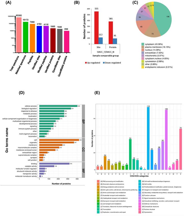
Bioscience Reports (2021) 41 BSR20211532
                                                                                                                                               https://doi.org/10.1042/BSR20211532

Table 1 Clinical information in OACC tumor and adjacent normal tissues
                                                                                                            Pathological                                            Lymph node
      Gender               Age (years)             Tumor size (cm)             Tumor location                diagnosis               Neuro recidivist                metastasis

      Female                     32                         1.5               Right submandibular                 ACC                          Yes                         No
       Male                      58                         3.0                Left submandibular                 ACC                          Yes                         Yes
      Female                     23                         1.0                       Palate                      ACC                          Yes                         No
       Male                      64                         2.5              Parotid gland and neck               ACC                          Yes                         Yes

                 the covalent conjugation in proteins by conserved small protein ubiquitin and ubiquitin-like (Ubl) proteins either
                 as a monomer or as a polyubiquitin chain. [29] Ubiquitin-activating protein E1, ubiquitin-conjugating protein E2
                 and ubiquitin-ligase E3 are involved to form a transient reaction, which can be reversed by deubiquitylating en-

                                                                                                                                                                                                 Downloaded from http://portlandpress.com/bioscirep/article-pdf/41/8/BSR20211532/919622/bsr-2021-1532.pdf by guest on 31 August 2021
                 zymes (DUBs) [30–32]. The modified substrates are degraded after binding to a multisubunit protease complex [29].
                 The dynamic changes of ubiquitination form an enzymatic and complete ordered system to control subcellular pro-
                 cesses [33], which affects pathophysiological states in cancer under various conditions [34,35]. Both oncogenes and
                 tumor-suppressor genes undergo ubiquitination [36]. Several studies mentioned the potential of ubiquitination re-
                 lated proteins as OACC biomarkers. From Nanostring nCounter miRNA assay, ubiquitin-like modifier activating
                 enzyme 2 (UBA2) was identified increased in primary and recurrent tumors than normal tissue, revealing the po-
                 tential connection between UBA2 and tumor recurrence and metastasis [37]. The expression of Ubiquitin-specific
                 protease 22 (USP22) in salivary ACC (SACC) was higher in the tumor group than in the adjacent normal group,
                 which was associated with a poor prognosis [38]. The low expression of the tumor suppressor gene cylindromato-
                 sis (CYLD), which has deubiquitinating enzyme activity, corrected with salivary gland tumor progression through
                 NF-κB pathway [39]. Notwithstanding, there is lack of relevant research on the molecular mechanisms and related
                 pathogenesis of ubiquitination network in OACC.
                    A global and comprehensive information about ubiquitin system is difficult due to the challenge for
                 high-throughput analysis [40]. Another obstacle is that these revisable modifications can be easily lost or not easily
                 detected during the experiment. The mass spectrometry (MS)-based proteomic approaches have been widely used in
                 qualitative and quantitative analysis of cellular biology [41]. The organic combination of non-standard quantitative
                 and MS-based proteomic technology can effectively identify ubiquitination substrates and modification network in
                 OACC.
                    In our project, 4D label-free quantitative ubiquitination proteomics was carried out. A total of 4152 ubiquitination
                 sites were identified on 1993 proteins, in which 1648 loci of 859 proteins contained quantitative information. We also
                 conducted a systematic bioinformatics analysis, including protein annotation, functional classification, functional
                 enrichment and cluster analysis based on functional enrichment proteins. The proteomic methodologies in our work
                 illustrating ubiquitination landscape in OACC can be applied to the search and identification of novel molecular
                 biomarkers, provide valuable information for diagnosis and help discovering novel therapeutic anticancer strategies.

                 Materials and methods
                 Sample preparation
                 Four pairs of OACC tumor and adjacent normal tissues were collected from Shenzhen People’s Hospital. The present
                 study was carried out following the Declaration of Helsinki and approved by the Medical Ethics Committee of Shen-
                 zhen People’s Hospital (No. LL-KY-2019173). Participants were informed about the introduction of the present study,
                 signed an informed consent, and agreed to take tissue samples after surgical resection for scientific research. Parts of
                 the clinical information of patients are listed in Table 1.

                 Protein extraction
                 Appropriate number of samples were added with liquid nitrogen to grind to powder and added with four-times of
                 powder lysis buffer (1% Triton X-100, 1% protease inhibitor, 50 μm PR-619, 3 μm TSA, 50 mm NAM) for ultrasonic
                 pyrolysis. After centrifugation in 12000×g for 10 min, the supernatant was transferred to a new centrifuge tube. The
                 protein concentration was determined by BCA kit.

                 Trypsin digestion
                 TCA was slowly added in each sample, followed by precipitating at 4◦ C. The supernatant was discarded after centrifu-
                 gation in 4500×g for 5 min. The precipitate was washed with pre-cooled acetone, dried in the air and dissolved by

2         © 2021 The Author(s). This is an open access article published by Portland Press Limited on behalf of the Biochemical Society and distributed under the Creative Commons Attribution
          License 4.0 (CC BY).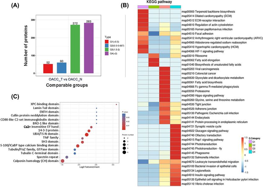
Bioscience Reports (2021) 41 BSR20211532
https://doi.org/10.1042/BSR20211532

200 mm TEAB buffer. Trypsin was added and the reaction was maintained overnight. After the incubation by dithio-
threitol at 56◦ C for 30 min, iodoacetamide (IAA) was added. The samples were then incubated at room temperature
for 15 min in dark.

MS
The digested peptides were dissolved by liquid chromatography mobile phase A (containing 0.1% formic acid and 2%
acetonitrile) and separated by NanoElute ultra performance liquid chromatography system. The flow rate was 450.00
nl/min, and the gradient of mobile phase B (containing 0.1% formic acid and 100% acetonitrile) was set as follows:
6–22%: 0–43 min; 22–30%: 43–56 min; 30–80%: 56–58 min; 80%: 58–60 min. The peptides were separated by Ultra
Performance Liquid Chromatography (UPLC) and injected into Capillary Ion Source for ionization to be analyzed
by Tims-TOF Pro MS. The voltage of ion source was 2.0 kV, and the peptide parent ion and its secondary fragments
were detected and analyzed by high-resolution TOF. The scanning range of secondary MS is set to 100–1700. The data

                                                                                                                                                                                           Downloaded from http://portlandpress.com/bioscirep/article-pdf/41/8/BSR20211532/919622/bsr-2021-1532.pdf by guest on 31 August 2021
acquisition mode is PASEF. After the collection of a first-order mass spectrum, the second-order mass spectrum with
charge number of parent ion in the range of 0–5 was collected ten-times in PASEF mode, and the dynamic exclusion
time of tandem MS scanning was set to 30 s.

Database searching
Maxquant 1.6.6.0 was used to retrieve the secondary MS data. The proteins were detected in Homo sapiens 9606
(20366 sequences) with the common contamination database. The additional reverse database was added to calculate
the false discovery rate (FDR) caused by random matching. The FDR and peptide spectrum matches (PSM) was set to
1%. Enzyme digestion: trypsin/P. Number of missed sites: 4. Minimum length: 7. Maximum modification number: 5.
Mass error tolerance of primary parent ion: 20.0 ppm in first search and 20 ppm in main search. Mass error tolerance
of secondary fragment ion: 20.0 ppm. Cysteine alkylation carbamidomethyl (c) was set as fixed modification, and the
variable modification was [‘acetyl (protein N-term)’, ‘oxidation (m)’, ‘glygly (k)’]. The quantitative method was set to
LFQ.

Results
Systematic profiling of protein ubiquitination in OACC samples
In order to globally reveal the involvement of ubiquitin in the progression and regulation of OACC, we performed 4D
label-free quantitative ubiquitination proteomics study through comparing OACC tumor samples (OACC T) with
the adjacent normal samples (OACC N) in four patients who had not received any drug treatment before operation.
The identification data were filtered as localization probability > 0.75. After MS analysis and database search, a total
of 63282 secondary spectra were obtained, of which 15172 were available. The relative quantitative value was obtained
according to the intensity of the modified site between different samples. According to this method, 7956 peptides and
4116 modified peptides were identified. Among 4152 ubiquitination sites in 1993 proteins, 1648 sites in 859 proteins
were quantified (Figure 1A). OACC T showed 555 ubiquitination sites up-regulated (≥1.5-fold, P-value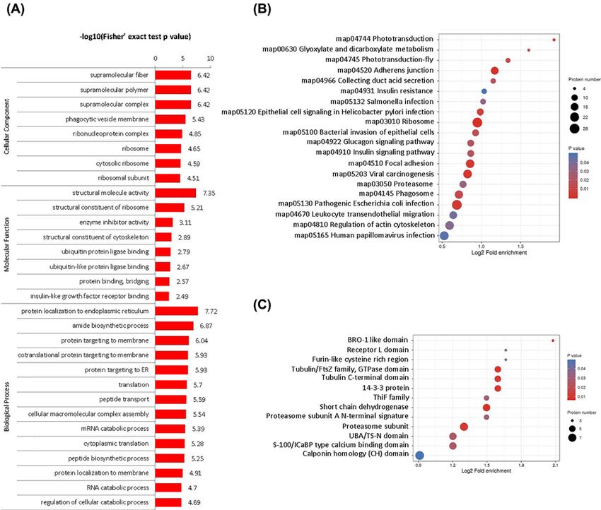
Bioscience Reports (2021) 41 BSR20211532
                                                                                                                                         https://doi.org/10.1042/BSR20211532

                                                                                                                                                                                           Downloaded from http://portlandpress.com/bioscirep/article-pdf/41/8/BSR20211532/919622/bsr-2021-1532.pdf by guest on 31 August 2021

           Figure 1. Identification of protein ubiquitination
           (A) Number of proteins and modification sites identified according to MS data. (B) Differentially modified sites and proteins in tumor
           and normal samples, respectively (filtered with threshold value of expression fold change > 1.5 and P-value
Bioscience Reports (2021) 41 BSR20211532
https://doi.org/10.1042/BSR20211532
Table 2 Top 20 up- and down-regulated proteins and the corresponding sequence (normalized modification sites
quantitation)
Protein accession Gene name Modified sequence OACC T/OACC N ratio Regulated type
P62979 RPS27A IQDK(1)EGIPPDQQR 26.637 Up
O43175 PHGDH AWAGSPK(1)GTIQVITQGTSLK 16.252 Up
Q8NBN3 TMEM87A FAFSPLSEEEEEDEQK(0.944)EPMLK(0.056) 11.91 Up
P54725 RAD23A IDEK(1)NFVVVMVTK 8.567 Up
P54727 RAD23B IDEK(1)NFVVVMVTK 8.17 Up
P54727 RAD23B IDIDPEETVK(0.996)ALK(0.004) 8.17 Up
P68363 TUBA1B AYHEQLSVAEITNACFEPANQMVK(1)CDPR 8.154 Up
P06213 INSR GGK(1)GLLPVR 6.307 Up
P62736 ACTA2 K(1)DLYANNVLSGGTTMYPGIADR 6.263 Up
Downloaded from http://portlandpress.com/bioscirep/article-pdf/41/8/BSR20211532/919622/bsr-2021-1532.pdf by guest on 31 August 2021
P68363 TUBA1B TIGGGDDSFNTFFSETGAGK(1)HVPR 6.208 Up
P62873 GNB1 K(1)ACADATLSQITNNIDPVGR 6.169 Up
Q5BJD5 TMEM41B AVK(1)WSQQVER 6.09 Up
P43121 MCAM SDK(1)LPEEMGLLQGSSGDK 6.085 Up
P63261 ACTG1 K(1)DLYANTVLSGGTTMYPGIADR 5.928 Up
P08670 VIM ETNLDSLPLVDTHSK(1)R 5.9 Up
Q9Y487 ATP6V0A2 VTK(1)TFVK 5.834 Up
P15311 EZR LTPK(1)IGFPWSEIR 5.442 Up
O75881 CYP7B1 DDFLK(0.999)FDDK(0.001) 5.421 Up
P83731 RPL24 AITGASLADIMAK(1)R 5.359 Up
P55287 CDH11 K(1)EPLIVFEEEDVR 5.357 Up
P05023 ATP1A1 AAVPDAVGK(1)CR 0.031 Down
P68363 TUBA1B LSVDYGK(0.98)K(0.946)SK(0.074) 0.033 Down
Q13797 ITGA9 YK(1)EIIEAEK 0.062 Down
Q8WZ42 TTN ITNYIVEK(1)CATTAER 0.175 Down
Q13797 ITGA9 EIIEAEK(1)NR 0.202 Down
Q9HCU0 CD248 WVIHAGSK(1)SPTEPMPPR 0.219 Down
P62841 RPS15 EAPPMEKPEVVK(1)THLR 0.244 Down
P15104 GLUL K(1)DPNK(1)LVLCEVFK 0.245 Down
Q99808 SLC29A1 SLTAVFMWPGK(1)DSR 0.267 Down
P49411 TUFM K(1)YEEIDNAPEER 0.268 Down
Q05086 UBE3A AAAK(1)HLIER 0.268 Down
Q7L1W4 LRRC8D DGEQAK(1)ALFEK 0.291 Down
O43707 ACTN4 K(1)HEAFESDLAAHQDR 0.294 Down
P23229 ITGA6 EIK(0.003)DEK(0.997)YIDNLEK 0.324 Down
P08133 ANXA6 PANDFNPDADAK(1)ALR 0.331 Down
P35555 FBN1 GQCIK(1)PLFGAVTK 0.331 Down
Q96D96 HVCN1 LK(1)QMNVQLAAK 0.334 Down
P09525 ANXA4 ISQK(1)DIEQSIK 0.335 Down
P42167 TMPO YVPLADVK(0.95)SEK(0.05) 0.336 Down
P19388 POLR2E GQVVK(1)IIR 0.343 Down
Functional enrichment analysis of DUPs
We conducted the functional enrichment analyses with GO annotation and Kyoto Encyclopedia of Genes and
Genomes (KEGG) pathway enrichment analysis. Followed by phagocytic vesicle membrane, cytosolic ribosome and
ribosomal subunit most differed in CC category. In MF category, the most DUPs were concentrated in insulin-like
growth factor receptor binding, structural constituent of ribosome and structural constituent of cytoskeleton. Mean-
while, cytoplasmic translation, protein localization to endoplasmic reticulum, co-translational protein targeting
membrane and protein targeting ER were most altered in BP category (Figure 2A; Supplementary Table S1).
According to the KEGG pathway analysis, we identified 25 pathways from up-regulated and 10 pathways from
up-regulated DUPs. The most significantly up-regulated were phototransduction (map04744), glyoxylate and di-
carboxylate metabolism (map00630), and phototransduction-fly (map04745). Meanwhile, aldosterone-regulated
sodium reabsorption (map04960), fatty acid elongation (map00062) and terpenoid backbone biosynthesis
(map00900) were the mostly down-regulated (Figure 2B; Supplementary Table S2). Protein domain is the unit of
© 2021 The Author(s). This is an open access article published by Portland Press Limited on behalf of the Biochemical Society and distributed under the Creative Commons Attribution 5
License 4.0 (CC BY).Bioscience Reports (2021) 41 BSR20211532
https://doi.org/10.1042/BSR20211532
Downloaded from http://portlandpress.com/bioscirep/article-pdf/41/8/BSR20211532/919622/bsr-2021-1532.pdf by guest on 31 August 2021
Figure 2. Functional classification of DUPs
(A) GO enrichment analysis, (B) KEGG pathway analysis and (C) protein domain enrichment of DUPs. In the bubble chart, the vertical
axis is the functional classification or pathway, and the horizontal axis is the log2 converted value of the proportion of different
proteins in the functional type compared with the proportion of identification proteins. The circle color indicates the enrichment of
significant P-value, and the circle size indicates the number of differential proteins in functional class or pathway.
protein evolution, which has similar sequence, structure and function. We performed enrichment analysis on the
domain level (Figure 2C). Regarding the BRO1-like domain, the most significantly up-regulated domains contained
ALIX V-shaped domain binding to HIV, regulated-SNARE-like domain and XPC-binding domain. Furthermore,
PLD-like domain was the most significantly down-regulated domain (Supplementary Table S3).
Cluster analysis of differential modified proteins
In order to test the rationality and accuracy of the identified Figure 2B, we used cluster analysis to aggregate the
proteins according to the trend of expression. In order to find the correlation of protein functions in different fold
change, we divide DUPs into four clusters according to OACC T/OACC N ratio, which were called Q1 (ratio < 0.5),
Q2 (ratio between 0.5 and 0.667), Q3 (ratio between 1.5 and 2) and Q4 (ratio > 2) (Figure 3A). In GO enrichment
analysis, chromaffin granule membrane, cytosolic small ribosomal subunit, myelin sheath and polysome differed the
most in each cluster by CC category. In MF category of each cluster, the most enriched were acid-ammonia (or amide)
ligase activity, neurexin family protein binding, ion channel binding and ADP binding. In addition, filopodium as-
sembly, mitochondrial RNA metabolic process, positive regulation of stress fiber assembly and sarcomere organi-
zation were the top regulated in BP category in each cluster (Supplementary Table S4). According to the P-value
6 © 2021 The Author(s). This is an open access article published by Portland Press Limited on behalf of the Biochemical Society and distributed under the Creative Commons Attribution
License 4.0 (CC BY).Bioscience Reports (2021) 41 BSR20211532
https://doi.org/10.1042/BSR20211532
Downloaded from http://portlandpress.com/bioscirep/article-pdf/41/8/BSR20211532/919622/bsr-2021-1532.pdf by guest on 31 August 2021
Figure 3. Cluster analysis of differential modified proteins
(A) Protein number in each cluster. (B) KEGG pathway enrichment analysis of all clusters by heatmap. The color blocks correspond-
ing to the functional description of the differentially expressed proteins in different groups indicated the degree of enrichment. Red
indicates strong enrichment and blue indicates weak enrichment. (C) Protein domain enrichment analysis in Q3 cluster.
in Fisher’s exact test obtained by enrichment analysis, hierarchical clustering method is used to gather the related
functions in different groups and draw a Heatmap. Regarding KEGG analysis, terpenoid backbone biosynthesis
(map00900), fatty acid elongation (map00062), glyoxylate and dicarboxylate metabolism (map00630), and photo-
transduction (map04744) enriched most in each cluster (Figure 3B; Supplementary Table S5). XPC-binding domain,
BRO1-like domain, CD80-like C2-set immunoglobulin domain, cullin protein neddylation domain, ENTH domain
and lamin tail domain were mostly enriched in Q3 cluster according to protein domain enrichment analysis (Figure
3C; Supplementary Table S6).
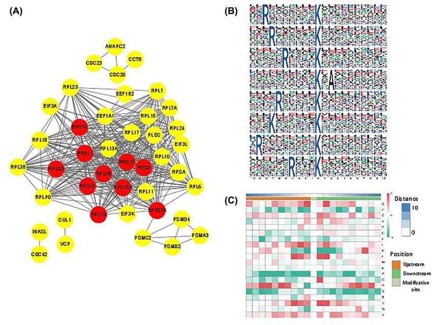
Protein–protein interaction network of differential modified proteins
Protein–protein interaction (PPI) network is composed of proteins through their interactions. In the network dia-
gram, nodes represent proteins, and nodes are labeled with the names of these proteins. The interaction between two
proteins is connected by wires. According to the protein interaction database of STRING (v.10.5) [42], we performed
PPI network by selecting the top 50 proteins based on degree. The interaction relationship of DUPs was extracted ac-
cording to the confidence score > 0.7 (high confidence) (Figure 4A; Supplementary Table S7). According to analysis
by software Cytoscape, the top ten hub proteins were listed (Table 3).
Motif analysis of protein modification
Protein motif analysis calculates the regular trend of amino acid sequences in the region of modification sites. This
kind of analysis can find the sequence characteristics, so as to speculate or determine the modification-related en-
zymes. We identified eight conserved ubiquitination motifs analyzed by Motif-X (Figure 4B). The enrichment of
specific amino acids neighboring the ubiquitination sites were exhibited (Figure 4C).
© 2021 The Author(s). This is an open access article published by Portland Press Limited on behalf of the Biochemical Society and distributed under the Creative Commons Attribution 7
License 4.0 (CC BY).Bioscience Reports (2021) 41 BSR20211532
https://doi.org/10.1042/BSR20211532
Downloaded from http://portlandpress.com/bioscirep/article-pdf/41/8/BSR20211532/919622/bsr-2021-1532.pdf by guest on 31 August 2021
Figure 4. PPI network and Motif analysis of ubiquitination sites
(A) PPI network analyses of differentially expressed ubiquitinated proteins analyzed by STRING database. The red circles marked
top ten hub proteins based on degree value analyzed by software Cytoscape. (B) Significantly enriched ubiquitination motifs by
Motif-X. (C) Motif enrichment heat map of ubiquitination. Red indicates that the amino acid is significantly enriched near the ubiq-
uitination sites, and green indicates that the amino acid is significantly reduced near the ubiquitination sites.
Table 3 Top ten hub proteins in PPI network based on degree value
Betweenness Neighborhood
Name Degree centrality Closeness centrality connectivity Clustering coefficient
RPS16 28 0.021698041 0.804878049 23.21428571 0.82010582
RPS11 28 0.021698041 0.804878049 23.21428571 0.82010582
RPS3 28 0.021698041 0.804878049 23.21428571 0.82010582
RPS17 28 0.021698041 0.804878049 23.21428571 0.82010582
RPS15A 27 0.020608616 0.785714286 23.2962963 0.823361823
RPS15 27 0.017387074 0.76744186 23.48148148 0.84045584
RPS27A 26 0.222127935 0.825 24.15384615 0.886153846
RPS23 26 0.00803387 0.76744186 24.38461538 0.907692308
RPS14 26 0.010250981 0.75 24.11538462 0.886153846
RPL19 25 0.002430965 0.75 25 0.96
Discussion
ACC of the salivary glands performs the properties of slow-growing, local and/or distant spread, nodal positivity
and high mortality, as well as high rate of occurrence and metastasis [43,44]. Several specific prognostic factors had
been identified the association with ACC [45]. Meanwhile, most of the related studies are based on the statistics and
8 © 2021 The Author(s). This is an open access article published by Portland Press Limited on behalf of the Biochemical Society and distributed under the Creative Commons Attribution
License 4.0 (CC BY).Bioscience Reports (2021) 41 BSR20211532
https://doi.org/10.1042/BSR20211532
correlation analysis of clinical cases, and lack of more detailed and in-depth biological mechanism research on the
occurrence and progress of the disease. The ubiquitination modification plays a significant role in cancer pathology
[46]. MS can be used to analyze the role of protein PTMs in human diseases, and PTM-based protein variants can be
explored as deeply as possible.
Ribosomal protein S27a (RPS27A) is the top differentially up-regulated protein in our identification. RPS27A per-
forms multifunction in ribosome biogenesis and protein PTMs, contributing to progression of leukemia or solid
tumors [47,48]. A ubiquitin-fused RPS27A protein (Uba80) was reported related to apoptotic cell death and overex-
pressed in colon and renal cancer [49–52]. However, the molecular mechanism of RPS27A-related ubiquitination in
tumors remains to be studied. Phosphoglycerate dehydrogenase (PHGDH) is the rate-limiting enzyme of de novo
serine biosynthesis pathway, which is closely related to the occurrence and development of many kinds of tumors
[53,54]. The serine synthesis during cancer progression was suppressed when PHGDH went through Parkin-related
ubiquitination and degradation [55,56]. PHGDH is also a ubiquitination substrate of RNF5 in the study of breast
Downloaded from http://portlandpress.com/bioscirep/article-pdf/41/8/BSR20211532/919622/bsr-2021-1532.pdf by guest on 31 August 2021
cancer cells [57]. The PHGDH ubiquitination in OACC has not been reported. TMEM87A, also named as Elkin1,
is important in cell–cell adhesin and metastasis with limited studies [58]. The insulin receptor (INSR) is a key reg-
ulator in metabolic homeostasis through diverse signal pathways including PI3K/AKT and MAPK [59]. Although
phosphorylation is critical in INSR-dependent signal cascade, the ubiquitin/proteasome system modulate degrada-
tion of transducers in this pathway [60,61]. The biological function of these targets in OACC and their combination
with metabolic abnormalities and immune regulation will drive us to further study how gene changes modulate the
behavior of cancer cells, so as to unlock more effective treatment methods.
In addition, ATP1A1, Tubulin α1b (TUBA1B) and integrin subunit α9 (ITGA9) are the top down-regulated ac-
cording to OACC T/OACC N ratio. As a membrane-bound ion pump, Na+ /K+ -ATPase shows tissue-specific profile
[62]. The overexpression of α1 subunit of Na+ /K+ -ATPase (ATP1A1) were observed in esophageal squamous cell
carcinoma [63], non-small-cell lung cancer and hepatocellular carcinoma, contributing to cancer proliferation and
migration [64,65]. However, ATP1A1 were significantly down-regulated in prostate cancer [66,67], colorectal can-
cer [68] and renal cell carcinoma [69]. The function and PTM regulation of ATP1A1 in OACC is not yet fully clear.
TUBA1B belongs to cytoskeleton compartment with a central function in cell shape maintenance and cellular process
regulation, especially in cell division [70]. The higher expression of TUBA1B and poor prognosis were reported in
hepatocellular carcinoma [71]. Interesting, the ubiquitination at different sites of TUBA1B shows different up- and
down-regulation trends in tumor tissues (Table 2). This suggests that TUBA1B ubiquitination regulation may have
different biological functions. With the continuous deepening of cytoskeleton-related research, the importance of
microtubules in tumor metastasis has begun to become prominent. The ITGA9 belongs to integrin protein family
and a partner of β1 subunit facilitating the interaction of cell–cell and cell–extracellular matrix [72]. Depletion of
ITGA9 suppressed breast cancer progression and metastasis through GSK3/β-catenin pathway [73]. The decreased
expression of ITGA9 in lung cancer indicated potential genetic and epigenetic regulation mechanism [74]. Besides
TUBA1B and ITGA9, other cell cytoskeleton and cell adhesion associated proteins were also identified in our results,
including ACTA2, MCAM, ACTG1, VIM and CDH11. Due to the high recurrence and metastasis of OACC, research
on the modification of cytoskeleton and cell adhesion will help to further clarify the mechanism of OACC metastasis.
Considering the hub genes of interaction regulation by Cytoscape analysis, we found they all belong to riboso-
mal proteins. Ribosomal proteins are abnormally expressed in a variety of tumors, which affect the apoptosis, aging,
growth, invasion, drug and radiation resistance [75]. The expression level of ribosomal protein has become a poten-
tial indicator of tumor diagnosis, treatment and prognosis [76]. Through the in-depth study of abnormal expression
and ubiquitination modification of ribosomal proteins in tumor tissue, we can further understand the role of high
expression of ribosomal protein gene in malignant tumor.
Protein ubiquitination plays a very important role in cellular processes such as subcellular localization, growth,
apoptosis and metabolism. The ubiquitination modification omics based on MS has been used for disease biomarkers
and pathogenic mechanism analysis [77,78]. In this project, we studied the ubiquitin proteomics of OACC tumor and
adjacent normal tissues, identified the different ubiquitin modification sites, analyzed the function of the identified
ubiquitinated proteins, and obtained eight protein clusters. Our work enriched the scope of OACC research and
became a reference for the development of novel targets. These results also supported the possible functions of the
ubiquitination of cell skeleton and extracellular matrix associated proteins in OACC development and metastasis.
However, whether the selected differential protein can be used as a therapeutic target for OACC still needs more
detailed in vivo and in vitro experiments to verify, so as to better carry out precise treatment for OACC, add further
knowledge specifically for patients and explore more promising combination therapy.
© 2021 The Author(s). This is an open access article published by Portland Press Limited on behalf of the Biochemical Society and distributed under the Creative Commons 9
Attribution License 4.0 (CC BY).Bioscience Reports (2021) 41 BSR20211532
https://doi.org/10.1042/BSR20211532
Data Availability
All data included in the present study are available upon request by contact with the corresponding authors.
Competing Interests
The authors declare that there are no competing interests associated with the manuscript.
Funding
The present study was supported by the Science and Technology Planning Project of Shenzhen [grant numbers
JCYJ20190807145815129, JCYJ20180306140810282]; the National Natural Science Foundation of China [grant number
82003114]; and the Applied Basic Research Project of Sichuan Science and Technology Department [grant number 2020YJ0174].
CRediT Author Contribution
Downloaded from http://portlandpress.com/bioscirep/article-pdf/41/8/BSR20211532/919622/bsr-2021-1532.pdf by guest on 31 August 2021
Wen Li: Data curation, Formal analysis, Funding acquisition, Validation, Investigation, Visualization, Methodology,
Writing—original draft, Writing—review and editing. Xiaobin Wang: Data curation, Software, Formal analysis, Validation, Inves-
tigation, Writing—review and editing. Qian Zhang: Data curation, Validation, Investigation, Methodology,. Hanlin Wang: Data
curation, Validation, Investigation, Visualization, Methodology. Wenxin Zuo: Data curation, Formal analysis, Project administra-
tion. Hongliang Xie: Resources, Data curation, Software. Jianming Tang: Resources, Data curation, Software, Methodology.
Mengmeng Wang: Data curation, Investigation, Visualization. Zhipeng Zeng: Formal analysis, Methodology. Wanxia Cai: Con-
ceptualization, Resources, Project administration. Donge Tang: Data curation, Formal analysis, Supervision, Funding acquisition,
Validation. Yong Dai: Resources, Data curation, Formal analysis, Supervision, Funding acquisition, Validation, Writing—review
and editing.
Ethics Approval and Consent to Participate
The study was carried out following the Declaration of Helsinki for experiments involving humans and was approved by the Medi-
cal Ethics Committee of Shenzhen People’s Hospital.
Abbreviations
ACC, adenoid cystic carcinoma; BP, biological process; CC, cellular component; COG, clusters of original groups; CYLD ,
cylindromatosis; DUP, differentially ubiquitinated protein; ER , Endoplasmic Reticulum; FDR , false discovery rate; GO, Gene
Ontology; IAA , iodoacetamide; INSR, insulin receptor; ITGA9, integrin subunit α9; KEGG, Kyoto Encyclopedia of Genes and
Genomes; MF, molecular function; MS, mass spectrometry; OACC, oral ACC; PHGDH, phosphoglycerate dehydrogenase; PPI,
protein–protein interaction; PSM , peptide-spectrum matches; PTM, post-translational modification; RPS27A, ribosomal protein
S27a; SACC , salivary adenoid cystic carcinoma; TCA , Trichloroaceticacid; TUBA1B, Tubulin α1b; UBA2, ubiquitin-like modifier
activating enzyme 2; UPLC , Ultra Performance Liquid Chromatography; USP22 , Ubiquitin-specific protease 22.
References
1 Stell, P.M. (1986) Adenoid cystic carcinoma. Clin. Otolaryngol. Allied Sci. 11, 267–291, https://doi.org/10.1111/j.1365-2273.1986.tb01928.x
2 Bradley, P.J. (2004) Adenoid cystic carcinoma of the head and neck: a review. Curr. Opin. Otolaryngol. Head Neck Surg. 12, 127–132,
https://doi.org/10.1097/00020840-200404000-00013
3 Li, N., Xu, L., Zhao, H., El-Naggar, A.K. and Sturgis, E.M. (2012) A comparison of the demographics, clinical features, and survival of patients with
adenoid cystic carcinoma of major and minor salivary glands versus less common sites within the Surveillance, Epidemiology, and End Results registry.
Cancer Am. Cancer Soc. 118, 3945–3953, https://doi.org/10.1002/cncr.26740
4 Jones, A.S., Hamilton, J.W., Rowley, H., Husband, D. and Helliwell, T.R. (1997) Adenoid cystic carcinoma of the head and neck. Clin. Otolaryngol. Allied
Sci. 22, 434–443, https://doi.org/10.1046/j.1365-2273.1997.00041.x
5 Spiro, R.H. and Huvos, A.G. (1992) Stage means more than grade in adenoid cystic carcinoma. Am. J. Surg. 164, 623–628,
https://doi.org/10.1016/S0002-9610(05)80721-4
6 Bradley, P.J. (2017) Adenoid cystic carcinoma evaluation and management: progress with optimism!. Curr. Opin. Otolaryngol. Head Neck Surg. 25,
147–153, https://doi.org/10.1097/MOO.0000000000000347
7 Lorini, L., Ardighieri, L., Bozzola, A., Romani, C., Bignotti, E., Buglione, M. et al. (2021) Prognosis and management of recurrent and/or metastatic head
and neck adenoid cystic carcinoma. Oral Oncol. 115, 105213, https://doi.org/10.1016/j.oraloncology.2021.105213
8 Li, Q., Huang, P., Zheng, C., Wang, J. and Ge, M. (2017) Prognostic significance of p53 immunohistochemical expression in adenoid cystic carcinoma of
the salivary glands: a meta-analysis. Oncotarget 8, 29458–29473, https://doi.org/10.18632/oncotarget.15297
9 Thierauf, J., Weissinger, S.E., Veit, J.A., Affolter, A., Laureano, N.K., Beutner, D. et al. (2018) Low SOX2 expression marks a distinct subset of adenoid
cystic carcinoma of the head and neck and is associated with an advanced tumor stage. PLoS ONE 13, e194989,
https://doi.org/10.1371/journal.pone.0194989
10 Bazarsad, S., Kim, J.Y., Zhang, X., Kim, K.Y., Lee, D.Y., Ryu, M.H. et al. (2018) Ataxia-telangiectasia-mutated protein expression as a prognostic marker
in adenoid cystic carcinoma of the salivary glands. Yonsei Med. J. 59, 717–726, https://doi.org/10.3349/ymj.2018.59.6.717
10 © 2021 The Author(s). This is an open access article published by Portland Press Limited on behalf of the Biochemical Society and distributed under the Creative Commons
Attribution License 4.0 (CC BY).Bioscience Reports (2021) 41 BSR20211532
https://doi.org/10.1042/BSR20211532
11 Li, H., Liao, X., Liu, Y., Shen, Z., Gan, X., Li, H. et al. (2015) The expression of MACC1 and its role in the proliferation and apoptosis of salivary adenoid
cystic carcinoma. J. Oral Pathol. Med. 44, 810–817, https://doi.org/10.1111/jop.12309
12 Liu, C., Jiang, Y.H., Zhao, Z.L., Wu, H.W., Zhang, L., Yang, Z. et al. (2019) Knockdown of histone methyltransferase WHSC1 induces apoptosis and
inhibits cell proliferation and tumorigenesis in salivary adenoid cystic carcinoma. Anticancer Res. 39, 2729–2737,
https://doi.org/10.21873/anticanres.13399
13 Lee, S.J., Chung, K.Y., Kwon, J.E., Yoon, S.O. and Kim, S.K. (2018) Expression of EpCAM in adenoid cystic carcinoma. Pathology 50, 737–741,
https://doi.org/10.1016/j.pathol.2018.08.013
14 Klein, N.T., Valstar, M.H., Smit, L.A., Smeele, L.E., Zuithoff, N., de Keizer, B. et al. (2020) Prostate-specific membrane antigen (PSMA) expression in
adenoid cystic carcinoma of the head and neck. BMC Cancer 20, 519, https://doi.org/10.1186/s12885-020-06847-9
15 Liang, Y., Jiao, J., Liang, L., Zhang, J., Lu, Y., Xie, H. et al. (2018) Tumor necrosis factor receptor-associated factor 6 mediated the promotion of salivary
adenoid cystic carcinoma progression through Smad-p38-JNK signaling pathway induced by TGF-beta. J. Oral Pathol. Med. 47, 583–589,
https://doi.org/10.1111/jop.12709
16 Chen, W., Ren, X., Wu, J., Gao, X., Cen, X., Wang, S. et al. (2018) HSP27 associates with epithelial-mesenchymal transition, stemness and
Downloaded from http://portlandpress.com/bioscirep/article-pdf/41/8/BSR20211532/919622/bsr-2021-1532.pdf by guest on 31 August 2021
radioresistance of salivary adenoid cystic carcinoma. J. Cell. Mol. Med. 22, 2283–2298, https://doi.org/10.1111/jcmm.13510
17 Jiang, Y.P., Tang, Y.L., Wang, S.S., Wu, J.S., Zhang, M., Pang, X. et al. (2020) PRRX1-induced epithelial-to-mesenchymal transition in salivary adenoid
cystic carcinoma activates the metabolic reprogramming of free fatty acids to promote invasion and metastasis. Cell Prolif. 53, e12705,
https://doi.org/10.1111/cpr.12705
18 de Mendonca, R.P., Chemelo, G.P., Mitre, G.P., Branco, D.C., Da, C.N., Tuji, F.M. et al. (2020) Role of hypoxia-related proteins in adenoid cystic
carcinoma invasion. Diagn. Pathol. 15, 47, https://doi.org/10.1186/s13000-020-00967-3
19 Sakane, T., Murase, T., Okuda, K., Saida, K., Masaki, A., Yamada, T. et al. (2019) A mutation analysis of the EGFR pathway genes, RAS, EGFR, PIK3CA,
AKT1 and BRAF, and TP53 gene in thymic carcinoma and thymoma type A/B3. Histopathology 75, 755–766, https://doi.org/10.1111/his.13936
20 Stenman, G., Persson, F. and Andersson, M.K. (2014) Diagnostic and therapeutic implications of new molecular biomarkers in salivary gland cancers.
Oral Oncol. 50, 683–690, https://doi.org/10.1016/j.oraloncology.2014.04.008
21 Park, S., Vora, M., van Zante, A., Humtsoe, J., Kim, H.S., Yom, S. et al. (2020) Clinicopathologic implications of Myb and Beta-catenin expression in
adenoid cystic carcinoma. J. Otolaryngol. Head Neck Surg. 49, 48, https://doi.org/10.1186/s40463-020-00446-1
22 Togashi, Y., Dobashi, A., Sakata, S., Sato, Y., Baba, S., Seto, A. et al. (2018) MYB and MYBL1 in adenoid cystic carcinoma: diversity in the mode of
genomic rearrangement and transcripts. Mod. Pathol. 31, 934–946, https://doi.org/10.1038/s41379-018-0008-8
23 Warner, K.A., Oklejas, A.E., Pearson, A.T., Zhang, Z., Wu, W., Divi, V. et al. (2018) UM-HACC-2A: MYB-NFIB fusion-positive human adenoid cystic
carcinoma cell line. Oral Oncol. 87, 21–28, https://doi.org/10.1016/j.oraloncology.2018.10.012
24 Xie, J., Lin, L.S., Huang, X.Y., Gan, R.H., Ding, L.C., Su, B.H. et al. (2020) The NOTCH1-HEY1 pathway regulates self-renewal and
epithelial-mesenchymal transition of salivary adenoid cystic carcinoma cells. Int. J. Biol. Sci. 16, 598–610, https://doi.org/10.7150/ijbs.36407
25 Branco, K., Ribeiro, A., de Mendonca, R.P., de Jesus, V.P.J., Da, S.K.M., Arnaud, M. et al. (2018) Abnormal activation of the Akt signaling pathway in
adenoid cystic carcinoma. Eur. Arch. Otorhinolaryngol. 275, 3039–3047, https://doi.org/10.1007/s00405-018-5182-2
26 Ouyang, D.Q., Liang, L.Z., Ke, Z.F., Zheng, G.S., Weng, D.S., Yang, W.F. et al. (2017) Association between high expression of phosphorylated Akt and
mammalian target of rapamycin and improved survival in salivary gland adenoid cystic carcinoma. Head Neck 39, 1145–1154,
https://doi.org/10.1002/hed.24732
27 Walsh, C.T., Garneau-Tsodikova, S. and Gatto, G.J. (2005) Protein posttranslational modifications: the chemistry of proteome diversifications. Angew.
Chem. Int. Ed. Engl. 44, 7342–7372, https://doi.org/10.1002/anie.200501023
28 Santos, A.L. and Lindner, A.B. (2017) Protein posttranslational modifications: roles in aging and age-related disease. Oxid. Med. Cell. Longev. 2017,
5716409, https://doi.org/10.1155/2017/5716409
29 Hochstrasser, M. (2009) Origin and function of ubiquitin-like proteins. Nature 458, 422–429, https://doi.org/10.1038/nature07958
30 Lee, I. and Schindelin, H. (2008) Structural insights into E1-catalyzed ubiquitin activation and transfer to conjugating enzymes. Cell 134, 268–278,
https://doi.org/10.1016/j.cell.2008.05.046
31 Amerik, A.Y. and Hochstrasser, M. (2004) Mechanism and function of deubiquitinating enzymes. Biochim. Biophys. Acta 1695, 189–207,
https://doi.org/10.1016/j.bbamcr.2004.10.003
32 Nijman, S.M., Luna-Vargas, M.P., Velds, A., Brummelkamp, T.R., Dirac, A.M., Sixma, T.K. et al. (2005) A genomic and functional inventory of
deubiquitinating enzymes. Cell 123, 773–786, https://doi.org/10.1016/j.cell.2005.11.007
33 Eisenberg-Lerner, A., Ciechanover, A. and Merbl, Y. (2016) Post-translational modification profiling - a novel tool for mapping the protein modification
landscape in cancer. Exp. Biol. Med. (Maywood) 241, 1475–1482, https://doi.org/10.1177/1535370216651732
34 Schwartz, A.L. and Ciechanover, A. (2009) Targeting proteins for destruction by the ubiquitin system: implications for human pathobiology. Annu. Rev.
Pharmacol. Toxicol. 49, 73–96, https://doi.org/10.1146/annurev.pharmtox.051208.165340
35 Scheffner, M., Huibregtse, J.M., Vierstra, R.D. and Howley, P.M. (1993) The HPV-16 E6 and E6-AP complex functions as a ubiquitin-protein ligase in the
ubiquitination of p53. Cell 75, 495–505, https://doi.org/10.1016/0092-8674(93)90384-3
36 Popovic, D., Vucic, D. and Dikic, I. (2014) Ubiquitination in disease pathogenesis and treatment. Nat. Med. 20, 1242–1253,
https://doi.org/10.1038/nm.3739
37 Feng, X., Matsuo, K., Zhang, T., Hu, Y., Mays, A.C., Browne, J.D. et al. (2017) MicroRNA profiling and target genes related to metastasis of salivary
adenoid cystic carcinoma. Anticancer Res. 37, 3473–3481
38 Dai, W., Yao, Y., Zhou, Q. and Sun, C.F. (2014) Ubiquitin-specific peptidase 22, a histone deubiquitinating enzyme, is a novel poor prognostic factor for
salivary adenoid cystic carcinoma. PLoS ONE 9, e87148, https://doi.org/10.1371/journal.pone.0087148
© 2021 The Author(s). This is an open access article published by Portland Press Limited on behalf of the Biochemical Society and distributed under the Creative Commons 11
Attribution License 4.0 (CC BY).Bioscience Reports (2021) 41 BSR20211532
https://doi.org/10.1042/BSR20211532
39 Fukuda, M., Fukuda, F., Horiuchi, Y., Oku, Y., Suzuki, S., Kusama, K. et al. (2006) Expression of CYLD, NF-kappaB and NF-kappaB-related factors in
salivary gland tumors. In Vivo 20, 467–472
40 Yen, H.C., Xu, Q., Chou, D.M., Zhao, Z. and Elledge, S.J. (2008) Global protein stability profiling in mammalian cells. Science 322, 918–923,
https://doi.org/10.1126/science.1160489
41 Kirkpatrick, D.S., Denison, C. and Gygi, S.P. (2005) Weighing in on ubiquitin: the expanding role of mass-spectrometry-based proteomics. Nat. Cell Biol.
7, 750–757, https://doi.org/10.1038/ncb0805-750
42 Szklarczyk, D., Franceschini, A., Wyder, S., Forslund, K., Heller, D., Huerta-Cepas, J. et al. (2015) STRING v10: protein-protein interaction networks,
integrated over the tree of life. Nucleic Acids Res. 43, D447–D452, https://doi.org/10.1093/nar/gku1003
43 Huang, M., Ma, D., Sun, K., Yu, G., Guo, C. and Gao, F. (1997) Factors influencing survival rate in adenoid cystic carcinoma of the salivary glands. Int. J.
Oral Maxillofac. Surg. 26, 435–439, https://doi.org/10.1016/S0901-5027(97)80008-2
44 Khafif, A., Anavi, Y., Haviv, J., Fienmesser, R., Calderon, S. and Marshak, G. (2005) Adenoid cystic carcinoma of the salivary glands: a 20-year review
with long-term follow-up. Ear Nose Throat J. 84, 662,664–667, https://doi.org/10.1177/014556130508401016
45 Terhaard, C.H., Lubsen, H., Van der Tweel, I., Hilgers, F.J., Eijkenboom, W.M., Marres, H.A. et al. (2004) Salivary gland carcinoma: independent
Downloaded from http://portlandpress.com/bioscirep/article-pdf/41/8/BSR20211532/919622/bsr-2021-1532.pdf by guest on 31 August 2021
prognostic factors for locoregional control, distant metastases, and overall survival: results of the Dutch head and neck oncology cooperative group.
Head Neck 26, 681–692, 692-693, https://doi.org/10.1002/hed.10400
46 Foot, N., Henshall, T. and Kumar, S. (2017) Ubiquitination and the regulation of membrane proteins. Physiol. Rev. 97, 253–281,
https://doi.org/10.1152/physrev.00012.2016
47 Wang, H., Yu, J., Zhang, L., Xiong, Y., Chen, S., Xing, H. et al. (2014) RPS27a promotes proliferation, regulates cell cycle progression and inhibits
apoptosis of leukemia cells. Biochem. Biophys. Res. Commun. 446, 1204–1210, https://doi.org/10.1016/j.bbrc.2014.03.086
48 Gunasekaran, V.P. and Ganeshan, M. (2014) Inverse correlation of ribosomal protein S27A and multifunctional protein YB-1 in hepatocellular carcinoma.
Clin. Biochem. 47, 1262–1264, https://doi.org/10.1016/j.clinbiochem.2014.05.004
49 Kirschner, L.S. and Stratakis, C.A. (2000) Structure of the human ubiquitin fusion gene Uba80 (RPS27a) and one of its pseudogenes. Biochem. Biophys.
Res. Commun. 270, 1106–1110, https://doi.org/10.1006/bbrc.2000.2568
50 Han, X.J., Lee, M.J., Yu, G.R., Lee, Z.W., Bae, J.Y., Bae, Y.C. et al. (2012) Altered dynamics of ubiquitin hybrid proteins during tumor cell apoptosis. Cell
Death Dis. 3, e255, https://doi.org/10.1038/cddis.2011.142
51 Barnard, G.F., Mori, M., Staniunas, R.J., Begum, N.A., Bao, S., Puder, M. et al. (1995) Ubiquitin fusion proteins are overexpressed in colon cancer but
not in gastric cancer. Biochim. Biophys. Acta 1272, 147–153, https://doi.org/10.1016/0925-4439(95)00079-8
52 Kanayama, H., Tanaka, K., Aki, M., Kagawa, S., Miyaji, H., Satoh, M. et al. (1991) Changes in expressions of proteasome and ubiquitin genes in human
renal cancer cells. Cancer Res. 51, 6677–6685
53 Yoshino, H., Enokida, H., Osako, Y., Nohata, N., Yonemori, M., Sugita, S. et al. (2020) Characterization of PHGDH expression in bladder cancer: potential
targeting therapy with gemcitabine/cisplatin and the contribution of promoter DNA hypomethylation. Mol. Oncol. 14, 2190–2202,
https://doi.org/10.1002/1878-0261.12697
54 Song, Z., Feng, C., Lu, Y., Lin, Y. and Dong, C. (2018) PHGDH is an independent prognosis marker and contributes cell proliferation, migration and
invasion in human pancreatic cancer. Gene 642, 43–50, https://doi.org/10.1016/j.gene.2017.11.014
55 Pencheva, R. and Todorov, T. (1988) Genetic analysis of heteroclones of Streptomyces erythreus. I. Determination of the arrangement of genetic loci on
its map. Acta. Microbiol. Bulg. 23, 3–10
56 Dalton, W.B. (2020) Parkin on serine: a Parkinson disease gene suppresses serine synthesis in cancer. J. Clin. Invest. 130, 2820–2822,
https://doi.org/10.1172/JCI137411
57 Wang, C., Wan, X., Yu, T., Huang, Z., Shen, C., Qi, Q. et al. (2020) Acetylation stabilizes phosphoglycerate dehydrogenase by disrupting the interaction
of E3 ligase RNF5 to promote breast tumorigenesis. Cell Rep. 32, 108021, https://doi.org/10.1016/j.celrep.2020.108021
58 Patkunarajah, A., Stear, J.H., Moroni, M., Schroeter, L., Blaszkiewicz, J., Tearle, J.L. et al. (2020) TMEM87a/Elkin1, a component of a novel
mechanoelectrical transduction pathway, modulates melanoma adhesion and migration. eLife 9, e53308, https://doi.org/10.7554/eLife.53308
59 Payankaulam, S., Raicu, A.M. and Arnosti, D.N. (2019) Transcriptional regulation of INSR, the insulin receptor gene. Genes (Basel) 10,
https://doi.org/10.3390/genes10120984
60 Balaji, V., Pokrzywa, W. and Hoppe, T. (2018) Ubiquitylation pathways in insulin signaling and organismal homeostasis. Bioessays 40, e1700223,
https://doi.org/10.1002/bies.201700223
61 Nagarajan, A., Petersen, M.C., Nasiri, A.R., Butrico, G., Fung, A., Ruan, H.B. et al. (2016) MARCH1 regulates insulin sensitivity by controlling cell surface
insulin receptor levels. Nat. Commun. 7, 12639, https://doi.org/10.1038/ncomms12639
62 Lingrel, J.B. and Kuntzweiler, T. (1994) Na+,K(+)-ATPase. J. Biol. Chem. 269, 19659–19662, https://doi.org/10.1016/S0021-9258(17)32067-7
63 Wu, I.C., Chen, Y.K., Wu, C.C., Cheng, Y.J., Chen, W.C., Ko, H.J. et al. (2016) Overexpression of ATPase Na+/+ transporting alpha 1 polypeptide, ATP1A1,
correlates with clinical diagnosis and progression of esophageal squamous cell carcinoma. Oncotarget 7, 85244–85258,
https://doi.org/10.18632/oncotarget.13267
64 Mijatovic, T., Roland, I., Van Quaquebeke, E., Nilsson, B., Mathieu, A., Van Vynckt, F. et al. (2007) The alpha1 subunit of the sodium pump could
represent a novel target to combat non-small cell lung cancers. J. Pathol. 212, 170–179, https://doi.org/10.1002/path.2172
65 Zhuang, L., Xu, L., Wang, P., Jiang, Y., Yong, P., Zhang, C. et al. (2015) Na+/K+-ATPase alpha1 subunit, a novel therapeutic target for hepatocellular
carcinoma. Oncotarget 6, 28183–28193, https://doi.org/10.18632/oncotarget.4726
66 Mobasheri, A., Fox, R., Evans, I., Cullingham, F., Martin-Vasallo, P. and Foster, C.S. (2003) Epithelial Na, K-ATPase expression is down-regulated in
canine prostate cancer; a possible consequence of metabolic transformation in the process of prostate malignancy. Cancer Cell Int. 3, 8,
https://doi.org/10.1186/1475-2867-3-8
12 © 2021 The Author(s). This is an open access article published by Portland Press Limited on behalf of the Biochemical Society and distributed under the Creative Commons
Attribution License 4.0 (CC BY).Bioscience Reports (2021) 41 BSR20211532
https://doi.org/10.1042/BSR20211532
67 Li, Z., Zhang, Z., Xie, J.X., Li, X., Tian, J., Cai, T. et al. (2011) Na/K-ATPase mimetic pNaKtide peptide inhibits the growth of human cancer cells. J. Biol.
Chem. 286, 32394–32403, https://doi.org/10.1074/jbc.M110.207597
68 Sakai, H., Suzuki, T., Maeda, M., Takahashi, Y., Horikawa, N., Minamimura, T. et al. (2004) Up-regulation of Na(+),K(+)-ATPase alpha 3-isoform and
down-regulation of the alpha1-isoform in human colorectal cancer. FEBS Lett. 563, 151–154, https://doi.org/10.1016/S0014-5793(04)00292-3
69 Zhang, D., Zhang, P., Yang, P., He, Y., Wang, X., Yang, Y. et al. (2017) Downregulation of ATP1A1 promotes cancer development in renal cell carcinoma.
Clin. Proteomics 14, 15, https://doi.org/10.1186/s12014-017-9150-4
70 Glotzer, M. (2009) The 3Ms of central spindle assembly: microtubules, motors and MAPs. Nat. Rev. Mol. Cell Biol. 10, 9–20,
https://doi.org/10.1038/nrm2609
71 Lu, C., Zhang, J., He, S., Wan, C., Shan, A., Wang, Y. et al. (2013) Increased alpha-tubulin1b expression indicates poor prognosis and resistance to
chemotherapy in hepatocellular carcinoma. Dig. Dis. Sci. 58, 2713–2720, https://doi.org/10.1007/s10620-013-2692-z
72 Hoye, A.M., Couchman, J.R., Wewer, U.M., Fukami, K. and Yoneda, A. (2012) The newcomer in the integrin family: integrin alpha9 in biology and
cancer. Adv. Biol. Regul. 52, 326–339, https://doi.org/10.1016/j.jbior.2012.03.004
73 Wang, Z., Li, Y., Xiao, Y., Lin, H.P., Yang, P., Humphries, B. et al. (2019) Integrin alpha9 depletion promotes beta-catenin degradation to suppress
Downloaded from http://portlandpress.com/bioscirep/article-pdf/41/8/BSR20211532/919622/bsr-2021-1532.pdf by guest on 31 August 2021
triple-negative breast cancer tumor growth and metastasis. Int. J. Cancer 145, 2767–2780, https://doi.org/10.1002/ijc.32359
74 Anedchenko, E.A., Dmitriev, A.A., Krasnov, G.S., Kondrat’Eva, T.T., Kopantsev, E.P., Vinogradova, T.V. et al. (2008) [Down-regulation of RBSP3/CTDSPL,
NPRL2/G21, RASSF1A, ITGA9, HYAL1 and HYAL2 genes in non-small cell lung cancer]. Mol. Biol. (Mosk.) 42, 965–976,
https://doi.org/10.1134/S0026893308060058
75 Lessard, F., Brakier-Gingras, L. and Ferbeyre, G. (2019) Ribosomal proteins control tumor suppressor pathways in response to nucleolar stress.
Bioessays 41, e1800183, https://doi.org/10.1002/bies.201800183
76 Panda, A., Yadav, A., Yeerna, H., Singh, A., Biehl, M., Lux, M. et al. (2020) Tissue- and development-stage-specific mRNA and heterogeneous CNV
signatures of human ribosomal proteins in normal and cancer samples. Nucleic Acids Res. 48, 7079–7098
77 Sun, Y., Zheng, X., Yuan, H., Chen, G., Ouyang, J., Liu, J. et al. (2020) Proteomic analyses reveal divergent ubiquitylation patterns in hepatocellula
carcinoma cell lines with different metastasis potential. J. Proteomics 225, 103834, https://doi.org/10.1016/j.jprot.2020.103834
78 Liu, M., Yan, M., Lv, H., Wang, B., Lv, X., Zhang, H. et al. (2020) Macrophage K63-linked ubiquitination of YAP promotes its nuclear localization and
exacerbates atherosclerosis. Cell Rep. 32, https://doi.org/10.1016/j.celrep.2020.107990
© 2021 The Author(s). This is an open access article published by Portland Press Limited on behalf of the Biochemical Society and distributed under the Creative Commons Attribution 13
License 4.0 (CC BY).You can also read


