Drosophila sessile hemocyte clusters are true hematopoietic tissues that regulate larval blood cell differentiation
←
→
Page content transcription
If your browser does not render page correctly, please read the page content below
RESEARCH ARTICLE
           elifesciences.org

                                      Drosophila sessile hemocyte clusters are
                                      true hematopoietic tissues that regulate
                                      larval blood cell differentiation
                                      Alexandre B Leitão1*, Élio Sucena1,2*
                                      1
                                       Instituto Gulbenkian de Ciência, Oeiras, Portugal; 2Departamento de Biologia
                                      Animal, Universidade de Lisboa, Lisbon, Portugal

                                      Abstract Virtually all species of coelomate animals contain blood cells that display a division of
                                      labor necessary for homeostasis. This functional partition depends upon the balance between
                                      proliferation and differentiation mostly accomplished in the hematopoietic organs. In Drosophila
                                      melanogaster, the lymph gland produces plasmatocytes and crystal cells that are not released until
                                      pupariation. Yet, throughout larval development, both hemocyte types increase in numbers.
                                      Mature plasmatocytes can proliferate but it is not known if crystal cell numbers increase by
                                      self-renewal or by de novo differentiation. We show that new crystal cells in third instar larvae
                                      originate through a Notch-dependent process of plasmatocyte transdifferentiation. This process
                                      occurs in the sessile clusters and is contingent upon the integrity of these structures. The existence of
                                      this hematopoietic tissue, relying on structure-dependent signaling events to promote blood
                                      homeostasis, creates a new paradigm for addressing outstanding questions in Drosophila
                                      hematopoiesis and establishing further parallels with vertebrate systems.
                                      DOI: 10.7554/eLife.06166.001

                                      Introduction
*For correspondence: aleitao@         In insects, the functions of hemocytes (blood cells) are very diverse and include phagocytosis,
igc.gulbenkian.pt (ABL);
                                      extracellular matrix deposition, AMP production, encapsulation, and melanization. Similarly to what
esucena@igc.gulbenkian.pt (ÉS)
                                      happens in vertebrates, the different functions performed by insect hemocytes are, to some degree,
Competing interests: The              compartmentalized into different cell types (Honti et al., 2014). Some mature blood cells retain the
authors declare that no               ability to divide when in circulation, but the majority of blood cell proliferation and differentiation
competing interests exist.            occurs in the hematopoietic organs (Grigorian and Hartenstein, 2013). These organs provide the
Funding: See page 14                  correct cellular and molecular environment for the control of cell proliferation and differentiation,
                                      namely in the so-called stem cell niches (Koch and Radtke, 2007; Martinez-Agosto et al., 2007).
Received: 19 December 2014
                                      Thus, the study of hematopoietic organs structure and function is essential to understand how
Accepted: 03 February 2015
                                      different mature blood cells arise and how their absolute and relative numbers are controlled.
Published: 04 February 2015
                                          In Drosophila melanogaster, embryonic hematopoiesis produces two different types of mature
Reviewing editor: Utpal               hemocytes: plasmatocytes and crystal cells. Plasmatocytes are phagocytic cells often functionally
Banerjee, University of California,   compared to vertebrate macrophages (Evans et al., 2003). Crystal cells are non-phagocytic cells
Los Angeles, United States
                                      known to produce prophenoloxidase, an essential component of the melanization cascade (Binggeli
    Copyright Leitão and Sucena.     et al., 2014). Both plasmatocytes and crystal cells generated during embryogenesis persist into larval
This article is distributed under     stages. Hemocytes in the larva can be found in circulation but the majority of them are attached to the
the terms of the Creative             cuticular epidermis as sessile cells (Lanot et al., 2001; Kurucz et al., 2007b; Makhijani et al., 2011).
Commons Attribution License,          Hemocytes attached to the epidermis are not randomly dispersed but form stereotypical clusters of
which permits unrestricted use        cells in every segment of the larva (Zettervall et al., 2004; Makhijani et al., 2011) indicating that
and redistribution provided that
                                      some signal must direct hemocytes to these locations. In fact, it has been recently shown that
the original author and source are
                                      peripheral nervous system (PNS) neurons attract hemocytes and provide unknown trophic molecules
credited.

Leitão and Sucena. eLife 2015;4:e06166. DOI: 10.7554/eLife.06166                                                                        1 of 16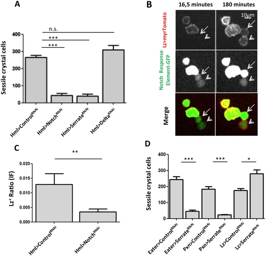
Research article                                                                            Developmental biology and stem cells

                                  eLife digest Insects have several different types of blood cell, many of which are unable to divide
                                  to form new cells once they have matured. Instead, fresh blood cells are normally generated in
                                  specialized ‘hematopoietic’ organs, such as the lymph gland in the Drosophila species of fruit fly. The
                                  structure of these organs plays an important role in controlling how new blood cells develop.
                                      Drosophila embryos make two types of blood cell: plasmatocytes and crystal cells. Both defend
                                  against harmful microorganisms, but in different ways. Plasmatocytes engulf and destroy invaders,
                                  whereas crystal cells release chemicals that encase microbes in a hardened gel. The blood cells made
                                  in the Drosophila embryo are still present once the fly enters its larval stages. At this stage of
                                  development, most of the blood cells are found in clusters attached to the cuticle that covers the
                                  larva’s surface, but a few circulate freely around the larva’s body.
                                      As a Drosophila larva develops, the number of blood cells in the larva increases. However,
                                  previous work has shown these additional blood cells are not normally released from the lymph gland
                                  of the larva. Furthermore, mature crystal cells do not appear to form new cells by dividing in two.
                                      Leitão and Sucena now show that the stationary clusters of blood cells produce new crystal cells in
                                  Drosophila larvae. Within the clusters, plasmatocytes are made to turn into crystal cells via
                                  a signaling pathway controlled by a protein called Notch. This pathway was already known to be
                                  essential for forming crystal cells. Leitão and Sucena also show that the structure of the clusters
                                  influences whether crystal cells are made, which means that the clusters can be considered to be
                                  hematopoietic tissue.
                                      It is now important to compare how the production of the same cell type is controlled in two
                                  distinct hematopoietic structures: the clusters and the lymph gland. From this comparison, general
                                  principles may be drawn and tested in other systems, including vertebrates.
                                  DOI: 10.7554/eLife.06166.002

                                  for their survival (Makhijani et al., 2011). The larva also possesses a hematopoietic organ, the lymph
                                  gland, where plasmatocytes and crystal cells differentiate from prohemocytes (Crozatier and
                                  Meister, 2007). Prohemocytes residing in the medullary zone of the lymph gland are instructed by
                                  cells from the posterior signaling center (PSC) to maintain their quiescent state or to differentiate into
                                  mature plasmatocytes or crystal cells (Crozatier et al., 2004; Mandal et al., 2007). During the
                                  differentiation process, it has been suggested that cells migrate and occupy the most cortical zone of
                                  the lymph gland (Jung et al., 2005; Krzemien et al., 2010b). An essential aspect of the Drosophila
                                  larval hematopoiesis is that hemocytes produced in the lymph gland do not disperse from the organ
                                  until pupariation or upon injury such as parasitoid wasp egg infection (Holz et al., 2003; Honti et al.,
                                  2010). Hence, in homeostatic conditions, differentiated hemocytes in the lymph gland do not
                                  contribute to the circulating and sessile hemocyte population. Nonetheless, the hemocyte population
                                  found in circulation and in sessile patches expands throughout larval development. Plasmatocytes are
                                  mitotically active cells (Rizki, 1957; Lanot et al., 2001) expanding during larval development by self
                                  renewal (Makhijani et al., 2011). On the other hand, all reports thus far concur in that mature crystal
                                  cells do not divide during larval stages (Krzemien et al., 2010b; Lanot et al., 2001; Rizki, 1957),
                                  although they have been shown to proliferate during embryogenesis (Lebestky et al., 2000). Further
                                  characterization of a yet unknown source and undetermined mechanism of crystal cell differentiation is
                                  required to understand how its number increases during larval development.
                                      Although little is known on how crystal cells are formed outside the lymph gland, it has been shown
                                  that Notch signaling is necessary to form these cells (Duvic et al., 2002; Lebestky et al., 2003). In the
                                  lymph gland, the role of Notch signaling in crystal cell formation is cell autonomous (Mukherjee et al.,
                                  2011). Notch activation is sufficient in hemocytes to induce the expression of lozenge, the first known
                                  transcription factor in crystal cell development (Lebestky et al., 2000). One particularity of Notch
                                  signaling is that it requires cell contact since the two known Drosophila Notch ligands, Serrate and
                                  Delta, are membrane bound proteins (Fiúza and Arias, 2007). In the lymph gland, Serrate-positive
                                  hemocytes induce neighboring cells to adopt crystal cell fates (Lebestky et al., 2003; Mukherjee
                                  et al., 2011; Ferguson & Martinez-agosto 2014). Outside the lymph gland, only in sessile clusters
                                  may we observe hemocytes establishing stable cell–cell contacts between them (Lanot et al., 2001).
                                  In fact, hemocytes in clusters are densely packed and linked through interdigitations (Lanot et al., 2001),

Leitão and Sucena. eLife 2015;4:e06166. DOI: 10.7554/eLife.06166                                                                      2 of 16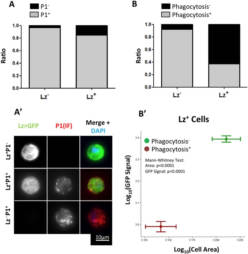
Research article                                                                             Developmental biology and stem cells

                                  particularly in the last two abdominal larval segments, the putative posterior hematopoietic tissue
                                  (PHT) (Kurucz et al., 2007).
                                      Indeed, in recent years, the idea that hematopoietic properties must reside outside of the lymph
                                  gland has been put forward explicitly by the Andó laboratory (Márkus et al., 2009). Firstly, in a
                                  descriptive endeavor by Kurucz et al. (2007) where an operational posterior hematopoietic tissue
                                  (PHT) consisting of the sessile hemocyte clusters in the last two abdominal segments is postulated;
                                  and later in a set of experiments showing that hemocytes taken from these clusters have the ability to
                                  differentiate into lamellocytes upon transfer to a different larva (Márkus et al., 2009; Honti et al.,
                                  2010). Importantly, sessile hemocytes in clusters constitute the biggest compartment of hemocytes in
                                  the larva (Lanot et al., 2001; Makhijani et al., 2011), contained within epidermal and muscle tissue in
                                  a structure that has been called hematopoietic pockets (Makhijani et al., 2011). Moreover, the sessile
                                  plasmatocytes in such clusters have a higher division rate than those in circulation (Makhijani et al.,
                                  2011). However, to consider the hemocyte clusters as a bona fide hematopoietic tissue, evidence is
                                  needed that their structure is necessary to control cell proliferation and/or cell fate decisions.
                                      In this study, we directly test the hypothesis that the hemocyte clusters constitute a hematopoietic
                                  tissue by addressing systematically the following questions: (i) are crystal cells differentiating in these
                                  clusters? (ii) is the structure/architecture of these clusters necessary for this function? and (iii) what is
                                  the role of the Notch pathway in this hematopoietic role?

                                  Results
                                  Crystal cell numbers increase during larval development through de
                                  novo differentiation
                                  In homeostatic conditions, the embryonic-derived hemocyte population consists of plasmatocytes and
                                  crystal cells. It is possible to distinguish these two cell types with several combinations of cell markers
                                  (Lebestky et al., 2000) such as the two live genetic drivers: HemolectinΔ-nuclearDsRed (Clark et al.,
                                  2011) and Lozenge-GAL4 in combination with UAS-EGFP/mCD8GFP (Lebestky et al., 2000). With
                                  this combination of markers we can distinguish across the larval cuticle, Hml+Lz− from Hml+Lz+ sessile
                                  hemocytes (Figure 1A,A′). Lozenge is the first marker known in the genetic cascade that leads to
                                  crystal cell differentiation and its expression is maintained as the cell matures (Lebestky et al., 2000).
                                  Hemolectin promoter has been used in different Drosophila transgenic lines to mark the majority of
                                  hemocytes (Sinenko and Mathey-Prevot, 2004; Clark et al., 2011). Hence, Hml+Lz+ cells are fully
                                  mature crystal cells or differentiating crystal cells while Hml+Lz− cells are plasmatocytes. During
                                  maturation, crystal cells loose HmlΔ-GAL4 expression (Mukherjee et al., 2011). The same is observed
                                  with HmlΔ-nuclearDsRed but only in rare cells. This difference may be explained by the different
                                  degradation times of nuclearDsRed and cytoplasmatic GFP. In fact, it is possible to detect a higher
                                  proportion of Hml−Lz+ cells with HmlΔ-cytoplasmaticDsRed (Figure 1—figure supplement 1).
                                  Another characteristic that distinguishes plasmatocytes from crystal cells is that the former tend to
                                  increase in size as they mature (Terriente-Felix et al., 2013). Measuring hemocytes cell areas in the
                                  three different populations of cells, we can observe a clear difference in size distributions between
                                  Hml+Lz−, Hml+Lz+, and Hml−Lz+ cells (Figure 1—figure supplement 2).
                                      When quantifying total hemocyte counts throughout larva development, it is undisputed that both
                                  plasmatocytes and crystal cell numbers increase (Rizki, 1957; Lanot et al., 2001; Makhijani et al.,
                                  2011). Here, we focus on third instar larvae because this is the developmental window in which the
                                  majority of larval hemocytes are originated (Lanot et al., 2001; Makhijani et al., 2011). Moreover, at
                                  this stage we could develop reliable in vivo imaging procedures that render our analysis and
                                  interpretations more pertinent (see below). It has been suggested repeatedly that during larval stages
                                  mature crystal cells are post-mitotic (Rizki, 1957; Lanot et al., 2001) making it reasonable to assume
                                  that new crystal cells differentiate as development proceeds. This can be achieved by inducing new
                                  crystal cell precursors, proliferation of crystal cell precursors or simply by maturation of precursor cells
                                  already present in the larval body cavity. The earliest known marker predictive of crystal cell dif-
                                  ferentiation is Lozenge and it has been reported that during embryogenesis Lz+ cells can proliferate
                                  (Lebestky et al., 2000). We checked whether Lz+ cells increase in number throughout third instar
                                  larval development or if their number is fixed and crystal cells mature from these precursors. We
                                  counted the total number of sessile hemocytes in the HmlΔ-nuclearDsRed LzGAL4>GFP/mCD8GFP
                                  larvae (see ‘Materials and methods’). We could confirm that Hml+ cells increase as third instar larval

Leitão and Sucena. eLife 2015;4:e06166. DOI: 10.7554/eLife.06166                                                                        3 of 16
Research article Developmental biology and stem cells
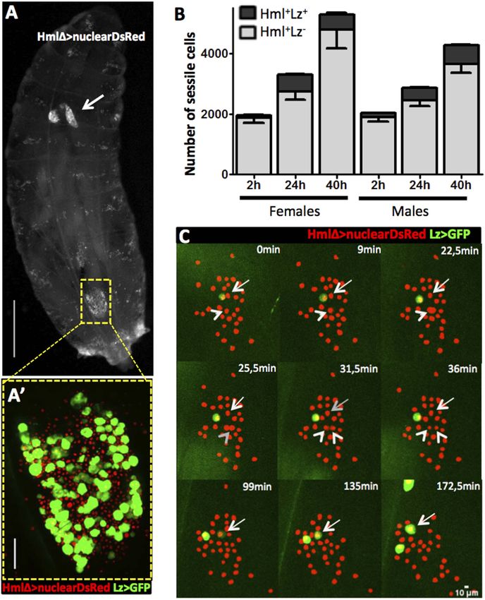
Figure 1. Hml+Lz+ cells increase during larval development by de novo differentiation. (A) Dorsal view of a third
instar larva with hemocyte nuclei marked using HmlΔ-nuclearDsRed. The lymph gland (arrow) and sessile hemocytes
along the body axis are visible, particularly in a big cluster on the A7 segment (square). Scale bar = 1 mm. (A′)
Magnification of the A7 hemocyte cluster showing that it is constituted of Hml+Lz− and Hml+Lz+ cells. Scale bar = 50
μm. (B) Throughout third instar larval development of both females and males, Hml+ sessile cells (grey bars) increase
accompanied by an increment of sessile Lz+ cells (black bars), n = 10 per time point, error bars represent the SEM.
(C) Still images of a 3-hr video showing hemocytes marked by HmlΔ-nuclearDsRed; Lz>EGFP/CD8GFP. It is possible
to observe cell divisions in Hml+Lz− (arrow heads) and GFP induction (arrows).
DOI: 10.7554/eLife.06166.003
The following figure supplements are available for figure 1:
Figure supplement 1. Example of a sessile hemocyte cluster (abdominal segment A7) in a HmlΔ-DsRed;
Lz>mCD8GFP larva. It is possible to observe small HmlhighLzlow cells (arrows) and Hml−Lz+ cells (asterisks).
DOI: 10.7554/eLife.06166.004
Figure supplement 2. Probability density plots for the different cell type sizes found in sessile clusters of HmlΔ-
DsRed; Lz>mCD8GFP larvae (n = 8 samples).
DOI: 10.7554/eLife.06166.005
Figure supplement 3. Throughout the 3-hr period covered in our videos, we can observe that GFP intensity in Lz+ cells
increases, as measured by mean grey value of the cell at the beginning (0 min) and at the end (180 min) of the video.
DOI: 10.7554/eLife.06166.006
Figure 1. continued on next page
Leitão and Sucena. eLife 2015;4:e06166. DOI: 10.7554/eLife.06166 4 of 16Research article Developmental biology and stem cells
Figure 1. Continued
Figure supplement 4. Quantification of GFP intensity and cell area of Lz+ cells in hemolymph smears of HmlΔ-
DsRed; Lz>mCD8GFP larvae, shows a strong positive correlation between cell size and GFP intensity.
DOI: 10.7554/eLife.06166.007
development proceeds, both in males and females (Figure 1B). The number of Lz+ cells also increases
in the same time period (Figure 1B). In females there is no difference in the number of Lz+ in the last
16 hr of development. With this, late third stage larva females have less crystal cells than males which
is not common in the majority of fly stocks where females tend to have a higher number of crystal cells
than males (see ‘Results’ below). Nevertheless, the results clearly show that committed crystal cells
(Hml+Lz+) are increasing in number during third instar larval development, in parallel with an
expansion of the plasmatocyte (Hml+Lz−) population.
Next, we wanted to distinguish if Lz+ cells expand by cell proliferation or by de novo differentiation
from Hml+Lz− cells. A way to achieve this is to directly visualize hemocyte clusters with live time-lapse
imaging and calculate the proliferation and differentiation rates for each cell type. To that purpose,
we developed a method for imaging epidermal hemocyte clusters in live larvae for periods of 3 hr.
HmlΔ-nuclearDsRed; Lz-GAL4>EGFP/mCD8GFP early L3 male larvae (EGFP hemocytes in cells in our videos is ∼3.5% in 3 hr. Knowing the
a dorsal cluster. Examples of Hml+Lz− hemocytes in differentiation rate of Hml+Lz− into Hml+Lz+,
division are highlighted with green circles and examples
we can extrapolate the number of Hml +Lz +
of Hml+Lz− hemocytes differentiating into Hml+Lz+
cells differentiated from Hml +Lz − cells at given
hemocytes identified by a red circle. Scale bar = 10 μm.
DOI: 10.7554/eLife.06166.008
time (see ‘Materials and methods’). The differen-
tiation rate calculated in our video analysis is
Leitão and Sucena. eLife 2015;4:e06166. DOI: 10.7554/eLife.06166 5 of 16Research article Developmental biology and stem cells
sufficient to explain the increase in Hml+Lz+ cell number observed in the first 24 hr of third instar larval
development. The mean increase of Lz+ cells measured during development is 256.4 cells (Figure 1B)
and the extrapolation gives an increase of 447 Lz+ cells. On the whole, the analysis of these videos
shows that Hml+Lz+ cells differentiate from Hml+Lz− cells in hemocyte clusters at a rate that is sufficient
to explain the increase in this cell type observed in the first 24 hr of L3 development.
Plasmatocytes transdifferentiate into crystal cells
Only a small proportion of Hml+Lz− cells become Hml+Lz+ in the course of third instar larval
development. An important element to clarify is whether all Hml+Lz− cells have the capacity to
become Hml+Lz+ cells or whether this property is exclusive of a subpopulation of Hml+Lz− cells. In the
time window of interest, hemocytes can be divided into two sub-populations, NimrodC1+ and
NimrodC1− (Kurucz et al., 2007; Csordás et al., 2014). During lymph gland development, Nimrod is
only detectable in mature plasmatocytes and it is not expressed in Lz+ cells (Terriente-Felix et al.,
2013; Ferguson & Martinez-agosto 2014). Thus, in the lymph gland Lz+ cells differentiate from Hml+
Nimrod− cells. To test if Nimrod− hemocytes in sessile clusters are the precursors of Lz+ cells, we
checked for Nimrod protein (P1 antibody, see details in ‘Materials and methods’) in Lz+ sessile cells. It
is worth noticing that, because crystal cells burst after bleeding (Bidla et al., 2007), there is a bias in
immunofluorescence stainings in favor of more immature crystal cells. Contrarily to the lymph gland,
the majority of Lz+ cells are also Nimrod+ (Figure 2A–A″). This result suggests that Lz+ cells
differentiate from a pool of mature plasmatocytes.
Interestingly, it has been shown before that plasmatocytes transdifferentiate into lamellocytes
(Honti et al., 2010; Meister and Ferrandon, 2011). In this case, cells that are phagocytically active
become non-phagocytic and start to express lamellocyte markers (Honti et al., 2010). Given this, we
proceeded to test if the observed transition between Lz− and Lz+ corresponds to a change of fate
from plasmatocyte (phagocytic) to crystal cell (non-phagocytic). From our results, it is evident that
phagocytosis index is higher in Lz− hemocytes but it is still non-negligible in Lz+ hemocytes
(Figure 2B). When we characterize cell area and GFP intensity in Lz+ cells, it is possible to observe that
the smaller and GFPlow expressing cells are able to phagocyte bacteria (Figure 2B′). Large cells that
have GFPhigh expression are virtually non-phagocytic cells. This indicates that induced Lz+ cells are
plasmatocytes with phagocytic activity, which is lost as they mature into crystal cells. Altogether,
these results support the transdifferentiation of mature phagocytically active plasmatocytes into
non-phagocytic crystal cells.
Serrate expression in plasmatocytes is essential for crystal cell
differentiation
As mentioned above, crystal cell numbers are reduced in larvae raised at a restrictive temperature in
a thermo sensitive Notch allele background (Duvic et al., 2002). This reduction is visible in sessile
hemocytes and in the lymph gland (Duvic et al., 2002; Lebestky et al., 2003). In the lymph gland
Notch signaling has a cell autonomous role on Hml+Lz− cells (Mukherjee et al., 2011). To test for the
role of the Notch pathway in the differentiation of crystal cells in the clusters, we used RNAi exclusively
in hemocytes by way of the HmlΔGAL4 driver. Firstly, we establish that Notch downregulation reduces
the number of sessile crystal cells both in females (Figure 3A) and males (Figure 3—figure
supplement 1). As mentioned above, in this experiment it is possible to observe that sessile crystal
cell numbers are higher in females (compare controls in Figure 2A and Figure 3—figure supplement 1).
In Drosophila, Notch is activated by two different ligands: Serrate and Delta (Fiúza and Arias, 2007).
Only Serrate mutants have reduced numbers of embryonically derived crystal cell (Duvic et al., 2002;
Lebestky et al., 2003). Similarly, knocking down Serrate in hemocytes (HmlΔ-GAL4>UAS-Serrate RNAi)
reduces the number of crystal cells to a similar level than found using Notch RNAi, as opposed to
disrupting Delta function (Figure 3A). This indicates that Serrate, the ligand necessary to induce Notch
signaling in hemocytes, interestingly, it is expressed in the hemocytes themselves.
Importantly, the knockdown of Notch does not disrupt the hemocyte clusters nor changes the
concentration of hemocytes in circulation (Figure 3—figure supplement 2,3). In the lymph gland,
Notch activation is essential to induce lozenge upregulation (Lebestky et al., 2003). As observed in
the lymph gland (Terriente-Felix et al., 2013), we can detect Notch enhancer GFP reporters in Lz+
sessile hemocytes and, using video analysis, we can observe induction of lozenge in Notch activated
cells (Figure 3B). To confirm that Notch knockdown inhibits the induction of lozenge in sessile
Leitão and Sucena. eLife 2015;4:e06166. DOI: 10.7554/eLife.06166 6 of 16Research article Developmental biology and stem cells
Figure 2. Lz+ cells derive from mature plasmatocytes. (A) P1 immunofluorescence (IF) staining of sessile hemocytes
marks the majority of Lz− and Lz+ cells. Bars represent the mean ratio of P1+ and P1− cells in these two population of
cells (n = 10 samples) (A′) Examples of P1+ Lz− plasmatocyte, P1+ Lz+ crystal cell and P1−Lz+ crystal cell. (B) Part of the
Lz+ cells are capable of doing phagocytosis. Bars represent the mean ratio between phagocytic and non-phagocytic
cells (n = 5 samples). (B′) Phagocytic capacity in Lz+ cells correlates negatively with both cell size and GFP intensity
(measured by mean grey value of the picture). Points represent the mean and error bars represent the SEM.
DOI: 10.7554/eLife.06166.009
hemocytes and not the maturation of Lz+ cells into crystal cells, we measured the proportion of Lz+ cells
with anti-Lozenge antibody while inhibiting Notch expression in all hemocytes (HmlΔ-
GAL4>NotchRNAi). The proportion of Lz + cells in this case is clearly reduced (Figure 3C).
Because increased crystal cell apoptosis could also explain the reduced crystal cell numbers, we
estimated hemocyte apoptosis upon Notch knockdown and could not find any significant
difference to controls (Figure 3—figure supplement 4). Altogether these results confirm that
Notch activation is essential to induce lozenge expression in larval hemocytes that will mature
into crystal cells.
Since we used the HmlΔ-GAL4 driver, we could not distinguish if Serrate is necessary in Hml+Lz−, in
Hml+Lz+, or in both cell types. To test these alternatives, we performed knockdown of Serrate with the
Lz-GAL4 driver and found no reduction in the number of crystal cells (Females in Figure 3D, males in
Figure 3—figure supplement 5). In addition, two other GAL4 drivers expressed in plasmatocytes,
Eater-GAL4 and Pxn-GAL4, reduce the number of sessile crystal cells when driving Serrate RNAi
Leitão and Sucena. eLife 2015;4:e06166. DOI: 10.7554/eLife.06166 7 of 16Research article Developmental biology and stem cells
Figure 3. Serrate downregulation in plasmatocytes leads to a reduction in sessile crystal cell number. (A) Notch RNAi
driven in all hemocytes reduces the number of melanized sessile crystal cells observed upon heat shock treatment to the
whole larva. A similar level of reduction is seen with SerrateRNAi but not with DeltaRNAi (n = 20). (B) Still images of a 3-hr
video showing the induction of lozenge reporter expression in Notch activated hemocytes (arrows). (C) NotchRNAi reduces
the proportion of Lz+ cells in sessile hemocytes quantified with P1 immunofluorescence (IF) staining (n = 11 samples).
(D) Serrate RNAi driven only in Lz+ cells does not reduce the number of melanized sessile crystal cells seen upon heat
shock treatment contrarily to two other drivers expressed in plasmatocytes, Eater-GAL4 and Pxn-GAL4 (n = 20 samples). In
all graphics only female larvae are shown, error bars represent SEM, n.s. = non significant p-value, **p < 0.01, ***p < 0.001.
DOI: 10.7554/eLife.06166.010
The following figure supplements are available for figure 3:
Figure supplement 1. NotchRNAi and SerrateRNAi but not DeltaRNAi driven in all hemocytes reduce the number of
melanized sessile crystal cells observed upon heat shock treatment to the whole larva (males are shown, n = 20).
DOI: 10.7554/eLife.06166.011
Figure supplement 2. The localization of hemocyte in sessile clusters is not affected by Notch pathway
manipulation through RNAi induction under HmlΔ-GAL4 control.
DOI: 10.7554/eLife.06166.012
Figure supplement 3. Notch pathway manipulation through RNAi induction under HmlΔ-GAL4 control, does not
change hemocyte concentration in hemolymph.
DOI: 10.7554/eLife.06166.013
Figure supplement 4. Notch knockdown through RNAi under HmlΔ-GAL4 control does not increase cell death as
measured by a flow cytometry Propidium Iodide (PI) exclusion assay , error bars represent the SEM. n.s. = p ≥ 0.05.
DOI: 10.7554/eLife.06166.014
Figure supplement 5. Serrate RNAi driven only in Lz+ cells does not reduce the number of melanized sessile crystal
cells seen upon heat shock treatment contrarily to two other drivers expressed in plasmatocytes, Eater-GAL4 and
Pxn-GAL4 (males are shown, n = 20 samples).
DOI: 10.7554/eLife.06166.015
Leitão and Sucena. eLife 2015;4:e06166. DOI: 10.7554/eLife.06166 8 of 16Research article Developmental biology and stem cells
Figure 4. Cluster structure is necessary for crystal cell development. (A) Sessile hemocytes in Lz>mCD8GFP HmlΔ-
cytoplasmic DsRed larvae were scored for the number of contacts. Probability of a cell being Lz+ increases linearly
with the number of cells it is in contact with. (n = 8) (B) In early third instar larvae, the continued disruption of clusters
for a 10-hr period leads to a reduction in the proportion of Lz+ cells (circulating and sessile cells were quantified).
Error bars represent SEM, ***p < 0.001.
DOI: 10.7554/eLife.06166.016
The following figure supplements are available for figure 4:
Figure supplement 1. In HmlΔ-nuclearDsRed; Lz-GAL4, UAS-GFP larvae, the dorsal cluster in the A7 segment is
easily observed (left panel).
DOI: 10.7554/eLife.06166.017
Figure supplement 2. Flow cytometry analysis for cell viability and Dot-GAL4 lineage tracing in cluster disrupted
larvae.
DOI: 10.7554/eLife.06166.018
Figure supplement 3. It is possible to detect lymph gland derived hemocytes by flow cytometry with Dot-GAL4
lineage-traced hemocytes in pupa (blue line) or when larvae are infected with parasitoid wasps (green line).
DOI: 10.7554/eLife.06166.019
(females in Figure 3D, males in Figure 3—figure supplement 5). Hence, we conclude that
plasmatocyte are responsible for Serrate signaling to activate Notch in other plasmatocytes and start
crystal cell development.
Hemocyte cluster structure is necessary for Notch-dependent larval
hematopoiesis
For Notch pathway activation, cells need to be in contact because the ligand Serrate is membrane
bound (Guruharsha et al., 2012). The fact that Serrate expression in Hml+Lz− hemocytes is necessary
for crystal cell development suggests that Lz+ cells are induced when hemocytes are in contact within
the clusters. If this is the case, we can predict that the probability of an Hml+ cell to be also Lz+
increases with the number of cell contacts it establishes with Hml+Lz− cells. We quantified the
number of cell contacts that each cell type makes within the sessile clusters and compared cells within
the same size range (i.e., limited to the maximum size of Hml+Lz− cells). As predicted, the proportion
of Hml+Lz+/ Hml+Lz− increases with the number of Hml+Lz− cells with which it is in contact (Figure 4A).
Given that the clustering of hemocytes is important for hematopoietic decisions, we tested this
idea further by assessing how the absence of clusters would affect crystal cell differentiation.
A misexpression screen identified some genes that when overexpressed in hemocytes can disrupt
hemocyte clusters (Stofanko et al., 2008). However, we did not use these lines to test for crystal
cell/plasmatocyte ratio alterations because it would be difficult to discern between the effect of not
having the clusters and the effect of gene up-regulation in hemocytes. We opted to disrupt the
hemocyte clusters by manipulating physically the larvae (Makhijani et al., 2011). By rolling groups of
larvae between two cover slides, it is possible to force the hemocytes to detach from the epidermis
and enter hemolymph circulation. After cluster disruption, hemocytes start to aggregate again,
gradually (Makhijani et al., 2011) (Figure 4—figure supplement 1). To maintain hemocytes in
circulation for a period of 10 hr, we disrupted hemocyte clusters in larvae every 1 hr 30 min
Leitão and Sucena. eLife 2015;4:e06166. DOI: 10.7554/eLife.06166 9 of 16Research article Developmental biology and stem cells
(see ‘Materials and methods’). Using flow cytometry, we measured the proportion of Hml+Lz+/Hml+Lz−
cells at the end of this treatment. The relative number of Hml+Lz+ cells decreases in the treatment
group (Figure 4B), indicating that clusters are necessary for crystal cell differentiation.
However, the cluster disruption treatment could also disrupt the lymph gland and/or change the
rate of apoptosis differentially between Hml+Lz+ and Hml+Lz− cells. We tested both possibilities.
Firstly, using a PI exclusion assay, we determined if the rate of apoptosis changes upon cluster
disruption. There is no significant difference between control and treatment groups (Figure 4—figure
supplement 2). Secondly, to check if cluster disruption affects the lymph gland, we made use of
DotGAL4, a driver expressed in lymph gland hemocytes but not in circulating and sessile hemocytes
(Honti et al., 2010). Through lineage tracing analysis with Dot-GAL4 driver, we can check cells that
are derived from the lymph gland. Indeed, using flow cytometry, we can observe lymph gland-derived
hemocytes in the circulating pool of wasp-infected larvae and pupae (Figure 4—figure supplement 3).
When the same technique is used in cluster-disrupted larvae, there is no detectable lymph gland-
derived hemocytes in circulation (Figure 4—figure supplement 2). Thus, the integrity of the cluster is
necessary for crystal cell differentiation. Strikingly, in agreement with our observations, recent work
shows that in Eater mutants that do not form hemocyte clusters, sessile crystal cells are absent
(Bretscher et al., 2015).
Altogether these results support that Notch signaling is necessary for crystal cell differentiation
and depends on cluster structure. It is important to note, though, that hemocytes form clusters of
different sizes and at different locations along the larval body (Kurucz et al., 2007; Makhijani et al., 2011).
Our cluster disruption procedure affects sessile hemocytes indiscriminately such that we cannot determine
the relative importance of these cluster features on crystal cell differentiation.
Discussion
Our motivation to carry out this work was to explain the increase of circulating and sessile crystal cell
numbers during Drosophila larval development. This phenomenon is lined with an apparent paradox:
no mature crystal cell has been seen dividing during larval stages (Rizki, 1957; Lanot et al., 2001;
Krzemien et al., 2010) and crystal cells in the lymph gland do not enter circulation in homeostatic
conditions (Holz et al., 2003). Crystal cell number increase may rely upon a population of pro-crystal
cells that proliferates in the larva before cell maturation or that that exists in sufficient numbers at the
beginning of development to mature into crystal cells throughout development. The first known
upregulated gene diagnostic of crystal cell development is the transcription factor lozenge (Lz)
(Lebestky et al., 2000). That is, a cell will be Lz+ before it matures into crystal cell and maintains this
expression upon differentiation (Lebestky et al., 2000). We have shown that throughout third instar
development, the number of Lz+ cells in the sessile population increases. This observation excludes
the possibility that a population of Lz+ cells exists in fixed number and matures into crystal cell. Lz+ cells
have been reported to proliferate during embryogenesis (Lebestky et al., 2000). Surprisingly, we do
not see Lz+ cell division in our video analysis. Although, with our results, we cannot exclude that a small
proportion of Lz+ cells proliferate during larval stages, we can show that lozenge induction in Hml+Lz− is
sufficient to explain the increase in Hml+ Lz+ cells during third instar larva.
The activation of lozenge in hemocytes is Notch-dependent with Serrate acting as the ligand
(Duvic et al., 2002; Lebestky et al., 2003). When we ablate serrate expression only in hemocytes
using the HmlΔ-GAL4 driver coupled to a UAS-SerRNAi, the number of differentiated crystal cells is
severely reduced within the sessile population. This indicates that hemocytes are the cells responsible
for crystal cell induction in sessile clusters. Moreover, the hemocytes inducing crystal cell development
are themselves Lz− because SerrateRNAi driven by Lz-GAL4 driver does not decrease the number of
crystal cells. For Notch to be activated it requires that a Serrate expressing cell is in contact for
a certain period of time (Guruharsha et al., 2012). We show that this contact is a property of the
clusters where Hml+Lz+ cells are induced from Hml+Lz− cells. This observation establishes an important
parallel between sessile and lymph gland crystal cell development. In both cases, the precursor to
a crystal cell is an Hml+Lz− hemocyte (Mukherjee et al., 2011). However, there is a fundamental
difference between these two hematopoietic events regarding the cells where lozenge is first
expressed. Although hemocytes only activate lozenge expression in the cortical zone of the lymph
gland (Lebestky et al., 2000), the work of Krzemien et al. suggest that in the medullary zone of the
lymph gland cells are already committed to become crystal cells at late second instar (Krzemien et al.,
2010). This suggests that medullary zone cells migrating to the cortical zone can be considered pro-crystal
Leitão and Sucena. eLife 2015;4:e06166. DOI: 10.7554/eLife.06166 10 of 16Research article Developmental biology and stem cells
cells. In line with this observation, in the lymph gland, no co-localization between plasmatocyte marker P1
and crystal cell marker Lz is ever observed (Terriente-Felix et al., 2013; Ferguson & Martinez-agosto
2014). In contrast, our analyses in hemocyte clusters suggest that lozenge induction occurs in mature
plasmatocytes. Firstly, they derive from P1 cells. Secondly, Lz+GFPlow cells can phagocyte as opposed to
Lz+GFPhigh cells, which loose this capacity. Lz+GFPlow cells are, according to our video analysis, the initial
stages of crystal cell differentiation as virtually all Lz+GFPlow become Lz+GFPhigh. Throughout time cells
increase their GFP expression and become larger. Altogether our results suggest that mature
plasmatocytes can differentiate into crystal cells.
This conclusion may help explaining some disparate results in the literature. Firstly, circulating
crystal cells in the larva derive from cells that express the plasmatocyte-specific marker croquemort
during the embryonic stage (Franc et al., 1999; Honti et al., 2010). Secondly, Lebestky et al. consider
that a small fraction of the Lz-GAL4 positive cells gives rise to plasmatocytes defined by morphology
and croquemort expression (Lebestky et al., 2000). In light of our results, we propose that the
plasmatocyte-like cells expressing lozenge are plasmatocytes in route to become crystal cells. To our
knowledge, the hypothesis that plasmatocytes give rise to crystal cells was put forward by Rizky in
1957, with the purpose of explaining how crystal cells increase in number without proliferation (Rizki,
1957). Our work provides the first body of evidence that puts this idea to test and validates this
hypothesis.
It is possible that, contrary to the lymph gland, hemocyte clusters are not regionalized structures
(Honti et al., 2014). Yet, with the results shown here, we propose that hemocyte clusters work as
a true hematopoietic tissue. Their presence and integrity are necessary for the proper establishment
of Hml+Lz+/Hml+Lz− numbers during larva development. Interestingly, the hemocytes in clusters are in
dynamic relation with circulating hemocytes (Babcock et al., 2008; Welman et al., 2010; Makhijani
et al., 2011). This is confirmed in our videos where cells can be observed entering circulation from the
patches and leaving circulation to become sessile. This dynamics opens the possibility of a more
complex number and cell type regulation mechanism operating at the whole-organism level.
Secondly, another interesting property of sessile plasmatocytes consists of their higher division rate
with respect to their circulating counterparts (Makhijani et al., 2011). This could happen because
there is a different molecular ‘environment’ in hemocyte clusters (Makhijani et al., 2011) and/or
because a sessile cell has an increased probability of entering cell division. We argue that the
existence of these two characteristics, cell proliferation control and cell differentiation, is sufficient to
consider the hemocyte clusters as hematopoietic tissues. In brief, hemocyte clusters enhance
hemocyte proliferation and provide structure as to guarantee the necessary cell contacts that engage
the signaling events behind cell fate decisions. Noticeably, hemocytes in clusters can be mobilized to
circulation upon immune challenge (Zettervall et al., 2004), a process that is in part dependent on
the small GTPase Rac1 (Xavier and Williams, 2011). The role of hemocyte clusters is most likely
restricted to larval stages because, once pupariation starts, a peak of ecdysone promotes the
dispersion of hemocytes throughout the epidermis (Regan et al., 2013).
The differentiation of crystal cells from plasmatocytes within sessile clusters creates, in our view, an
interesting parallel with macrophage development in vertebrates. Macrophages are the most plastic
cells in the vertebrate’s hematopoietic tissue and their specialization in vivo depends on the local
microenvironment provided by the tissue they colonize (Ostuni and Natoli, 2011; Wynm et al.,
2013). Similarly, here we show that in Drosophila larvae the microenvironment provided by hemocyte
clusters is necessary to induce crystal cell differentiation from plasmatocytes, namely through
a cell-contact mechanism involving Notch-Serrate.
A putative important difference between hemocyte clusters and the lymph gland concerns the
mechanisms in control of cell proliferation and differentiation. In support of this notion, misexpression
of some genes in hemocytes can disrupt hemocyte clusters without affecting lymph gland morphology
(Stofanko et al., 2008). Tightly linked to this question is one other fundamental aspect that remains
unaddressed: the control of proportions between different cell types. Throughout homeostatic
development, it is commonly observed, both in vertebrates (Almeida et al., 2005) and invertebrates
(Rizki, 1957), that blood cell types respect fixed relative numbers. Also, it is now evident that
plasmatocytes are very plastic cells and may represent a rare case of functionally mature cells
transdifferentiating into other cell types: lamellocytes (Honti et al., 2010) and crystal cells.
Transdifferentiation, the process where a cell changes its cell fate without passing through a less
differentiated state, is used recurrently in cell culture assays but rarely seen in vivo (Jopling
Leitão and Sucena. eLife 2015;4:e06166. DOI: 10.7554/eLife.06166 11 of 16Research article Developmental biology and stem cells
et al., 2011). How recurrent this mechanism may be in animal development presents itself as one
important question for the future. We consider that acknowledging this novel hematopoietic organ,
dynamically attached to the circulating hemocyte population and relying on structure-dependent
signaling events to promote blood homeostasis, brings us a step closer to addressing these outstanding
fundamental questions of Drosophila hematopoiesis.
Materials and methods
Fly stocks and parasitoid maintenance
All fly stocks were maintained in standard fly food at room temperature. Experiments were performed
at 25˚C except for RNAi experiments that were performed at 29˚C. The following stocks were obtained
from the Bloomington Stock Center: Lz-GAL4 UAS-mCD8GFP (#6314); Lz-GAL4 UAS-GFP (#6313);
Notch Responsive Element (#30727); UAS-FLP UbiFRTSTOPStinger (#28282); UAS-myrtdTomato
(32221); HmlΔGAL4 UAS-EGFP (30140). The following stocks were obtained from the Vienna
Drosophila Resource Center: CG9313RNAi (#103600), NotchRNAi (#100002), SerrateRNAi (#108348),
DeltaRNAi (#109491) (Dietzl et al., 2007). The line Cg9313RNAi was used as control for RNAi experiments
since it is a gene specifically expressed in male testis (Paulo Navarro, personal communication). The line
HmlΔ-nuclearDsRed was a kind gift from Marc Dionne (Clark et al., 2011). The line HmlΔ-DsRed was
a kind gift from Utpal Banerjee (Makhijani et al., 2011). The Eater-GAL4 (II) was a generous gift from
Robert A Schultz (Tokusumi et al., 2009). Leptopilina boulardi females of the strain G486 (a kind gift
from Fernando Roch) were allowed to lay eggs on second instar Drosophila Dif mutants at room
temperature. Adult parasitoids were maintained in fly food vials with a drop of honey.
Larva staging
Around 20 female flies were placed in a cage with a food plate containing yeast. Egg lays took place at
25˚C for 6 hr. At ∼72 hr midpoint after egg lay, second instar larvae were selected based on spiracle
morphology and transferred into a new food plate. After 2 hr, larvae that molted into third instar were
selected and transferred into a food tube. This first time point is referred to as 2 hr after third instar.
Flow cytometry analysis and cell viability assay
Larvae were bled in 200–400 μl of Ringer’s solution. The number of larvae greatly depends on the
experiment but at least 10 larvae were used in each sample. The hemocyte dilution was passed through
a 30-μm filter to exclude cell aggregates. The samples were maintained on ice until acquisition.
Acquisition was performed in CyAn ADP cytometry Analyzer (DAKO Cytomation, Beckman Coulter)
with Summit software (DAKO). Hemocyte population was gated in Forward Scatter (FSC) and Side
Scatter (SSC) channels and single events in FSC and Pulse Width channels. GFP and DsRed were
measured in the appropriate channels. To analyze results, it was used the Flowing Software (version
2.5.0). To analyze cell viability a stock solution of Propidium Iodide (PI) was diluted in 200 μl of Ringer’s
solution to a final concentration of 2 μg/ml. Positive events for PI were considered dead or dying cells.
Video preparation and analysis
Male larvae were selected as described above, briefly washed in Ringer’s solution, dried on filter
paper and attached to double-sided tape on a cover slip. A second cover slip is placed on top of the
larva (dorsal side) so that the larva is stuck between two cover slips. The larva does not move but stays
alive for at least 12 hr in a humid chamber. The pressure from the cover slip affects the A7 dorsal
cluster, most probably because of the disruption of normal hemolymph circulation. Hence, we imaged
more anterior epidermal clusters that were not so affected. The larva was mounted in an inverted
confocal spinning microscope (Andor Revolution xD). The temperature of the slide chamber was
maintained at 25˚C and 95% relative humidity. A Z-stack of pictures ranging 28 μm was taken every 1
min 30 s for the GFP and RFP channel throughout a period of 3 hr. At the end of the video each larva
was checked for viability by observing the beating dorsal vessel and mouthparts movement. Only one
larva died during the process. Z-stacks were then analyzed manually in FIJI software (Schindelin et al.,
2012). With video analysis, we estimated the Differentiation Proportion during 3 hr. To extrapolate
the number of Hml+Lz+ induced cells for other time points, we used the formula:
Leitão and Sucena. eLife 2015;4:e06166. DOI: 10.7554/eLife.06166 12 of 16Research article Developmental biology and stem cells
Hml+ Lz+t+1 = Hml+ Lz+t0 + Hml+ Lz−t0 × ðDifferentiation ProportionÞ:
Total sessile hemocyte counts, total hemocyte load, and crystal cell
counts
To count the total number of sessile hemocytes in all larvae, pictures were taken in four different
angles, using the appropriate fluorescence markers for hemocytes, under a fluorescence stereoscope
(Zeiss SteREO Lumar.V12). The last body segment of the larva was excluded because it is difficult to
image. To estimate hemocyte concentration in the hemolymph, six wandering male or female larvae
were briefly washed in Ringer’s solution, dried in filter paper, pooled in a glass well and bled
by rupturing the cuticle in the ventral side (to avoid disturbance of sessile hemocytes in the dorsal
part where they are more abundant). The hemolymph was collected and pooled into a 0.5 ml
microcentrifuge tube and diluted 1:10 with Ringer’s solution. 9.5 μl of diluted hemolymph was loaded
into a Neubauer chamber and hemocytes counted in squares of 1 mm2. This way the hemocyte
concentration can be estimated by the formula: [number of counted cells] × 105 cells/ml. To count
sessile crystal cells in all larvae, we performed a 70˚C heat shock treatment for 10 min (Neyen et al.,
2014). With this treatment mature crystal cells melanize and it is possible to count sessile mature crystal
cell numbers trough the cuticle.
Hemocyte immunohistochemistry
To collect circulating hemocytes larvae were bled in Ringer’s solution from the ventral side to minimize
disruption of sessile clusters. To collect sessile hemocytes the dorsal part of the cuticle was dissected,
washed twice in Ringer’s solution and hemocytes were removed by gently passing the forceps in the
cuticle. Hemocytes were allowed to settle to slide glass reaction well (Ø 5 mm Marienfeld, Lauda-
Königshofen, Germany) in a humid chamber for 10 min and fixed with 4% formaldehyde solution for
20 min. After fixation cells were washed three times with PBS and blocked with PBST (PBS + 0.1%
Triton X + 1% normal goat serum) for 30 min. After washing the cells with PBS, the primary antibody was
added at the correct dilution and cells incubated overnight at 4˚C. Cells were then washed three times
with PBS for 15 min and the secondary antibody added in the correct concentration. Cells were
incubated for 3 hr at room temperature or at 4˚C overnight with the secondary antibody. The secondary
antibody was washed three times with PBS. DAPI was added and incubated for 3 min followed by three
washes with PBS. Slides were mounted with 80% glycerol solution and kept at 4˚C before image
acquisition. Secondary antibodies used: AlexaFluor 488 and AlexaFluor 546 (1:1000 dilution, Life
Technologies, NY, USA). Primary antibodies used: anti-lozenge (1:100 dilution, Developmental Studies
Hybridoma Bank, University of Iowa, US) anti-NimrodC1 (P1 antibody, kind gift from Istvan Andó, 1:100
dilution). Some D. melanogaster lines have a deletion in NimrodC1 locus and the epitope for P1
antibody is not present (Honti et al., 2013). Because we could not detect P1 staining on Lz-GAL4
UAS-mCD8GFP line hemocytes, we crossed it with Oregon R males and tested the F1.
Cell imaging
Larvae were rolled to take pictures from different angles. Hemolymph smears with live or fixed cells
were imaged in a Leica DMRA2 microscope coupled with a CoolSNAP HQ CCD camera.
Phagocytosis assay
To test phagocytosis, early third stage larvae were injected with 69 nl of pHrodo Red E. coli
BioParticles (1 mg/ml; Molecular Probes). Injected larvae were maintained in yeast for 1 hr before ∼10
larvae were bled in 20 μl PBS (pH = 7.4). Hemocytes were allowed to settle for 20 min at room
temperature in a humid chamber, washed with PBS and pictures were taken immediately.
Cluster disruption assay
Pools of ∼20 early third instar male larvae were transferred to fresh yeast on a plastic petri dish placed
in a humid chamber. Every 1 hr 30 min, larvae were taken from yeast, cleaned in Ringer’s solution and
dried in filter paper. Groups of ∼5 larvae were rolled several times by pressing a cover glass to disrupt
hemocyte clusters. Control larvae were maintained on yeast. Before larva sampling the two groups
were subjected to cluster disruption to sample both circulating and sessile hemocytes.
Leitão and Sucena. eLife 2015;4:e06166. DOI: 10.7554/eLife.06166 13 of 16Research article Developmental biology and stem cells
Statistical analysis
Every experiment was repeated at least twice to check for reproducibility. Samples were tested for
normality with Shapiro–Wilk test and the appropriate statistical test was then applied. Student’s
t-tests were used to compare two treatments. ANOVA (or Kruskal–Wallis as non-parametric test)
was performed when several comparisons were necessary and Dunnett’s multiple comparison
tests (or Dunn’s multiple comparison test) were applied to test differences between pairs of
treatments. Statistical test and graphics were performed in Prism v5.01 (GraphPad Software) and
R v2.15.2.
Acknowledgements
We thank all members of the laboratories of Élio Sucena, Patrı́cia Beldade, and Christen Mirth for
important discussions throughout this project. We are indebted to Bruno Lemaitre for sharing
unpublished results. We thank Marc Dionne, Robert A Schultz, and István Andó for fly lines and/or
reagents, the VDRC (Vienna, Austria) for the RNAi fly lines and the Hybridoma Bank (Iowa, US)
for monoclonal antibodies. This work was supported by Fundação Calouste Gulbenkian/Instituto
Gulbenkian de Ciência and by Fundação para a Ciência e a Tecnologia (SFRH/BD/51175/2010
to ABL).
Additional information
Funding
Funder Grant reference number Author
Fundação para a Ciência e SFRH/BD/51175/2010 Alexandre B Leitão
a Tecnologia
Calouste Gulbenkian Alexandre B Leitão, Élio
Foundation Sucena
The funders had no role in study design, data collection and interpretation, or the
decision to submit the work for publication.
Author contributions
ABL, Conception and design, Acquisition of data, Analysis and interpretation of data, Drafting or
revising the article, Contributed unpublished essential data or reagents; ÉS, Conception and design,
Analysis and interpretation of data, Drafting or revising the article, Contributed unpublished
essential data or reagents
References
Almeida AR, Rocha B, Freitas AA, Tanchot C. 2005. Homeostasis of T cell numbers: from thymus production to
peripheral compartmentalization and the indexation of regulatory T cells. Seminars in Immunology 17:239–249.
doi: 10.1016/j.smim.2005.02.002.
Babcock DT, Brock AR, Fish GS, Wang Y, Perrin L, Krasnow MA, Galko MJ. 2008. Circulating blood cells function as
a surveillance system for damaged tissue in Drosophila larvae. Proceedings of the National Academy of Sciences
of USA 105:10017–10022. doi: 10.1073/pnas.0709951105.
Bidla G, Dushay MS, Theopold U. 2007. Crystal cell rupture after injury in Drosophila requires the JNK pathway,
small GTPases and the TNF homolog Eiger. Journal of Cell Science 120:1209–1215. doi: 10.1242/jcs.03420.
Binggeli O, Neyen C, Poidevin M, Lemaitre B. 2014. Prophenoloxidase activation is required for survival to
microbial infections in Drosophila. PLOS Pathogens 10:e1004067. doi: 10.1371/journal.ppat.1004067.
Bretscher AJ, Honti V, Binggeli O, Burri O, Poidevin M, Kurucz É, Zsámboki J, Andó I, Lemaitre B. 2015. The
Nimrod transmembrane receptor Eater is required for hemocyte attachment to the sessile compartment in
Drosophila melanogaster. Biology Open doi: 10.1242/bio.201410595.
Clark RI, Woodcock KJ, Geissmann F, Trouillet C, Dionne MS. 2011. Multiple TGF-β superfamily signals modulate
the adult Drosophila immune response. Current Biology 21:1672–1677. doi: 10.1016/j.cub.2011.08.048.
Crozatier M, Meister M. 2007. Drosophila haematopoiesis. Cellular Microbiology 9:1117–1126. doi: 10.1111/j.
1462-5822.2007.00930.x.
Crozatier M, Ubeda JM, Vincent A, Meister M. 2004. Cellular immune response to parasitization in Drosophila
requires the EBF orthologue collier. PLOS Biology 2:E196. doi: 10.1371/journal.pbio.0020196.
Csordás G, Varga GI, Honti V, Jankovics F, Kurucz É, Andó I. 2014. In Vivo Immunostaining of hemocyte
compartments in Drosophila for live imaging. PLOS ONE 9:e98191. doi: 10.1371/journal.pone.0098191.
Leitão and Sucena. eLife 2015;4:e06166. DOI: 10.7554/eLife.06166 14 of 16Research article Developmental biology and stem cells
Dietzl G, Chen D, Schnorrer F, Su KC, Barinova Y, Fellner M, Gasser B, Kinsey K, Oppel S, Scheiblauer S, Couto A,
Marra A, Keleman K, Dickson BJ. 2007. A genome-wide transgenic RNAi library for conditional gene inactivation
in Drosophila. Nature 448:151–156. doi: 10.1038/nature05954.
Duvic B, Hoffmann JA, Meister M, Royet J. 2002. Notch signaling controls lineage specification during Drosophila
larval hematopoiesis. Current Biology 12:1923–1927. doi: 10.1016/S0960-9822(02)01297-6.
Evans CJ, Hartenstein V, Banerjee U. 2003. Thicker than blood: conserved mechanisms in Drosophila and
vertebrate hematopoiesis. Developmental Cell 5:673–690. doi: 10.1016/S1534-5807(03)00335-6.
Ferguson GB, Martinez-agosto JA. 2014. Yorkie and Scalloped signaling regulates Notch-dependent lineage
specification during Drosophila hematopoiesis. Current Biology 24:2665–2672. doi: 10.1016/j.cub.2014.09.081.
Fiúza UM, Arias AM. 2007. Cell and molecular biology of Notch. The Journal of Endocrinology 194:459–474.
doi: 10.1677/JOE-07-0242.
Franc NC, Heitzler P, Ezekowitz RA, White K. 1999. Requirement for croquemort in phagocytosis of apoptotic cells
in Drosophila. Science 284:1991–1994. doi: 10.1126/science.284.5422.1991.
Grigorian M, Hartenstein V. 2013. Hematopoiesis and hematopoietic organs in arthropods. Development Genes
and Evolution 223:103–115. doi: 10.1007/s00427-012-0428-2.
Guruharsha KG, Kankel MW, Artavanis-Tsakonas S. 2012. The Notch signalling system: recent insights into the
complexity of a conserved pathway. Nature Reviews Genetics 13:654–666. doi: 10.1038/nrg3272.
Holz A, Bossinger B, Strasser T, Janning W, Klapper R. 2003. The two origins of hemocytes in Drosophila.
Development 130:4955–4962. doi: 10.1242/dev.00702.
Honti V, Cinege G, Csordás G, Kurucz E, Zsámboki J, Evans CJ, Banerjee U, Andó I. 2013. Variation of NimC1
expression in Drosophila stocks and transgenic strains. Fly 7:263–266. doi: 10.4161/fly.25654.
Honti V, Csordás G, Kurucz É, Márkus R, Andó I. 2014. The cell-mediated immunity of Drosophila melanogaster:
hemocyte lineages, immune compartments, microanatomy and regulation. Developmental and Comparative
Immunology 42:47–56. doi: 10.1016/j.dci.2013.06.005.
Honti V, Csordás G, Márkus R, Kurucz E, Jankovics F, Andó I. 2010. Cell lineage tracing reveals the plasticity of the
hemocyte lineages and of the hematopoietic compartments in Drosophila melanogaster. Molecular Immunology
47:1997–2004. doi: 10.1016/j.molimm.2010.04.017.
Jopling C, Boue S, Izpisua Belmonte JC. 2011. Dedifferentiation, transdifferentiation and reprogramming: three
routes to regeneration. Nature Reviews Molecular Cell Biology 12:79–89. doi: 10.1038/nrm3043.
Jung SH, Evans CJ, Uemura C, Banerjee U. 2005. The Drosophila lymph gland as a developmental model of
hematopoiesis. Development 132:2521–2533. doi: 10.1242/dev.01837.
Koch U, Radtke F. 2007. Haematopoietic stem cell niche in Drosophila. BioEssays 29:713–716. doi: 10.1002/bies.20613.
Krzemien J, Crozatier M, Vincent A. 2010a. Ontogeny of the Drosophila larval hematopoietic organ, hemocyte
homeostasis and the dedicated cellular immune response to parasitism. The International Journal of
Developmental Biology 54:1117–1125. doi: 10.1387/ijdb.093053jk.
Krzemien J, Oyallon J, Crozatier M, Vincent A. 2010b. Hematopoietic Progenitors and hemocyte lineages in the
Drosophila lymph gland. Developmental biology 346:310–319. doi: 10.1016/j.ydbio.2010.08.003.
Kurucz E, Márkus R, Zsámboki J, Folkl-Medzihradszky K, Darula Z, Vilmos P, Udvardy A, Krausz I, Lukacsovich T,
Gateff E, Zettervall CJ, Hultmark D, Andó I. 2007a. Nimrod, a putative phagocytosis receptor with EGF repeats in
Drosophila plasmatocytes. Current Biology 17:649–654. doi: 10.1016/j.cub.2007.02.041.
Kurucz E, Váczi B, Márkus R, Laurinyecz B, Vilmos P, Zsámboki J, Csorba K, Gateff E, Hultmark D, Andó I. 2007b.
Definition of Drosophila hemocyte subsets by cell-type specific antigens. Acta Biologica Hungarica 58:
Suppl95–111. doi: 10.1556/ABiol.58.2007.Suppl.8.
Lanot R, Zachary D, Holder F, Meister M. 2001. Postembryonic hematopoiesis in Drosophila. Developmental
Biology 230:243–257. doi: 10.1006/dbio.2000.0123.
Lebestky T, Chang T, Hartenstein V, Banerjee U. 2000. Specification of Drosophila hematopoietic lineage by
conserved transcription factors. Science 288:146–149. doi: 10.1126/science.288.5463.146.
Lebestky T, Jung SH, Banerjee U. 2003. A Serrate-expressing signaling center controls Drosophila hematopoiesis.
Genes & Development 17:348–353. doi: 10.1101/gad.1052803.
Makhijani K, Alexander B, Tanaka T, Rulifson E, Brückner K. 2011. The peripheral nervous system supports blood
cell homing and survival in the Drosophila larva. Development 138:5379–5391. doi: 10.1242/dev.067322.
Mandal L, Martinez-Agosto JA, Evans CJ, Hartenstein V, Banerjee U. 2007. A Hedgehog- and Antennapedia-
dependent niche maintains Drosophila haematopoietic precursors. Nature 446:320–324. doi: 10.1038/
nature05585.
Márkus R, Laurinyecz B, Kurucz E, Honti V, Bajusz I, Sipos B, Somogyi K, Kronhamn J, Hultmark D, Andó I. 2009.
Sessile hemocytes as a hematopoietic compartment in Drosophila melanogaster. Proceedings of the National
Academy of Sciences of USA 106:4805–4809. doi: 10.1073/pnas.0801766106.
Martinez-Agosto JA, Mikkola HK, Hartenstein V, Banerjee U. 2007. The hematopoietic stem cell and its niche:
a comparative view. Genes & Development 21:3044–3060. doi: 10.1101/gad.1602607.
Meister M, Ferrandon D. 2011. Immune cell transdifferentiation: a complex crosstalk between circulating immune
cells and the haematopoietic niche. EMBO Reports 13:3–4. doi: 10.1038/embor.2011.238.
Mukherjee T, Kim WS, Mandal L, Banerjee U. 2011. Interaction between Notch and Hif-alpha in development and
survival of Drosophila blood cells. Science 332:1210–1213. doi: 10.1126/science.1199643.
Neyen C, Bretscher AJ, Binggeli O, Lemaitre B. 2014. Methods to study Drosophila immunity. Methods 68:
116–128. doi: 10.1016/j.ymeth.2014.02.023.
Ostuni R, Natoli G. 2011. Transcriptional control of macrophage diversity and specialization. European Journal of
Immunology 41:2486–2490. doi: 10.1002/eji.201141706.
Leitão and Sucena. eLife 2015;4:e06166. DOI: 10.7554/eLife.06166 15 of 16You can also read