Cytotoxic and anticancer properties of the Malaysian mangrove pit viper (Trimeresurus purpureomaculatus) venom and its disintegrin ...
←
→
Page content transcription
If your browser does not render page correctly, please read the page content below
RESEARCH OPEN ACCESS
ISSN 1678-9199
www.jvat.org
Cytotoxic and anticancer properties of the
Malaysian mangrove pit viper (Trimeresurus
purpureomaculatus) venom and its disintegrin
(purpureomaculin)
Choo Hock Tan1,* , Jia Lee Liew1, Suerialoasan Navanesan2, Kae Shin Sim2, Nget Hong Tan3, Kae Yi Tan3
1
Department of Pharmacology, Faculty of Medicine, University of Malaya, Kuala Lumpur, Malaysia.
2
Institute of Biological Sciences, Faculty of Science, University of Malaya, Kuala Lumpur, Malaysia.
3
Department of Molecular Medicine, Faculty of Medicine, University of Malaya, Kuala Lumpur, Malaysia.
Abstract
Background: The Asiatic pit vipers from the Trimeresurus complex are medically
important venomous snakes. These pit vipers are often associated with snakebite that
leads to fatal coagulopathy and tissue necrosis. The cytotoxic venoms of Trimeresurus
Keywords: spp.; however, hold great potential for the development of peptide-based anticancer drugs.
Shore pit viper Methods: This study investigated the cytotoxic effect of the venom from Trimeresurus
Trimeresurus purpureomaculatus purpureomaculatus, the mangrove pit viper (also known as shore pit viper) which is
native in Malaysia, across a panel of human cancer cell lines from breast, lung, colon
Disintegrin
and prostate as well as the corresponding normal cell lines of each tissue.
Selective cytotoxicity
Results: The venom exhibited dose-dependent cytotoxic activities on all cell lines tested,
Anti-neoplastic activity with median inhibition concentrations (IC50) ranging from 0.42 to 6.98 µg/mL. The venom
has a high selectivity index (SI = 14.54) on breast cancer cell line (MCF7), indicating
that it is significantly more cytotoxic toward the cancer than to normal cell lines.
Furthermore, the venom was fractionated using C18 reversed-phase high-performance
liquid chromatography and the anticancer effect of each protein fraction was examined.
Fraction 1 that contains a hydrophilic low molecular weight (approximately 7.5 kDa)
protein was found to be the most cytotoxic and selective toward the breast cancer cell
line (MCF7). The protein was identified using liquid chromatography-tandem mass
spectrometry as a venom disintegrin, termed purpureomaculin in this study.
Conclusion: Taken together, the findings revealed the potent and selective cytotoxicity
of a disintegrin protein isolated from the Malaysian T. purpureomaculatus venom and
suggested its anticancer potential in drug discovery.
* Correspondence: tanch@um.edu.my
https://doi.org/10.1590/1678-9199-JVATITD-2020-0013
Received: 04 February 2020; Accepted: 29 May 2020; Published online: 17 July 2020
On-line ISSN 1678-9199 © The Author(s). 2020 Open Access This article is distributed under the terms of the Creative Commons Attribution 4.0 International License (http://
creativecommons.org/licenses/by/4.0/), which permits unrestricted use, distribution, and reproduction in any medium, provided you give appropriate credit to the original author(s) and
the source, provide a link to the Creative Commons license, and indicate if changes were made. The Creative Commons Public Domain Dedication waiver (http://creativecommons.org/
publicdomain/zero/1.0/) applies to the data made available in this article, unless otherwise stated.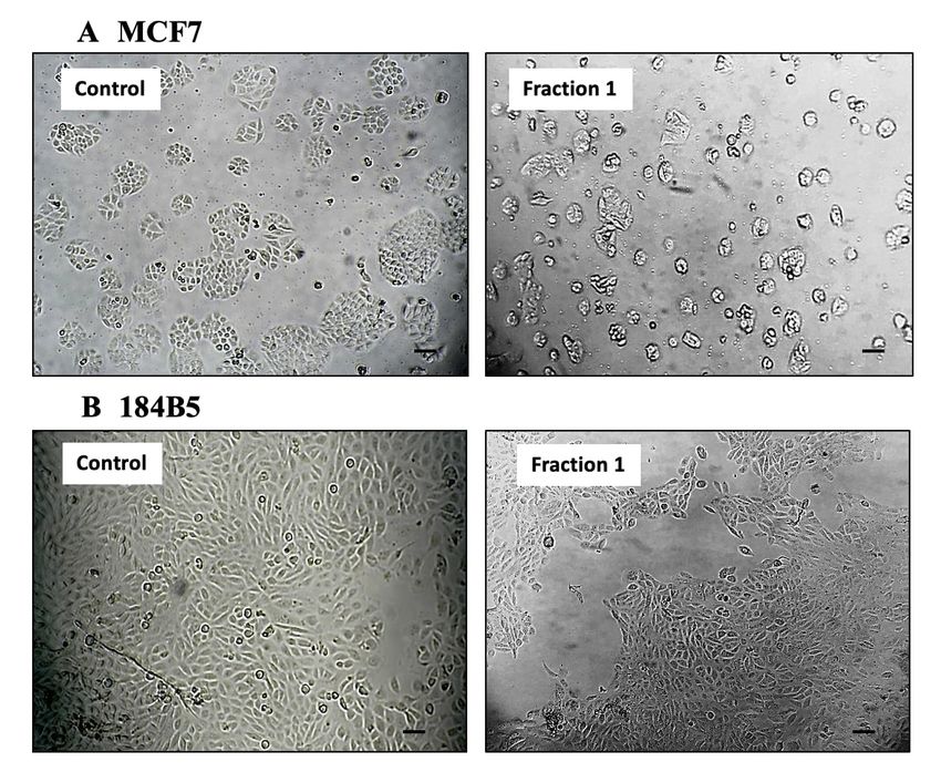
Tan et al. J Venom Anim Toxins incl Trop Dis, 2020, 26:e20200013                                                               Page 2 of 14

Background                                                            receptor that regulates cell motility, survival, proliferation,
Snake venom is widely regarded as an advanced “biochemical            angiogenesis and cell invasion [21, 22]. The antineoplastic
weapon” that exists in nature. Venomous snakes rely on venom          potential of snake venom disintegrins have been shown in
for survival, where it is employed for predation (primary             several studies, e.g. contortrostatin (disintegrin isolated from
function), digestion and defense [1]. Rapid evolution drives          Agkistrodon contortrix venom) [23], saxatilin (disintegrin from
the emergence of diverse pharmacologically active snake venom         Gloydius saxatilis venom) [24], PAIEM (disintegrin isolated
proteins that are adapted to target the normal physiology of          from Echis multisquamatis venom) [25] and a disintegrin from
prey by exerting various toxic effects [2]. The prey of venomous      Crotalus durissus collilineatus venom [26]. The disintegrins act by
snakes consists in parts of small mammals such as rodents             inhibiting angiogenesis, invasion and migration of cancer cells.
but may also include birds, lizards and amphibians [3, 4]. It is      The finding of disintegrin in the Malaysian T. purpureomaculatus
noteworthy that the mammalian system-targeting property of            venom is significant as the peptide could be a promising local
snake venom proteins is often specific and selective, thus making     source of anticancer candidate that has not been characterized
them a natural repertoire of therapeutic molecules [5, 6].            previously.
   At present, advanced cancers that are fast-growing and                In this study, the Malaysian T. purpureomaculatus venom
capable of metastasizing is a major cause of mortality globally       was suggested to be cytotoxic to a wider range of cancer cell
[7, 8]. In this context, snake venom proteins have the potential      lines including those of the human breast, lung, colon and
for drug discovery in line with the search for novel peptide-         prostate. The selective anticancer activity, if any, could be partly
based anticancer agents with high efficacy and selectivity            contributed by the disintegrin present in the venom. Hence,
in targeting cancer cells [9, 10]. Notable examples typically         this study aimed to investigate the cytotoxicity of Malaysian
involved venom proteins from vipers and pit vipers (family            T. purpureomaculatus venom across four common human
Viperidae), e.g. disintegrin from Agkistrodon contortrix venom        cancerous and the corresponding normal cell lines. In addition,
[11], phospholipases A2 from Cerastes cerastes [12] and L-amino       the disintegrin was purified from the venom, and its amino acid
acid oxidases from Bothrops sp. [13].                                 sequence as well as anticancer properties were characterized.
   A common pharmacological feature of the venoms of
viperid snakes, including the Asian arboreal pit vipers from the      Methods
Trimeresurus complex, is their ability to derange hemostasis and
induce local tissue necrosis [14–16]. The tissue-necrotizing effect
                                                                      Venom Sample
associated with Trimeresurus pit viper envenomation suggests
cytotoxic activity in the venom that may be further explored          The venom of mangrove pit viper (Trimeresurus purpu
for anticancer potential. A recent study with Thai Trimeresurus       reomaculatus, MTP) was a pooled sample from ten adult snake
purpureomaculatus venom reported high cytotoxicity of the             specimens (four males; six females) from Peninsular Malaysia.
venom toward SHSY5Y cell line, and moderate cytotoxicity              The venom obtained was freeze-dried and stored at −20 °C
of BPP-related peptides, PLA2 and peptide-rich fraction of the        until use. Venom stock used in each cytotoxic experiment was
venom [17]. Another publication reported that L-amino acid            prepared freshly from lyophilized venom and subjected to a
oxidase (LAAO, 55−60 kDa) from Malaysian Trimeresurus                 quick spin for 15 seconds prior to the treatment.
purpureomaculatus venom induced cytotoxicity in three colon
cell lines (SW480, SW620 and CCD-18Co), but no selectivity was        Chemicals and Materials
observed between the cancerous cell lines (SW480, SW620) and          All chemicals and reagents used in the studies were of analytical
the non-cancerous, normal cell line (CCD-18Co) [18].                  grade. 3,(4,5-dimethythiazol-2-yl)-2,3-diphenyl tetrazolium
   Our recent quantitative proteomic analysis of the Malaysian        bromide (MTT) was purchased from Sigma Aldrich (Missouri,
T. purpureomaculatus venom (MTP) revealed that LAAO                   USA) and dimethyl sulfoxide (DMSO) was supplied by Merck
constituted about 3% of the total venom proteins, while               (Darmstadt, Germany). Trypsin-EDTA was supplied by Nacalai
disintegrin, a compound with potential inhibitory effect on           Tesque (Kyoto, Japan). The positive control in the MTT assay
cancer cell growth, was present at a higher abundance in the          was 5-fluorouracil (5-FU) (Sigma-Aldrich, Missouri, USA).
venom (approximately 14%) [19]. The LAAO content reported             The reversed-phase HPLC column LiChrospher® WP 300 RP-
previously for the Malaysian T. purpureomaculatus venom could         18 (5 μm) was purchased from Merck (Darmstadt, Germany)
not be compared as it was estimated with a qualitative method         and HPLC solvents were from Thermo Scientific ™ Pierce ™
[20]. In a more recent study, relative protein abundances of          (Massachusetts, USA). Trifluoroacetic acid (TFA) was purchased
0.91% LAAO and 0.10% disintegrin were identified in the Thai          from Sigma Aldrich (Missouri, USA). Ammonium bicarbonate,
T. purpureomaculatus venom [17].                                      dithiothreitol (DTT) and iodoacetamide (IAA), which were
   Disintegrin is a much smaller (4-15 kDa, monomer or dimer)         used in protein digestion, were purchased from Sigma-Aldrich
cysteine-rich and RGD-containing polypeptide that targets             (Missouri, USA), Trypsin protease (MS grade) was purchased
specifically on integrin – a ubiquitously expressed cell surface      from Thermo Scientific Pierce ™ (Massachusetts, USA) and
Tan et al. J Venom Anim Toxins incl Trop Dis, 2020, 26:e20200013                                                                           Page 3 of 14

desalting C18 pipette tips was purchased from Merck Millipore       previous medium. The plate was covered with aluminum foil
ZipTip® (MilliporeSigma, Massachusetts, USA). Protein ladder        and returned to the CO2 incubator for 3 hours incubation. The
of PM2700 ExcelBand™ 3-color Broad Range Protein Marker was         MTT solution was pipetted out gently after the incubation and
purchased from SMOBIO Technology, Inc. (Hsinchu, Taiwan).           200 μL of dimethyl sulfoxide (DMSO) was added into each well
                                                                    to solubilize the formazan crystal. Absorbance was measured
Cell Culture                                                        using Hidex Plate CHAMELEON™ V (Hidex, Turku, Finland)
Both normal and cancer cell lines were obtained from American       multilabel microplate reader at 570 nm. The median inhibition
Type Culture Collection (ATCC, Virginia, USA). MCF7 (ATCC®          concentration (IC50) was determined from the dose-response
HTB-22™; human breast adenocarcinoma cell line), HT-29              curve plotted with percentage of cell viability against venom
(ATCC® HTB-38™; human colon colorectal adenocarcinoma cell          concentration (μg/mL). In the assay, 5-fluorouracil (5-FU) was
line), A549 (ATCC® CCL-185™; human lung carcinoma cell line)        used as a positive reference. The percentage of cell viability was
and PC-3 (ATCC® CRL-1435™; human prostate adenocarcinoma            calculated with the following formula:
cell line) were used as the cancer cell panel, while 184B5 (ATCC®
CRL-8799™; human breast normal cell line), CCD-18Co (ATCC®                                 Average OD of treated cells – Average OD of blank
                                                                    Cell viability (%) =                                                        × 100%
CRL-1459 ™; human colon normal cell line), MRC5 (ATCC ®                                    Average OD of control cells – Average OD of blank
CCL-171™; human lung normal cell line) and RWPE-1 (ATCC ®
CRL-11609™; human prostate normal cell line) were used as the       Selectivity Index Determination
normal cell panel in the cytotoxic assay.                           The degrees of selective cytotoxicity of the venom in cancer cell
   MCF7 and HT-29 cell lines were cultured in Dulbecco’s            lines were indicated with selectivity index (SI), determined as
modified Eagle’s medium (DMEM) (Nacalai Tesque, Kyoto,              follows:
Japan), supplemented with 10% fetal bovine serum (FBS) (TICO                                                  (IC50 in normal cell line)
                                                                                 Selectivity index (SI) =
Europe, Amstelveen, Netherlands) and 100 µg/mL penicillin-                                                    (IC50 in cancer cell line)
streptomycin (Nacalai Tesque, Kyoto, Japan), while A549
and PC-3 were maintained using RPMI 1640 medium with                Fractionation of T. purpureomaculatus Venom and
L-glutamine (Lonza, Verviers, Belgium), with 10% FBS and 100        Bioassay-Guided Cytotoxicity Study
µg/mL penicillin-streptomycin. Normal cell lines such as CCD-       Two hundred microliters of T. purpureomaculatus venom
18Co and MRC5 were maintained in Eagle’s minimum essential          solution (10 mg/mL in ultrapure water) were injected into
medium (EMEM) (Sigma-Aldrich, Missouri, USA), with 10%              LiChrosper® WP 300-RP-18 reversed-phase column (5 μm
FBS and 100 µg/mL penicillin-streptomycin. The 184B5 was            column particle size) through the Shimadzu LC-20AD high
maintained in mammary epithelial cell growth medium (MEGM)          performance liquid chromatography system. The column was
with bullet kit (Lonza, Basel, Switzerland), supplemented with      pre-equilibrated with solvent B [0.1% trifluoroacetic acid (TFA)
10% FBS and 100 µg/mL penicillin-streptomycin, while RWPE-          in acetonitrile] followed by solvent A (0.1% TFA in water). The
1 was maintained in Keratinocyte-SFM (serum-free medium)            elution began with the stepwise linear gradient (0-5% of B for
(Thermo Scientific, Massachusetts, USA), with only 100 µg/mL        10 min, followed by 5-15% B for 20 min, 15-60% B for the next
penicillin-streptomycin added to the medium. The cell lines         180 minutes, 60-70% B for 10 minutes and 75-100% of B over
were cultured in a 5% carbon dioxide (CO2) incubator (Shel Lab,     245 min) in 0.1% TFA in acetonitrile (ACN) for 245 minutes
Oregon, USA) at 37 ºC, with pH of 7.2 - 7.5 and relative humidity   (flow rate: 1 mL/min). Protein elution was monitored at 215 nm.
of about 95%. The growth of cells was monitored routinely by           The protein fractions were collected manually, subsequently
observing cell morphology under an inverted microscope (Leica       freeze-dried and stored at −20 °C until use. Each protein
Microsystems, Wetzlar, Germany). All of the cell-related work       fraction was reconstituted in ultrapure water and the protein
was carried out using aseptic techniques under sterile condition.   concentration was estimated by Nanodrop Spectrophotometer
                                                                    2000 (ThermoFisher ™ , Massachusetts, USA) prior to the
Cell Viability Assay                                                experiment. The cytotoxic fraction of T. purpureomaculatus
Cell viability was studied with the 3-(4,5-dimethylthiazol-2-yl)-   venom was screened with a bioassay-guided method modified
2,5-diphenyltetrazolium bromide (MTT) assay. The cells of the       from Shahbazi [27]. The cytotoxic activities of the venom fraction
respective cell lines were detached from the culture flasks using   were tested at a standard dose of 20 µg each on human breast
an appropriate amount of trypsin-EDTA and desired number            cancer cell line (MCF7) according to the cell viability assay
of cells were seeded in a 96-well microplate (15,000-150,000        described above. The amount of protein from each venom
cells/mL) for overnight incubation. The attached cells were then    fraction tested (20 µg) was referred from a previous study
treated with serial dilutions (ranging from 0.10-31.62 μg/mL) of    by Bradshaw [28]. Cell images were captured 72 hours after
T. purpureomaculatus venom for 72 hours. After treatment, 10%       treatment using inverted microscope (Leica Microsystems,
of MTT reagent was added into each well without removing the        Wetzlar, Germany).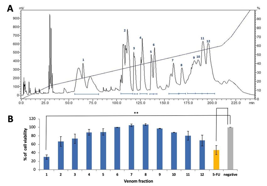
Tan et al. J Venom Anim Toxins incl Trop Dis, 2020, 26:e20200013                                                           Page 4 of 14

Sodium Dodecyl Sulphate-Polyacrylamide Gel                           for cysteine carbamidomethylation is set as a fixed modification,
Electrophoresis (SDS-PAGE)                                           while methionine oxidation is set as variable modification.
Gel electrophoresis was carried out according to the protocol           The mass spectrometry derived peptide masses were searched
of Laemmli [29]. Approximately 10 μg of protein fraction was         against a non-redundant NCBI database of Serpentes (taxid:
loaded onto a 15% acrylamide gel and the electrophoresis was         8570) combined with the venom-gland transcriptome database
carried out under reducing conditions at 90 V for 2 hours. Protein   of T. purpureomaculatus. The protein identification was validated
ladder PM2700 ExcelBand™ 3-color Broad Range Protein Marker          by the following filters: protein score > 20, peptide score > 10
was used as molecular weight standards in the electrophoresis        and score peak intensity (SPI) > 70%. Proteins with “Distinct
(5-245 kDa). Coomassie Brilliant Blue R-250 staining was used        Peptide” ≥ 2 were considered significant matches.
for the visualization of protein.
                                                                     Bioinformatic Analyses
Protein Digestion and Mass Spectrometry Analysis
of the Cytotoxic-Contributing Fraction of MTP                        Sequence analysis of disintegrin
Venom
                                                                     Multiple sequence alignment of disintegrins was performed using
The most cytotoxic protein fraction of MTP venom was subjected       MUSCLE program and Jalview software v2.11 to indicate the
to in-solution tryptic digestion and nano-ESI LC-MS/MS analysis      region of similarity. The percentage of similarity was calculated
as previously reported [30]. Ten micrograms of the protein (10 µL    using EMBL-EBI Clustal Omega (https://www.ebi.ac.uk/
of 1 mg/mL protein concentration) first underwent reduction and      Tools/msa/clustalo/). The disintegrin sequence of Malaysian
alkylation by addition of 15 μL of 50 mM ammonium bicarbonate        Trimeresurus purepureomaculatus (MTP) venom was derived
and 1.5 μL of 100 mM dithiothreitol (DTT). The mixture was           from the venom-gland transcriptome (entry is available in
heated at 95 ºC for 5 min before addition of 3 μL of 100 mM          NCBI GenBank database, accession ID: QJA41976.1). Other
iodoacetamide (IAA). The mixture was later incubated in dark         related disintegrin sequences were retrieved from Uniprot
at room temperature for another 20 min. Digestion was carried        Knowledgebase (https://www.uniprot.org/) [31].
out by adding one microliter of 0.1 mg/mL trypsin solution to the
reaction tube and incubated at 37 ºC for 3 hours. Additional 1 µL    In silico physicochemical characterization of
of trypsin solution was added for overnight incubation at 30 ºC.     disintegrin
After digestion, the digested peptides were desalted for removal
                                                                     The physicochemical properties of the disintegrin from MTP
of salts and contaminants.
                                                                     venom were characterized using the Expasy ProtParam online
   The desalted peptides (approximately 10 µg) were then
                                                                     tool (https://web.expasy.org/protparam/) according to Roly et
reconstituted in 7 µL of 0.1% formic acid in water. One microliter
                                                                     al. [32]. Parameters including theoretical molecular weight,
of the solution (containing approximately 1.4 µg of peptides) was
                                                                     isoelectric point (pI), instability index and grand average of
subjected to nano-electrospray ionization MS/MS via Agilent
                                                                     hydropathicity (GRAVY) value were determined.
1260 HPLC-Chip/MS Interface coupled with Agilent 6550
Accurate-Mass Q-TOF LC/MS system (Agilent Technologies,
Santa Clara, California, USA). The sample peptides were              Statistical Analysis
separated in a large capacity chip Zorbax 300 Å, C18, 160 nl         The median inhibition concentration (IC50) value was determined
enrichment column, 75 μm × 150 mm analytical column and              using GraphPad Prism 5 statistical software (GraphPad Software
5 μm particles (Agilent part no. G4240–62010). Parameters            Inc., California, USA) and the values were expressed as mean ±
were set as follows: injection volume at 1 µL per sample, flow       S.E.M. of three replicates. Comparative data were statistically
rate from capillary pump at 4 µL/min and 0.4 µL/min from             analyzed using Student’s unpaired t-test (at 95% confidence
Nano pump (G2226A), gradient used: 5-50% solution B (0.1%            interval) with GraphPad Prism 5 software (GraphPad Software
formic acid in acetonitrile) for 11 min, 50-70% B for next 4         Inc., California, USA).
min, and 70% B for 3 min and ion polarity was set to positive
ionization mode. Drying gas flow and temperature were set at         Results
11 L/min and 290 ºC. Fragmentor voltage was set at 175 V while
for capillary voltage, it was set at 1800 V. MS scan range of 200-
3000 m/z and MS/MS scan range of 50-3200 m/z were acquired           Cytotoxicity of Trimeresurus purpureomaculatus
in the tandem mass spectrometry mode, with precursor charge          Venom
selection set as doubly charge state and above with the exclusion    MTP venom exhibited dose-dependent cytotoxic effects toward
of precursor 1221.9906 m/z (z = 1) for internal mass calibration     all cancer cell lines tested (median inhibition concentrations,
and reference ions set at 299.2944 (z = 1). Data with a MH+          IC50 = 0.42−2.50 µg/mL). The effects were generally stronger in
mass range between 50 and 3200 were extracted and analyzed           the cancer cells compared to the corresponding normal cells
in Agilent Spectrum Mill MS Proteomics Workbench software            (IC50 = 0.70-6.98 µg/mL) (Table 1). The venom cytotoxicity was
packages (Agilent Technologies, Santa Clara, CA, USA). Setting       most potent to the colorectal adenocarcinoma cell line (IC50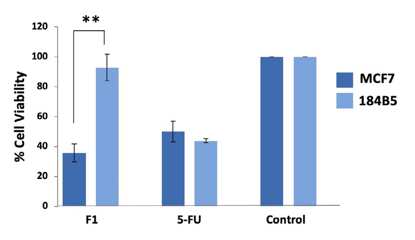
Tan et al. J Venom Anim Toxins incl Trop Dis, 2020, 26:e20200013                                                                                         Page 5 of 14

= 0.42 ± 0.06 µg/mL), followed by the breast cancer cell line                         fold more cytotoxic to MCF7 than it was to the corresponding
(IC50 = 0.48 ± 0.02 µg/mL) and prostate cancer cell line (IC50 =                      normal cell line (184B5) (Table 1, Figure 1).
1.60 ± 0.18 µg/mL). The venom was least cytotoxic to the lung
cancer cell line (IC50 = 2.50 ± 0.20 µg/mL). The venom was also                       Cytotoxicity of the Chromatographic Fractions of
significantly more potent compared to 5-FU in inhibiting the                          Trimeresurus purpureomaculatus Venom
growth of breast, lung and prostate cancer cells (p < 0.05). In                       MTP venom was resolved using C18 reversed-phase high
comparison to the IC50 in normal cell lines, the cytotoxic effect                     performance liquid chromatography (RP-HPLC) into 12
was not selective toward colon, lung and prostate cancer cells as                     fractions as shown in Figure 2A. The cytotoxic effect of each
indicated by a selectivity index (SI) ≤ 10. The selectivity index                     fraction was further tested on MCF7 cell line in view of the
of the venom was, however, much higher (SI = 14.54) for the                           high selectivity index (SI) of MTP venom in breast cancer cell
breast cancer cell line (MCF7), implying that the venom was 15-                       line. MCF7 cells treated with protein from Fraction 1 (20 µg)

Table 1. Median inhibition concentrations (IC50) (µg/mL) and selectivity indices (SI) of Malaysian T. purpureomaculatus (MTP) venom for different cell lines after
72-hour treatment in comparison to 5-fluorouracil (5-FU).

                                                               MTP                                                              5-FU
                                               IC50                                                             IC50
Cell Lines                                                                       SI                                                                SI
                                             (µg/mL)                                                          (µg/mL)
Breast
MCF7                                       0.48 ± 0.02                         14.54                         8.26 ± 0.77                          0.16
184B5                                      6.98 ± 0.53                                                       1.34 ± 0.14
Colon
HT-29                                      0.42 ± 0.06                          1.67                         0.12 ± 0.02                       >258.33
CCD-18Co                                   0.70 ± 0.15                                                         > 31.62
Lung
A549                                       2.50 ± 0.20                          1.44                           > 31.62                            NA
MRC5                                       3.61 ± 0.14                                                         > 31.62
Prostate
PC3                                        1.60 ± 0.18                          1.19                        12.83 ± 1.52                          0.22
RWPE-1                                     1.91 ± 0.53                                                       2.79 ± 0.66

IC50 values were derived from triplicates ± S.E.M.
Selectivity index ≥ 10 is considered as the threshold for a compound having cancer-selective cytotoxic effect [28].
NA: not available, MCF7: human breast cancer cell line, 184B5: human breast normal cell line, HT-29: human colon cancer cell line, CCD-18Co: human colon
normal cell line, A549: human lung cancer cell line, MRC5: human lung normal cell line, PC3: human prostate cancer cell line, RWPE-1: human prostate normal
cell line.

Figure 1. Dose-dependent growth inhibitory effect of Trimeresurus purpureomaculatus (MTP) venom in human breast cell lines. Median inhibition concentrations
(IC50) were determined from the dose-response curve. Values were presented as means ± S.E.M. (n = 3). MCF7: human breast cancer cell line, 184B5: human
breast normal cell line.
Tan et al. J Venom Anim Toxins incl Trop Dis, 2020, 26:e20200013 Page 6 of 14
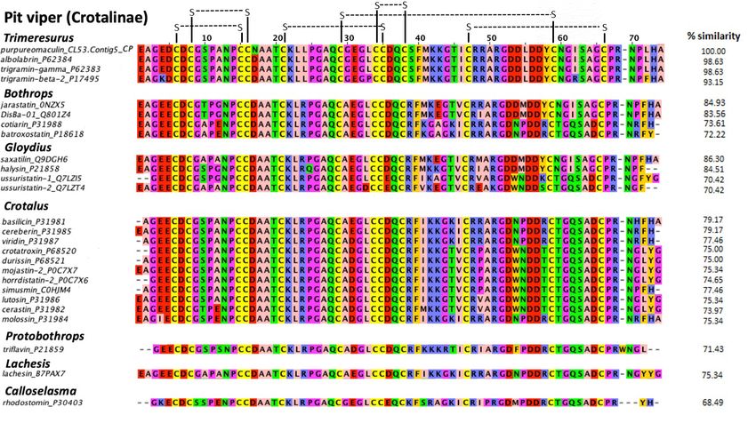
showed the most potent inhibitory effect (cell viability reduced by effect was observed in the corresponding normal breast cells
65%) compared to the non-treated control (**p < 0.01). Proteins (184B5 cell line) (Figure 4). Microscopic examination revealed
from Fraction 2 and 12 exhibited moderate cytotoxic effect (cell the presence of detached, clumped and rounded floater cells in
viability reduced by 25−30%) whereas other fractions did not the purpureomaculin-treated breast cancer cells following a
show significant cytotoxicity in the MCF7 cell line (Figure 2B). treatment period of 72 hours. On the contrary, the normal cell
In comparison, 5-FU at IC50 of 9 µg/mL induced a 50% reduction line (184B5) treated with purpureomaculin was observed to
in the cell viability study. remain healthy and viable, with slight reduced growth compared
to the untreated control (Figure 5).
Identification and Cytotoxicity of Disintegrin from
Trimeresurus purpureomaculatus Venom Sequence Analysis and in silico Characterization of
Figure 3 shows the SDS-PAGE profile of the cytotoxic protein Purpureomaculin, the Cytotoxic Disintegrin from T.
isolated in Fraction 1. A homogenous protein band was observed purpureomaculatus Venom
and the molecular mass was estimated to be 7585 Da by its The tryptic peptides of purpureomaculin (K)LLPGAQCGE
relative migration distance (Rf). LC-MS/MS identified Fraction GLCCDQCSFMKK(G) and (R)ARGDDLDDYCNGISAGCPR(N)
1 to contain only one protein, which is a disintegrin of snake from Fraction 1 were matched to t he disintegrin
venom and it is termed as “purpureomaculin” in the present transcript “CL53.Contig5_CP” from the Malaysian
study. The mass spectrometry data file of purpureomaculin is T. purpureomaculatus venom-gland transcriptomic
available in Additional file 1, and the primary data was deposited database. A disintegrin with full amino acid sequence was
to the ProteomeXchange Consortium via the iProX partner identified. It is a mature chain polypeptide of 73 amino acid
repository [33] with the dataset identifier PXD018463. residues: AGEDCDCGSPA NPCCNA ATCK LLPGAQ
Purpureomaculin at 20 µg significantly inhibited the growth of CGEGLCCDQCSFMKKGTICRRARGDDLDDYCNGISAG
MCF7 cell line by 65% (**p < 0.01), while no significant cytotoxic CPRNPLHA.
Figure 2. (A) C18 reversed-phase high performance liquid chromatography of venom from the Malaysian T. purpureomaculatus. (B) Cell viability of human breast
cancer cells (MCF7) after 72-hour treatment with the HPLC fractions of Malaysian T. purpureomaculatus venom. Positive and negative controls were 5-fluorouracil
(5-FU) and treatment-free, respectively, in the assay.Tan et al. J Venom Anim Toxins incl Trop Dis, 2020, 26:e20200013 Page 7 of 14
Figure 4. Cell viability of human breast cell lines, MCF7 (cancerous) and
184B5 (normal) after 72-hour treatment with 20 µg of Fraction 1 (F1,
purpureomaculin) purified from Trimeresurus purpureomaculatus venom.
Positive control: 5-fluorouracil (5-FU); negative control: treatment-free well.
following positions: (6, 15), (8, 16), (21, 35), (29, 59), (34, 38),
(47, 66). Purpureomaculin also harbored the tripeptide RGD, a
short sequence motif of biological interest formed by amino acid
residues 51−53. Compared with the disintegrin sequences of other
viperids, high similarity (> 70%) was observed in phylogenetically
related pit viper genera and species, e.g. Trimeresurus complex.
Table 2 shows the physicochemical properties of
purpureomaculin analyzed in silico using the ProtParam Tool.
Purpureomaculin has an estimated molecular weight of 7572.52
Da and is weakly acidic (pI = 4.79).
Table 2. Physicochemical properties of purpureomaculin from the Malaysian
Trimeresurus purpureomaculatus venom.
Properties Values
Residues 73
Mr 7572.52
pI 4.79
Instability Index (II)a 25.23
Stability Stable
Aliphatic Index (AI)b 48.36
Figure 3. Protein content of Fraction 1 (F1) of Malaysian T. purpureomaculatus GRAVYc -0.390
venom was validated under 15% reducing gel electrophoresis. Protein ladder Extinction coefficients*d 2240
PM2700 ExcelBand™ 3-color Broad Range Protein Marker was used for
Extinction coefficients* (reduced)e 1490
molecular weight standards (5−245 kDa).
Mr: Molecular weight (Da); pI: Isoelectric point; GRAVY: Grand average of
Multiple sequence alignment showed that purpureomaculin is hydropathicity.
highly homologous (98.6%) with albolabrin from Trimeresurus
a
Estimate of protein stability (instability index ofTan et al. J Venom Anim Toxins incl Trop Dis, 2020, 26:e20200013 Page 8 of 14 Figure 5. Morphological changes in human breast cell lines, (A) MCF7 (cancerous) and (B) 184B5 (normal) after 72-hour treatment of Fraction 1 (purpureomaculin) of Trimeresurus purpureomaculatus venom. Scale bars = 30 µm. Figure 6. Multiple sequence alignment of purpureomaculin (CL53.Contig5_CP) with other disintegrin sequences of Crotalinae. Purpureomaculin is a disintegrin isolated from the Malaysian Trimeresurus purpureomaculatus venom, while other disintegrin sequences were obtained from UniProtKB. The percentages of similarity (% similarity) of disintegrin sequences in comparison to purpureomaculin were calculated using Omega Clustal software. Zappo color scheme indicates residue properties: pink: aliphatic/hydrophobic; orange: aromatic; blue: positive; red: negative; green: hydrophilic; magenta: proline/glycine (conformationally special); yellow: cysteine. Disulfide bonds were illustrated in black connecting lines.
Tan et al. J Venom Anim Toxins incl Trop Dis, 2020, 26:e20200013                                                                Page 9 of 14

Discussion                                                              hence, all venom samples used in the present study were freshly
In this study, the potent cytotoxicity of Malaysian T.                  reconstituted from lyophilized stock sample prior to treating
purpureomaculatus venom was shown in a panel of cancer                  the cells in the MTT experiment. The additional measure,
(MCF7, HT-29, PC3, A549) and normal (184B5, CCD-18Co,                   presumably, avoided any possible protein degradation [38] or
RWPE-1, MRC5) cell lines. MTP venom was particularly toxic              inactivation of the venom bioactive components by unfavorable
to breast cancer cell line (MCF7) and colon cell lines (HT-29 and       pH or temperature [39].
CCD-18Co), indicating high specificity of the venom toward these           In addition, previous studies had demonstrated variable
tested cells. The venom also demonstrated selective cytotoxicity,       anticancer activities for venom disintegrin from pit vipers. For
particularly in the human breast cancer cells (MCF7). This              instance, lebein (disintegrin isolated from Macrovipera lebetina
suggests that the venom contains toxin(s) or protein(s) that could      venom) has been shown to significantly reduce the cell viability
be cancer-specific in breast tumor, thus potentiating the venom         of colon adenocarcinoma cell lines after 72-hour incubation [40].
cytotoxicity at a lower dose in the cancer cells compared to the        A more recent study demonstrated that disintegrins isolated
corresponding normal cells. The cytotoxic effect of Malaysian           from Cerastes cerastes venom displayed varying IC50 values
T. purpureomaculatus venom also appeared to be more potent              (1.60−8.17 µg/mL) in SHSY5Y neuroblastoma cell line [17].
than other pit viper venoms, including venoms of Protobothrops          Tzabcanin (disintegrin isolated from Crotalus simus tzabcan
flavoviridis [35], Trimeresurus macrops and Trimeresurus hageni         venom) exhibited low cytotoxicity against Colo-205 cells and was
[36], albeit different human cell lines were utilized in each study.    not cytotoxic to MCF7 cells [41]. On the contrary, the disintegrin
   Further investigation revealed that most of the venom                purpureomaculin of T. purpureomaculatus was found to be
components (fractionated with HPLC) showed variable anticancer          potent in inhibiting the growth of MCF7 cells (current study).
activity, implying that the overall venom cytotoxicity could be         The anticancer activities of disintegrin from many closely related
a result of synergistic interactions of multiple components in          species such as trigramin (disintegrin isolated from Trimeresurus
the venom. The most potent anti-neoplastic activity was found           gramineus venom) and albolabrin (disintegrin isolated from
in the protein fraction containing snake venom disintegrin.             Trimeresurus albolabris venom) remain unknown to date, and
This was a purified protein validated through SDS-PAGE and              warrant investigation for insights in the anticancer potential
LC-MS/MS, and termed purpureomaculin in line with the                   of the venom peptide from this large Asiatic pit viper complex.
naming of most snake venom-derived disintegrin protein (prefix             The multiple sequence alignment revealed that the venom
“species-” + post-fix “-in”). The finding of purpureomaculin            disintegrins of pit vipers (Crotalinae) are mainly of intermediate
anticancer activity is consistent with earlier studies that reported    length with 12 conserved cysteine residues, forming 6 disulfide
the inhibitory activity of snake venom disintegrins on the              bonds that support the folding and stability of the protein
proliferation, metastasis and adhesion of cancer cells [21, 25, 37].    [42]. Disintegrins from Trimeresurus, Bothrops, Gloydius,
The selective cytotoxic effect of purpureomaculin in breast             Crotalus, Protobothrops and Lachesis complexes in particular
cancer cells (MCF7) corroborates the selective cytotoxicity of          showed a higher rate of amino acid conservation, indicating
MTP venom in the same cancer cell line observed in this study.          the phylogenetic relationship among different genera within
   The present findings varied when compared with previous              the Crotalinae subfamily. At the intra-generic level of
studies on other cancer cells using T. purpureomaculatus venoms         Trimeresurus, purpureomaculin sequence was found almost
from Malaysia and Thailand [17, 18]. IC50 values with wide              identical (similarity: 98.6%) to albolabrin and trigramin-gamma
discrepancy were observed between previous studies (Malaysian           (disintegrins from Trimeresurus albolabris and Trimeresurus
T. purpureomaculatus IC50 (72 hours): 15.99−29.43 µg/mL in human        gramineus). Like albolabrin and trigramin, purpureomaculin
colon cell lines [18]; Thai T. purpureomaculatus (IC50 (48 hours):      is structurally categorized as the medium-sized disintegrins
0.25−5.24 µg/mL in human neuroblastoma, cervical, colon,                which contain about 70 amino acids and 6 disulfide cysteine
breast, bladder, glioblastoma and kidney cell lines [17]) and the       bonds, as described by Calvete [42]. The high similarity of
present work (Malaysian T. purpureomaculatus: IC50 (72 hours): 0.42-    purpureomaculin to these disintegrins indicates structural
6.98 µg/mL in breast, lung, colon and prostate cell lines) (Table 3).   and functional resemblance between these disintegrins, and
Nevertheless, the cytotoxic activity of the present Malaysian T.        that purpureomaculin is likely to exert its cytotoxic activity
purpureomaculatus venom in human colon cells appeared to                similar to trigramin and albolabrin by inhibiting cell-matrix
be comparable to the Thai venom sample reported by Ozverel              adhesion through the binding of RGD tripeptide domain
[17]. In contrast, much higher IC50 values (by a difference of >        to the cellular integrin receptors [43, 44]. One remarkable
10 folds) in human colon cells were reported previously in the          difference of purpureomaculin is the amino acid substitution
other Malaysian T. purpureomaculatus venom sample [18]. The             at the 17th position. It is noted that amino acid at this position
discrepancy could be due to the variation in venom composition,         is highly conserved throughout the pit viper genera, and the
the use of different colon cell lines, or the influence of the          substitution of the aspartic acid (D) to the neutral asparagine (N)
condition of venom preparation. In our experience, repeated             in purpureomaculin may have a unique evolutionary implication
freeze-thaw cycles of the MTP venom led to inconsistent and             on the role of the protein expressed. The consequence of the
somewhat deteriorating venom cytotoxic activity (unpublished);          amino acid substitution deserves further investigation.
Table 3. Comparison of cytotoxicity studies of T. purpureomaculatus venoms from different locations (Malaysia and Thailand).
Trimeresurus Trimeresurus Trimeresurus
Species purpureomaculatus purpureomaculatus purpureomaculatus
(current work) [18] [34]
Origin Malaysia (wild) Malaysia Thailand (kept in Turkey)
Whole venom study
Treatment
72 hours 72 hours 48 hours
duration
Tissue-organ
Cancerous Normal Selectivity Cancerous Normal Selectivity Cancerous Normal Selectivity
and cell line
MCF7, IC50 s> 50 µg/mL
MCF7, IC50 = 184B5, IC50 = Yes
Breast N.D. N.D. N.D. MDA-MB-231, IC50 = N.D. N.D.
0.48 ± 0.02 µg/mL 6.98 ± 0.53 µg/mL (S.I. = 14.54)
Tan et al. J Venom Anim Toxins incl Trop Dis, 2020, 26:e20200013
1.77 ± 0.21 µg/mL
SW480, IC50 =
HT-29, IC50 = CCD-18Co, IC50 = 29.43 ± 0.48 µg/mL CCD-18Co, IC50 = CaCo-2, IC50 =
Colon No No N.D. N.D.
0.42 ± 0.06 µg/mL 0.70 ± 0.15 µg/mL SW620, IC50 = 15.99 ± 1.20 µg/mL 3.10 ± 1.40 µg/mL
23.19 ± 1.57 µg/mL
A549, IC50 = MRC5, IC50 = A549, IC50 =
Lung No N.D. N.D. N.D. N.D. N.D.
2.50 ± 0.20 µg/mL 3.61 ± 0.14 µg/mL 4.65 ± 0.19 µg/mL
PC3, IC50 = RWPE-1, IC50 =
Prostate No N.D. N.D. N.D. N.D. N.D. N.D.
1.60 ± 0.18 µg/mL 1.91 ± 0.53 µg/mL
SHSY5Y (neuroblastoma),
IC50 = 0.25 ± 0.05 µg/mL
Nerve N.D. N.D. N.D. N.D. N.D. N.D. U87MG N.D. N.D.
(glioblastoma),IC50 =
1.32 ± 0.03 µg/mL
HeLa, IC50 =
Cervix N.D. N.D. N.D. N.D. N.D. N.D. N.D. N.D.
2.15 ± 1.50 µg/mL
253-JBV, IC50 =
Urinary bladder N.D. N.D. N.D. N.D. N.D. N.D. N.D. N.D.
5.24 ± 0.77 µg/mL
HEK293, IC50 =
Kidney N.D. N.D. N.D. N.D. N.D. N.D. N.D. N.D.
2.04 ± 0.21 µg/mL
Page 10 of 14Table 3. Cont.
Trimeresurus Trimeresurus Trimeresurus
Species purpureomaculatus purpureomaculatus purpureomaculatus
(current work) [18] [34]
Venom fraction study
Fractionation
Reversed-phase HPLC Size-exclusion & Reversed-phase HPLC Reversed-phase HPLC
method
Treatment
72 hours 72 hours 48 hours
duration
Tan et al. J Venom Anim Toxins incl Trop Dis, 2020, 26:e20200013
Tissue-organ
Cancerous Normal Selectivity Cancerous Normal Selectivity Cancerous Normal Selectivity
and cell line
MCF7; disintegrin at 184B5; disintegrin at 20
Breast 20 µg reduced cell µg did not significantly Yes N.D. N.D. N.D. N.D. N.D. N.D.
viability by 65% reduced cell viability
SW480; LAAO IC50 =
CCD-18Co;
13.56 ± 0.58 µg/mL
Colon N.D. N.D. N.D. LAAO IC50 = No N.D. N.D. N.D.
SW620; LAAO IC50 =
14.98 ± 2.67 µg/mL
13.17 ± 0.77 µg/mL
SHSY5Y (neuroblastoma);
BPP-RP IC50 =
37.01 ± 3.30 µg/mL
& 78.39 ± 5.38 µg/mL
Nerve N.D. N.D. N.D. N.D. N.D. N.D. N.D. N.D.
PLA2 IC50 =
43.79 ± 3.40 µg/mL
peptide-rich fraction
IC50 = 34.07 ± 0.07 µg/mL
HPLC: high performance liquid chromatography, IC50: median inhibition concentration, S.I.: selectivity index, N.D.: not determined, LAAO: L-amino acid oxidase, BPP-RP: bradykinin-potentiating peptide, PLA2:
phospholipase A2.
Page 11 of 14Tan et al. J Venom Anim Toxins incl Trop Dis, 2020, 26:e20200013 Page 12 of 14
The sequence similarities between purpureomaculin and other modification e.g. nano-carrier conjugation in the advanced
disintegrin sequences are lower (69−86%) when comparing with development of peptide-based anticancer agent [54, 55].
venom disintegrins of different genera other than Trimeresurus.
All pit viper disintegrin sequences (including purpureomaculin)
contains RGD-flanking motif, a tripeptide domain known
Conclusion
to bind to a wide range of integrin receptors. In particular, The present study demonstrated the potent cytotoxicity of the
disintegrins with RGD tripeptides were known to bind key Malaysian T. purpureomaculatus venom and unveiled its selective
integrin receptors such as α2β1 [45], α4β1 [46], αvβ3 [47, 48] and anticancer activity in the human breast cancer cell line (MCF7).
αvβ5 [49], leading to inhibition of integrin-mediated functions Purpureomaculin, a disintegrin identified from the Malaysian
in cancer cells. Among these, the high expression of integrins T. purpureomaculatus venom, was found to be most cytotoxic
αvβ3 and αvβ5 are commonly associated with tumorigenesis and selective among several protein fractions of the venom,
[50]. A distinct feature of MCF7 cell line is that it lacks integrin presumably due to its inhibitory action on cell-cell adhesion. In
αvβ3 receptor, but showed hyperexpression of integrins αvβ5 silico characterization of purpureomaculin sequence predicted
receptor [51]. We hypothesized that the selective cytotoxicity a stable, hydrophilic small polypeptide molecule, with features
of purpureomaculin is at least in part mediated through the that may favorably unleash its pharmaceutical potential in
binding of integrin receptors (e.g. αvβ5 receptor) that leads to the the development of a peptide-based anticancer therapeutic.
inhibition of cancer cell adhesion, angiogenesis and metastasis. Nevertheless, further studies are required to investigate the
This explains the microscopic observation in the present study, peptide delivery system and immunogenicity of the peptide,
where purpureomaculin caused significant detachment of the which are the main obstacles of most current peptide-based
treated breast cancer cells (MCF7), forming round-shape floaters therapeutics.
while sparing the architecture of the normal breast cells (184B5).
In addition, the primary sequence of purpureomaculin was Abbreviations
analyzed in silico to further understand the physicochemical 5-FU: 5-fluorouracil; ACN: acetonitrile; ATCC: American
properties of the identified disintegrin. Purpureomaculin was Type Culture Collection; DMEM: Dulbecco’s modified Eagle’s
computed to have a molecular weight of 7572.5 Da, similar to medium; DMSO: dimethyl sulfoxide; DTT: dithiothreitol; EMEM:
that determined from the reducing SDS-PAGE of the protein Eagle’s minimum essential médium; FBS: fetal bovine serum;
(7585 Da). The molecular mass determined, however, may be IAA: iodoacetamide; IC50: median inhibition concentration;
slightly varied from that examined by intact mass profiling LAAO: L-amino acid oxidase; MEGM: mammary epithelial cell
using a top-down mass spectrometry analysis. The intact mass
growth medium; MTP: Malaysian T. purpureomaculatus; pI:
profiling of purpureomaculin hence should be carried out in
isoelectric point; RP-HPLC: reversed-phase high performance
the future to establish the exact mass. On the other hand, the
liquid chromatography; SI: selectivity index; SPI: score peak
computed instability index (II) that falls below 40 indicates that
intensity; TFA: trifluoroacetic acid.
purpureomaculin is theoretically stable in vitro. The instability
index could be applied to estimate the in vivo half-life of a protein
Availability of data and material
[52]: Proteins whose II < 40 have an in vivo half-life beyond 16
h, whereas proteins whose II > 40 are usually eliminated faster Data generated and analyzed during this study were included
from the body with an in vivo half-life of less than 5 h [53]. The II in this published article.
value obtained for purpureomaculin in this work was about 25,
predicting a long in vivo half-life beyond 16 h for this disintegrin. Funding
The aliphatic index (AI) represents the relative volume of The present study was supported by research grants RF007C-2018
aliphatic side chains (alanine, valine, isoleucine and leucine) and PV058-2018 from the University of Malaya.
in the amino acid sequence of a protein. The AI value computed
for purpureomaculin reflects high protein thermostability. Competing interests
On the other hand, the GRAVY parameter predicts for the
The authors declare no conflict of interest.
feature hydrophobic (positive values) or hydrophilic (negative
values) of a protein. Purpureomaculin has a negative value of
GRAVY, implying that it is more of a hydrophilic protein – this Authors’ Contributions
is also consistent with the early elution of this protein from the CHT and KYT conceived the study and designed experiments.
reversed-phase HPLC. Together, the physicochemical parameters NHT, KSS and SN participated in the study design and
predict that purpureomaculin is a relatively stable protein with methodology. CHT, KYT and KSS provided resources. JLL
an extended in vivo half-life, and readily dissolves in a polar performed experiments and analysis of data. CHT, KYT and
solvent. Moreover, the small molecular size of the disintegrin JLL interpreted and curated the data. CHT and JLL wrote the
peptide (7572.52 Da) implies that it is likely less antigenic, more main article. All authors read, revised and approved the final
accessible to cancer cell environment and amenable to structural manuscript.Tan et al. J Venom Anim Toxins incl Trop Dis, 2020, 26:e20200013 Page 13 of 14
Ethics approval 15. Hutton RA, Looareesuwan S, Ho M, Silamut K, Chanthavanich P, Karbwang
J, et al. Arboreal green pit vipers (genus Trimeresurus) of South-east
Not applicable. Asia: bites by T. albolabris and T. macrops in Thailand and a review of
the literature. Transactions of the Royal Society of Tropical Medicine
and Hygiene. 1990;84(6):866-74. doi: https://doi.org/10.1016/0035-
Consent for publication
9203(90)90111-Q.
Not applicable. 16. Chan T, Hung LK. Digital gangrene following a green pit viper bite.
The Southeast Asian journal of tropical medicine and public health.
2010;41(1):192-4. Epub 2010/06/29. PubMed PMID: 20578498.
Supplementary material
17. Ozverel CS, Damm M, Hempel B-F, Göçmen B, Sroka R, Süssmuth
Additional file 1. LC-MS/MS of purpureomaculin isolated from RD, et al. Investigating the cytotoxic effects of the venom proteome of
Malaysian Trimeresurus purpureomaculatus venom (Fraction 1). two species of the Viperidae family (Cerastes cerastes and Cryptelytrops
purpureomaculatus) from various habitats. Comparative Biochemistry
and Physiology part C: Toxicology & Pharmacology. 2019;220:20-30.
doi: https://doi.org/10.1016/j.cbpc.2019.02.013.
References 18. Zainal Abidin SA, Rajadurai P, Hoque Chowdhury ME, Othman I, Naidu
1. Casewell NR, Wuster W, Vonk FJ, Harrison RA, Fry BG. Complex cocktails: R. Cytotoxic, anti-proliferative and apoptosis activity of l-amino acid
the evolutionary novelty of venoms. Trends in ecology & evolution. oxidase from Malaysian Cryptelytrops purpureomaculatus (CP-LAAO)
2013;28(4):219-29. Epub 2012/12/12. doi: 10.1016/j.tree.2012.10.020. venom on human colon cancer cells. Molecules. 2018;23(6).
PubMed PMID: 23219381. 19. Liew JL, Tan NH, Tan CH. Proteomics and preclinical antivenom
2. Ferraz CR, Arrahman A, Xie C, Casewell NR, Lewis RJ, Kool J, et al. neutralization of the mangrove pit viper (Trimeresurus purpureomaculatus,
Multifunctional toxins in snake venoms and therapeutic implications: From Malaysia) and white-lipped green pit viper (Trimeresurus albolabris, Thailand)
pain to hemorrhage and necrosis. Frontiers in ecology and evolution. venoms. Acta tropica. 2020;209:105528. doi: https://doi.org/10.1016/j.
2019;7(218). doi: 10.3389/fevo.2019.00218. actatropica.2020.105528.
3. Das I. A field guide to the reptiles of South-East Asia: Bloomsbury 20. Zainal Abidin SA, Rajadurai P, Chowdhury ME, Ahmad Rusmili MR,
Publishing; 2015. Othman I, Naidu R. Proteomic characterization and comparison of
4. Marlon R. 107+ ular Indonesia: panduan visual dan identifikasi lapangan Malaysian Tropidolaemus wagleri and Cryptelytrops purpureomaculatus
2014. venom using shotgun-proteomics. Toxins 2016;8(10). Epub 2016/10/21.
5. Mohamed Abd El-Aziz T, Garcia Soares A, Stockand JD. Snake venoms doi: 10.3390/toxins8100299. PubMed PMID: 27763534; PubMed Central
in drug discovery: Valuable therapeutic tools for life saving. Toxins. PMCID: PMCPmc5086659.
2019;11(10):564. doi: 10.3390/toxins11100564. PubMed PMID: 31557973. 21. Arruda Macêdo JK, Fox JW, de Souza Castro M. Disintegrins from snake
6. Munawar A, Ali S, Akrem A, Betzel C. Snake venom peptides: Tools of venoms and their applications in cancer research and therapy. Current
biodiscovery. Toxins. 2018;10(11):474. protein & peptide science. 2015;16(6):532-48. doi: 10.2174/1389203716
7. Siegel RL, Miller KD, Jemal A. Cancer Statistics, 2017. CA Cancer J Clin. 666150515125002. PubMed PMID: 26031306.
2017;67(1):7-30. doi: 10.3322/caac.21387. PubMed PMID: 28055103. 22. Rivas-Mercado EA, Garza-Ocañas L. Disintegrins obtained from snake
8. Ferlay J, Soerjomataram I, Dikshit R, Eser S, Mathers C, Rebelo M, et venom and their pharmacological potential. University medicine magazine.
al. Cancer incidence and mortality worldwide: sources, methods and 2017;19(74):32-7. doi: https://doi.org/10.1016/j.rmu.2017.02.004.
major patterns in GLOBOCAN 2012. International journal of cancer. 23. Swenson S, Costa F, Ernst W, Fujii G, Markland FS. Contortrostatin, a
2015;136(5):E359-86. Epub 2014/09/16. doi: 10.1002/ijc.29210. PubMed snake venom disintegrin with anti-angiogenic and anti-tumor activity.
PMID: 25220842. Pathophysiology of haemostasis and thrombosis. 2005;34(4-5):169-76.
9. Li L, Huang J, Lin YH. Snake venoms in cancer therapy: Past, present and Epub 2006/05/19. doi: 10.1159/000092418. PubMed PMID: 16707922.
future. Toxins. 2018;10(9):346. doi: 10.3390/toxins10090346. PubMed 24. Hong SY, Koh YS, Chung KH, Kim DS. Snake venom disintegrin, saxatilin,
PMID: 30158426. inhibits platelet aggregation, human umbilical vein endothelial cell
10. Boohaker RJ, Lee MW, Vishnubhotla P, Perez JM, Khaled AR. The proliferation, and smooth muscle cell migration. Thrombosis research.
use of therapeutic peptides to target and to kill cancer cells. Current 2002;105(1):79-86. Epub 2002/02/28. PubMed PMID: 11864711.
medicinal chemistry. 2012;19(22):3794-804. Epub 2012/06/26. doi: 25. Chernyshenko V, Petruk N, Korolova D, Kasatkina L, Gornytska O,
10.2174/092986712801661004. PubMed PMID: 22725698; PubMed Platonova T, et al. Antiplatelet and antiproliferative action of disintegrin
Central PMCID: PMCPmc4537071. from Echis multisquamatis snake venom. Croatian medical journal.
11. Trikha M, De Clerck YA, Markland FS. Contortrostatin, a snake venom 2017;58(2):118-27.
disintegrin, inhibits beta 1 integrin-mediated human metastatic melanoma 26. de Oliveira IS, Manzini RV, Ferreira IG, Cardoso IA, Bordon KdCF, Machado
cell adhesion and blocks experimental metastasis. Cancer research. ART, et al. Cell migration inhibition activity of a non-RGD disintegrin
1994;54(18):4993-8. Epub 1994/09/15. PubMed PMID: 7520832. from Crotalus durissus collilineatus venom. Journal of venomous animals
12. Zouari-Kessentini R, Srairi-Abid N, Bazaa A, El Ayeb M, Luis J, and toxins including tropical diseases. 2018;24(1):28.
Marrakchi N. Antitumoral potential of Tunisian snake venoms secreted 27. Shahbazi B, Najafabadi ZS, Goudarzi H, Sajadi M, Tahoori F, Bagheri
phospholipases A2. Biomed Res Int. 2013;2013:391389. Epub 2013/03/20. M. Cytotoxic effects of Pseudocerastes persicus venom and its HPLC
doi: 10.1155/2013/391389. PubMed PMID: 23509718; PubMed Central fractions on lung cancer cells. Journal of venomous animals and toxins
PMCID: PMCPMC3581298. including tropical diseases. 2019;25.
13. Costa TR, Burin SM, Menaldo DL, de Castro FA, Sampaio SV. Snake 28. Bradshaw MJ, Saviola AJ, Fesler E, Mackessy SP. Evaluation of cytotoxic
venom L-amino acid oxidases: an overview on their antitumor effects. activities of snake venoms toward breast (MCF-7) and skin cancer (A-
J Venom Anim Toxins Incl Trop Dis. 2014;20:23. Epub 2014/06/19. doi: 375) cell lines. Cytotechnology. 2016;68(4):687-700. Epub 2014/11/20.
10.1186/1678-9199-20-23. PubMed PMID: 24940304; PubMed Central doi: 10.1007/s10616-014-9820-2. PubMed PMID: 25407733; PubMed
PMCID: PMCPMC4060840. Central PMCID: PMCPmc4960119.
14. Mong R, Tan HH. Snakebite by the shore pit viper (Trimeresurus 29. Laemmli UK. Cleavage of structural proteins during the assembly of the
purpureomaculatus) treated with polyvalent antivenom. Wilderness head of bacteriophage T4. Nature. 1970;227(5259):680.
& environmental medicine. 2016;27(2):266-70. Epub 2016/04/12. doi: 30. Tan CH, Tan KY, Ng TS, Quah ESH, Ismail AK, Khomvilai S, et al. Venomics
10.1016/j.wem.2016.01.001. PubMed PMID: 27061038. of Trimeresurus (Popeia) nebularis, the Cameron Highlands pit viper fromTan et al. J Venom Anim Toxins incl Trop Dis, 2020, 26:e20200013 Page 14 of 14
Malaysia: Insights into venom proteome, toxicity and neutralization of 43. Knudsen KA, Tuszynski GP, Huang T-F, Niewiarowski S. Trigramin, an RGD-
antivenom. Toxins. 2019;11(2):95. containing peptide from snake venom, inhibits cell-substratum adhesion
31. Consortium TU. UniProt: a worldwide hub of protein knowledge. Nucleic of human melanoma cells. Experimental cell research. 1988;179(1):42-9.
acids research. 2018;47(D1):D506-D15. doi: 10.1093/nar/gky1049. doi: https://doi.org/10.1016/0014-4827(88)90346-1.
32. Roly ZY, Islam MM, Reza MA. A comparative in silico characterization of 44. Soszka T, Knudsen KA, Beviglia L, Rossi C, Poggi A, Niewiarowski S.
functional and physicochemical properties of 3FTx (three finger toxin) Inhibition of murine melanoma cell-matrix adhesion and experimental
proteins from four venomous snakes. Bioinformation. 2014;10(5):281-7. metastasis by albolabrin, an RGD-containing peptide isolated from
doi: 10.6026/97320630010281. PubMed PMID: 24966535. the venom of Trimeresurus albolabris. Experimental cell research.
1991;196(1):6-12. Epub 1991/09/01. doi: 10.1016/0014-4827(91)90449-
33. Ma J, Chen T, Wu S, Yang C, Bai M, Shu K, et al. iProX: an integrated
5. PubMed PMID: 1879472.
proteome resource. Nucleic acids research. 2019;47(D1):D1211-D7. doi:
10.1093/nar/gky869. PubMed PMID: 30252093. 45. Eble JA, Niland S, Dennes A, Schmidt-Hederich A, Bruckner P, Brunner
G. Rhodocetin antagonizes stromal tumor invasion in vitro and other
34. Ozverel CS, Damm M, Hempel B-F, Göçmen B, Sroka R, Süssmuth RD,
alpha2beta1 integrin-mediated cell functions. Journal of the international
et al. Investigating the cytotoxic effects of the venom proteome of two
society for matrix biology. 2002;21(7):547-58. Epub 2002/12/12. PubMed
species of the Viperidae family (Cerastes cerastes and Cryptelytrops
PMID: 12475639.
purpureomaculatus) from various habitats. Comp Biochem Physiol
46. Danen EHJ, Marcinkiewicz C, Cornelissen IMHA, van Kraats AA, Pachter
C Toxicol Pharmacol. 2019;220:20-30. doi: https://doi.org/10.1016/j.
JA, Ruiter DJ, et al. The disintegrin eristostatin interferes with integrin
cbpc.2019.02.013.
α4β1 function and with experimental metastasis of human melanoma
35. Damm M, Hempel B-F, Nalbantsoy A, Süssmuth RD. Comprehensive cells. Experimental cell research. 1998;238(1):188-96. doi: https://doi.
snake venomics of the Okinawa Habu pit viper, Protobothrops flavoviridis, org/10.1006/excr.1997.3821.
by complementary mass spectrometry-guided approaches. Molecules.
47. Chung KH, Kim SH, Han Ky, Sohn YD, Chang SI, Baek KH, et al. Inhibitory
2018;23(8):1893.
effect of salmosin, a Korean snake venom derived disintegrin, on the
36. Kumkate S, Chanhome L, Thiangtrongjit T, Noiphrom J, Laoungboa P, Khow integrin αv‐mediated proliferation of SK‐Mel‐2 human melanoma cells.
O, et al. Venomics and cellular toxicity of Thai pit vipers (Trimeresurus Journal of pharmacy and pharmacology. 2003;55(11):1577-82.
macrops and T. hageni). Toxins 2020;12(1). Epub 2020/01/23. doi: 10.3390/ 48. Ramos OH, Kauskot A, Cominetti MR, Bechyne I, Salla Pontes CL,
toxins12010054. PubMed PMID: 31963345; PubMed Central PMCID: Chareyre F, et al. A novel alpha(v)beta (3)-blocking disintegrin containing
PMCPMC7020458. the RGD motive, DisBa-01, inhibits bFGF-induced angiogenesis and
37. Hammouda MB, Montenegro MF, Sánchez-del-Campo L, Zakraoui O, melanoma metastasis. Clinical & experimental metastasis. 2008;25(1):53-
Aloui Z, Riahi-Chebbi I, et al. Lebein, a snake venom disintegrin, induces 64. Epub 2007/10/24. doi: 10.1007/s10585-007-9101-y. PubMed PMID:
apoptosis in human melanoma cells. Toxins. 2016;8(7):206. 17952617.
38. Mills JB, Mant CT, Hodges RS. One-step purification of a recombinant 49. Zhou Q, Nakada MT, Brooks PC, Swenson SD, Ritter MR, Argounova
protein from a whole cell extract by reversed-phase high-performance S, et al. Contortrostatin, a homodimeric disintegrin, binds to integrin
liquid chromatography. Journal of chromatography A. 2006;1133(1- alphavbeta5. Biochemical and biophysical research communications.
2):248-53. Epub 09/01. doi: 10.1016/j.chroma.2006.08.042. PubMed 2000;267(1):350-5. Epub 2000/01/07. doi: 10.1006/bbrc.1999.1965. PubMed
PMID: 16945380. PMID: 10623623.
39. Kang TS, Georgieva D, Genov N, Murakami MT, Sinha M, Kumar RP, 50. Bianconi D, Unseld M, Prager GW. Integrins in the spotlight of cancer. Int
et al. Enzymatic toxins from snake venom: structural characterization J Mol Sci. 2016;17(12):2037. doi: 10.3390/ijms17122037. PubMed PMID:
and mechanism of catalysis. The FEBS journal. 2011;278(23):4544-76. 27929432.
Epub 2011/04/08. doi: 10.1111/j.1742-4658.2011.08115.x. PubMed PMID: 51. Taherian A, Li X, Liu Y, Haas T. Differences in integrin expression and
21470368. signaling within human breast cancer cells. BMC cancer. 2011;11:293. doi:
40. Zakraoui O, Marcinkiewicz C, Aloui Z, Othman H, Grepin R, Haoues M, 10.1186/1471-2407-11-293.
et al. Lebein, a snake venom disintegrin, suppresses human colon cancer 52. Guruprasad K, Reddy BB, Pandit MW. Correlation between stability of
cells proliferation and tumor-induced angiogenesis through cell cycle a protein and its dipeptide composition: a novel approach for predicting
arrest, apoptosis induction and inhibition of VEGF expression. Molecular in vivo stability of a protein from its primary sequence. Protein Eng.
carcinogenesis. 2017;56(1):18-35. Epub 2016/01/30. doi: 10.1002/mc.22470. 1990;4(2):155-61.
PubMed PMID: 26824338. 53. Rogers S, Wells R, Rechsteiner M. Amino acid sequences common to rapidly
41. Saviola AJ, Modahl CM, Mackessy SP. Disintegrins of Crotalus simus tzabcan degraded proteins: the PEST hypothesis. Science. 1986;234(4774):364-8.
venom: Isolation, characterization and evaluation of the cytotoxic and Epub 1986/10/17. doi: 10.1126/science.2876518. PubMed PMID: 2876518.
anti-adhesion activities of tzabcanin, a new RGD disintegrin. Biochimie. 54. Mishra J, Panda JJ. Short peptide-based smart targeted cancer
2015;116:92-102. nanotherapeutics: a glimmer of hope. Therapeutic delivery. 2019;10(3):135-
42. Calvete JJ, Schaefer W, Soszka T, Lu W, Cook JJ, Jameson BA, et al. 8. Epub 2019/03/27. doi: 10.4155/tde-2019-0005. PubMed PMID: 30909857.
Identification of the disulfide bond pattern in albolabrin, an RGD- 55. Bhawani SA, Husaini A, Ahmad FB, Asaruddin MR. Polymer based protein
containing peptide from the venom of Trimeresurus albolabris: Significance therapeutics. Current protein & peptide science. 2018;19(10):972-82.
for the express of platelet aggregation inhibitory activity. Biochemistry. Epub 2017/08/23. doi: 10.2174/1389203718666170821162823. PubMed
1991;30(21):5225-9. PMID: 28828988.You can also read