Toca-1 Mediates Cdc42-Dependent Actin Nucleation by Activating the N-WASP-WIP Complex
←
→
Page content transcription
If your browser does not render page correctly, please read the page content below
Cell, Vol. 118, 203–216, July 23, 2004, Copyright 2004 by Cell Press
Toca-1 Mediates Cdc42-Dependent Actin Nucleation
by Activating the N-WASP-WIP Complex
Hsin-Yi Henry Ho,1,3 Rajat Rohatgi,1,3,4 lular cytoplasmic milieu. Actin polymerization is globally
Andres M. Lebensohn,1 Le Ma,1,5 Jiaxu Li,2 inhibited but can be locally activated by signals such
Steven P. Gygi,2 and Marc W. Kirschner1,* as activated Cdc42 or phosphatidylinositol 4,5-bisphos-
1
Department of Systems Biology phate (PIP2) (Ma et al., 1998a). Our prior work in high-
2
Department of Cell Biology speed supernatant fractions of Xenopus egg extracts
Harvard Medical School (called “Xenopus HSS” throughout the text) has deline-
Boston, Massachusetts 02115 ated an actin nucleation pathway composed of PIP2,
Cdc42, N-WASP, and the Arp2/3 complex (Ma et al.,
1998b; Rohatgi et al., 1999). Using purified components,
Summary we showed that Cdc42 and PIP2 can synergistically acti-
vate N-WASP, which in turn stimulates the actin-nucle-
An important signaling pathway to the actin cytoskele- ating activity of the Arp2/3 complex (Rohatgi et al., 1999).
ton links the Rho family GTPase Cdc42 to the actin- (This system will be referred to as the “purified system”
throughout the text.) This work, along with work in sev-
nucleating Arp2/3 complex through N-WASP. Never-
eral other systems, converged to demonstrate that the
theless, these previously identified components are
WASP family of proteins relays signals from Rho family
not sufficient to mediate Cdc42-induced actin poly-
GTPases to actin nucleation through the Arp2/3 com-
merization in a physiological context. In this paper, we
plex (Machesky et al., 1999; Rohatgi et al., 1999). In
describe the biochemical purification of Toca-1 (trans-
addition to the ubiquitously expressed N-WASP, the
ducer of Cdc42-dependent actin assembly) as an essen-
WASP family includes the founding member Wiskott-
tial component of the Cdc42 pathway. Toca-1 binds both
Aldrich syndrome protein (WASP), mutated in the epony-
N-WASP and Cdc42 and is a member of the evolution-
mous pediatric disease, and the Scar/WAVE proteins,
arily conserved PCH protein family. Toca-1 promotes
implicated in linking the Rac pathway to the Arp2/3 com-
actin nucleation by activating the N-WASP-WIP/CR16
plex (Derry et al., 1994; Miki et al., 1998).
complex, the predominant form of N-WASP in cells.
Given that WASP family proteins function as integra-
Thus, the cooperative actions of two distinct Cdc42
tion nodes for signals to the Arp2/3 complex, it is impor-
effectors, the N-WASP-WIP complex and Toca-1, are
tant to understand how these proteins are themselves
required for Cdc42-induced actin assembly. These
regulated. Biochemical and structural studies of WASP/
findings represent a significantly revised view of
N-WASP have shown that these molecules exist in an
Cdc42-signaling and shed light on the pathogenesis
autoinhibited conformation, in which the N-terminal reg-
of Wiskott-Aldrich syndrome.
ulatory domain blocks the activity of the C-terminal
Arp2/3 activating domain (Kim et al., 2000; Rohatgi et
Introduction al., 2000). Under purified conditions in vitro, this autoin-
hibitory interaction can be relieved by a myriad of poten-
The actin cytoskeleton drives dynamic cellular pro- tial upstream activators, including Cdc42, PIP2, Src-
cesses such as cell motility, cytokinesis, and vesicular homology 3 (SH3) domain containing proteins such as
movement. The proper execution of these complex pro- Nck and Grb2, and tyrosine kinases (Carlier et al., 2000;
cesses requires that cells integrate intra- and extracellu- Cory et al., 2003; Rohatgi et al., 1999, 2001; Suetsugu
lar signals to control actin assembly (and disassembly) et al., 2002; Torres and Rosen, 2003). Nevertheless, it is
with a high degree of temporal and spatial specificity. not clear which activators are physiologically important.
Phosphoinositides and members of the Rho family of Despite the insights gained by studying N-WASP un-
small GTP binding proteins, whose founding members der purified conditions, there are important differences
include Rac, Rho, and Cdc42, have been implicated as between the previously described purified system and
important signaling intermediates that link cell surface the more physiological Xenopus extract system. First,
signals to the actin cytoskeleton (Etienne-Manneville while activated Cdc42 alone is sufficient to induce ro-
and Hall, 2002; Yin and Janmey, 2003). Over the past bust actin polymerization in the putatively lipid-free Xen-
decade, a great deal of effort has focused on identifying opus HSS (which is a 100,000 ⫻ g supernatant), both
the pathways that link Rho family GTPases and phos- Cdc42 and PIP2 are required for the full activation of
phoinositides to the control of actin polymerization. N-WASP in the purified system (Rohatgi et al., 1999).
We have successfully used cytoplasmic extracts This suggests that additional factor(s) control N-WASP
made from Xenopus eggs to investigate how the Rho activation in extracts. Second, most of the N-WASP in
family protein Cdc42 regulates actin nucleation (Ma et extracts is in a complex with WIP (WASP interacting
al., 1998a). These extracts faithfully recapitulate the cel- protein) or a related protein, CR16, rather than as free
N-WASP (Ho et al., 2001). We suspected that the
N-WASP-WIP/CR16 complex might have a different
*Correspondence: marc@hms.harvard.edu
3
mode of regulation from that of free N-WASP, as ob-
These authors contributed equally to this work.
4
Present address: Department of Oncology, Stanford University
served in the purified system. Finally, a PIP2 binding
Medical Center, Stanford University, Stanford, California 94305. mutant of N-WASP that cannot respond to PIP2 in the
5
Department of Biological Sciences, Stanford University, Stanford, purified system is capable of partially rescuing PIP2-
California 94305. induced actin polymerization in N-WASP-depleted ex-
Cell 204 Figure 1. Biochemical Purification of Toca-1 (A) Cdc42-induced F-actin foci formation. High-speed supernatants (HSS) made from brain extracts were supplemented with rhodamine-actin (3 M) and either activated (GTP␥S loaded) or inactive (GDP loaded) Cdc42. The formation of F-actin foci was assayed by fluorescence mi- croscopy. (B) Fractionation scheme used for the separation of three MCAP (mediators of Cdc42-dependent actin polymerization) activities from bovine brain HSS. (C) All three MCAPs are required to reconstitute Cdc42-induced actin polymerization. MCAP1 (in the form of purified Arp2/3 complex) and MCAP2A (a crude fraction containing N-WASP) are not sufficient. (D) N-WASP is an essential component of MCAP2A. Cdc42-induced actin assembly is abolished by immunodepletion of N-WASP from MCAP2A (left panel) but can be rescued by purified N-WASP (50 nM N-WASP, right panel). (E) Active fractions from the final sucrose gradient step of MCAP2B purification were analyzed by SDS-PAGE. The activity profile across the sucrose gradient is shown below the gel. MCAP2B activity perfectly cofractionated with an ⵑ60 kDa protein band (indicated by an arrowhead) in the gel. (F) Purified recombinant human Toca-1 fully complements the activity of MCAP2B in the Cdc42-induced F-actin foci formation assay. tracts, suggesting that the major function of PIP2 in ex- mogeneity. The importance of Toca-1 is underscored by tracts is not at the level of N-WASP activation, as sug- the finding that it is also required both for PIP2-induced gested by the purified system (Rohatgi et al., 2000). actin polymerization and for the actin-driven motility of These observations indicate that the previously de- endomembrane vesicles. Toca-1 is a Cdc42 binding pro- scribed purified system (consisting of Cdc42, PIP2, free tein and is a member of the PCH (pombe Cdc15 homol- N-WASP, and the Arp2/3 complex) is incomplete. ogy) protein family highly conserved across eukaryotes In order to better understand the Cdc42 pathway, we (Lippincott and Li, 2000). Based on analysis of interac- fractionated Xenopus HSS with the goal of identifying tions between Toca-1 and the previously described all of the required components. In this paper, we report components, we construct a revised model of this im- the purification of an essential component called Toca-1 portant pathway. This model substantially clarifies the (transducer of Cdc42-dependent actin assembly) to ho- molecular logic of Cdc42-actin signaling, suggests a
Toca-1 Mediates Cdc42-Dependent Actin Nucleation
205
Table 1. Purification Table for MCAP2B
Fraction Activity (unit) Protein (mg) Specific Activity (unit/mg) Fold Purification Yield (%)
High-speed supernatant — 30,000 — — —
Butyl eluate 200,000 3600 56 1 100
Resource S 100,000 200 500 9 50
Cibacron blue 80,000 70 1140 20 40
Superose 6 40,000 21 1900 35 20
Mono Q 30,000 4 7500 135 15
Mono S 12,000 0.3 40,000 720 6
Mini S 6000 0.15 40,000 720 3
Sucrose gradient 4000 0.015 270,000 4800 2
Starting material, 25 bovine calf brains. Activity unit is defined by the amount (volume) of MCAP2B-containing fraction required to support
Cdc42-induced actin foci formation in a 7L reaction volume. Given that the microscopic assay described in Figure 1 is inherently qualitative,
the activity levels are estimated based on end-point dilution and a four-point visual scoring system. Activity in the high-speed supernatant
was too low to be assayed quantitatively and thus was not included in the computation of final fold purification. If this step is included, we
estimate the net purification to be at least 25,000-fold.
biochemical function for the emerging PCH family of MCAP2A (data not shown). Immunodepletion of N-WASP
proteins, and provides insights into the molecular patho- from MCAP2A eliminated the ability of MCAP2A to sup-
genesis of Wiskott-Aldrich syndrome. port Cdc42-induced actin assembly when combined
with the Arp2/3 complex and MCAP2B (Figure 1D). The
Results activity could be restored by adding recombinant
N-WASP to the depleted fraction (Figure 1D). Thus, in
A Previously Unidentified Activity Is Required agreement with our prior results in the Xenopus system
for Cdc42-Dependent Actin Assembly (Rohatgi et al., 1999), N-WASP is an essential compo-
in Cell Extracts nent of MCAP2A.
We previously reported the reconstitution of Cdc42- We next focused on the identification of MCAP2B, an
induced actin assembly in high-speed supernatants activity that likely represented a previously unknown
made from Xenopus egg extracts (“Xenopus HSS”) (Ma component of the Cdc42 pathway, as it did not cofractio-
et al., 1998a). The Arp2/3 complex and N-WASP are two nate with either the Arp2/3 complex or N-WASP (Figure
components essential for this activity; however, one ad- 1C). Early on in the purification, we noticed that the
ditional chromatographically distinct activity is also re- activity in MCAP2B had affinity for Cdc42. The activity
quired. In developing a purification strategy, we discov- can be depleted from MCAP2B fractions by Cdc42-
ered that this activity could be more easily isolated from GTP␥S beads but not Cdc42-GDP beads (data not
bovine brain extracts. As the Xenopus HSS is much shown). Yet, attempts to identify MCAP2B by screening
more amenable to quantitative monitoring of actin as- for known Cdc42 binding proteins or by affinity purifica-
sembly, we return to it after the purification for biochemi- tion using immobilized Cdc42 were not successful.
cal analysis of the pathway. Therefore, we attempted to purify MCAP2B to homoge-
High-speed supernatants made from bovine brain ex- neity by conventional fractionation techniques.
tracts (brain HSS) behave much like Xenopus HSS with
respect to actin assembly. Actin polymerization is initi- Purification of MCAP2B from Bovine Brain Extracts
ated by the addition of recombinant Cdc42-GTP␥S to To purify MCAP2B, soluble proteins derived from 25
brain HSS supplemented with rhodamine-labeled actin, bovine calf brains were sequentially fractionated over
resulting in the rapid formation of F-actin (cytochalasin eight steps shown in Table 1. At each step, column
B-sensitive) foci (Figure 1A). As expected, the activity fractions were tested for their ability to support the for-
depends on the GTP bound state of Cdc42. This path- mation of Cdc42-induced F-actin foci by combining
way is also specific to Cdc42. Other small G proteins them with purified Arp2/3 complex (MCAP1) and par-
such as Rac and Rho cannot induce F-actin foci (data tially purified MCAP2A (as shown in Figure 1C). Through-
not shown). This rhodamine-actin based microscopic out the entire purification, ␣-Arp3 and ␣-N-WASP immu-
assay is quick and consumes only a small amount of noblotting was performed to confirm that MCAP2B was
material, making it an ideal assay for following the activ- distinct from these previously identified components
ity of this pathway during protein purification. (data not shown).
As in the Xenopus system, fractionation of bovine After an estimated 25,000-fold purification, summa-
brain HSS revealed that Cdc42-induced actin polymer- rized in Table 1, the MCAP2B activity perfectly cofrac-
ization requires at least three chromatographically dis- tionated with an ⵑ60 kDa protein (Figure 1E), which
tinct activities, or MCAPs (mediators of Cdc42-induced was unambiguously identified by liquid chromatogra-
actin polymerization) (Figures 1B and 1C) (Ma et al., phy-coupled tandem mass spectrometry (LC/MS/MS).
1998b). We have previously described the purification Twenty-three tryptic peptides matched an unnamed pu-
of MCAP1 as the Arp2/3 complex (Ma et al., 1998b; tative human protein product encoded by Sequence 3
Rohatgi et al., 1999). ␣-N-WASP immunoblotting re- from Patent WO0075321 (GenBank accession number
vealed that the majority of N-WASP cofractionated with AX058596_1) with a predicted molecular weight of 63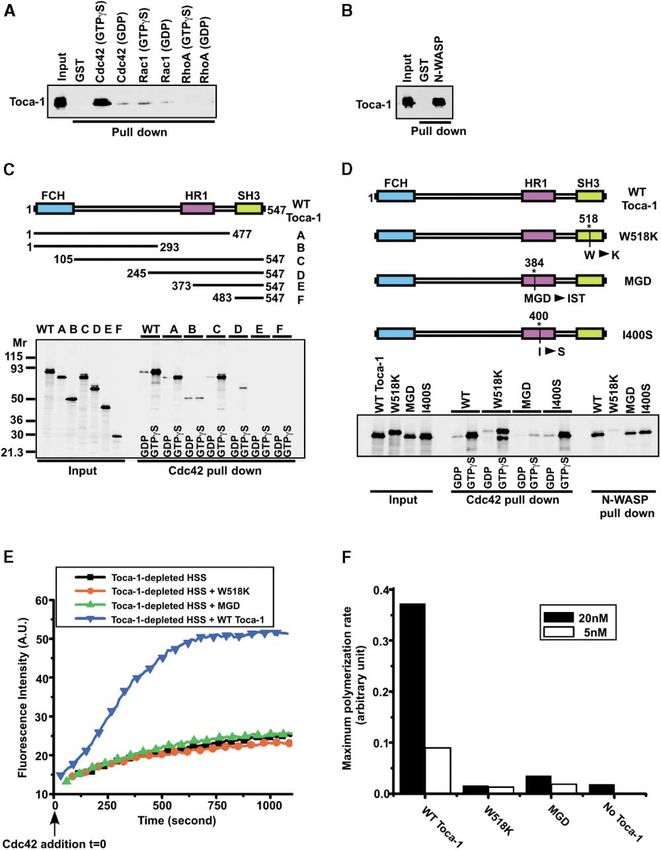
Cell
206

Figure 2. Toca-1 Belongs to the PCH Protein Family and Is Highly Conserved Across Eukaryotes
(A) The amino acid sequence and domain structure of human Toca-1. The FCH domain is highlighted blue, the HR1 domain purple, and the
SH3 domain yellow.
(B) Phylogenetic analysis of Toca-1 and other PCH family proteins. H.s., Homo sapiens; X.t., Xenopus tropicalis; D.m., Drosophila melanogaster;
C.e., Caenorhabditis elegans; S.c., Saccharomyces cerevisiae; S.p., Schizosaccharomyces pombe. Percent sequence identity to human Toca-1
for each protein is shown in parentheses.
(C) Schematic representation of Toca-1 and other PCH proteins showing their domain organization.
(D) Comparison of HR1 domains from PCH family proteins and from selected RhoA binding proteins. Residues similar among all HR1 domains
are highlighted purple, residues similar among PCH proteins are highlighted yellow, and residues similar among RhoA binding proteins are
highlighted blue. The MGD, I, and W residues mutated in the Cdc42- and N-WASP binding mutants of Toca-1 are boxed in (A) and (D).

kDa. We named this protein Toca-1 for transducer of                        silent mutations into the Toca-1 cDNA (described in
Cdc42-dependent actin assembly-1 (Figure 2A).                              Supplemental Data at http://www.cell.com/cgi/content/
   To confirm the nucleotide sequence encoding human                       full/118/2/203/DC1).
Toca-1, we sequenced four independent PCR products                            After cloning Toca-1, we produced recombinant hu-
derived from human fetal brain cDNA pools. All four                        man Toca-1 in insect (SF9) cells (Figure 3B) and found
sequencing reactions yielded identical sequences,                          that this protein could fully complement the bovine
which differed from that of Patent WO0075321 at ten                        MCAP2B activity (Figure 1F). Thus, Toca-1 is responsi-
different nucleotide positions. Our nucleotide sequence                    ble for the activity of MCAP2B.
(deposited in GenBank as accession number AY514449)
is more likely the accurate one, because it was derived                    Toca-1 Belongs to the PCH Protein Family
from four independent cDNA amplification and sequenc-                      and Is Conserved Across Eukaryotes
ing reactions. Molecular cloning of Toca-1 was quite                       Sequence analysis revealed that Toca-1 is structurally
difficult because the cDNA encoding human Toca-1 is                        related to proteins of the PCH (pombe Cdc15 homology)
lethal to E. coli, and initially we were unable to express                 family, which have been implicated recently in a wide
the protein. We overcame this difficulty by introducing                    variety of actin-dependent processes, including cytoki-
Toca-1 Mediates Cdc42-Dependent Actin Nucleation 207 Figure 3. Toca-1 Is Required for Cdc42- and PIP2-Induced Actin Assembly in Xenopus Egg Extracts (A) Immunodepletion of Toca-1 from Xenopus HSS. Extracts were immunodepleted with ␣-Toca-1 or nonspecific (mock) antibodies and analyzed by ␣-Toca-1 immunoblotting. (B) A purified preparation of recombinant human Toca-1 analyzed by SDS-PAGE and Coomassie blue staining. (C) Comparison of actin assembly stimulated by Cdc42-GTP␥S (250 nM) in untreated HSS, mock-depleted HSS, Toca-1-depleted HSS, and Toca-1-depleted HSS reconstituted with 5 nM recombinant Toca-1. Polymerization kinetics were monitored in the HSS using the pyrene- actin assay. (D) Dose-response curve showing the variation in the maximum actin polymerization rate as a function of increasing concentrations of Toca-1 added back to Toca-1-depleted HSS. The maximum polymerization rate was calculated from the linear phase of polymerization curves of the type shown in (C). The curve is a nonlinear least squares fit of a single-site binding isotherm to the data points. The red lines denote the rate of filament elongation in untreated HSS stimulated with the same concentration of Cdc42. (E) Comparison of actin assembly stimulated by PIP2-containing vesicles (10 M) in mock-depleted HSS, Toca-1-depleted HSS, and Toca-1- depleted HSS rescued with 10 nM recombinant Toca-1. nesis, membrane trafficking, and cellular morphogene- (SH3) domains at the C terminus (Figure 2C) (Lippincott sis (Lippincott and Li, 2000). This protein family is con- and Li, 2000). The FCH domain is found in a large number served throughout eukaryotic evolution and includes of proteins involved in signal transduction, but its func- human formin binding protein 17 (FBP17), human tion is largely unknown. In addition, many PCH proteins Cdc42-interacting protein 4 (CIP4), human syndapins, are also predicted to contain coiled-coil domains. In D. melanogaster RE39037, C. elegans CE27939, S. cere- the case of Toca-1, FBP17, CIP4, and D. melanogaster visiae Bzz1p, and S. pombe Cdc15 (Figure 2B). Members RE39037, one of these coiled-coil regions has homology of this protein family are defined by a common domain to a domain called HR1 (protein kinase C-related kinase structure that includes a FER/CIP4 homology (FCH) do- homology region 1), which was originally identified as a main at the N terminus and one or two Src homology 3 Rho-interactive module in several RhoA binding proteins
Cell
208

Figure 4. Direct Binding of Toca-1 to Cdc42 and N-WASP Is Required for Its Function
(A) Toca-1 directly interacts with Cdc42 in a GTP-dependent manner. GST-Cdc42, GST-Rac1, and GST-RhoA loaded with GTP␥S or GDP
were immobilized on glutathione-Sepharose beads and tested for their ability to bind purified recombinant Toca-1. Three percent of the input
and 30% of the pulled down material were analyzed by ␣-Toca-1 immunoblotting. (B) Toca-1 directly interacts with N-WASP. The ability of
GST-N-WASP to interact with Toca-1 was tested in the same type of pull-down assay as in (A). Five percent of the input and 5% of the pulled
down material were analyzed by ␣-N-WASP immunoblotting. (C) (Top) A summary of the nomenclature and amino acid boundaries of the
various human Toca-1 fragments used to map the Cdc42 binding site. (Bottom) The indicated protein constructs were synthesized as Myc-
tagged, 35S-labeled proteins by in vitro translation and tested for their abilities to bind specifically to GST-Cdc42-GTP␥S immobilized on beads.
Two percent of the input and 30% of the pulled down material were analyzed by SDS-PAGE and autoradiography. (D) (Top) Schematic diagram
of the MGD to IST, the W518K, and the I400S mutants of human Toca-1 showing the sites of the mutations. The MGD, W518, and I400 residues
are shown (boxed) in Figures 2A and 2D. (Bottom) The MGD, W518K, and I400S mutants were synthesized as 35S-labeled proteins by in vitro
translation and tested for their abilities to bind specifically to GST-Cdc42-GTP␥S and to GST-N-WASP. Wild-type Toca-1 was used as the
positive control. Two percent of the input and 30% (Cdc42 binding reactions) or 2% (N-WASP binding reactions) of the pulled down material
were analyzed. The W518K doublet in the Cdc42 pull-down is likely due to partial proteolysis that occurred during the binding experiment
(the level of the lower band is not reproducible). (E) Wild-type Toca-1, the MGD, and W518K mutants (5 nM each) were tested for their abilities
to restore Cdc42-induced actin assembly in Toca-1-depleted Xenopus HSS. (F) Comparison of the abilities of wild-type Toca-1, the MGD,
and the W518K mutants at 5 nM or 20 nM to rescue actin polymerization in Toca-1-depleted Xenopus HSS. Curves of the type shown in (E)
are quantitated by measuring the maximum polymerization rate.
Toca-1 Mediates Cdc42-Dependent Actin Nucleation
209

(Figures 2C and 2D). The functional conservation of          reported interaction between the SH3 domain of CIP4
Toca-1 across species is highlighted by the finding that     and WASP (Tian et al., 2000).
Toca-1 homologs from X. tropicalis and D. melanogaster          To map the region of Toca-1 required for binding to
can complement the MCAP2B activity in our assay sys-         Cdc42, Toca-1 fragments were tested for their ability to
tem (data not shown).                                        selectively interact with Cdc42-GTP␥S (Figure 4C). This
                                                             deletion analysis identified the region between amino
Toca-1 Is Essential for Cdc42- and PIP2-Induced              acids 245 and 477, a region that contains the HR1 do-
Actin Polymerization                                         main, as minimally essential for Cdc42 binding. The re-
We returned to the complete Xenopus extract system for       gion between amino acids 105 and 244 contributes to the
biochemical analysis of Toca-1 in the Cdc42 pathway. In      efficiency of this interaction, perhaps by promoting the
this system, the kinetics of actin polymerization can be     proper folding of Toca-1. These regions overlap with
monitored quantitatively using pyrene-labeled actin, a       the previously mapped Cdc42 binding site in CIP4 (CIP4
fluorescent derivative of actin that exhibits a dramatic     amino acid residues 383–417, corresponding to residues
increase in fluorescence intensity upon polymerization       385–417 in Toca-1) (Tian et al., 2000). Although the HR1
(Ma et al., 1998a). Using an affinity-purified ␣-Toca-1      domain was not yet clearly defined and thus not recog-
polyclonal antibody, we immunodepleted ⬎95% of               nized in CIP4 and FBP17 at the times these proteins
endogenous Toca-1 from Xenopus HSS (Figure 3A).              were first described, it is now clear that both CIP4 and
Cdc42-induced actin polymerization was drastically re-       FBP17 also contain HR1 domains (see Figures 2C and
duced in Toca-1-depleted HSS (Figure 3C). Importantly,       2D). The HR1 domain was originally implicated in the
the activity could be rescued by adding back purified        interactions of several RhoA binding proteins, including
recombinant Toca-1 at 5 nM (Figures 3B and 3C). In-          PRK1/PKN, rhotekin, rhophilin, and p160ROCK, with
creasing the amount of Toca-1 resulted in a dose-            RhoA (Flynn et al., 1998). Interestingly, whereas the first
dependent and saturable increase in the maximum              HR1 (HR1a) domain from the regulatory N-terminal re-
polymerization rate. The concentration versus polymer-       gion of PRK1 interacts specifically with RhoA, the sec-
ization data was well fit by a hyperbolic binding isotherm   ond HR1 (HR1b) from the same protein has been shown
that assumes a single binding site (Figure 3D). The con-     recently to interact specifically with Rac1 (Flynn et al.,
centration of Toca-1 required for half-maximal activation    1998; Owen et al., 2003). Both HR1a and HR1b domains
(Kact) is 14 nM, and the maximal polymerization rate at      from PRK1 adopt an antiparallel coiled-coil finger fold,
saturation (Pmax) is 0.33/s. Comparing this Pmax value to    which makes direct contacts with RhoA or Rac1 (Mae-
the polymerization rate in untreated extracts (0.082/s)      saki et al., 1999; Owen et al., 2003).
suggests that Toca-1 is present at a concentration (ⵑ5          Our results clearly define a new class of HR1 domains,
nM) significantly lower than the Kact. This result agrees    including those found in Toca-1, CIP4, and likely FBP17,
with the Toca-1 concentration determined by quantita-        that mediates specific interactions with Cdc42 (Figures
tive immunoblotting in these extracts (Toca-1, ⵑ10 nM;       2D and 4A). Secondary structure modeling of the Toca-1
N-WASP, ⵑ100 nM). We confirmed that immunodeple-             HR1 domain suggests that its structure is very similar
tion of Toca-1 from Xenopus HSS had no effect on the         to those described for the PRK1 HR1s (Maesaki et al.,
                                                             1999; Owen et al., 2003). Thus, the HR1 antiparallel
endogenous levels of N-WASP and the Arp2/3 complex,
                                                             coiled-coil finger structure appears to be a general scaf-
suggesting that the effect of Toca-1 depletion was spe-
                                                             fold for interactions with Rho-family GTPases, and
cific (data not shown).
                                                             amino acid differences between the HR1 domains likely
   Toca-1 is also required for PIP2-induced actin assem-
                                                             determine specificity for different Rho GTPases (Figure
bly. Synthetic lipid vesicles containing PIP2 induce actin
                                                             2D). In addition to its presence in several PCH family
polymerization in Xenopus HSS, and this activity de-
                                                             proteins, the HR1 domain can be found in a large number
pends on the Cdc42-N-WASP-Arp2/3 pathway (Ma et
                                                             of other proteins in GenBank. Thus, HR1 domains should
al., 1998a; Rohatgi et al., 1999). PIP2-induced actin as-
                                                             be thought of as a new class of small G protein binding
sembly is eliminated in Xenopus HSS immunodepleted
                                                             domains (such as the CRIB domain) that define a group
of Toca-1, and the activity can be rescued by adding         of proteins linked to small GTPase signaling.
back 10 nM recombinant Toca-1 protein (Figure 3E).              To test the functional significance of Toca-1’s interac-
Thus, Toca-1 is required for both Cdc42- and PIP2-           tions with Cdc42 and N-WASP, we generated Toca-1
induced actin polymerization.                                mutants defective in these activities. As shown in Figure
                                                             4D, the MGD mutant, with three conserved residues
Direct Binding of Toca-1 to Cdc42 and N-WASP                 (MGD) in the HR1 domain substituted with IST, is signifi-
Is Required for Its Function                                 cantly impaired in its ability to bind Cdc42 (but not
We expected that Toca-1 would directly bind to Cdc42-        N-WASP). Notably, the I398S mutation in CIP4 has been
GTP, because the MCAP2B activity from brain HSS can          shown to abrogate binding to Cdc42 (Tian et al., 2000);
be depleted with Cdc42-GTP␥S-coated beads and be-            however, the analogous mutant of Toca-1 (I400S) is un-
cause CIP4, a protein related to Toca-1, is a known          affected (Figure 4D). The W518K mutant, with a con-
Cdc42 binding protein (Aspenstrom, 1997). When re-           served tryptophan in the SH3 domain mutated to a ly-
combinant Toca-1 was incubated with Cdc42-GTP␥S or           sine, no longer binds to N-WASP but can still bind Cdc42
Cdc42-GDP immobilized on beads, it bound specifically        with high affinity (Figure 4D).
to Cdc42-GTP␥S (Figure 4A). No specific interaction was         The MGD (HR1) and W518K (SH3) mutants were tested
detected between Toca-1 and Rac1 or RhoA under the           for their ability to restore Cdc42-induced actin assembly
same conditions. Purified Toca-1 directly interacts with     in extracts depleted of endogenous Toca-1. While wild-
N-WASP as well (Figure 4B), consistent with a previously     type Toca-1 at 5 nM effectively rescued actin polymer-
Cell 210 Figure 5. Toca-1 Is Required for Cdc42-Dependent Activation of the Native N-WASP-WIP Complex (A) The pyrene-actin assay was used to compare the effect of Toca-1 (10 nM) on actin polymerization (2 M total G-actin; 35% pyrene labeled) in the presence of Arp2/3 complex (30 nM), recombinant N-WASP (100 nM), and Cdc42-GTP␥S (250 nM). All reactions contain actin and the Arp2/3 complex, and the other factors are present as indicated. (B) A constitutively active N-WASP mutant (Act. NW) can induce actin polymerization in Xenopus HSS depleted of both endogenous N-WASP and Toca-1. The polymerization kinetics in mock-depleted HSS with or without Cdc42-GTP␥S stimulation are shown as controls. (C) The fractionation scheme used to purify the native N-WASP-WIP complex from Xenopus egg HSS. During the purification, the complex was followed by ␣-N-WASP immunoblotting. (D) Purified preparations of native N-WASP-WIP complex and recombinant untagged N-WASP used in the actin polymerization experiments shown in (A), (E), and (F) are shown on a 4%–20% SDS-polyacrylamide gel. Xenopus WIP copurified with N-WASP as a stoichiometric complex and was identified by mass spectrometry. (E) The pyrene-actin assay was used to compare the activation of recombinant free N-WASP to that of the purified native N-WASP-WIP complex at several different concentrations by Cdc42-GTP␥S (250 nM) alone or Cdc42-GTP␥S (250 nM) ⫹ Toca-1 (10 nM). All reactions contained 2 M G actin (35% pyrene labeled) and 30 nM Arp2/3 complex. The fold increase (above a background reaction lacking any N-WASP) in the maximum polymerization rate is plotted as a function of N-WASP and N-WASP-WIP concentrations. (F) The pyrene-actin assay was used to compare the activation of the purified N-WASP-WIP complex (6 nM) by Cdc42-GTP␥S (250 nM) in the presence or absence of Toca-1 (10 nM). Cdc42-GDP (250 nM) is completely inactive in inducing actin polymerization in the presence of both the N-WASP-WIP complex and Toca-1. All reactions contained 2 M G actin (35% pyrene labeled) and 30 nM Arp2/3 complex.
Toca-1 Mediates Cdc42-Dependent Actin Nucleation
211

ization in these extracts, the MGD (HR1) and the W518K         as a complex with N-WASP (data not shown). Therefore,
(SH3) mutants were both completely inactive at this con-       we conventionally purified the native N-WASP-WIP com-
centration (Figures 4E and 4F). At 20 nM, the MGD mu-          plex from Xenopus eggs (Figures 5C and 5D). Using an
tant exhibited a slight activity (ⵑ15% of wild-type at         in vitro purified system consisting of the native N-WASP-
the same concentration) (Figure 4F), likely reflecting its     WIP complex from Xenopus eggs or recombinant
residual affinity for Cdc42 (Figure 4D). The W518K mu-         N-WASP from SF9 cells, Cdc42-GTP␥S, and the Arp2/3
tant was still completely inactive at 20 nM (Figure 4F).       complex, the behavior of the N-WASP-WIP complex was
In summary, the ability of Toca-1 to interact with both        directly compared to that of recombinant free N-WASP
Cdc42 and N-WASP is required for its function. Since           over a wide range of concentrations (Figure 5E). Again,
SH3 domains display promiscuous interactions with              Toca-1 had only a small stimulatory effect on the activa-
proline-rich proteins (such as N-WASP) under purified          tion of recombinant free N-WASP by Cdc42-GTP␥S (Fig-
conditions, we cannot exclude the possibility that the         ure 5E). In contrast, activation of the native N-WASP-
SH3 domain of Toca-1 interacts with proteins other than        WIP complex by Cdc42-GTP␥S strongly depended on
N-WASP in extracts.                                            the presence of Toca-1 (Figures 5E and 5F). Further-
                                                               more, while recombinant free N-WASP exhibits signifi-
Toca-1 Is Required for Cdc42 to Activate                       cant basal activity even in the absence of Cdc42, the
the Native N-WASP-WIP Complex                                  N-WASP-WIP complex displays virtually no basal activ-
Thus far, we have established the requirement of Toca-1        ity and absolutely depends on the presence of both
for Cdc42- and PIP2-induced actin nucleation in cell           Cdc42 and Toca-1 for activation (Figures 5E and 5F).
extracts. Surprisingly, when we added Toca-1 to the            Noticeably, the native N-WASP-WIP complex has a spe-
purified system, consisting of Cdc42-GTP␥S, recombi-           cific activity approximately one order of magnitude
nant N-WASP, and the Arp2/3 complex, there was only            higher than that of recombinant N-WASP (Figure 5E).
a small effect on the actin polymerization kinetics (Figure    We conclude that Toca-1 is required for Cdc42-GTP to
5A). Again, this confirms the initial observation that led     activate the N-WASP-WIP/CR16 complex, the predomi-
us to pursue the purification of Toca-1—the Cdc42 path-        nant form of N-WASP in cells. This provides a mechanis-
way in extracts is significantly different from the purified   tic explanation for the requirement of Toca-1 in mediat-
system. There are two possible explanations for this           ing Cdc42-induced actin polymerization in extracts.
difference in the requirement for Toca-1 between these
systems. First, N-WASP activity in extracts might be           Toca-1 Is Required for PMA-Induced Actin
subject to an additional level of inhibition not present       Comet Formation and Vesicle Motility
in the purified system, and Toca-1 is involved in relieving    in Xenopus Extracts
this inhibition. Alternatively, the extracts contain an in-    Work in many systems has suggested that N-WASP-
hibitor of actin polymerization, such as a filament-cap-       and Arp2/3-dependent actin nucleation is directly linked
ping activity (Huang et al., 1999) not present in the puri-    to membrane trafficking (Schafer, 2002; Sokac et al.,
fied system, and Toca-1 is required to antagonize such         2003). Actin comet tails, similar to those assembled by
an inhibitor.                                                  Listeria, Shigella, and Vaccinia virus, have been shown
   If the requirement of Toca-1 were at the level of           to power the intracellular motility of pinosomes, lyso-
N-WASP activation, this requirement would be bypassed          somes, and endosomes in a variety of cell types (Allen,
by replacing endogenous N-WASP with a constitutively           2003; Kaksonen et al., 2003; Merrifield et al., 1999; Ro-
active form of N-WASP. However, if Toca-1 antagonizes          zelle et al., 2000). The propulsive movement of endocytic
an inhibitor of actin polymerization, it should still be       vesicles by actin comets has also been observed in
required for actin polymerization induced by the consti-       whole Xenopus eggs and shown to be triggered by a
tutively active N-WASP. Consistent with the former pre-        protein kinase C (PKC)-mediated signaling cascade
diction, Toca-1 is no longer required for actin polymer-       in vivo (Taunton et al., 2000). This process has been
ization when endogenous N-WASP in extract is replaced          reconstituted in a cell-free system in Xenopus egg ex-
with a constitutively active mutant of N-WASP (Figure          tracts stimulated with the PKC activator PMA and de-
5B; see Supplemental Data on Cell web site for a com-          pends on Cdc42, N-WASP, and the Arp2/3 complex
plete description and characterization of the constitu-        (Taunton et al., 2000).
tively active N-WASP mutant). We have previously found            Since Toca-1 is an essential component of the Cdc42
that native N-WASP exists in a tight complex with CR16         pathway, we tested whether Toca-1 is also required for
in bovine brain (Ho et al., 2001). A similar complex be-       actin comet-based vesicle motility using this assay. As
tween WASP and WIP, a protein closely related to CR16,         previously reported, PMA can stimulate the assembly
has also been reported (Ramesh et al., 1997). Impor-           of actin comet tails on vesicle surfaces in Xenopus HSS
tantly, WIP can suppress the activation of recombinant         supplemented with endomembranes isolated from HeLa
N-WASP by Cdc42 in vitro (Martinez-Quiles et al., 2001).       cells (Figure 6A). PMA-induced actin comet formation
Thus, we speculated that the requirement for Toca-1 in         in these extracts was abolished by antibody depletion
extracts might reflect its ability to activate the endoge-     of Toca-1 (Figure 6A). The ␣-Toca-1 antibody (30 nM)
nous N-WASP-WIP/CR16 complex, a requirement that               was maintained in the reaction to neutralize the Toca-1
is not present in the purified system since it uses recom-     activity carried over from HeLa lysates. Addback of re-
binant N-WASP not bound to WIP/CR16.                           combinant Toca-1 at 150 nM completely rescued actin
   WIP and CR16 have unusually high (ⵑ30%) proline con-        comet tail formation. Higher concentrations of Toca-1
tents and are difficult to express as soluble recombinant      resulted in the formation of longer actin comet tails,
proteins that are fully functional in our assay system, even   presumably due to an increased rate of actin nucleation
Cell
212
Figure 6. Toca-1 Is Required for PMA-Induced Actin Comet Tail Assembly on the Surface of Intracellular Vesicles
(A) Comparison of PMA-induced actin comet tail formation on the surface of HeLa endomembrane vesicles in Xenopus HSS, mock-depleted
HSS, Toca-1-depleted HSS, Toca-1-depleted HSS rescued with 150 nM Toca-1, and Toca-1-depleted HSS rescued with 600 nM Toca-1. Note
that the ␣-Toca-1 antibody (30 nM) was maintained in the Toca-1-depleted HSS to neutralize the Toca-1 activity carried over from HeLa
lysates. DMSO was used as the negative control for PMA stimulation.
(B) A high-magnification view of a comet tail emanating from a vesicle surface (arrowhead).
and a constant rate of filament disassembly (Figure acts with both Toca-1 and the N-WASP-WIP complex,
6A). As previously reported (Taunton et al., 2000), PMA- and these interactions lead to the activation of N-WASP,
induced comets were associated with membrane vesi- which in turn stimulates actin nucleation through the
cles, suggesting actin is nucleated on vesicle surfaces Arp2/3 complex (Figure 7).
(Figure 6B). A special feature of this Cdc42-dependent signaling
network is the control of actin nucleation by coupling
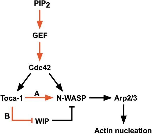
Discussion two Cdc42-dependent regulatory branches (Figure 7),
mediated by the Cdc42-N-WASP and Cdc42-Toca-1 in-
A Revised View of Cdc42 Signaling teractions. This arrangement allows for increased fidel-
to the Actin Cytoskeleton ity as well as regulatory flexibility in this pathway. For
In this paper, we have demonstrated that the previously instance, since Cdc42 is coupled to several other path-
identified components of the Cdc42-signaling pathway, ways in the cell, Toca-1, present at concentrations well
namely N-WASP and the Arp2/3 complex, are not suffi- below saturation (Figure 3D), is ideally suited to regulate
cient to mediate actin assembly in a physiological con- flux down the Cdc42-N-WASP-Arp2/3 pathway. We ex-
text. Instead, the PCH family protein Toca-1 is required pect to find other signaling pathways that directly modu-
to activate the N-WASP-WIP complex present in cell late the activity of Toca-1.
extracts. In this revised model, activated Cdc42 inter- An intriguing question raised by our data is whetherToca-1 Mediates Cdc42-Dependent Actin Nucleation
213
Toca-1 and Cdc42 (Ho et al., 2001). However, Toca-1
and PIP2 are not redundant in the more physiological
milieu of extracts, because Toca-1 immunodepletion ab-
rogates PIP2-induced actin assembly. This finding sug-
gests that the major role of PIP2 in extracts is upstream
of N-WASP, perhaps at the level of Cdc42 activation
(Figure 7).
Functional Significance of the N-WASP-WIP
Interaction: Insight into the Molecular
Pathogenesis of Wiskott-Aldrich Syndrome
Direct comparison of purified native N-WASP-WIP com-
plex and recombinant free N-WASP in the actin polymer-
ization assay suggests that WIP can suppress not only
the activation of N-WASP by Cdc42 alone but also the
basal activation of N-WASP in the absence of any activa-
tors (Figures 5E and 5F), consistent with previous find-
ings (Martinez-Quiles et al., 2001). It is probably physio-
logically important for WIP to suppress the activity of
Figure 7. Model for the Regulation of Actin Nucleation by Cdc42 N-WASP. Without WIP, a small but significant amount of
See Discussion for details. Black arrows indicate confirmed func- N-WASP likely populates the active state, since recombi-
tional interactions. Red arrows indicate speculative interactions. nant N-WASP exhibits basal Arp2/3-activating activity
Toca-1 can either directly activate N-WASP (A) or indirectly activate even without activators like Cdc42 (Figures 5A and 5E).
N-WASP by inhibiting WIP (B). GEF, guanine nucleotide exchange
This unregulated stimulation of actin assembly would
factor.
be dangerous to the cell. We propose that WIP functions
by stabilizing the autoinhibited conformation of N-WASP
in the absence of genuine activating signals. Alterna-
Cdc42 can bind both N-WASP and Toca-1 simultane-
tively, posttranslational modification of the N-WASP-
ously. Though we have found that a CRIB-containing
WIP complex or additional substoichiometric factors
fragment of N-WASP does not compete with Toca-1 for
present in the N-WASP-WIP preparation could also po-
binding to Cdc42 (data not shown), additional analyses
tentially contribute to the regulation of this complex by
are needed to elucidate the precise physical arrange-
Toca-1.
ment of the Cdc42-Toca-1 and Cdc42-N-WASP interac-
We envision two possible mechanisms by which
tions.
Toca-1 can regulate the N-WASP-WIP complex. First,
This work reveals an important biochemical function
Toca-1 could directly activate N-WASP by destabilizing
for a PCH family member, namely the stimulation of
the inhibitory intramolecular interaction. Alternatively,
actin nucleation. Our finding that Toca-1 is required for
Toca-1 could indirectly contribute to N-WASP activation
both Cdc42 and N-WASP-dependent actin nucleation
by antagonizing the suppressive activity of WIP. In the
and the actin comet-based propulsive movement of en-
latter case, the simultaneous activation and derepres-
domembrane vesicles provides a functional link between
sion of N-WASP, both through Cdc42, would generate
actin assembly and membrane trafficking (Schafer, 2002).
a sharp temporal transition between the inactive and the
More generally, PCH family proteins may serve to link
active states of N-WASP. In addition, the requirement
various cellular processes to actin nucleation mediated
for two or possibly more Cdc42 interactions (Cdc42-
by WASP family members.
N-WASP and Cdc42-Toca-1) in this pathway would per-
mit a spatially synergistic response at sites of local
The Role of PIP2 in Cdc42-Dependent Cdc42 activation. We await a more quantitative exami-
Actin Assembly nation of the pathway to test these possibilities.
We have previously suggested two roles for PIP2 in the An important question concerning the regulation of
Cdc42 pathway: one at the level of N-WASP activation the N-WASP-WIP complex is whether binding of Toca-1
and the second at a more upstream point at the level of to the complex (either through N-WASP or WIP or both)
Cdc42 activation through a guanine nucleotide exchange leads to the dissociation of the complex. Due to the
factor. These conclusions are based on two observations: inability to produce functional free WIP, we have not
PIP2 can bind and activate N-WASP in cooperation with been able to test the possible interaction between WIP
Cdc42; however, a mutant of N-WASP that cannot bind and Toca-1. Consequently, even though Toca-1 coated
or respond to PIP2 in vitro can largely restore PIP2- beads, when mixed with the native N-WASP-WIP com-
induced actin assembly to N-WASP-depleted extracts plex, can pull down both N-WASP and WIP (data not
(Rohatgi et al., 2000). The latter observation suggests shown), it is not clear whether these proteins are bound
that the major function of PIP2 in extracts is not directly to Toca-1 beads as an intact complex or as dissoci-
at the level of N-WASP activation but rather at a more ated subunits.
upstream step. An important question raised by this Our model may have important implications for un-
work is whether PIP2 and Toca-1 are redundant activa- derstanding the mechanism of the pediatric disease
tors of N-WASP. In fact, the N-WASP-WIP complex can Wiskott-Aldrich syndrome (WAS). WAS is an X-linked
be fully activated by Cdc42 and PIP2 just as it can by recessive disorder characterized by thrombocytopenia,Cell
214
eczema, and immunodeficiency, all phenotypes that expressed sequence tag (EST) clone (GenBank accession number
have been linked to misregulation of the actin cytoskele- AL655809) by PCR. Mutagenesis was performed by PCR or by using
the GeneEditor Mutagenesis Kit (Promega). All constructs were con-
ton (Snapper and Rosen, 2003). A majority (28/35) of
firmed by sequencing.
disease-causing missense mutations in WASP are pre- For in vitro translation, full-length or fragments of Toca-1 were
dicted to disrupt the WASP-WIP interactions, based on cloned into the pCS2⫹ or pCS2⫹MT vectors. For baculoviral con-
a recently solved NMR structure of the interface (Volk- struction, Toca-1 was cloned into the pFastBacHT vector, which
man et al., 2002). Thus, one important outstanding ques- contains a hexahistidine tag located N-terminal to the cloning sites.
tion in this disease is understanding the consequence
of disrupting the WASP-WIP interaction. Our results pro- Preparation of Recombinant Proteins
vide a possible answer to this question—disruption of GST-Cdc42, GST-Rac1, and GST-RhoA were prepared from Sf9
cells and loaded with different nucleotides (GTP␥S or GDP) while still
the WASP-WIP interaction would eliminate the Toca-1 bound to glutathione-Sepharose beads according to established
input into this pathway and prevent appropriate tempo- protocols (Ma et al., 1998a).
ral and spatial regulation of actin assembly in response Hexahistidine-tagged wild-type or mutant Toca-1 proteins were
to Cdc42 signals. In fact, lymphocytes from WIP knock- expressed in SF9 cells and affinity purified on nickel-Sepharose
out mice have severe deficits in signal-induced changes beads. Proteins were eluted in 50 mM Na-phosphate (pH 7.8), 400
mM NaCl, 260 mM imidazole, 5 mM -ME.
in their cortical actin cytoskeleton (Anton et al., 2002).
Untagged rat N-WASP expressed in SF9 cells was purified as
In summary, we propose the following view of the previously described (Rohatgi et al., 1999).
Cdc42 signaling pathway (Figure 7). Formation of PIP2 Protein concentrations were determined by the Bradford assay
on membranes (such as a vesicle surface) leads to the (Bio-Rad) or by densitometry of GelCode Blue (Pierce) stained gels,
recruitment and activation of Cdc42. Prenylated Cdc42 using BSA as a standard in both cases.
inserts into the membrane and forms high avidity sites In vitro translations were performed using the Promega TNT kit
that recruit Toca-1 and the N-WASP-WIP complex. Acti- and 35S-methionine according to manufacturer’s instructions.
vation of N-WASP then could proceed through one of
Preparation of Antibodies
two paths: both Cdc42 and Toca-1 could cooperate to
Purified full-length human Toca-1 was used to raise antisera in rab-
activate the N-WASP-WIP complex, or Toca-1 could bits (Cocalico, Reamstown, PA). The antibodies were affinity purified
function indirectly by relieving the inhibition of N-WASP according to established protocols (Harlow and Lane, 1999).
by WIP. Toca-1 is ideally positioned to be an important
regulatory node for the Cdc42 pathway. The function of Immunodepletion
Toca-1 suggests a specific mechanism by which PCH Affinity-purified ␣-Toca-1 or nonspecific rabbit IgG (7 g) was first
family proteins can influence actin nucleation in a wide incubated with 25 L Protein A-Dynabeads (Dynal) in PBS plus 0.1%
variety of cellular processes such as vesicle motility Triton X-100. Beads were washed twice with PBS plus 0.1% Triton
X-100 and three times with XB (Xenopus extract buffer, 30 mM
and cytokinesis. Important future questions include the
HEPES [pH 7.7], 100 mM KCl, 1 mM MgCl2). Xenopus HSS (100 L)
precise biochemical mechanism by which the N-WASP- were incubated with antibody-coated beads and rotated at 4⬚C for
WIP complex is activated by Toca-1 and Cdc42, as well 1.5 hr. Beads were removed by centrifugation, and the supernatants
as investigation into the regulation of Toca-1 itself by were used for actin polymerization within 6 hr.
other signals.
Protein Binding Assays
Experimental Procedures For GST pull-down assays using purified recombinant Toca-1, 8 g
of GST-tagged protein (Cdc42, Rac1, RhoA, or N-WASP) immobi-
Conventional Protein Fractionation lized on 8 L glutathione-Sepharose was incubated at 4⬚C with 0.5
Bovine brain Toca-1, Arp2/3 complex, and Xenopus N-WASP-WIP g of purified human Toca-1 in 50 L of XB containing 1 mg/ml of
complex were purified using conventional protein fractionation tech- chicken egg albumin. The beads were washed once with XB plus
niques. Experimental details are described in Supplemental Data. 0.25% Triton X-100, once with XB plus 200mM KCl and 0.1% Triton
X-100, and once with XB plus 0.1% Triton X-100. Proteins bound
Protein Identification by Tandem Mass Spectrometry to the beads were eluted with SDS sample buffer and analyzed
Protein identification by tandem mass spectrometry was performed by immunoblotting.
as described (Gygi et al., 1999). GST pull-down assays using 35S-labeled proteins were performed
as described above. The reticulocyte lysate (10 L) containing the
35
S-labeled proteins were used in each binding reaction. The labeled
Actin Polymerization Assays
proteins were visualized using a PhosphorImager (Bio-Rad).
Rhodamine-actin microscopic assays using bovine brain extracts
were performed as previously described for Xenopus HSS (Ma et
al., 1998b). PMA-Induced Vesicle Motility Assays
Pyrene-actin was used to follow actin polymerization in Xenopus PMA-induced vesicle motility assays were performed as described
extracts as described previously (Ma et al., 1998b). Polymerization (Taunton et al., 2000). Actin comet tails were induced by addition
was initiated by addition of 250 nM GST-Cdc42-GTP␥S or 10 M of PMA (2 M final) to Xenopus HSS supplemented with HeLa post-
lipid vesicles containing 10% PI(4,5)P2, 45% phosphatidylcholine, nuclear supernatant containing endomembranes. HSS was immu-
and 45% phosphatidylinositol. nodepleted with ␣-Toca-1 or nonspecific IgG. An additional 30 nM
Actin polymerization assays using purified components were per- of the respective antibodies was added to the final reactions to
formed as previously described (Rohatgi et al., 1999). All reactions neutralize Toca-1 carried over from the HeLa cell endomembranes.
contained 2 M purified rabbit muscle actin (35% pyrene labeled),
30 nM purified bovine Arp2/3 complex, 250 nM purified prenylated Data Analysis
GST-Cdc42 produced in insect cells, and indicated concentrations All kinetic analyses were performed using Origin (Microcal Soft-
of Toca-1, N-WASP, or the N-WASP-WIP complex. ware). Maximum elongation rates from pyrene-actin polymerization
reactions were calculated from the slopes of the linear, elongation
Molecular Biology phase of the actin assembly curves. All data shown in the figures
Cloning of human Toca-1 cDNA is described in Supplemental Data. were taken from experiments performed at least twice. Toca-1 dose-
The cDNA encoding Xenopus tropicalis Toca-1 was isolated from an response data were fitted by least squares nonlinear regressionToca-1 Mediates Cdc42-Dependent Actin Nucleation
215
using GraphPad Prism 4. Protein sequence analyses were per- (2000). Autoinhibition and activation mechanisms of the Wiskott-
formed using ScanSite and Clustal W. Aldrich syndrome protein. Nature 404, 151–158.
Lippincott, J., and Li, R. (2000). Involvement of PCH family proteins in
Acknowledgments cytokinesis and actin distribution. Microsc. Res. Tech. 49, 168–172.
Ma, L., Cantley, L.C., Janmey, P.A., and Kirschner, M.W. (1998a).
We thank Andrej Shevchenko for identifying several Toca-1 peptides Corequirement of specific phosphoinositides and small GTP-bind-
by mass spectrometry earlier in our attempts to purify Toca-1 and ing protein Cdc42 in inducing actin assembly in Xenopus egg ex-
Ethan Lee for the Drosophila RE39037 cDNA clone. We thank Tim tracts. J. Cell Biol. 140, 1125–1136.
Mitchison, Lew Cantley, Narayanaswamy Ramesh, and members of
Ma, L., Rohatgi, R., and Kirschner, M.W. (1998b). The Arp2/3 com-
the Kirschner lab for helpful discussion and Lew Cantley, Nagi Ayad,
plex mediates actin polymerization induced by the small GTP-bind-
Greg Hoffman, Kristen Kwan, and Mike Springer for comments on
ing protein Cdc42. Proc. Natl. Acad. Sci. USA 95, 15362–15367.
the manuscript. We thank Greg Hoffman for modeling the structure
of the Toca-1 HR1 domain. R.R. was a member of the Medical Machesky, L.M., Mullins, R.D., Higgs, H.N., Kaiser, D.A., Blanchoin,
Scientist Training Program at Harvard Medical School during his L., May, R.C., Hall, M.E., and Pollard, T.D. (1999). Scar, a WASp-
work on this project. This work is supported in part by grants from related protein, activates nucleation of actin filaments by the Arp2/3
the National Institute of Health to M.W.K. (GM026875-27) and complex. Proc. Natl. Acad. Sci. USA 96, 3739–3744.
S.P.G. (HG00041). Maesaki, R., Ihara, K., Shimizu, T., Kuroda, S., Kaibuchi, K., and
Hakoshima, T. (1999). The structural basis of Rho effector recogni-
Received: March 4, 2004 tion revealed by the crystal structure of human RhoA complexed
Revised: May 28, 2004 with the effector domain of PKN/PRK1. Mol. Cell 4, 793–803.
Accepted: May 28, 2004 Martinez-Quiles, N., Rohatgi, R., Anton, I.M., Medina, M., Saville,
Published: July 22, 2004 S.P., Miki, H., Yamaguchi, H., Takenawa, T., Hartwig, J.H., Geha,
R.S., and Ramesh, N. (2001). WIP regulates N-WASP-mediated actin
polymerization and filopodium formation. Nat. Cell Biol. 3, 484–491.
References
Merrifield, C.J., Moss, S.E., Ballestrem, C., Imhof, B.A., Giese, G.,
Allen, P.G. (2003). Actin filament uncapping localizes to ruffling la- Wunderlich, I., and Almers, W. (1999). Endocytic vesicles move at
mellae and rocketing vesicles. Nat. Cell Biol. 5, 972–979. the tips of actin tails in cultured mast cells. Nat. Cell Biol. 1, 72–74.
Anton, I.M., de la Fuente, M.A., Sims, T.N., Freeman, S., Ramesh, Miki, H., Suetsugu, S., and Takenawa, T. (1998). WAVE, a novel
N., Hartwig, J.H., Dustin, M.L., and Geha, R.S. (2002). WIP deficiency WASP-family protein involved in actin reorganization induced by
reveals a differential role for WIP and the actin cytoskeleton in T Rac. EMBO J. 17, 6932–6941.
and B cell activation. Immunity 16, 193–204. Owen, D., Lowe, P.N., Nietlispach, D., Brosnan, C.E., Chirgadze,
Aspenstrom, P. (1997). A Cdc42 target protein with homology to the D.Y., Parker, P.J., Blundell, T.L., and Mott, H.R. (2003). Molecular
non-kinase domain of FER has a potential role in regulating the actin dissection of the interaction between the small G proteins Rac1 and
cytoskeleton. Curr. Biol. 7, 479–487. RhoA and protein kinase C-related kinase 1 (PRK1). J. Biol. Chem.
278, 50578–50587.
Carlier, M.F., Nioche, P., Broutin-L’Hermite, I., Boujemaa, R., Le
Clainche, C., Egile, C., Garbay, C., Ducruix, A., Sansonetti, P., and Ramesh, N., Anton, I.M., Hartwig, J.H., and Geha, R.S. (1997). WIP, a
Pantaloni, D. (2000). GRB2 links signaling to actin assembly by en- protein associated with Wiskott-Aldrich syndrome protein, induces
hancing interaction of neural Wiskott-Aldrich syndrome protein actin polymerization and redistribution in lymphoid cells. Proc. Natl.
(N-WASp) with actin-related protein (ARP2/3) complex. J. Biol. Acad. Sci. USA 94, 14671–14676.
Chem. 275, 21946–21952. Rohatgi, R., Ma, L., Miki, H., Lopez, M., Kirchhausen, T., Takenawa,
Cory, G.O., Cramer, R., Blanchoin, L., and Ridley, A.J. (2003). Phos- T., and Kirschner, M.W. (1999). The interaction between N-WASP
phorylation of the WASP-VCA domain increases its affinity for the and the Arp2/3 complex links Cdc42-dependent signals to actin
Arp2/3 complex and enhances actin polymerization by WASP. Mol. assembly. Cell 97, 221–231.
Cell 11, 1229–1239. Rohatgi, R., Ho, H.Y., and Kirschner, M.W. (2000). Mechanism of
N-WASP activation by CDC42 and phosphatidylinositol 4,5-bis-
Derry, J.M., Ochs, H.D., and Francke, U. (1994). Isolation of a novel
phosphate. J. Cell Biol. 150, 1299–1310.
gene mutated in Wiskott-Aldrich syndrome. Cell 78, 635–644.
Rohatgi, R., Nollau, P., Ho, H.Y., Kirschner, M.W., and Mayer, B.J.
Etienne-Manneville, S., and Hall, A. (2002). Rho GTPases in cell
(2001). Nck and phosphatidylinositol 4,5-bisphosphate synergisti-
biology. Nature 420, 629–635.
cally activate actin polymerization through the N-WASP-Arp2/3
Flynn, P., Mellor, H., Palmer, R., Panayotou, G., and Parker, P.J. pathway. J. Biol. Chem. 276, 26448–26452.
(1998). Multiple interactions of PRK1 with RhoA. Functional assign-
Rozelle, A.L., Machesky, L.M., Yamamoto, M., Driessens, M.H., In-
ment of the Hr1 repeat motif. J. Biol. Chem. 273, 2698–2705.
sall, R.H., Roth, M.G., Luby-Phelps, K., Marriott, G., Hall, A., and Yin,
Gygi, S.P., Han, D.K., Gingras, A.C., Sonenberg, N., and Aebersold, H.L. (2000). Phosphatidylinositol 4,5-bisphosphate induces actin-
R. (1999). Protein analysis by mass spectrometry and sequence based movement of raft-enriched vesicles through WASP-Arp2/3.
database searching: tools for cancer research in the post-genomic Curr. Biol. 10, 311–320.
era. Electrophoresis 20, 310–319.
Schafer, D.A. (2002). Coupling actin dynamics and membrane dy-
Harlow, E., and Lane, D. (1999). Using Antibodies: A Laboratory namics during endocytosis. Curr. Opin. Cell Biol. 14, 76–81.
Manual (Cold Spring Harbor, NY: Cold Spring Harbor Laboratory
Snapper, S.B., and Rosen, F.S. (2003). A family of WASPs. N. Engl.
Press).
J. Med. 348, 350–351.
Ho, H.Y., Rohatgi, R., Ma, L., and Kirschner, M.W. (2001). CR16 Sokac, A.M., Co, C., Taunton, J., and Bement, W. (2003). Cdc42-
forms a complex with N-WASP in brain and is a novel member of dependent actin polymerization during compensatory endocytosis
a conserved proline-rich actin-binding protein family. Proc. Natl. in Xenopus eggs. Nat. Cell Biol. 5, 727–732.
Acad. Sci. USA 98, 11306–11311.
Suetsugu, S., Hattori, M., Miki, H., Tezuka, T., Yamamoto, T., Miko-
Huang, M., Yang, C., Schafer, D.A., Cooper, J.A., Higgs, H.N., and shiba, K., and Takenawa, T. (2002). Sustained activation of N-WASP
Zigmond, S.H. (1999). Cdc42-induced actin filaments are protected through phosphorylation is essential for neurite extension. Dev. Cell
from capping protein. Curr. Biol. 9, 979–982. 3, 645–658.
Kaksonen, M., Sun, Y., and Drubin, D.G. (2003). A pathway for asso- Taunton, J., Rowning, B.A., Coughlin, M.L., Wu, M., Moon, R.T.,
ciation of receptors, adaptors, and actin during endocytic internal- Mitchison, T.J., and Larabell, C.A. (2000). Actin-dependent propul-
ization. Cell 115, 475–487. sion of endosomes and lysosomes by recruitment of N-WASP. J.
Kim, A.S., Kakalis, L.T., Abdul-Manan, N., Liu, G.A., and Rosen, M.K. Cell Biol. 148, 519–530.You can also read