LncRNA DANCR represses Doxorubicin-induced apoptosis through stabilizing MALAT1 expression in colorectal cancer cells - Nature
←
→
Page content transcription
If your browser does not render page correctly, please read the page content below
Xiong et al. Cell Death and Disease (2021)12:24
                                    https://doi.org/10.1038/s41419-020-03318-8                                                                                                             Cell Death & Disease

                                     ARTICLE                                                                                                                                                    Open Access

                                    LncRNA DANCR represses Doxorubicin-induced
                                    apoptosis through stabilizing MALAT1 expression
                                    in colorectal cancer cells
                                    Minmin Xiong1, Mengshi Wu1, Dan Peng1, Weijun Huang1, Zehong Chen2, Haoxian Ke1, Zewen Chen3, Wu Song2,
                                    Yonghua Zhao4, Andy P. Xiang 1 and Xiaomin Zhong 1

                                      Abstract
                                      Long non-coding RNA (lncRNA) DANCR has been reported to participate in key processes such as stem cell
                                      differentiation and tumorigenesis. In a high throughput screening for lncRNAs involved in Doxorubicin-induced
                                      apoptosis, we found DANCR was suppressed by Doxorubicin and it acted as an important repressor of apoptosis in
                                      colorectal cancer. Further studies demonstrated that DANCR promoted the oncogenic lncRNA MALAT1 expression via
                                      enhancing the RNA stability of MALAT1 to suppress apoptosis. MALAT1 could efficiently mediate the suppressive
                                      function of DANCR on apoptosis. Mechanistic studies found the RNA-binding protein QK served as an interacting
                                      partner of both DANCR and MALAT1, and the protein level of QK was subjected to the regulation by DANCR.
                                      Furthermore, QK was able to modulate the RNA stability of MALAT1, and the interaction between QK and MALAT1 was
                                      controlled by DANCR. In addition, QK could mediate the function of DANCR in regulating the expression of MALAT1
                                      and suppressing apoptosis. These results revealed DANCR played a critical role in Doxorubicin-induced apoptosis in
                  1234567890():,;
1234567890():,;
                  1234567890():,;
1234567890():,;

                                      colorectal cancer cells, which was achieved by the interaction between DANCR and QK to enhance the expression of
                                      MALAT1.

                                    Introduction                                                                                        been reported to be involved in the control of cellular
                                      Long non-coding RNA (lncRNA) is a category of RNA                                                 apoptosis. LncRNAs such as lincRNA-p217, PANDA8,
                                    molecules arbitrarily defined as longer than 200 nucleo-                                             DINO9, PURPL10, NEAT111,12, and PINCR13, are tran-
                                    tides, which usually has little protein-coding potential.                                           scriptionally regulated by the tumor suppressor TP53 in
                                    Functions of lncRNAs have been widely studied in dif-                                               response to DNA damage and apoptosis. In addition,
                                    ferent biological systems, and they were found critical to                                          lncRNAs H19, GAS5, TUG1, etc., were also found to be
                                    many physiological processes, such as embryonic devel-                                              critical regulators of apoptosis in different cellular con-
                                    opment and human diseases, especially cancers, by reg-                                              texts14–17. Although these studies implicate the impor-
                                    ulating cell proliferation, apoptosis, migration,                                                   tance of lncRNAs in modulating apoptosis, the identities
                                    metabolism, and differentiation1–6. Among the vast                                                  and biological functions of the majority of apoptosis-
                                    population of lncRNA species, a subset of lncRNAs have                                              regulating lncRNAs remain to be elucidated under dif-
                                                                                                                                        ferent physiological conditions, e.g. chemotherapeutic
                                                                                                                                        drugs-induced apoptosis in cancer cells.
                                    Correspondence: Xiaomin Zhong (zhongxm23@mail.sysu.edu.cn)
                                    1                                                                                                     Doxorubicin (Dox) is an anthracycline glycoside anti-
                                     Key Laboratory for Stem Cells and Tissue Engineering, Ministry of Education,
                                    Center for Stem Cell Biology and Tissue Engineering, Zhongshan School of                            biotic that has been widely used as a chemotherapeutic
                                    Medicine, Sun Yat-Sen University, 510080 Guangzhou, China
                                    2
                                                                                                                                        drug for the treatment of a wide range of cancers for over
                                     Center of Gastrointestinal Surgery, Center of Gastric Cancer, The First Affiliated
                                                                                                                                        30 years18. Dox treatment generally elicits multiple phy-
                                    Hospital, Sun Yat-Sen University, 510080 Guangzhou, China
                                    Full list of author information is available at the end of the article                              siological responses, such as apoptosis, autophagy, and
                                    Edited by G. Calin

                                    © The Author(s) 2021
                                                        Open Access This article is licensed under a Creative Commons Attribution 4.0 International License, which permits use, sharing, adaptation, distribution and reproduction
                                                        in any medium or format, as long as you give appropriate credit to the original author(s) and the source, provide a link to the Creative Commons license, and indicate if
                                    changes were made. The images or other third party material in this article are included in the article’s Creative Commons license, unless indicated otherwise in a credit line to the material. If
                                    material is not included in the article’s Creative Commons license and your intended use is not permitted by statutory regulation or exceeds the permitted use, you will need to obtain
                                    permission directly from the copyright holder. To view a copy of this license, visit http://creativecommons.org/licenses/by/4.0/.

                                    Official journal of the Cell Death Differentiation Association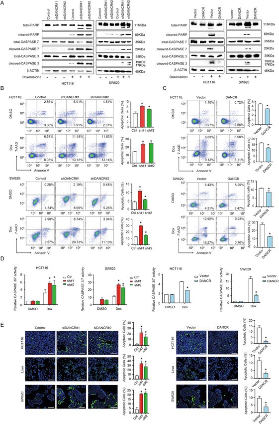
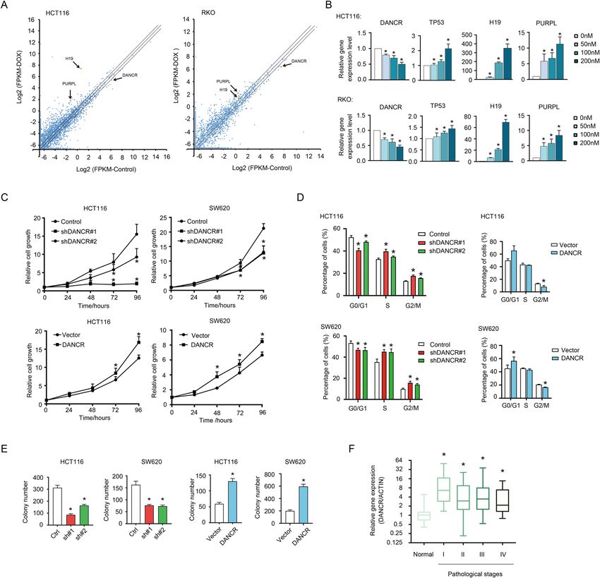
Xiong et al. Cell Death and Disease (2021)12:24                                                                Page 2 of 17

necrosis. Apoptosis represents one of the typical pheno-        colorectal cancer34. Zeng group reported that the over-
types induced by Dox. The mechanism involved in Dox-            expression of DANCR in colorectal cancer was correlated
induced apoptosis is especially important for resolving the     with cell proliferation and cancer metastasis35. Tao et al.
problem of drug resistance to Dox that evolves in               found that down regulation of DANCR induced cell death
advanced tumors frequently19. Previous research indi-           and the expression of apoptotic markers36. Although
cated Dox activates AMP-activated protein kinase                these studies provided early evidence depicting the
(AMPK-inducing apoptosis), TP53, and the Bcl-2/Bax              oncogenic role of DANCR in colorectal cancer, the major
apoptosis pathway to induce apoptosis20–22. Recently,           function and detailed working mechanism of DANCR in
studies found lncRNAs were emerging as novel regulators         the disease need further exploration.
in Dox-induced apoptosis. Chang group identified                   In this work, we reported lncRNA DANCR repressed by
lncRNA DINO served as a TP53-controlled transcript              Dox was closely correlated with colorectal cancer cell
upon Dox treatment. DINO was necessary for TP53-                apoptosis. The suppressive function of DANCR on
dependent cell cycle arrest and apoptosis by binding to         apoptosis was attributed to regulating the expression of
and stabilizing the TP53 protein9. Similarly, Lal group         MALAT1. Furthermore, the interaction between DANCR
found lncRNA PURPL formed a regulatory loop with                and the RNA-binding protein QK contributed to the
TP53 in Dox-induced apoptosis and modulated tumor-              regulatory function of DANCR on MALAT1 expression
igenesis in colorectal cancer cells10.                          and suppression of apoptosis. In summary, these findings
  To get a full insight of lncRNAs involved in the              provide new insight into the function and the working
mechanism controlling Dox-induced apoptosis, this study         mechanism of DANCR in colorectal cancer cells.
took advantage of the RNA sequencing data from Lal
group10 to identify novel lncRNA regulators. We found           Results
the expression of differentiation antagonizing non-protein      DANCR was overexpressed in colorectal cancer with a
coding RNA (DANCR) was suppressed upon Dox treat-               regulatory function in cell growth
ment, implicating it might be involved in Dox-induced             To identify novel lncRNAs involved in Dox-induced
apoptosis in colorectal cancer cells. LncRNA DANCR was          apoptosis, data from the GEO dataset GSE79249 was
initially identified as a suppressor of epidermal progenitor     retrieved and analyzed to detect changes in gene expres-
cell differentiation3. Studies indicated DANCR could            sion upon Dox treatment in wild type HCT116 cells
affect osteoblast differentiation through association with      (GSM2089687/GSM2089688: Control vs. GSM2089689/
enhancer of zeste homolog 2 (EZH2) to repress the               GSM2089690: Dox), and in wild type RKO cells
expression of RUNX2 gene23. In addition, DANCR was              (GSM2089693: Control vs. GSM2089694: Dox)10.
reported to overexpress in multiple types of cancers,           LncRNA genes with a cutoff of 1.5-fold change were
implicating its important function in tumorigenesis24–30.       sorted out for further investigation. Results of HCT116
For example, in prostate cancer DANCR was identified as          and RKO cells were compared. The two cell lines had 449
a downstream gene of C-MYC, and DANCR promoted                  upregulated and 154 downregulated lncRNAs in common
cell cycle progression and cell proliferation by repressing     (Fig. 1A, Table S2). LncRNAs previously demonstrated to
the expression of CDKN1A24. In hepatocellular cancer,           be correlated with apoptosis regulation, such as PURPL
DANCR elevated the expression of CTNNB1 to maintain             and H1910,14,37,38, were identified upregulated upon Dox
the cancer stem cell population and induced the devel-          treatment (Fig. 1A). And DANCR turned out to be one of
opment of xenograft liver cancer in mouse model29. And          the downregulated genes in both HCT116 and RKO cells
DANCR was reported to interact with the HIF1α pathway           (Fig. 1A), which could be independently verified by
to enhance the metastasis of nasopharyngeal carcinoma30.        treating the two cell lines with increasing dosages of Dox
In addition, DANCR had also been demonstrated to be             (Fig. 1B). As controls, the expression levels of TP53, H19,
involved in the proliferation, metastasis, angiogenesis, and    and PURPL were also validated to be positively correlated
differentiation in various cancer types, such as osteo-         to Dox dosage. These results implicated a close relation-
sarcoma, glioma, breast cancer, cervical cancer, bladder        ship of DANCR with Dox-induced apoptosis. To explore
cancer, etc.25–28,31–33. Based on these studies, DANCR is       the role of DANCR in cell growth and apoptosis, we
emerging as an oncogenic lncRNA in cancers.                     constructed the loss-of-function and gain-of-function
  However, the contribution of DANCR to the tumor-              stable cell lines with both HCT116 and SW620 cells
igenesis of colorectal cancer and its working mechanism         (Fig. S1A, S1B). When DANCR was silenced with two
have not been fully elucidated. So far only a few studies       independent shRNAs, both HCT116 and SW620 cells first
have explored the function of DANCR in colorectal can-          showed retarded proliferation (Fig. 1C, top), and subse-
cer. A meta-analysis report based on a Chinese population       quently severe cell death. And overexpression of DANCR
showed that a high expression level of DANCR was cor-           promoted the growth of both HCT116 and SW620 cells
related to the progression and poor prognosis of                moderately (Fig. 1C, bottom). Furthermore, DANCR

Official journal of the Cell Death Differentiation Association
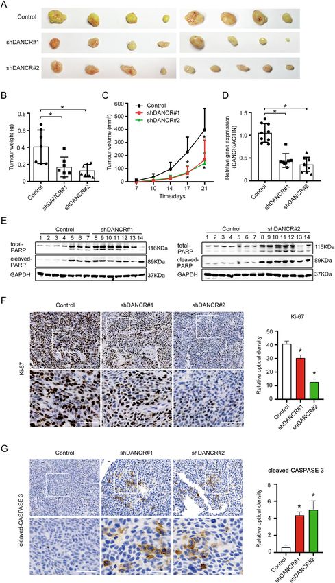
Xiong et al. Cell Death and Disease (2021)12:24                                                                                                  Page 3 of 17

  Fig. 1 DANCR was closely correlated with cell growth and tumor progression in colorectal cancer. A Changes in lncRNA expression in HCT116
  and RKO cells upon Dox treatment were presented in Scatter Dot Plots. X axis: log2(FPKM) of control cells; Y axis: log2(FPKM) of Dox-treated cells.
  Arrows indicated lncRNAs DANCR, PURPL, and H19, whose fold changes were higher than 1.5-fold. B The expression levels of DANCR, TP53, H19, and
  PURPL were detected by qPCR in HCT116 (top) and RKO (bottom) cells treated with increasing dosages of Dox (0, 50, 100, 200 nM) for 24 h. *p < 0.05.
  C DANCR knockdown inhibited HCT116 and SW620 cells proliferation (top), whereas DANCR overexpression promoted the growth of HCT116 and
  SW620 cells detected by CCK8 assays (bottom). *p < 0.05. D Cell cycle progression was analyzed using flow cytometry in HCT116 and SW620 cells
  with shDANCR or DANCR overexpression vectors. *p < 0.05. E The anchorage-independent growth capacity of HCT116 and SW620 cells was impaired
  upon DANCR knockdown, and it was enhanced in HCT116 and SW620 cells overexpressing DANCR. *p < 0.05. F Relative expression levels of DANCR
  in colorectal cancer tissues of different pathological stages. Normal, adjacent noncancerous tissues (n = 18). I–IV: TNM stages I (n = 21), II (n = 40), III
  (n = 44), and IV (n = 21). *p < 0.05.

knockdown resulted in decreased cell numbers in G0/G1                             capacity in anchorage-independent growth of HCT116
phase but elevated cell numbers in S and G2/M phases                              and SW620 cells (Figs. 1E and S1D). However, DANCR
(Fig. 1D, left; Fig. S1C), and overexpression of DANCR                            had minor effects on the migration activity of HCT116
had an opposite effect in both HCT116 and SW620 cells                             and SW620 cells (Fig. S1E, S1F). To further evaluate the
(Fig. 1D, right; Fig. S1C). In addition, colony forming                           contribution of DANCR to colorectal cancer progression,
assays indicated DANCR significantly promoted the                                  an expression profiling for DANCR was performed with

Official journal of the Cell Death Differentiation Association
Xiong et al. Cell Death and Disease (2021)12:24 Page 4 of 17
commercially available cDNA arrays derived from human weights (Fig. 3B) and tumor volumes (Fig. 3C). The
colon cancer tissues (cancerous tissues of TNM Stages silencing efficiency of DANCR expression was validated in
I–IV, n = 126; adjacent normal tissues, n = 18). DANCR the xenograft tumors (Fig. 3D), indicating the difference
expression was significantly higher in malignant tissues, in tumor size was specifically correlated with DANCR
especially in Stage I, than in normal tissues (Fig. 1F). expression level. To ensure cellular apoptosis was
However, no significant correlations between DANCR responsible for the negative effect that DANCR exerted to
expression and regional lymph node metastasis, neither tumor formation in vivo, the expression of PARP, Ki-67,
distant metastasis, were observed (data not shown). These and CASPAPSE 3 was examined in the xenograft tumor
results indicated the high expression level of DANCR samples. Results showed that cleaved PARP expressed at a
might contribute to the early development of colorectal higher level in shDANCR groups than in the control
cancer. group (Fig. 3E). Immunohistochemistry (IHC) also
demonstrated that shDANCR groups had significantly
DANCR-regulated apoptosis of colorectal cancer cells fewer Ki-67-positive cells, and much more cleaved CAS-
in vitro PASE 3-positive cells than the control group (Fig. 3F, G).
As mentioned above, severe cell death was observed These data suggested that DANCR enhanced tumor for-
following growth retardation upon loss-of-function of mation in vivo through regulating apoptosis.
DANCR. We hypothesized cell death represented the
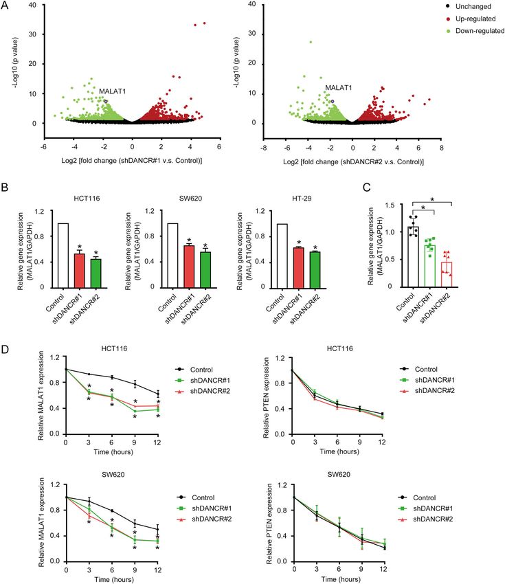
major phenotype resulted from DANCR knockdown. To DANCR modulated MALAT1 expression by enhancing the
verify the hypothesis, protein markers of apoptosis (PARP, RNA stability of MALAT1
CASPASE 7, and CASPASE 3) were examined when In order to investigate the anti-apoptotic mechanism of
HCT116 and SW620 cells were treated with Dox (Fig. DANCR and to get a genome-wide insight of the mole-
2A). We found the cleaved forms of PARP, CASPASE 7, cular changes induced by DANCR knockdown, we per-
and CASPASE 3 were all elevated in shDANCR cell lines formed a transcriptome sequencing using the shDANCR
of HCT116, SW620, and HT-29 compared to control cells and control HCT116 cell lines. DANCR knockdown led to
in the presence of Dox (Fig. 2A, left; Fig. S2A). On the changes in the expression of a large number of genes (Fig.
other hand, overexpression of DANCR in HCT116 and 4A and Table S3), including some genes known to be
SW620 cells decreased the expression of cleaved PARP, correlated with cell proliferation and apoptosis, such as
CASPASE 7, and CASPASE 3 (Fig. 2A, right). Annexin V MALAT1, XIAP, SOCS2, CCND1, etc. Among the altered
flow cytometry assay further confirmed DANCR knock- genes, we were particularly interested in MALAT1, whose
down significantly increased (Figs. 2B and S2B), and expression was significantly downregulated upon DANCR
overexpression of DANCR decreased (Fig. 2C), the knockdown (Fig. 4A), due to its widely studied functions
number of apoptotic cells in HCT116, SW620, and HT-29 in cancers, including promotion of cell growth and
cell lines. An in vitro assay to measure the enzymatic metastasis, and suppression of apoptosis39–45. As shown
activity of CASPASE 3/7 from cell lysates displayed that in Fig. 4B, the regulation to MALAT1 expression by
DANCR knockdown increased, while DANCR over- DANCR could be consistently validated in HCT116,
expression decreased, the enzymatic activity in the pre- SW620, and HT-29 cell lines. Furthermore, in xenograft
sence of Dox in both HCT116 and SW620 cells (Fig. 2D). tumor tissues (as shown in Fig. 3), DANCR knockdown
In addition, TUNEL assay showed that HCT116, LoVo, also dramatically impaired MALAT1 expression (Fig. 4C).
and SW620 cells with DANCR siRNAs had a higher To explore whether DANCR modulated MALAT1
number of apoptotic cells than control cells (Fig. 2E, left). expression transcriptionally, reporter assays using a
Nevertheless, DANCR overexpression had an opposite MALAT1 promoter-driven Luciferase plasmid were per-
effect (Fig. 2E, right). From the above data, we concluded formed in shDANCR HCT116 cells. However, DANCR
that repression of apoptosis was a major biological role of knockdown only had minor effects on the transcriptional
DANCR in colorectal cancer cells in vitro. activity of MALAT1 promoter (Fig. S3). Subsequently, the
RNA stability of MALAT1 was examined in HCT116 and
DANCR suppressed apoptosis of colorectal cancer cells SW620 cell lines with shDANCR vectors (Fig. 4D, left).
in vivo Surprisingly, MALAT1 in shDANCR cells degraded much
To verify whether DANCR suppresses apoptosis in vivo faster than in control cells. The difference in the turn-over
to promote tumor growth, we performed xenograft tumor rate of MALAT1 transcript could already be seen as early
formation assays by implanting HCT116 shDANCR cells as 3 h post-treatment of Actinomycin D. However, an
or control cells subcutaneously to nude mice. Tumor irrelevant mRNA PTEN did not manifest differences in
samples harvested 3 weeks post-implantation indicated the degradation dynamics between shDANCR cells and
shDANCR cells formed tumors with dramatic decrease in control cells (Fig. 4D, right), indicating DANCR regulated
the average size (Fig. 3A), as well as decreases in tumor the RNA stability of MALAT1 specifically.
Official journal of the Cell Death Differentiation Association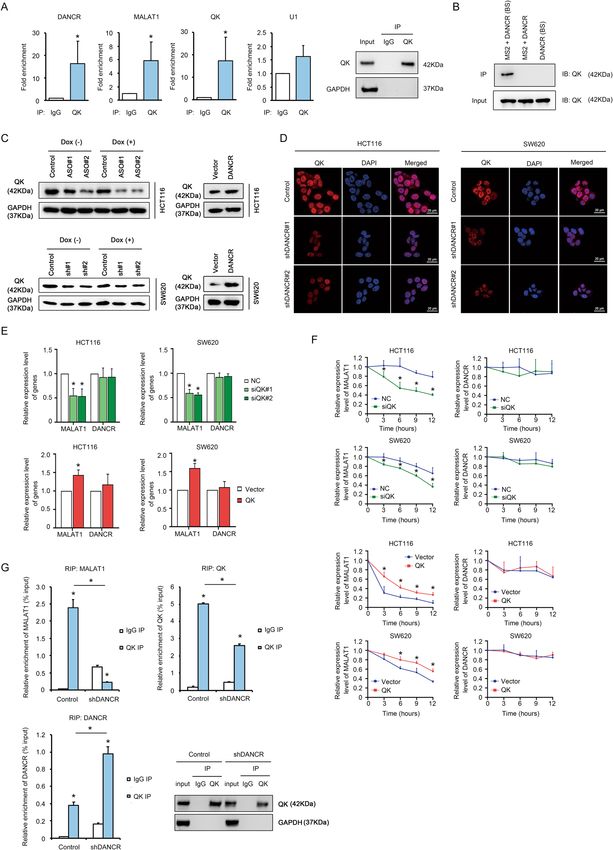
Xiong et al. Cell Death and Disease (2021)12:24                                                                                         Page 5 of 17

  Fig. 2 DANCR regulated apoptosis of colorectal cancer cells in vitro. A Left: Western blotting showed increased expression of total and cleaved
  PARP, CASPASE 7, and CASPASE 3 upon treatment with 200 nM Doxorubicin for 24 h in HCT116 and SW620 cells with shDANCR. Right: DANCR
  overexpression in HCT116 and SW620 cells repressed the expression of cleaved PARP, cleaved CASPASE 7, and cleaved CASPASE 3 detected by
  Western blotting. B Representative images of Annexin V flow cytometry assays and data statistics showed increased apoptotic cell numbers upon
  DANCR silencing in HCT116 and SW620 cells, which was treated with DMSO or 200 nM Doxorubicin for 24 h. *p < 0.05. C Annexin V flow cytometry
  assays showed DANCR overexpression increased tolerance to apoptosis in HCT116 and SW620 cells treated with DMSO or 200 nM Doxorubicin for
  24 h. *p < 0.05. D The effects of DANCR depletion and DANCR overexpression on the CASPASE 3/7 activity in HCT116 and SW620 cells in the presence
  or absence of 200 nM Doxorubicin for 24 h. *p < 0.05. E TUNEL staining and statistical analysis in HCT116, LoVo, and SW620 cells with DANCR siRNAs
  (left) or with DANCR overexpressing vectors (right). Bar, 100 μm. *p < 0.05.

Official journal of the Cell Death Differentiation Association
Xiong et al. Cell Death and Disease (2021)12:24                                                                                         Page 6 of 17

  Fig. 3 DANCR suppressed apoptosis of colorectal cancer cells in vivo. A Morphology of xenograft tumors formed by HCT116 cells with DANCR
  shRNAs or control cells. B, C Comparison of the weight and the size of xenograft tumors as shown in A. *p < 0.05. D The expression of DANCR in
  xenograft tumors as shown in A was analyzed by qRT-PCR. *p < 0.05. E The expression of cleaved PARP in xenograft tumors was detected by Western
  blotting. F, G The expression of Ki-67 and cleaved CASPASE 3 in xenograft tumors was showed in representative immunohistochemical images (left).
  Relative optical density of the expression of Ki-67 and cleaved CASPASE 3 was analyzed (right). Bar, 100 μm (upper panel) and 50 μm (lower panel).
  *p < 0.05.

Official journal of the Cell Death Differentiation Association
Xiong et al. Cell Death and Disease (2021)12:24                                                                                    Page 7 of 17

  Fig. 4 DANCR modulated MALAT1 expression by enhancing the RNA stability of MALAT1. A Differentially expressed genes upon DANCR
  knockdown in HCT116 cells are shown in volcano plots. X axis: Log2 [fold change (shRNA vs. Control)]; Y axis: −Log10 (p value). B Decreased
  expression of MALAT1 upon DANCR knockdown was validated by qRT-PCR in HCT116, SW620, and HT-29 cells. *p < 0.05. C Decreased expression of
  MALAT1 upon DANCR knockdown was validated using qRT-PCR in xenograft tumor tissues (n = 7 for each group). *p < 0.05. D The expression levels
  of MALAT1 (left) and PTEN (right) upon DANCR knockdown in HCT116 and SW620 cells were detected by qRT-PCR at 0, 3, 6, 9, and 12 h post-
  treatment with Actinomycin D. *p < 0.05.

Official journal of the Cell Death Differentiation Association
Xiong et al. Cell Death and Disease (2021)12:24 Page 8 of 17
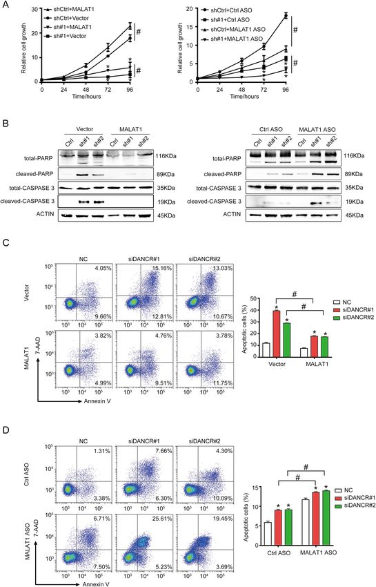
MALAT1 mediated the anti-apoptotic function of DANCR sought to confirm the interaction between DANCR and
As shown above, DANCR regulated the RNA stability of QK. The RNA-binding protein immunoprecipitation
MALAT1. To prove whether the regulation to MALAT1 (RIP) assay with a QK specific antibody showed significant
expression by DANCR had physiological functions, we enrichment of DANCR and MALAT1 by QK (Fig. 6A). As
tested the possibility of MALAT1 to mediate the anti- controls, QK mRNA and U1, respectively, displayed
apoptotic function of DANCR. MALAT1 was over- abundant and negligible enrichment compared to control
expressed or knockdown by antisense oligonucleotides IgG IP. This indicated the interaction between DANCR
(ASO) in HCT116 shDANCR cells (Fig. S4). Cell pro- and QK, as well as MALAT1 and QK, was highly specific.
liferation assays showed that overexpression of MALAT1 Subsequently, an MS2-DANCR RIP assay was developed
rescued the growth retardation induced by DANCR to validate the above result using the strategy that
knockdown (Fig. 5A, left). And simultaneously silencing DANCR transcript expressed with a tandem sequence of
MALAT1 further compromised the growth of shDANCR 12-copied MS2 protein-binding sites (DANCR (BS)),
cells (Fig. 5A, right). Meanwhile, the expression of cleaved together with the interacting partners of DANCR, can be
PARP and cleaved CASPASE 3 proteins in shDANCR specifically enriched by the MS2 protein52. Results
cells overexpressing MALAT1 was decreased to an almost showed that QK protein was efficiently captured in the
undetectable level compared to parental shDANCR cells presence of both MS2 protein and DANCR (BS), but not
(Fig. 5B, left). However, silencing MALAT1 in shDANCR in the negative control with MS2 protein and wild type
cells dramatically promoted the production of cleaved DANCR, nor the one with DANCR (BS) alone without
PARP and cleaved CASPASE 3 proteins (Fig. 5B, right). MS2 protein (Fig. 6B). Interestingly, QK mRNA and
Similarly, Annexin V assays showed that the population of MALAT1 were not significantly enriched in the MS2+
apoptotic cells induced by DANCR knockdown was sig- DANCR (BS) immunoprecipitate (Fig. S6). This impli-
nificantly decreased when simultaneously overexpressing cated QK protein, instead of its mRNA, was the inter-
MALAT1 (Fig. 5C). And silencing MALAT1 in siDANCR acting partner of DANCR, and MALAT1 might not have
cells further enhanced the number of apoptotic cells direct interaction with DANCR. To determine the phy-
compared to parental siDANCR cells (Fig. 5D). The above siological role of the interaction between QK protein and
data implicated that MALAT1 could mediate the sup- DANCR, we tested whether DANCR could affect the
pressive function of DANCR in apoptosis. expression of QK. When DANCR was knocked down by
either ASO or shRNA, the protein level of QK detected by
DANCR-associated protein QK promoted the stability of Western Blotting (Fig. 6C, left) and immunofluorescence
MALAT1 staining (Fig. 6D) was significantly diminished in both
Although DANCR was confirmed to repress apoptosis HCT116 and SW620 cells. And DANCR overexpression
by enhancing the expression of MALAT1, the question elevated the protein level of QK moderately (Fig. 6C,
how DANCR modulated MALAT1 stability remained right). This result indicated DANCR interacted with QK
unanswered. We hypothesized the regulation to MALAT1 to play a key role in maintaining the protein level of QK.
expression by DANCR was implemented by direct Since MALAT1 could also be enriched by QK (Fig. 6A),
RNA–RNA interaction. However, the results of RNA we next verified whether QK was responsible for main-
in situ hybridization in wide type HT29 and HCT116 cell taining the steady-state expression level and the RNA
lines did not support this hypothesis. Only a negligible stability of MALAT1. QK knockdown by siRNA
portion of DANCR, whose subcellular localization was remarkably decreased MALAT1 expression level, and QK
detected 80% in the cytoplasm and 20% in the nucleus, overexpression increased MALAT1 level (Figs. 6E and
overlapped with MALAT1 inside the nucleus (Fig. S5). S7). However, the expression level of DANCR was unal-
Considering the evidence that lncRNAs usually associate tered by QK. Subsequently, the effect of QK on the RNA
with proteins to perform their functions, we searched stability of MALAT1 was further investigated by treating
Starbase (http://starbase.sysu.edu.cn)46 for RNA-binding the QK knockdown and overexpression cell lines with
proteins that may interact with DANCR. Interestingly, we Actinomycin D. In accordance with the result of Fig. 6E,
found DANCR, as well as MALAT1, harbored multiple QK knockdown dramatically accelerated the turnover rate
binding sites of the RNA-binding protein QUAKING of MALAT1, and QK overexpression significantly
(QK), which is a widely reported regulator to RNA sta- enhanced the RNA stability of MALAT1 in HCT116 and
bility, RNA splicing, RNA transportation, and transla- SW620 cells (Fig. 6F, left). And under the same condi-
tional efficiency47–50. Furthermore, QK has been tions, QK did not affect the turnover of DANCR (Fig. 6F,
previously reported to be involved in apoptosis regula- right). Therefore, we concluded QK participated in the
tion51. Therefore, we hypothesized that QK served as an maintenance of MALAT1 expression level. To further
interacting partner of DANCR to mediate the regulatory characterize the mechanism through which QK regulated
function of DANCR in MALAT1 expression. First, we MALAT1 expression, RIP assays with a QK-specific
Official journal of the Cell Death Differentiation AssociationXiong et al. Cell Death and Disease (2021)12:24 Page 9 of 17 Fig. 5 MALAT1 mediated the anti-apoptotic function of DANCR. A MALAT1 overexpression could partially rescue the growth inhibition caused by DANCR silencing (left), and simultaneously silencing MALAT1 in shDANCR HCT116 cells further inhibited cellular proliferation (right). sh#1, DANCR shRNA#1. ASO antisense oligonucleotides. * and #, p < 0.05. B Western blotting detected MALAT1 overexpression decreased (left), while MALAT1 depletion further increased (right), the expression of cleaved PARP and cleaved CASPASE 3 induced by DANCR silencing. C MALAT1 overexpression decreased DANCR knockdown-induced apoptosis detected with Annexin V flow cytometry assay. Representative images and statistical analysis were shown. * and # p < 0.05. D MALAT1 depletion further increased DANCR knockdown-induced apoptosis detected with Annexin V flow cytometry assay. Representative images and statistical analysis were shown. * and #, p < 0.05. Official journal of the Cell Death Differentiation Association
Xiong et al. Cell Death and Disease (2021)12:24                 Page 10 of 17

  Fig. 6 (See legend on next page.)

Official journal of the Cell Death Differentiation Association
Xiong et al. Cell Death and Disease (2021)12:24 Page 11 of 17
(see figure on previous page)
Fig. 6 DANCR-associated protein QK promoted the stability of MALAT1. A Left: Fold enrichment of DANCR, MALAT1, QK mRNA, and U1 from
the RIP assay with a QK antibody in HCT116 cells was detected by qPCR. Right: Immunoprecipitation of QK protein from the RIP assay was shown by
Western Blotting. B The enrichment of QK protein from the MS2-DANCR RIP assay in HCT116 cells was detected by Western Blotting. MS2, the MS2
RNA-binding protein. DANCR (BS), ectopic expressed DANCR with a tandem sequence of 12-copied MS2-binding site. C The changes in QK protein
level upon DANCR knockdown (left) or overexpression (right) in HCT116 and SW620 cells was detected by Western Blotting in the presence or
absence of 200 nM Dox for 24 h. ASO, antisense oligonucleotide of DANCR. sh#1/#2, shDANCR#1/#2. D Immunofluorescence staining showed the
expression of QK protein in HCT116 and SW620 cells with control vector, shDANCR#1, and shDANCR#2. Red: QK, Blue: DAPI. Bar, 20 μm. E The
expression levels of MALAT1 and DANCR in siQK (top) and QK overexpression cells (bottom) of HCT116 and SW620 were detected by qPCR. *p < 0.05.
F The dynamic expression changes of MALAT1 and DANCR in siQK and QK overexpression cells of HCT116 and SW620 were detected by qRT-PCR at
0, 3, 6, 9, and 12 h post-treatment with Actinomycin D. *p < 0.05. G The relative enrichment of immunoprecipitated RNAs (% input) from control and
shDANCR HCT116 cell lines were compared by qPCR. QK protein in the cell lysate input and in the immunoprecipitate was detected by Western
Blotting. *p < 0.05.
antibody was performed in HCT116 cells with shDANCR number induced by siDANCR (Fig. 7C, top), and QK
(Figs. 6G and S8). In control cells, QK protein could overexpression in siDANCR cells reduced the apoptotic
efficiently enrich DANCR, MALAT1, and QK mRNA cell number to a level between the negative control and
compared to control IgG IP. Upon DANCR knockdown siDANCR alone (Fig. 7C, bottom). Summarized from the
by shRNA, the interaction between QK and MALAT1 was above data, we concluded that DANCR was a critical
abrogated to a large extent, which was comparable to the regulator to the protein level of QK, and QK served as a
enrichment of MALAT1 in control IgG IP group (Fig. 6G, mediator for DANCR to perform its function on stabi-
top left). Nevertheless, the enrichment of QK mRNA only lizing MALAT1 expression and suppressing apoptosis
decreased by about 50% in shDANCR cells (Fig. 6G, top (Fig. 7D).
right), which was proportional to the decrease in QK
protein upon DANCR knockdown (Fig. 6G, bottom right). Discussion
Consequently, we speculated the dramatic reduction in Dox is a widely used chemotherapeutic drug for treating
the interaction between QK and MALAT1 upon DANCR various types of cancers, and it triggers complicated
knockdown was probably due to the control by DANCR, physiological responses in different cellular contexts. The
more than due to the decrease in the input QK protein. downstream pathways and targets regulated by Dox need
These results implicated DANCR was a master regulator further elucidation to overcome the problem of Dox
to the association between QK and MALAT1, and hence resistance in advanced tumors. In this work, we revealed
the RNA stability of MALAT1 and the resistance to DANCR was a Dox-suppressed target, which was involved
apoptosis. in the regulation of apoptosis in colorectal cancer (Figs. 2
and 3). Nevertheless, how Dox controlled DANCR
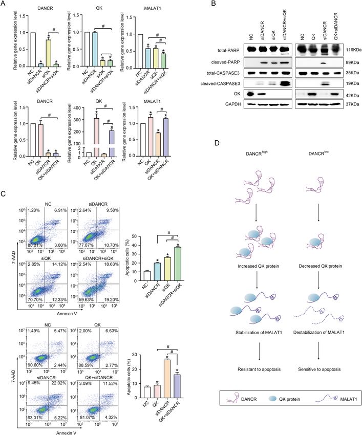
QK mediated the function of DANCR on regulating expression, as well as whether DANCR had functional
MALAT1 expression and suppressing apoptosis correlation with the previously known regulatory genes
To further characterize the functional correlation for apoptosis (such as TP53), remained questions unan-
between the regulation to QK protein by DANCR and the swered. Hopefully, this data added DANCR as a new
stabilization of MALAT1 by QK protein, we tested whe- effector in the downstream pathway of Dox, which may
ther QK could cooperate with DANCR in modulating help resolve the problem of drug resistance to Dox in
MALAT1 expression and suppressing apoptosis. In multiple cancer types.
HCT116 cells with siDANCR, simultaneously silencing Our work indicated that the major function of DANCR
QK further decreased MALAT1 expression to a level in colorectal cancer resided in the regulation of apoptosis.
much lower than in cells with siDANCR or siQK alone Interestingly, transcriptome-wide analysis revealed
(Fig. 7A, top right), and co-expressing a QK cDNA effi- DANCR was able to upregulate expression of MALAT1.
ciently rescued the downregulated expression of MALAT1 has been proven a potent repressor for cancer
MALAT1 to a level comparable to control cells (Fig. 7A, cell apoptosis41,45,53,54. Further studies revealed MALAT1
bottom right). The expression of apoptotic marker pro- could functionally mediate the suppressive function of
teins demonstrated that siQK further increased cleaved DANCR on apoptosis. However, the mechanism through
PARP and cleaved CASPASE 3 expression induced by which MALAT1 repressed apoptosis in colorectal cancer
siDANCR, and QK overexpression in siDANCR cells remains unclear. In multiple myeloma, MALAT1 was
abrogated the production of cleaved PARP and cleaved shown to repress apoptosis through binding to compo-
CASPASE 3 (Fig. 7B). Annexin V assays also showed that nents of the DNA damage repair complex to enhance
co-silencing QK significantly elevated the apoptotic cell alternative non-homologous end joining42, or controlling
Official journal of the Cell Death Differentiation AssociationXiong et al. Cell Death and Disease (2021)12:24 Page 12 of 17 Fig. 7 QK mediated the function of DANCR on regulating MALAT1 expression and suppressing apoptosis. A The expression of MALAT1 upon DANCR knockdown combined with siQK (upper panel), or with QK overexpression (lower panel) respectively, was detected by qRT-PCR in HCT116 cells. NC, negative control. *and #, p < 0.05. B Western blotting detected QK depletion further increased (left panel), while QK overexpression inhibited (right panel), the expression of cleaved PARP and cleaved CASPASE 3 induced by DANCR silencing in HCT116 cells. C QK depletion (upper panel) and QK overexpression (lower panel) further aggravated or alleviated siDANCR-induced apoptosis, which was detected with Annexin V flow cytometry assay in HCT116 cells. Representative images and statistical analysis were shown. *and #, p < 0.05. D A schematic diagram for the mechanism of apoptosis regulation by the DANCR/QK/MALAT1 axis in colorectal cancer cells. Official journal of the Cell Death Differentiation Association
Xiong et al. Cell Death and Disease (2021)12:24                                                              Page 13 of 17

the expression of proteasome subunits and anti-oxidation        Materials and methods
genes39. In our study, whether MALAT1 mediated the              Cell culture
anti-apoptotic function of DANCR through the same                 Human colorectal cancer cell lines HCT116, RKO,
mechanism remains an interesting question for further           SW620, HT-29, and LoVo were provided by the Cell
investigation.                                                  Bank, Type Culture Collection Committee, Chinese
  Our work also demonstrated DANCR suppressed                   Academy of Sciences (Shanghai, China). All cell lines were
apoptosis through enhancing the RNA stability of                authenticated by STR profiling and tested for mycoplasma
MALAT1. MALAT1 had been reported to form a stable               contamination. HCT116 and HT-29 cells were main-
RNA conformation through processing the 3′ end into a           tained in DMEM (Hyclone, Logan, UT, USA) supple-
triple helix55–58. The RNA stability of MALAT1 was              mented with 10% FBS (PAN-Biotech, Bavaria, Germany)
regulated by factors that can promote 3′ end processing         and 1% penicillin–streptomycin (Hyclone, Logan, UT,
and maturation, such as a natural antisense RNA                 USA). RKO, SW620, and LoVo cells were maintained in
TALAM157 and the Microprocessor Drosha–DGCR8                    RPMI 1640 (Hyclone, Logan, UT, USA) supplemented
complex59. In this work, we identified the RNA-binding           with 10% FBS and 1% penicillin–streptomycin. Cells were
protein QK as a mediator in transducing the positive            cultured at 37 °C in a humidified atmosphere with 5%
regulation of DANCR to MALAT1. Although we identi-              CO2.
fied QK could bind to both DANCR and MALAT1, it
remains an interesting question how QK promotes the             Plasmids
RNA stability of MALAT1. Whether QK participates in               Human DANCR and QK were amplified by PCR and
the 3′ end processing of MALAT1, or alternatively QK            cloned into the CD513B lentiviral vector (SBI System
interacts with TALAM1 and Drosha–DGCR8 complex,                 Biosciences, Palo Alto, CA, USA) to generate
need further investigation.                                     CD513B–DANCR and CD513B–QK overexpression
  In addition, DANCR controlled the protein level of            plasmids. MALAT1 full length cDNA and a Luciferase
QK, as well as the interaction between QK and                   reporter plasmid with MALAT1 promoter were obtained
MALAT1. However, the majority of DANCR localizes in             from Fenghui Biotechnology (Hunan, China). pMS2-GFP
the cytoplasm (Fig. S5), and QK protein predominantly           (#27121) and pSL-MS2-12X (#27119) were obtained from
localizes inside the nucleus (Fig. 6D). It constitutes          Addgene. The pPyCAGIP vector was a kind gift from Dr.
another interesting question how DANCR associates               Austin Smith. Sequences of primers, siRNAs, and DNA
with QK and affects QK protein level. We speculated             oligos for indicated applications are listed in Table S1.
that cytoplasmic DANCR might regulate the protein
stability of QK, or otherwise affect the transportation of      Cell transfection
QK from cytoplasm to nucleus, through direct interac-             For transient transfection, cells were seeded into six-
tion with QK protein. It is less likely that DANCR binds        well plate at 30% confluence. siRNA transfections were
to and stabilizes QK mRNA to promote the translation            performed with Lipofectamine RNAiMAX (Invitrogen,
of QK protein, because QK mRNA showed minor                     CA, USA) at a concentration of 90 nM. Plasmid trans-
enrichment in MS2-DANCR RIP assays (Fig. S6). And               fections were performed with MegaTran 1.0 transfection
the expression level of QK mRNA even elevated mod-              reagent (OriGene Technologies, Rockville, MD, USA)
erately upon DANCR knockdown (Fig. S8), which was               according to the manufacturer’s instructions.
opposite to the changes in QK protein level, probably
due to a negative feedback from the decrease in QK              RNA isolation and qRT-PCR
protein (Fig. 6G, bottom right). More evidence is needed           Total RNA was extracted from tissues and cells with
to address this question, which may help us have a deep         TRIzol reagent (Molecular Research Center, Cincinnati,
insight into the mechanism of apoptosis regulation by           OH, USA). RNA (1 μg) reverse transcription (RT) was
the DANCR/ QK/MALAT1 axis.                                      performed using a RevertAid First Strand cDNA Synthesis
  Altogether, this study identified a Dox-regulated              Kit (Thermo Scientific, Vilnius, Lithuania) in accordance
lncRNA DANCR and explored the suppressive function              with the manufacturer’s instructions. Quantitative real-
of DANCR on apoptosis. DANCR controlled expression              time polymerase chain reaction (qRT-PCR) was per-
of the RNA-binding protein QK to enhance the stability of       formed on Roche Light Cycler 480 detecting system using
MALAT1. Both MALAT1 and QK could efficiently                     LightCycler 480 SYBR Green Master (Roche, Indianapolis,
mediate the anti-apoptotic function of DANCR. These             IN, USA). Primer sequences for indicated genes were
data revealed new evidence for understanding the biolo-         listed in Table S1. Relative mRNA expression was calcu-
gical functions and the downstream regulatory network of        lated with the 2−ΔΔCt method, and gene expression levels
DANCR, which may help develop novel therapies tar-              were normalized to GAPDH. cDNA arrays derived from
geting DANCR in colorectal cancer.                              human colon cancer tissues (#HCRT101, #HCRT102, and

Official journal of the Cell Death Differentiation Association
Xiong et al. Cell Death and Disease (2021)12:24                                                                 Page 14 of 17

#HCRT104 purchased from OriGene Technologies, USA)              CASPASE 3/7 activity assay
were detected with the indicated primers according to the         CASPASE 3/7 activity was measured with a CASPASE
manufacturer’s instructions. The relative expression levels     3/7 Activity Assay Kit (Cell Signaling Technology, MA,
of DANCR in cancer tissues were calculated with meth-           USA). Briefly, cells with different treatments were col-
ods described above and compared to normal tissues.             lected in lysis buffer. 100 μg total proteins were added into
                                                                a black 96-well plate, mixed with substrate solution and
Western blotting                                                incubated at 37 °C in the dark. During the assay, activated
  Total protein was extracted from cells using RIPA lysis       CASPASE 3/7 cleaved the fluorescent substrate (Ac-
buffer (Millipore, Temecula, CA, USA). After quantification      DEVD-AMC), generating highly fluorescent AMC that
with a bicinchoninic acid (BCA) protein assay kit (Thermo       can be detected using a fluorescence reader with the
Scientific, Rockford, IL, USA), protein was denatured at         excitation at 380 nm and the emission at 450 nm. The
95 °C for 10 min, then separated by 10% or 15% SDS–PAGE         RFU were read with a fluorescence reader at 3 h post-
and transferred to 0.22 or 0.45 μm PVDF membranes               incubation and analyzed.
(Millipore, Co. Cork IRL). Then the membranes were
blocked in 5% nonfat milk and incubated with specific            Annexin V/7-AAD flow cytometry assay
primary antibodies at 4 °C overnight, followed by incubation      Cells with different treatments were collected and
with secondary antibody conjugated with horseradish per-        suspended in 100 μl binding buffer, containing 5 μl
oxidase (HRP) (1:5000, CST, MA, USA).                           Annexin V-PE and 5 μl 7-AAD. Samples were then
  Antibodies against ACTIN (cat. no. 4970), CASPASE 3           incubated for 30 min at room temperature in the dark,
(cat. no. 9662) and Cleaved CASPASE 3 (Asp175) (cat. no.        and the reaction was terminated with 400 μl binding
9661), CASPASE 7 (cat. no. 12827) and Cleaved CAS-              buffer. The apoptotic cell population was detected by a
PASE 7 (cat. no. 9491), PARP (cat. no. 9542) and Cleaved        flow cytometer (Becton Dickinson, San Diego,
PARP (Asp214) (cat. no. 5625), Anti-mouse (cat. no.             CA, USA).
7076) and anti-rabbit (cat. no. 7074) IgG HRP-linked
antibodies were all purchased from Cell Signaling Tech-         TdT-mediated dUTP nick-end labeling (TUNEL) assay
nology. QK antibody (cat. no. ab126742) was purchased             The TUNEL assay was performed with the In Situ Cell
from Abcam.                                                     Death Detection Kit (Roche, Mannheim, Germany; cat.
                                                                no. 11684795910). Cells were fixed with 4% paraf-
Cell proliferation assay                                        ormaldehyde for 1 h at room temperature and washed
  The Cell Counting Kit-8 (CCK-8) assay (Dojindo, KMJ,          twice with phosphate buffered saline (PBS). The samples
Japan) was used to measure cell proliferation. 1000 cells       were permeated with 0.1% Triton X-100 in sodium
with indicated treatment were seeded into 96-well plate.        citrate for 2 min on ice and rinsed twice in PBS. TUNEL
CCK8 regent was added at 0, 24, 48, 72, 96 h, and incubated     reaction mixture was incubated with samples in a
at 37 °C for 3 h in the dark before measuring the absorption    humidified atmosphere for 60 min at 37 °C in the dark.
at 450 nm with a Microplate Reader (Tecan, Australia).          The nuclei were stained with DAPI. The apoptosis ratio
                                                                was calculated by counting the TUNEL-positive cells.
Cell cycle analysis                                             Imaging was performed with Leica microsystems (Leica,
  Cell cycle was analyzed using a Cell Cycle Detection Kit      Germany).
(Beyotime, Shanghai, China). Briefly, cells were harvested
and fixed in 70% cold ethanol overnight at 4 °C. Fixed cells     Xenograft tumor formation
were washed twice in cold PBS and treated with RNase A            Four weeks old male BALB/c-nude (nu/nu) mice were
for 1 h. Cells were then stained with propidium iodide (PI)     obtained from the Laboratory Animal Center of Sun
for 30 min and immediately analyzed with a flow cyt-             Yat-Sen University and maintained in specific pathogen-
ometer (Becton Dickinson, CA, USA).                             free (SPF) environment. The procedure of animal
                                                                experiments was approved by the Institutional Animal
Soft agar assay                                                 Care and Use Committee of Sun Yat-Sen University. 3 ×
  The six-well plates were coated on the bottom with a          106 shDANCR cells or control cells were injected sub-
layer of complete DMEM medium (20% FBS, 2%                      cutaneously into BALB/c-nude (nu/nu) mouse (n = 7
penicillin–streptomycin) mixed with 1.2% low melting            for each group). Tumor volumes were monitored by
point agarose. 500–1000 cells were suspended in complete        measuring the tumor diameter every week and were
DMEM medium (20% FBS, 2% penicillin–streptomycin)               calculated using the formula (L*W2)/2. Four weeks later,
with 0.6% low melting point agarose and plated on top of        the mice were sacrificed to harvest the tumors, which
the wells. Two weeks later, the clones were stained with        were weighed and subjected to IHC staining for Ki-67
crystal violet and statistically analyzed.                      and cleaved CASPASE 3.

Official journal of the Cell Death Differentiation Association
Xiong et al. Cell Death and Disease (2021)12:24 Page 15 of 17
Luciferase reporter assay RNA-binding protein immunoprecipitation
To assess the function of DANCR on MALAT1 pro- RIP assay was performed with Magna RIP RIP Kit
moter activity, HCT116 cells with control shRNA or (Millipore, Billerica, MA, USA). For RIP with QK anti-
shDANCR were transfected with a Luciferase reporter body, 1 × 10⁷ HCT116 cells were rinsed twice with cold
plasmid containing MALAT1 promoter using MegaTran PBS and scraped off from plates and transferred to a
1.0 (OriGene Technologies, Rockville, MD, USA). Cell centrifuge tube. Cells were collected by centrifugation at
lysate was harvested 48 h post-transfection, and Dual- 1500 rpm at 4 °C and the supernatant was discarded.
Luciferase Reporter Assay System (Promega, Madison, 100 μl RIP lysis buffer was added to the cell pellet and
WI, USA) was used to measure the relative Luciferase incubated the lysate on ice for 5 min and subsequently
activity in accordance to the manufacturer’s instructions. stored at −80 °C overnight. Thawed the lysate and cen-
trifuged at 14,000 rpm for 10 min at 4 °C. Mixed 100 μl
RNA-sequencing analysis supernatant with 900 μl RIP immunoprecipitation buffer
Total RNA was extracted from cells using TRIzol composed of 860 μl RIP wash buffer, 35 μl 0.5 M EDTA,
reagent (Molecular Research Center, Cincinnati, OH, and 5 μl RNase inhibitor. Incubated QK antibody
USA). RNA samples passed through quality control (Abcam)-bound magnetic beads with cell lysate overnight
assessment were subjected to the second-generation high at 4 °C with gentle rotation. After washing for five times,
throughput sequencing (CapitalBio Technology). Gene immunoprecipitated RNAs and proteins were purified for
expression levels were calculated by StringTie software as analysis. Western blotting was used to verify the specifi-
fragments per kilo bases per million fragments (FPKM). city of QK antibody. qRT-PCR was applied to detect the
Differentially expressed genes were defined as fold RNAs enriched by QK.
change ≥ 1.5 and p < 0.05. Data of RNA sequencing can be For MS2-DANCR RIP, wild type HCT116 cells were
accessed in NCBI GEO database (GSE145407). first transfected with the following combinations of plas-
mids: (1) MS2+DANCR (BS) consisting of Flag-MS2-
Fluorescence in situ hybridization (FISH) GFP/pPyCAGIP and DANCR-MS2 BS (12X)/pPyCAGIP;
The RNA fluorescence in situ hybridization was carried (2) MS2+DANCR consisting of Flag-MS2-GFP/pPyCA-
out using the ACDBio RNAscope multiplex fluorescence GIP and DANCR/pPyCAGIP; (3) DANCR (BS) consisting
assay kit. RNA probes targeting DANCR (NR_024031.2) of DANCR-MS2 BS (12X)/pPyCAGIP alone. RIP assays
and MALAT1 (NR_002819.4) were synthesized by were performed as described above with the anti-FLAG
ACDBio Company. FISH was performed according to the M2 Magnetic Beads (Sigma).
manufacturer’s instructions. Briefly, cells cultured in an
eight-well chamber (Thermo Fisher Scientific, USA) were Statistical analysis
fixed in 4% PFA for 15 min at room temperature, and then All experiments shown were biologically replicated for
treated with hydrogen peroxide solution and Protease III more than five times. All results were shown as mean ±
for 10 min, respectively. MALAT1 probes (C2, Opal 520) standard deviation from at least three independent
were diluted 1:50 in DANCR probe (C1, Opal 570) and experiments. Two-tailed Student’s t-test was performed
pipetted into each well. Probe hybridization took place at for two groups, and one-way ANOVA for multi-group
40 °C for 2 h and cells were rinsed in 1× wash buffer. Cells comparison. The xenograft tumor weight changes were
were sequentially incubated in AMP1 for 30 min, AMP2 compared using a repeated measure ANOVA. Statistical
for 30 min and AMP3 for 15 min at 40 °C, rinsed with 1× analyses were conducted using SPSS 22.0 software (SPSS,
wash buffer between each incubation, for signal amplifi- Inc., Chicago, IL, USA) and p < 0.05 was considered
cation. Cells were also counterstained with DAPI to significant.
visualize nuclei. Samples were subsequently observed with
Acknowledgements
Zeiss LSM 880 confocal microscope. We thank Dr. Lin Zhang (University of Pennsylvania) for the great help on the
conception and design of this work. This work was supported by The National
Immunofluorescence staining Key Research and Development Program of China (2017YFA0105501 to X.Z.),
The Science and Technology Project of Guangdong Province
Cultured cells were fixed in 4% (v/v) paraformaldehyde (2015A020212019 to X.Z.), The National Key Research and Development
and permeabilized with 0.2% Triton X-100 for 15 min The Program of China (2017YFA0103802, 2018YFA0107200, and 2017YFA0103403
cells were first blocked with 10% normal goat serum for to A.P.X.), The National Natural Science Foundation of China (81730005 and
31771616 to A.P.X.), Key Scientific and Technological Program of Guangzhou
1 h at room temperature, incubated with QK antibody City (201803040011 to A.P.X.).
(1:100 dilution) overnight at 4 °C, and then incubated with
Alexa 555-labeled goat anti-rabbit secondary antibody Author details
1
(1:500 dilution) in the dark. Nuclei were visualized with Key Laboratory for Stem Cells and Tissue Engineering, Ministry of Education,
Center for Stem Cell Biology and Tissue Engineering, Zhongshan School of
DAPI (Fluka). Images were acquired using Zeiss 880 Medicine, Sun Yat-Sen University, 510080 Guangzhou, China. 2Center of
confocal microscope. Gastrointestinal Surgery, Center of Gastric Cancer, The First Affiliated Hospital,
Official journal of the Cell Death Differentiation AssociationXiong et al. Cell Death and Disease (2021)12:24 Page 16 of 17
Sun Yat-Sen University, 510080 Guangzhou, China. 3School of Biological 16. Mourtada-Maarabouni, M., Pickard, M. R., Hedge, V. L., Farzaneh, F. & Williams,
Science, University of Reading, Reading RG6 6AH, UK. 4State Key Laboratory of G. T. GAS5, a non-protein-coding RNA, controls apoptosis and is down-
Quality Research in Chinese Medicine, Institute of Chinese Medical Sciences, regulated in breast cancer. Oncogene 28, 195–208 (2009).
University of Macau, Taipa, Macao, 999078 Macao SAR, China 17. Yang, F. et al. Up-regulated long non-coding RNA H19 contributes to pro-
liferation of gastric cancer cells. FEBS J. 279, 3159–3165 (2012).
Author contributions 18. Carvalho, C. et al. Doxorubicin: the good, the bad and the ugly effect. Curr.
M.X., M.W., and D.P. performed experiments, collected, and analyzed the data. Med. Chem. 16, 3267–3285 (2009).
W.H. analyzed some of the data. Z.H.C., H.K., and Z.W.C. provided the 19. Kruh, G. D. & Goldstein, L. J. Doxorubicin and multidrug resistance. Curr. Opin.
methodology and technical support. W.S., Y.Z., and A.P.X. helped design the Oncol. 5, 1029–1034 (1993).
experiments and modify the manuscript. X.Z. designed and organized the 20. Childs, A. C., Phaneuf, S. L., Dirks, A. J., Phillips, T. & Leeuwenburgh, C. Doxor-
study, analyzed the data, and wrote the manuscript. ubicin treatment in vivo causes cytochrome C release and cardiomyocyte
apoptosis, as well as increased mitochondrial efficiency, superoxide dismutase
Conflict of interest activity, and Bcl-2:Bax ratio. Cancer Res. 62, 4592–4598 (2002).
The authors declare that they have no conflict of interest. 21. Tu, Y. et al. Upregulated expression of BCL-2 in multiple myeloma cells
induced by exposure to doxorubicin, etoposide, and hydrogen peroxide.
Blood 88, 1805–1812 (1996).
Ethics approval and consent to participate
22. Yeh, P. Y. et al. Phosphorylation of p53 on Thr55 by ERK2 is necessary for
Animal experiments were approved by the Institutional Animal Care and Use
doxorubicin-induced p53 activation and cell death. Oncogene 23, 3580–3588
Committee of Sun Yat-Sen University.
(2004).
23. Zhu, L. & Xu, P. C. Downregulated LncRNA-ANCR promotes osteoblast dif-
ferentiation by targeting EZH2 and regulating Runx2 expression. Biochem.
Publisher’s note Biophys. Res. Commun. 432, 612–617 (2013).
Springer Nature remains neutral with regard to jurisdictional claims in 24. Lu, Y. et al. MYC targeted long noncoding RNA DANCR promotes cancer in
published maps and institutional affiliations. part by reducing p21 levels. Cancer Res. 78, 64–74 (2018).
25. Jiang, N. et al. lncRNA DANCR promotes tumor progression and cancer
Supplementary Information accompanies this paper at (https://doi.org/ stemness features in osteosarcoma by upregulating AXL via miR-33a-5p
10.1038/s41419-020-03318-8). inhibition. Cancer Lett. 405, 46–55 (2017).
26. Li, Z. et al. The degradation of EZH2 mediated by lncRNA ANCR attenuated
Received: 26 August 2020 Revised: 2 December 2020 Accepted: 4 the invasion and metastasis of breast cancer. Cell Death Differ. 24, 59–71
December 2020 (2017).
27. Liang, H., Zhang, C., Guan, H., Liu, J. & Cui, Y. LncRNA DANCR promotes cervical
cancer progression by upregulating ROCK1 via sponging miR-335-5p. J. Cell.
Physiol. 234, 7266–7278 (2019).
28. Mao, Z. et al. LncRNA DANCR promotes migration and invasion through
References suppression of lncRNA-LET in gastric cancer cells. Biosci. Rep. 37, BSR20171070
1. Bartolomei, M. S., Zemel, S. & Tilghman, S. M. Parental imprinting of the mouse (2017).
H19 gene. Nature 351, 153–155 (1991). 29. Yuan, S. X. et al. Long noncoding RNA DANCR increases stemness features of
2. Clemson, C. M., McNeil, J. A., Willard, H. F. & Lawrence, J. B. XIST RNA paints the hepatocellular carcinoma by derepression of CTNNB1. Hepatology 63, 499–511
inactive X chromosome at interphase: evidence for a novel RNA involved in (2016).
nuclear/chromosome structure. J. Cell Biol. 132, 259–275 (1996). 30. Wen, X. et al. Long non-coding RNA DANCR stabilizes HIF-1alpha and pro-
3. Kretz, M. et al. Suppression of progenitor differentiation requires the long motes metastasis by interacting with NF90/NF45 complex in nasopharyngeal
noncoding RNA ANCR. Genes Dev. 26, 338–343 (2012). carcinoma. Theranostics 8, 5676–5689 (2018).
4. Orom, U. A. & Shiekhattar, R. Long noncoding RNAs usher in a new era in the 31. Jin, L. et al. Overexpression of long non-coding RNA differentiation antag-
biology of enhancers. Cell 154, 1190–1193 (2013). onizing non-protein coding RNA inhibits the proliferation, migration and
5. Prensner, J. R. & Chinnaiyan, A. M. The emergence of lncRNAs in cancer invasion and promotes apoptosis of renal cell carcinoma. Mol. Med. Rep. 16,
biology. Cancer Discov. 1, 391–407 (2011). 4463–4468 (2017).
6. Ulitsky, I. & Bartel, D. P. lincRNAs: genomics, evolution, and mechanisms. Cell 32. Chen, Z. et al. DANCR promotes metastasis and proliferation in bladder cancer
154, 26–46 (2013). cells by enhancing IL-11-STAT3 signaling and CCND1 expression. Mol. Ther. 27,
7. Huarte, M. et al. A large intergenic noncoding RNA induced by p53 mediates 326–341 (2019).
global gene repression in the p53 response. Cell 142, 409–419 (2010). 33. Lin, X. et al. LncRNA DANCR promotes tumor growth and angiogenesis in
8. Hung, T. et al. Extensive and coordinated transcription of noncoding RNAs ovarian cancer through direct targeting of miR-145. Mol. Carcinog. 58,
within cell-cycle promoters. Nat. Genet. 43, 621–629 (2011). 2286–2296 (2019).
9. Schmitt, A. M. et al. An inducible long noncoding RNA amplifies DNA damage 34. Liu, Y., Zhang, M., Liang, L., Li, J. & Chen, Y. X. Over-expression of lncRNA
signaling. Nat. Genet. 48, 1370–1376 (2016). DANCR is associated with advanced tumor progression and poor prognosis in
10. Li, X. L. et al. Long noncoding RNA PURPL suppresses basal p53 levels and patients with colorectal cancer. Int. J. Clin. Exp. Pathol. 8, 11480–11484 (2015).
promotes tumorigenicity in colorectal cancer. Cell Rep. 20, 2408–2423 (2017). 35. Wang, Y. et al. Long noncoding RNA DANCR promotes colorectal cancer
11. Adriaens, C. et al. p53 induces formation of NEAT1 lncRNA-containing para- proliferation and metastasis via miR-577 sponging. Exp. Mol. Med. 50, 57
speckles that modulate replication stress response and chemosensitivity. Nat. (2018).
Med. 22, 861–868 (2016). 36. Yang, X. J. et al. Silencing long non-coding RNA, differentiation antagonizing
12. Mello, S. S. et al. Neat1 is a p53-inducible lincRNA essential for transformation non-protein coding RNA promotes apoptosis and inhibits tumor growth in
suppression. Genes Dev. 31, 1095–1108 (2017). colon cancer. Oncol. Lett. 16, 2865–2872 (2018).
13. Chaudhary, R. et al. Prosurvival long noncoding RNA PINCR regulates a subset 37. Muller, V., Oliveira-Ferrer, L., Steinbach, B., Pantel, K. & Schwarzenbach, H.
of p53 targets in human colorectal cancer cells by binding to Matrin 3. Elife 6, Interplay of lncRNA H19/miR-675 and lncRNA NEAT1/miR-204 in breast can-
e23244 (2017). cer. Mol. Oncol. 13, 1137–1149 (2019).
14. Han, C. L. et al. Long non-coding RNA H19 contributes to apoptosis of hip- 38. Zhang, L. et al. H19 knockdown suppresses proliferation and induces apop-
pocampal neurons by inhibiting let-7b in a rat model of temporal lobe tosis by regulating miR-148b/WNT/beta-catenin in ox-LDL -stimulated vascular
epilepsy. Cell Death Dis. 9, 617 (2018). smooth muscle cells. J. Biomed. Sci. 25, 11 (2018).
15. Huang, M. D. et al. Long non-coding RNA TUG1 is up-regulated in hepato- 39. Amodio, N. et al. Drugging the lncRNA MALAT1 via LNA gapmeR ASO inhibits
cellular carcinoma and promotes cell growth and apoptosis by epigenetically gene expression of proteasome subunits and triggers anti-multiple myeloma
silencing of KLF2. Mol. Cancer 14, 165 (2015). activity. Leukemia 32, 1948–1957 (2018).
Official journal of the Cell Death Differentiation AssociationYou can also read