ScienceDirect Image-based cell phenotyping with deep learning Aditya Pratapa, Michael Doron and Juan C. Caicedo - Caicedo Lab
←
→
Page content transcription
If your browser does not render page correctly, please read the page content below
Available online at www.sciencedirect.com

                                             ScienceDirect

Image-based cell phenotyping with deep learning
Aditya Pratapa, Michael Doron and Juan C. Caicedo

Abstract                                                            access to optical, electronic, and chemical technology to
A cell’s phenotype is the culmination of several cellular pro-      image cellular structures at high spatial and temporal
cesses through a complex network of molecular interactions          resolution, which is instrumental in unveiling cellular
that ultimately result in a unique morphological signature.         states. Furthermore, computational methods are being
Visual cell phenotyping is the characterization and quantifica-     developed to extract the wealth of information
tion of these observable cellular traits in images. Recently,       contained in biological images [1].
cellular phenotyping has undergone a massive overhaul in
terms of scale, resolution, and throughput, which is attributable   Cellular phenotypes in images are inherently unstruc-
to advances across electronic, optical, and chemical technol-       tured and irregular, and in contrast to other probes or
ogies for imaging cells. Coupled with the rapid acceleration of     biological assays (which deliver specific biological
deep learning–based computational tools, these advances             quantities), images are made of pixels. Thus, the main
have opened up new avenues for innovation across a wide             challenge is to decode meaningful biological patterns
variety of high-throughput cell biology applications. Here, we      from pixel values with accuracy and sensitivity. Image
review applications wherein deep learning is powering the           processing and classical machine learning techniques
recognition, profiling, and prediction of visual phenotypes to      have been widely used to approach this challenge [2,3].
answer important biological questions. As the complexity and        However, these techniques have limited power to
scale of imaging assays increase, deep learning offers              realize the full potential that visual phenotypes have for
computational solutions to elucidate the details of previously      quantitative cell biology. Capturing the complexity and
unexplored cellular phenotypes.                                     subtlety of cellular phenotypes in images requires high-
                                                                    level understanding of important visual variations, a
Addresses                                                           capability that is more readily delivered by deep learning
Broad Institute of MIT and Harvard, United States
                                                                    [4,5].
Corresponding author: Caicedo, Juan C. (jcaicedo@broadinstitute.
org)                                                                The development of new deep learningebased com-
                                                                    puter vision methods has enabled a new wave of image
                                                                    analysis techniques to flourish. Deep learning has shown
Current Opinion in Chemical Biology 2021, 65:9–17
                                                                    tremendous promise in solving medical diagnostic tasks
This review comes from a themed issue on Machine Learning in        such as classification of radiographic and pathological
Chemical Biology                                                    clinical images [6,7], and has provided solutions to
Edited by Conor Coley and Xiao Wang                                 complex problems that were intractable before, such as
For a complete overview see the Issue and the Editorial             conditional image generation [8e10]. Deep learning has
                                                                    also been applied to analyzing cellular imaging tasks
https://doi.org/10.1016/j.cbpa.2021.04.001
                                                                    such as finding cells in images without manual inter-
                                                                    vention [11e14] and even powering industrial-level
1367-5931/© 2021 Elsevier Ltd. All rights reserved.
                                                                    drug discovery wherein thousands of cellular pheno-
                                                                    types need to be characterized [15,16]. The success of
Keywords                                                            these applications highlights the rich potential of im-
Deep learning, Cell phenotyping, Phenotypic screening, Image        aging coupled with efficient deep learning for capturing
analysis.
                                                                    and quantifying meaningful biological activity in a wide
                                                                    range of problems.
Introduction                                                        In this article, we use the term ‘visual phenotyping’ d
Visual phenotypic variations are everywhere in nature.
                                                                    or image-based cell phenotyping d to describe the
All living things have structural adaptations to their
                                                                    process of understanding relevant cellular traits in a
environments that can be visually recognized at macro-
                                                                    biological experiment through image analysis. This
scales and microscales. Microscopy became a funda-
                                                                    broad definition opens the spectrum of image analysis
mental tool for cell biology because it overcame the
                                                                    techniques to novel approaches that harness any
limitations of the human eye and allowed us to observe
                                                                    important characteristic of cellular phenotypes for
structural and molecular adaptations of single cells in
                                                                    advancing cell biology. Here, we survey cell biology
their microenvironments. Now, we have unprecedented

www.sciencedirect.com                                                            Current Opinion in Chemical Biology 2021, 65:9–17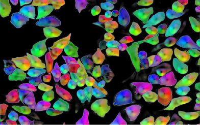
10 Machine Learning in Chemical Biology

 Box 1. Virtual screening of compound activity.                           Recognizing phenotypic traits of cells
                                                                          Given a visual phenotype of interest, how do we auto-
 Virtual screening aims to predict the outcome of a high-throughput       matically identify it in images? If such patterns can be
 biological assay to test the cell’s response to chemical perturba-
 tions. Traditional compound screens rely on specific molecular tests,
                                                                          formulated as explicit inputeoutput relationships, su-
 on specialized biochemical/functional assays, or on cell-based           pervised machine learning offers ways to train a
 assays with markers of specific activity such as proteins of inter-      computational model to identify the relevant visual
 est, DNA damage, or cell proliferation. In contrast, virtual screening   patterns in images. The trained models can then be
 with machine learning can use image features of cell morphology
                                                                          applied to new images to recognize and quantify the
 from a generic imaging assay (such as Cell Painting) to predict
 several of these assay outcomes simultaneously without physically        phenotypes of interest that correspond to various visual
 running them. This strategy decreases the number of experiments          patterns.
 needed to test an entire compound library with hundreds of assays
 [94–97], resulting in improved hit rates and accelerating the pace of    One application of supervised machine learning for
 drug discovery [95].
                                                                          recognizing visual phenotypes in cells is to identify
                                                                          different proteins and their locations. Using images from
 Methods for virtual screening of compounds usually follow an             the Human Protein Atlas [26], which capture the spatial
 image-based profiling approach, wherein the response of a popu-
 lation of cells is first characterized and aggregated for each com-
                                                                          distribution of proteins through immunofluorescence,
 pound (Profiling cell state), followed by supervised machine learning    several deep learning models have been successfully
 using ground truth data obtained for a few example compounds             trained to automatically recognize and annotate protein
 using specialized assays (Recognizing phenotypic traits of cells).       localization patterns [27,28]. In addition, alterations in
 This strategy allows researchers to perform virtual screening using
                                                                          the cellular structure can be a recognizable phenotypic
 image-based profiles in combination with chemical structures and
 gene expression [96,98] and also to extend it to other applications      trait of disease, such as cancer. A deep neural network
 such as predicting cell health phenotypes in Clustered Regularly         was shown to successfully classify the type of lung
 Interspaced Short Palindromic Repeats (CRISPR). based pertur-            cancer in tissue samples, with performance similar to
 bations of cancer cell lines [97].                                       trained human pathologists, and it was even able to
                                                                          predict the mutated genes [17]. Deep learning has been
 Deep learning has been explored for virtual screening using end-to-      found to recognize other cellular traits such as response
 end neural networks trained to predict assays directly from images,      to stress and cell age [29], as well as red blood cell
 demonstrating higher accuracy than using precomputed image-
 based profiles [94]. In addition, modeling the relationships be-
                                                                          quality [30,31].
 tween visual phenotypes and chemical structures using deep
 learning is an emerging research field that can complement assay         Visual phenotypes change over time in response to
 prediction in virtual screens. For example, the generation of mole-      cellular dynamics, revealing important information for
 cules that would produce desired morphological changes has been
                                                                          understanding biological processes. For instance,
 explored for de novo hit design [99], and in the opposite direction,
 generative models have been trained to predict morphological             Neumann et al. [32] leveraged high-throughput time-
 changes, given a specific molecule [100]. These advances in              lapse imaging of single-cell chromatin morphology and
 compound activity prediction are potentially transformative for drug     machine learning to enable a genome-wide siRNA
 discovery research and will likely continue to evolve with better        screen for genes regulating the cell cycle, yielding 12
 models and more data.
                                                                          newly validated and hundreds of novel candidate mitotic
                                                                          regulator genes. In a related study, a deep neural
                                                                          network model was trained to classify cell cycle stages
                                                                          using categorical labels, and the learned features
problems and applications wherein deep learning
                                                                          reconstructed their continuous temporal relationships
applied to images plays a central role. We review ad-
                                                                          [33]. Notably, deep learning models have also been
vances in visual phenotyping in cell classification
                                                                          trained to accurately predict the future lineage trajec-
(Recognizing phenotypic traits of cells) and cell seg-
                                                                          tory of differentiating progenitor cells in culture based
mentation (Box 1) and also consider new approaches
                                                                          on time-lapse, label-free light microscopy images up to
that are expanding our capacity to drive biological dis-
                                                                          three generations earlier than what previous methods
covery, including representation learning (Profiling cell
                                                                          had achieved [34,35]. These examples highlight the
state) and generative modeling (Predicting visual
                                                                          power of identifying temporal changes in visual pheno-
phenotypes). Outside this scope are many other
                                                                          types automatically.
important applications such as human tissue analysis in
pathology [17,18], clinical diagnostics based on
                                                                          A common aspect of the applications mentioned previ-
biomedical images [6,7,19], and live cell tracking [20e
                                                                          ously is the availability of ground truth data for training
23]. We do not cover the mathematical foundations of
                                                                          supervised learning models, which are collected manu-
deep learning [4,24] or aspects related to programming
                                                                          ally by experts or with the assistance of biological probes
frameworks and computing infrastructure [5,25]. Our
                                                                          (Figure. 1). This opens up the opportunity to train end-
goal is to provide an overview of the problems that deep
                                                                          to-end deep learning models that make predictions
learning applied to images can solve to advance ques-
                                                                          directly from image pixels without additional processing
tions in basic and applied biological research.

Current Opinion in Chemical Biology 2021, 65:9–17                                                                 www.sciencedirect.com
Image-based cell phenotyping with deep learning Pratapa et al.            11

or intervention, which is useful for automating tasks that             Box 2. Cell segmentation.
depend on visual phenotyping and for scaling up accu-
rate analyses. Beyond revealing basic cellular functions,              Cell segmentation, in essence, involves classifying pixels to belong
                                                                       to a biologically meaningful structure or the background (Fig. 2).
supervised learning for recognizing visual phenotypes is
                                                                       Machine learning for cell segmentation has been available for a long
also used to guide treatment design and drug discovery,                time in user-friendly software such as ilastik [101,102], and deep
wherein detecting specific traits is critical. A good                  learning is now aiming to achieve cell segmentation without any user
example of this is image-based assay prediction (Box 1).               intervention. Different types of deep learning models have been
Next, we discuss how to quantify visual phenotypes                     designed for cell segmentation including the U-Net model [91,92],
                                                                       DeepCell [103], and Mask RCNN [90,93], all of them able to achieve
when no ground truth data are available for training                   improved accuracy [11,104].
supervised learning models.
                                                                       There are two main challenges in building new cell segmentation
Profiling cell state                                                   tools: (1) manually annotated data need to be collected for training,
A major innovation in visual phenotyping is ‘image-based               and (2) the training of deep learning models on those data still re-
                                                                       quires substantial human effort (and data science skills) to tune
profiling,’ wherein phenotypes are considered contin-
                                                                       hyperparameters and to rectify faulty output. To address the first
uous rather than categorical. Building feature represen-               challenge, the collection of annotations for image analysis may be
tations that capture cell state from images is the main                crowdsourced, obtaining data quality close to that of experts [105],
goal, which enables us to match and compare phenotypes                 or obtained with fluorescent labels used only to gather training data
even if they are unknown ahead of time. This approach                  for segmenting unlabeled images [66]. To address the second
                                                                       challenge, new methodologies are being developed to automate
has many applications in cell biology, including charac-               neural network training, resulting in models that need little user
terizing chemical or genetic perturbations [16].                       intervention during training, while still achieving high segmentation
                                                                       accuracy [106].
A standard approach to high-throughput image-based
profiling of molecular perturbations (such as Cell Paint-              Segmentation models can be made generic enough to identify cells
ing [36,37]) is as follows. The samples are prepared in a              across many imaging experiments [11] by collecting a representa-
multiwell plate format and are subsequently exposed to a               tive set of manually annotated cells and training a single model once
                                                                       to be reused in many problems. NucleAIzer [12] and Cellpose [13]
systematic array of individual or combinatorial pertur-
                                                                       are tools that take this approach for nucleus and cytoplasm seg-
bations. The microscopy images are then acquired after                 mentation, respectively, requiring minimal user intervention.
staining or ‘painting’ the cells and subcellular compo-                Mesmer [14], a deep learning segmentation model trained on
nents by attaching different fluorescent tags. A key next              TissueNet, the largest data set of manually annotated cells collected
step is to extract quantitative multidimensional features,             so far, simultaneously identifies both the cytoplasm and the nucleus
                                                                       in microscopy images. Another approach to reusing existing deep
also known as cellular ‘profiles,’ from each image, either             learning–based models is the creation of model zoos — open-
at the individual-cell level or for entire fields of view [38].        access databases with user-friendly interfaces that facilitate
These image-based features play a similar role in repre-               sharing of ready-to-use trained models [107]. All these efforts are
senting the cellular phenotype as do gene expression                   successful examples of deep learning–based tools that bring the
measurements in transcriptional profiling [39].                        promise of fully automated cell segmentation closer to reality.

The last decade has seen a rapid development of                        Cell segmentation has many applications in biology, and deep
                                                                       learning–based models are already powering biological studies,
computational techniques for image-based profiling
                                                                       while introducing fewer quantification errors than classical ap-
[16,40]. Usually, the process involves identifying single              proaches [104]. Widespread adoption of such methods will depend
cells in images (Box 2) and then computing their                       on the availability of reliable pretrained models and user-friendly
morphology features for unbiased analysis (Figure. 2).                 tools for everyday usage in the laboratory. Some of the open chal-
Commercial microscopy software and open-source tools                   lenges in cell segmentation include the adoption of active learning to
                                                                       interactively annotate only the cells that the model finds challenging
such as CellProfiler [41], EBImage [42], and ImageJ                    to segment and the creation of open collaborative data sets for
[43] offer modules to segment individual cells and                     training larger robust models.
extract hundreds of features for each. These single-
cellelevel profiles can then be aggregated to obtain
population-level profiles. Several strategies for
computing aggregated profiles from segmented cells                   normalization [48] to training a generative model for
exist, but in practice, a simple median-based profile is             batch equalization [9].
known to be quite powerful [44,45]. Alternatively, some
tools also offer strategies to extract population-level              The central component of image-based profiling is
profiles directly from the field of view without the                 feature representation, which allows to match and
need for segmentation [46,47]. Similar to transcriptional            compare phenotypes using a similarity measure. There
profiling, image-based features are sensitive to technical           are two main strategies for computing feature repre-
variation, resulting in batch effects that can be detri-             sentations: (1) engineered features and (2) learned
mental to downstream analysis. Strategies to mitigate                features. Engineered features involve measuring the
batch effects include techniques ranging from variation              number of cells/subcellular components, classical

www.sciencedirect.com                                                                 Current Opinion in Chemical Biology 2021, 65:9–17
12 Machine Learning in Chemical Biology
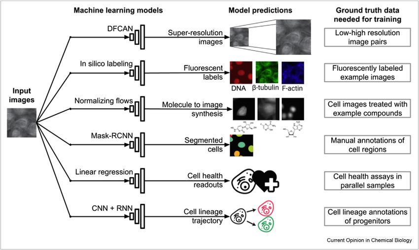
Figure. 1
Example applications of supervised machine learning models on microscopy images. These models can take input bright-field images, time-lapse mi-
croscopy sequences, Cell Painting images, or precomputed image features. Models can be trained to predict a diversity of phenotypic or perturbation
information, including higher resolution images [68,70], effects of compound treatments [56,89], segmentation maps [90–93], and fluorescence labels
[8,10,63]. While the training process requires input/output pairs using ground truth data (Recognizing phenotypic traits of cells), trained models can
predict these outputs from new images in an automated way for future analysis. DFCAN, deep Fourier channel attention network; Mask RCNN, Mask
Region-based Convolutional Neural Network; CNN + RNN, convolutional neural network + recurrent neural network.
Figure. 2
Current Opinion in Chemical Biology
Illustration of the typical computational workflow for image-based cell profiling. Single cells are first identified using image segmentation (Box 2), and then,
morphology features are computed to quantify cell state (Profiling cell state). The single-cell-level profiles can then be aggregated to obtain population-
level profiles. Image-based features can then be analyzed to identify and correct potential batch effects. These profiles generate follow up experimental
hypotheses and to obtain novel biological insights in the corresponding biological application.
Current Opinion in Chemical Biology 2021, 65:9–17 www.sciencedirect.com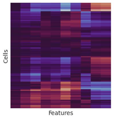
Image-based cell phenotyping with deep learning Pratapa et al.   13

cytometry-based features [49], and other coefficients           features from unlabeled images to estimate where the
such as Zernike shape features and Gabor texture fea-           stains would activate inside cells if physically applied.
tures [36]. Depending on the type of experiment, they           This approach has been successfully used to screen
can also represent normalized pixel intensities of fluo-        compounds for treating Alzheimer disease, improving
rescent markers after segmentation for their analysis           hit rates in a prospective evaluation [64]. It was also
[50]. Learned features commonly involve a deep                  shown to improve throughput in high-content screening
learning model, such as a convolutional neural network,         applications using reflectance microscopy, which results
trained to identify discriminatory or representative            in more accurate virtual stain predictions [65].
features directly from raw pixels. Because the ground
truth phenotype is unknown beforehand, representation           Label-free images may contain more information than
learning is usually either weakly supervised or unsu-           what can be recognized by the eye, expanding to other
pervised. The model can be trained on a library of              potential applications. The segmentation of the nucleus
cellular images corresponding to different perturbations        without a DNA stain is an example that follows a similar
or treatment conditions and then used for feature               approach. The fluorescent marker is first applied on
extraction [30,51,52]. Moreover, studies have also suc-         training images only, and then, trained models can detect
cessfully used features extracted from models trained on        the nucleus directly on bright-field images [66]. The use
natural images using transfer learning [48,53]. Another         of imaging flow cytometry for diagnosing leukemia usually
approach for self-supervised representation learning is         relies on several fluorescent markers, which could be used
to train a convolutional neural networkebased encoder           to train a model that detects the same phenotype using
to predict one of the channels in the image [54] or all         bright-field and dark-field images only [31].
channels through an autoencoder [55].
                                                                Deep learning models can also transform low-resolution
Taken together, these techniques played a major role in         visual phenotypes into high-resolution images [67]. The
furthering high-throughput image-based profiling to             frame-rate limits in super-resolution for single-molecule
assess and explore the effects of drug treatments               localization have been overcome without compromising
[51,56], RNA interference [57e59], or other pathogenic          accuracy using deep learning models that accelerate image
perturbations [60,61]. More recently, researchers have          reconstruction [68]. Similarly, super-resolution from
also used image-based profiling to investigate the              different microscopy modalities, such as transforming
functional effects of various chemical compounds in             confocal microscopy images to match the resolution of a
fighting against coronavirus disease 2019 [51,62]. In           stimulated emission depletion microscope, has been
contrast to visual phenotype recognition using super-           shown to be accurate [69]. There are still challenges with
vised learning (Recognizing phenotypic traits of cells),        using label-free images and reconstructing phenotypes
image-based profiling allows analysis of visual pheno-          computationally, and more research is still needed to
types in an unbiased way to formulate hypotheses and            determine the reliability of these approaches beyond
understand biological models and perturbations. This            proof-of-concept experiments [70].
approach is widely used in drug discovery and functional
genomics studies and is increasingly being adopted in           Future directions
other applications wherein images may reveal novel              Deep learning is expanding our ability to study cell
phenotypes that require quantification.                         biology with images in many different directions. In
                                                                addition to recognizing phenotypic traits, profiling cell
Predicting visual phenotypes                                    state, and generating new images, machine learning can
Images revealing high-quality visual phenotypes can             also power cell sorting in real time [71,72], guide
now also be generated using deep learning models.               optimal experimental design [73,74], report uncertainty
Given certain experimental observations, a model can            under the presence of novel phenotypes [75], and
estimate how cells would look like under specific               enable real-time volumetric reconstructions of cells
treatments or procedures. In this way, we can run sim-          [76]. Taken together, these applications exemplify how
ulations, make virtual experiments, or impute missing           visual phenotypes can support and automate experi-
data that are difficult to collect physically.                  mental decisions even before performing downstream
                                                                biological interpretation.
Fluorescence labeling reveals specific cellular structures
and can make visual phenotypes easier to be detected by         Imaging is a rich source of information that can be com-
the human eye. However, fluorescent markers also have           bined and connected with other existing biological data,
several limitations, including costs, toxicity for living       most notably high-resolution genetic data. Emerging
cells, or not even compatible with live imaging. An             technologies for sequencing single cells in tissues [77e
emerging trend in microscopy is the prediction of fluo-         79] offer the unique opportunity to study the relation-
rescence labels from transmitted bright-field images            ship between visual phenotypes, gene or protein expres-
[8,10,63], wherein deep learning models recognize               sion levels, and their spatial distribution (Box 2). Deep

www.sciencedirect.com                                                          Current Opinion in Chemical Biology 2021, 65:9–17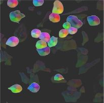
14 Machine Learning in Chemical Biology

learning has been recently used to predict local gene           Declaration of competing interest
expression patterns from tissue images [80,81] and bulk         The authors declare that they have no known competing
mRNA levels from whole-slide images [82], all based on          financial interests or personal relationships that could have
hematoxylin and eosinestained histology samples. These          appeared to influence the work reported in this paper.
results indicate that the correlation patterns between
morphology and gene expression can be extracted and             Acknowledgements
quantified, likely improving accuracy as more spatial           The authors would like to thank Anne Carpenter, Sami Farhi, Wolfgang
transcriptomics data are acquired.                              Pernice, and Shantanu Singh for valuable discussions and recommenda-
                                                                tions made to complete this manuscript. This work was supported by the
                                                                Broad Institute Schmidt Fellowship program.
Another promising tool for multimodal single-cell anal-
ysis is highly multiplexed imaging, which involves              References
measuring multiple fluorescent markers simultaneously           Papers of particular interest, published within the period of review,
                                                                have been highlighted as:
[83]. Unlike hematoxylin and eosin staining, these
techniques result in a comprehensive map of cellular             * of special interest
                                                                 * * of outstanding interest
organization within a tissue at single-cell resolution.
Highly multiplexed imaging has already been success-            1.   Meijering E, Carpenter AE, Peng H, Hamprecht FA, Olivo-
fully applied to obtain spatially resolved, single-cell level        Marin J-C: Imagining the future of bioimage analysis. Nat
measurements of transcriptional and protein landscape                Biotechnol 2016, 34:1250–1255.
within a tissue [84]. The computational pipeline for            2.   Sommer C, Gerlich DW: Machine learning in cell biology -
analyzing multiplexed imaging of tissues typically in-               teaching computers to recognize phenotypes. J Cell Sci
                                                                     2013, 126:5529–5539.
volves similar steps as in image-based profiling (Profiling
                                                                3.   Grys BT, Lo DS, Sahin N, Kraus OZ, Morris Q, Boone C, et al.:
cell state) [85]. The obtained features can then be used             Machine learning and computer vision approaches for
to classify cell types, analyze cellular neighborhoods, and          phenotypic profiling. J Cell Biol 2017, 216:65–71.
study cellecell interactions across phenotypes within a         4.   Gupta A, Harrison PJ, Wieslander H, Pielawski N, Kartasalo K,
tissue microenvironment [86]. Although still in early                Partel G, et al.: Deep learning in image cytometry: a review.
                                                                     Cytometry 2019, 95:366–380.
phases, deep learning tools may be used for integrating
cell morphology with changes in gene and protein                5.   Moen E, Bannon D, Kudo T, Graf W, Covert M, Van Valen D:
                                                                     Deep learning for cellular image analysis. Nat Methods 2019,
expression to study tumor microenvironments.                         16:1233–1246.
                                                                6.   Esteva A, Kuprel B, Novoa RA, Ko J, Swetter SM, Blau HM, et al.:
One of the main drawbacks of deep learning models is                 Dermatologist-level classification of skin cancer with deep
that they encode visual phenotypes in multiple layers of             neural networks. Nature 2017, 542:115–118.
feature maps that may not have any direct interpretation        7.   Rajpurkar P, Irvin J, Ball RL, Zhu K, Yang B, Mehta H, et al.:
or biological meaning. Developing methods that can                   Deep learning for chest radiograph diagnosis: a retrospec-
                                                                     tive comparison of the CheXNeXt algorithm to practicing
explain the patterns found in the data can facilitate                radiologists. PLoS Med 2018, 15, e1002686.
image analysis in supervised learning and profiling ap-         8.   Christiansen EM, Yang SJ, Ando DM, Javaherian A,
plications. The use of generative models has been                    Skibinski G, Lipnick S, et al.: In silico labeling: predicting
explored for explaining differences in phenotypes based              fluorescent labels in unlabeled images. Cell 2018, 173:
                                                                     792 – 803. e19.
on the underlying data distribution [55,87]. Additional
                                                                9.    Qian WW, Xia C, Venugopalan S, Narayanaswamy A, Dimon M,
work remains to be carried out in areas such as designing       *     Ashdown GW, et al.: Batch equalization with a generative
deep learning architectures that are inherently inter-                adversarial network. Bioinformatics 2020, 36:i875–i883.
pretable in a biological context [88], finding positive and     A generalizable deep learning based approach that uses StarGAN to
                                                                normalize images from different batches through equalization, while
negative examples associated with a phenotype of in-            also retaining their biological phenotypes.
terest, and using causal inference to identify the bio-         10. Ounkomol C, Seshamani S, Maleckar MM, Collman F,
logical effects of interventions.                                   Johnson GR: Label-free prediction of three-dimensional fluo-
                                                                    rescence images from transmitted-light microscopy. Nat
                                                                    Methods 2018, 15:917–920.
Developing and training deep learning models takes
                                                                11. Caicedo JC, Goodman A, Karhohs KW, Cimini BA, Ackerman J,
significant time and resources, including data collection           Haghighi M, et al.: Nucleus segmentation across imaging ex-
and computing power. The impact of visual phenotyping               periments: the 2018 data science bowl. Nat Methods 2019, 16:
based on deep learning can be amplified by sharing data,            1247–1253.
code, and trained models for other laboratories to              12. Hollandi R, Szkalisity A, Toth T, Tasnadi E, Molnar C, Mathe B,
                                                                    et al.: nucleAIzer: a parameter-free deep learning framework
reproduce and apply on their own research. Open data                for nucleus segmentation using image style transfer. Cell
and code will help advance methods, while model                     Systems 2020, 10:453–458.e6.
sharing will facilitate adoption of robust and generalist       13. Stringer C, Wang T, Michaelos M, Pachitariu M: Cellpose: a
solutions. The biological imaging community is making           * * generalist algorithm for cellular segmentation. Nat Methods
                                                                    2021, 18:100–106.
coordinated efforts to bring these technologies to all          A deep learning-based tool that performs precise 2D and 3D seg-
biological laboratories for supporting their research and       mentation across a wide range of cellular images without the need for
for making the power of visual phenotypes more acces-           model retraining and parameter tuning.
sible for biological discovery.
Current Opinion in Chemical Biology 2021, 65:9–17                                                                www.sciencedirect.com
Image-based cell phenotyping with deep learning Pratapa et al. 15
14. Greenwald NF, Miller G, Moen E, Kong A, Kagel A, Fullaway CC, 33. Eulenberg P, Köhler N, Blasi T, Filby A, Carpenter AE, Rees P,
* et al.: Whole-cell segmentation of tissue images with human- et al.: Reconstructing cell cycle and disease progression
level performance using large-scale data annotation and using deep learning. Nat Commun 2017, 8:463.
deep learning. Cold Spring Harbor Laboratory 2021, https://
doi.org/10.1101/2021.03.01.431313. 34. Buggenthin F, Buettner F, Hoppe PS, Endele M, Kroiss M,
A deep learning-based tool for cell segmentation in tissue imaging Strasser M, et al.: Prospective identification of hematopoietic
data; trained using TissueNet, a crowdsourced database of over 1 lineage choice by deep learning. Nat Methods 2017, 14:
million whole-cell and nuclear annotations for tissue images. 403–406.
15. Mullard A: Machine learning brings cell imaging promises into 35. Waisman A, La Greca A, Möbbs AM, Scarafía MA, Santín
focus. Nat Rev Drug Discov 2019, 18:653–655. * Velazque NL, Neiman G, et al.: Deep learning neural networks
highly predict very early onset of pluripotent stem cell dif-
16. Chandrasekaran SN, Ceulemans H, Boyd JD, Carpenter AE: ferentiation. Stem Cell Rep 2019, 12:845–859.
* * Image-based profiling for drug discovery: due for a machine- A demonstration of the ability of deep learning to recognize subtle
learning upgrade? Nat Rev Drug Discov 2020, https://doi.org/ morphological features from phase contrast images and predict early
10.1038/s41573-020-00117-w. onset of pluripotency without the need for fluorescent tagging.
A recent and comprehensive review of image-based profiling.
36. Bray M-A, Singh S, Han H, Davis CT, Borgeson B, Hartland C,
17. Coudray N, Ocampo PS, Sakellaropoulos T, Narula N, Snuderl M, et al.: Cell Painting, a high-content image-based assay for
Fenyö D, et al.: Classification and mutation prediction from morphological profiling using multiplexed fluorescent dyes.
non–small cell lung cancer histopathology images using Nat Protoc 2016, 11:1757–1774.
deep learning. Nat Med 2018, 24:1559–1567.
37. Gustafsdottir SM, Ljosa V, Sokolnicki KL, Anthony Wilson J,
18. Fu Y, Jung AW, Torne RV, Gonzalez S, Vöhringer H, Shmatko A, Walpita D, Kemp MM, et al.: Multiplex cytological profiling
et al.: Pan-cancer computational histopathology reveals mu- assay to measure diverse cellular states. PloS One 2013, 8,
tations, tumor composition and prognosis. Nat Canc 2020, 1: e80999.
800–810.
38. Caicedo JC, Singh S, Carpenter AE: Applications in image-
19. Mobadersany P, Yousefi S, Amgad M, Gutman DA, Barnholtz- based profiling of perturbations. Curr Opin Biotechnol 2016,
Sloan JS, Velázquez Vega JE, et al.: Predicting cancer out- 39:134–142.
comes from histology and genomics using convolutional
networks. Proc Natl Acad Sci U S A 2018, 115:E2970–E2979. 39. Wawer MJ, Li K, Gustafsdottir SM, Ljosa V, Bodycombe NE,
Marton MA, et al.: Toward performance-diverse small-mole-
20. Arts M, Smal I, Paul MW, Wyman C, Meijering E: Particle cule libraries for cell-based phenotypic screening using
mobility analysis using deep learning and the moment multiplexed high-dimensional profiling. Proc Natl Acad Sci U S
scaling spectrum. Sci Rep 2019, 9:17160. A 2014, 111:10911–10916.
21. Lugagne J-B, Lin H, Dunlop MJ: DeLTA: automated cell seg- 40. Caicedo JC, Cooper S, Heigwer F, Warchal S, Qiu P, Molnar C,
mentation, tracking, and lineage reconstruction using deep et al.: Data-analysis strategies for image-based cell profiling.
learning. PLoS Comput Biol 2020, 16, e1007673. Nat Methods 2017, 14:849–863.
22. Yao Y, Smal I, Grigoriev I, Akhmanova A, Meijering E: Deep- 41. McQuin C, Goodman A, Chernyshev V, Kamentsky L, Cimini BA,
learning method for data association in particle tracking. Karhohs KW, et al.: CellProfiler 3.0: next generation image
Bioinformatics 2020, 36:4935–4941. processing for biology. PLoS Comput Biol 2018, 16:e2005970,
https://doi.org/10.1371/journal.pbio.2005970.
23. Wang J, Su X, Zhao L, Zhang J: Deep reinforcement learning
for data association in cell tracking. Front Bioeng Biotechnol 42. Pau G, Fuchs F, Sklyar O, Boutros M, Huber W: EBImage–an R
2020, 8:298. package for image processing with applications to cellular
phenotypes. Bioinformatics 2010, 26:979–981.
24. Angermueller C, Pärnamaa T, Parts L, Stegle O: Deep learning
for computational biology. Mol Syst Biol 2016, 12:878. 43. Schneider CA, Rasband WS, Eliceiri KW: NIH Image to ImageJ:
25 years of image analysis. Nat Methods 2012, 9:671–675.
25. Bannon D, Moen E, Schwartz M, Borba E, Kudo T, Greenwald N,
et al.: DeepCell Kiosk: scaling deep learning-enabled cellular 44. Ljosa V, Caie PD, Ter Horst R, Sokolnicki KL, Jenkins EL,
image analysis with Kubernetes. Nat Methods 2021, 18:43–45. Daya S, et al.: Comparison of methods for image-based
profiling of cellular morphological responses to small-
26. Thul PJ, Åkesson L, Wiking M, Mahdessian D, Geladaki A, Ait molecule treatment. J Biomol Screen 2013, 18:1321–1329.
Blal H, et al.: A subcellular map of the human proteome. Sci-
ence 2017, 356, https://doi.org/10.1126/science.aal3321. 45. Rohban MH, Abbasi HS, Singh S, Carpenter AE: Capturing
single-cell heterogeneity via data fusion improves image-
27. Sullivan DP, Winsnes CF, Åkesson L, Hjelmare M, Wiking M, based profiling. Nat Commun 2019, 10:2082.
Schutten R, et al.: Deep learning is combined with massive-
scale citizen science to improve large-scale image classifi- 46. Rajaram S, Pavie B, Wu LF, Altschuler SJ: PhenoRipper: soft-
cation. Nat Biotechnol 2018, 36:820–828. ware for rapidly profiling microscopy images. Nat Methods
2012, 9:635–637.
28. Ouyang W, Winsnes CF, Hjelmare M, Cesnik AJ, Åkesson L,
Xu H, et al.: Analysis of the human protein Atlas image 47. Uhlmann V, Singh S, Carpenter AE, Charm CP-: segmentation-free
classification competition. Nat Methods 2019, 16:1254–1261. image classification made accessible. BMC Bioinf 2016, 17:51.
29. Mattiazzi Usaj M, Sahin N, Friesen H, Pons C, Usaj M, 48. Michael Ando D, McLean CY, Berndl M: Improving phenotypic
Masinas MPD, et al.: Systematic genetics and single-cell im- measurements in high-content imaging screens. Cold Spring
aging reveal widespread morphological pleiotropy and cell- Harbor Laboratory 2017:161422, https://doi.org/10.1101/161422.
to-cell variability. Mol Syst Biol 2020, 16:e9243.
49. Rodenacker K, Bengtsson E: A feature set for cytometry on
30. Doan M, Sebastian JA, Caicedo JC, Siegert S, Roch A, digitized microscopic images. Anal Cell Pathol 2003, 25:1–36.
Turner TR, et al.: Objective assessment of stored blood
quality by deep learning. Proc Natl Acad Sci U S A 2020, 117: 50. Jackson HW, Fischer JR, Zanotelli VRT, Ali HR, Mechera R,
21381–21390. * Soysal SD, et al.: The single-cell pathology landscape of
breast cancer. Nature 2020, 578:615–620.
31. Doan M, Case M, Masic D, Hennig H, McQuin C, Caicedo J, An application of highly multiplexed imaging to study spatially resolved
et al.: Label-free leukemia monitoring by computer vision. single-cell level phenotypic landscape in breast cancer.
Cytometry 2020, 97:407–414.
51. Cuccarese MF, Earnshaw BA, Heiser K, Fogelson B: Functional
32. Neumann B, Walter T, Hériché J-K, Bulkescher J, Erfle H, * * immune mapping with deep-learning enabled phenomics applied
Conrad C, et al.: Phenotypic profiling of the human genome to immunomodulatory and COVID-19 drug discovery. bioRxiv;
by time-lapse microscopy reveals cell division genes. Nature 2020, https://doi.org/10.1101/2020.08.02.233064.
2010, 464:721–727.
www.sciencedirect.com Current Opinion in Chemical Biology 2021, 65:9–1716 Machine Learning in Chemical Biology
A morphological database for studying the effect of over 1600 drug 68. Ouyang W, Aristov A, Lelek M, Hao X, Zimmer C: Deep learning
treatments on cells infected with SARS-CoV-2. massively accelerates super-resolution localization micro-
scopy. Nat Biotechnol 2018, 36:460–468.
52. Caicedo JC, McQuin C, Goodman A, Singh S, Carpenter AE:
Weakly supervised learning of single-cell feature embed- 69. Wang H, Rivenson Y, Jin Y, Wei Z, Gao R, Günaydõn H,
dings. IEEE Comput Soc Conf Comput Vis Pattern Recogn 2018, et al.: Deep learning enables cross-modality super-reso-
2018:9309–9318. lution in fluorescence microscopy. Nat Methods 2019, 16:
103 – 110.
53. Godec P, Pan cur M, Ileni
c N, Copar A, Stra
zar M, Erjavec A,
et al.: Democratized image analytics by visual programming 70. Belthangady C, Royer LA: Applications, promises, and pitfalls
through integration of deep models and small-scale machine * of deep learning for fluorescence image reconstruction. Nat
learning. Nat Commun 2019, 10:4551. Methods 2019, 16:1215–1225.
A comprehensive review of common approaches and pitfalls in deep
54. Lu AX, Kraus OZ, Cooper S, Moses AM: Learning unsupervised learning for predicting visual phenotypes with a focus on image
feature representations for single cell microscopy images reconstruction.
with paired cell inpainting. PLoS Comput Biol 2019, 15,
e1007348. 71. Nitta N, Sugimura T, Isozaki A, Mikami H, Hiraki K, Sakuma S,
et al.: Intelligent image-activated cell sorting. Cell 2018, 175:
55. Lafarge MW, Caicedo JC, Carpenter AE, Pluim JPW, Singh S, 266–276. e13.
Veta M: Capturing single-cell phenotypic variation via unsu-
pervised representation learning. In Proceedings of the 2nd 72. Lee J, Liu Z, Suzuki PH, Ahrens JF, Lai S, Lu X, et al.: Versatile
international conference on medical imaging with deep learning. phenotype-activated cell sorting. Sci Adv 2020, 6, https://
Edited by Cardoso MJ, Feragen A, Glocker B, Konukoglu E, doi.org/10.1126/sciadv.abb7438.
Oguz I, Unal G, et al., London, United Kingdom: PMLR; 2019:
315–325. 73. Rajaram S, Heinrich LE, Gordan JD, Avva J, Bonness KM,
Witkiewicz AK, et al.: Sampling strategies to capture single-
56. Caie PD, Walls RE, Ingleston-Orme A, Daya S, Houslay T, cell heterogeneity. Nat Methods 2017, 14:967–970.
Eagle R, et al.: High-content phenotypic profiling of drug
response signatures across distinct cancer cells. Mol Canc 74. Hou H, Gan T, Yang Y, Zhu X, Liu S, Guo W, et al.: Using deep
Therapeut 2010, 9:1913–1926. reinforcement learning to speed up collective cell migration.
BMC Bioinf 2019, 20:571.
57. Laufer C, Fischer B, Billmann M, Huber W, Boutros M: Mapping
genetic interactions in human cancer cells with RNAi and 75. Dürr O, Murina E, Siegismund D, Tolkachev V, Steigele S, Sick B:
multiparametric phenotyping. Nat Methods 2013:427–431, Know when you don’t know: a robust deep learning
https://doi.org/10.1038/nmeth.2436. approach in the presence of unknown phenotypes. Assay
Drug Dev Technol 2018, 16:343–349.
58. Horn T, Sandmann T, Fischer B, Axelsson E, Huber W,
Boutros M: Mapping of signaling networks through synthetic 76. Wang Z, Zhu L, Zhang H, Li G, Yi C, Li Y, et al.: Real-time
genetic interaction analysis by RNAi. Nat Methods 2011, 8: volumetric reconstruction of biological dynamics with light-
341–346. field microscopy and deep learning. Nat Methods 2021, https://
doi.org/10.1038/s41592-021-01058-x.
59. Fischer B, Sandmann T, Horn T, Billmann M, Chaudhary V,
Huber W, et al.: A map of directional genetic interactions in a 77. Ståhl PL, Salmén F, Vickovic S, Lundmark A, Navarro JF,
metazoan cell. Elife 2015, 4, https://doi.org/10.7554/eLife.05464. Magnusson J, et al.: Visualization and analysis of gene
expression in tissue sections by spatial transcriptomics.
60. Conrad C, Erfle H, Warnat P, Daigle N, Lörch T, Ellenberg J, Science 2016, 353:78–82.
et al.: Automatic identification of subcellular phenotypes on
human cell arrays. Genome Res 2004, 14:1130–1136. 78. Rodriques SG, Stickels RR, Goeva A, Martin CA, Murray E,
Vanderburg CR, et al.: Slide-seq: a scalable technology for
61. Perlman ZE, Slack MD, Feng Y, Mitchison TJ, Wu LF, measuring genome-wide expression at high spatial resolu-
Altschuler SJ: Multidimensional drug profiling by automated tion. Science 2019, 363:1463–1467.
microscopy. Science 2004, 306:1194–1198.
79. Eng C-HL, Lawson M, Zhu Q, Dries R, Koulena N, Takei Y, et al.:
62. White B, Komalo B, Nicolaisen L, Donne M, Marsh C, DeVay RM, Transcriptome-scale super-resolved imaging in tissues by
et al.: A multi-phenotype system to discover therapies for RNA seqFISH. Nature 2019, 568:235–239.
age-related dysregulation of the immune response to viral
infections. Cold Spring Harbor Laboratory 2020, https://doi.org/ 80. He B, Bergenstråhle L, Stenbeck L, Abid A, Andersson A, Borg Å,
10.1101/2020.07.30.223875. * * et al.: Integrating spatial gene expression and breast tumour
morphology via deep learning. Nat Biomed Eng 2020, 4:
63. Wang Z, Xie Y, Ji S: Global voxel transformer networks for 827–834.
augmented microscopy. Nat Machine Intelligence 2021, 3: A study that shows the promise of integrating morphology with gene
161–171. expression data and successfully predicts spatially resolved tran-
scriptome from existing histopathology images.
64. Wong DR, Conrad J, Johnson N, Ayers JI, Laeremans A, Lee JC,
* et al.: Trans-channel fluorescence learning improves high- 81. Schmauch B, Romagnoni A, Pronier E, Saillard C, Maillé P,
content screening for Alzheimer’s disease therapeutics. Cold Calderaro J, et al.: A deep learning model to predict RNA-Seq
Spring Harbor Laboratory 2021, https://doi.org/10.1101/ expression of tumours from whole slide images. Nat Commun
2021.01.08.425973. 2020, 11:3877.
A demonstration of the power of deep learning in constructing new
fluorescent images from pre-existing fluorescent markers. 82. Levy-Jurgenson A, Tekpli X, Kristensen VN, Yakhini Z: Spatial
transcriptomics inferred from pathology whole-slide images
65. Cheng S, Fu S, Kim YM, Song W, Li Y, Xue Y, et al.: Single-cell links tumor heterogeneity to survival in breast and lung
cytometry via multiplexed fluorescence prediction by label- cancer. Sci Rep 2020, 10:18802.
free reflectance microscopy. Sci Adv 2021, 7, https://doi.org/
10.1126/sciadv.abe0431. 83. Tan WCC, Nerurkar SN, Cai HY, Ng HHM, Wu D, Wee YTF,
et al.: Overview of multiplex immunohistochemistry/immu-
66. Sadanandan SK, Ranefall P, Le Guyader S, Wählby C: Auto- nofluorescence techniques in the era of cancer immuno-
mated training of deep convolutional neural networks for cell therapy. Canc Commun 2020, 40:135–153.
segmentation. Sci Rep 2017, 7:7860.
84. Bodenmiller B: Multiplexed epitope-based tissue imaging for
67. Qiao C, Li D, Guo Y, Liu C, Jiang T, Dai Q, et al.: Evaluation and discovery and healthcare applications. Cell Syst 2016, 2:
development of deep neural networks for image super- 225–238.
resolution in optical microscopy. Nat Methods 2021, 18:
194–202.
Current Opinion in Chemical Biology 2021, 65:9–17 www.sciencedirect.comImage-based cell phenotyping with deep learning Pratapa et al. 17
85. Schapiro D, Jackson HW, Raghuraman S, Fischer JR, morphology information for multitask bioactivity predictions.
Zanotelli VRT, Schulz D, et al.: histoCAT: analysis of cell J Chem Inf Model 2021, 61:1444–1456.
phenotypes and interactions in multiplex image cytometry
data. Nat Methods 2017, 14:873–876. 97. Way GP, Kost-Alimova M, Shibue T, Harrington WF, Gill S,
Piccioni F, et al.: Predicting cell health phenotypes using
86. Rashid R, Gaglia G, Chen Y-A, Lin J-R, Du Z, Maliga Z, et al.: image-based morphology profiling. Mol Biol Cell 2021,
Highly multiplexed immunofluorescence images and single- mbcE20120784.
cell data of immune markers in tonsil and lung cancer. Sci
Data 2019, 6:323. 98. Becker T, Yang K, Caicedo JC, Wagner BK, Dancik V,
Clemons P, et al.: Predicting compound activity from pheno-
87. Goldsborough P, Pawlowski N, Caicedo JC, Singh S, typic profiles and chemical structures. Cold Spring Harbor
Carpenter A: CytoGAN: generative modeling of cell images. bio- Laboratory 2020, https://doi.org/10.1101/2020.12.15.422887.
Rxiv; 2017:227645, https://doi.org/10.1101/227645.
99. Méndez-Lucio O, Zapata PAM, Wichard J, Rouquié D, Clevert D-
88. Ma J, Fong SH, Luo Y, Bakkenist CJ, Shen JP, Mourragui S, A: Cell morphology-guided de novo hit design by condition-
et al.: Few-shot learning creates predictive models of drug ing generative adversarial networks on phenotypic image
response that translate from high-throughput screens to in- features. ChemRxiv 2020, https://doi.org/10.26434/
dividual patients. Nat Canc 2021, 2:233–244. chemrxiv.11594067.v1.
89. Lapins M, Spjuth O: Evaluation of gene expression and 100.Yang K, Goldman S, Jin W, Lu A, Barzilay R, Jaakkola T, et al.:
phenotypic profiling data as quantitative descriptors for Improved conditional flow models for molecule to image syn-
predicting drug targets and mechanisms of action. Cold Spring thesis. arXiv [q-bio.BM]; 2020. Available: http://arxiv.org/abs/
Harbor Laboratory 2019:580654, https://doi.org/10.1101/580654. 2006.08532.
90. Vuola AO, Akram SU, Kannala J: Mask-RCNN and U-net ensem- 101. Sommer C, Straehle C, Köthe U, Hamprecht FA: Ilastik: inter-
bled for nuclei segmentation. In IEEE 16th international sympo- active learning and segmentation toolkit. In 2011 IEEE inter-
sium on biomedical imaging (ISBI 2019). 2019; 2019:208–212. national symposium on biomedical imaging: from nano to macro;
2011:230–233. ieeexplore.ieee.org; 2011.
91. Falk T, Mai D, Bensch R, Çiçek Ö, Abdulkadir A, Marrakchi Y, et al.:
U-Net: deep learning for cell counting, detection, and 102. Berg S, Kutra D, Kroeger T, Straehle CN, Kausler BX, Haubold C,
morphometry. Nat Methods 2018, https://doi.org/10.1038/ et al.: ilastik: interactive machine learning for (bio)image
s41592-018-0261-2. analysis. Nat Methods 2019, 16:1226–1232.
92. Ronneberger O, Fischer P, Brox T: U-net: convolutional net- 103. Van Valen DA, Kudo T, Lane KM, Macklin DN, Quach NT,
works for biomedical image segmentation. Med Image DeFelice MM, et al.: Deep learning automates the quantitative
Comput Comput Assist Interv 2015. Available: http://link.springer. analysis of individual cells in live-cell imaging experiments.
com/chapter/10.1007/978-3-319-24574-4_28; 2015. PLoS Comput Biol 2016, 12, e1005177.
93. Johnson JW: Adapting mask-RCNN for automatic nucleus seg- 104. Caicedo JC, Roth J, Goodman A, Becker T, Karhohs KW,
mentation. arXiv [cs.CV]; 2018. Available: http://arxiv.org/abs/ Broisin M, et al.: Evaluation of deep learning strategies for
1805.00500. nucleus segmentation in fluorescence images. Cytometry
2019, https://doi.org/10.1002/cyto.a.23863.
94. Hofmarcher M, Rumetshofer E, Clevert D-A, Hochreiter S,
* Klambauer G: Accurate prediction of biological assays with 105. Hughes AJ, Mornin JD, Biswas SK, Beck LE, Bauer DP,
high-throughput microscopy images and convolutional net- Raj A, et al.: Quanti.us: a tool for rapid, flexible, crowd-
works. J Chem Inf Model 2019, 59:1163–1171. based annotation of images. Nat Methods 2018, 15:
An application of high-throughput imaging for assay prediction using an 587 – 590.
end-to-end deep learning framework.
106. Isensee F, Jaeger PF, Kohl SAA, Petersen J, Maier-Hein KH:
95. Simm J, Klambauer G, Arany A, Steijaert M, Wegner JK, nnU-Net: a self-configuring method for deep learning-based
Gustin E, et al.: Repurposing high-throughput image assays biomedical image segmentation. Nat Methods 2021, 18:
enables biological activity prediction for drug discovery. Cell 203–211.
Chem Biol 2018:611–618, https://doi.org/10.1016/j.chem-
biol.2018.01.015. e3. 107. Ouyang W, Mueller F, Hjelmare M, Lundberg E, Zimmer C:
ImJoy: an open-source computational platform for the deep
96. Trapotsi M-A, Mervin LH, Afzal AM, Sturm N, Engkvist O, learning era. Nat Methods 2019, 16:1199–1200.
Barrett IP, et al.: Comparison of chemical structure and cell
www.sciencedirect.com Current Opinion in Chemical Biology 2021, 65:9–17You can also read