Interactions and Feedbacks in E-Cadherin Transcriptional Regulation
←
→
Page content transcription
If your browser does not render page correctly, please read the page content below
PERSPECTIVE
                                                                                                                                            published: 28 June 2021
                                                                                                                                     doi: 10.3389/fcell.2021.701175

                                            Interactions and Feedbacks in
                                            E-Cadherin Transcriptional
                                            Regulation
                                            Miguel Ramirez Moreno 1 , Przemyslaw A. Stempor 2 and Natalia A. Bulgakova 1
                                            1
                                             Department of Biomedical Science and Bateson Centre, The University of Sheffield, Sheffield, England, 2 SmartImmune
                                            Ltd., Cambridge, United Kingdom

                                            Epithelial tissues rely on the adhesion between participating cells to retain their
                                            integrity. The transmembrane protein E-cadherin is the major protein that mediates
                                            homophilic adhesion between neighbouring cells and is, therefore, one of the critical
                                            components for epithelial integrity. E-cadherin downregulation has been described
                                            extensively as a prerequisite for epithelial-to-mesenchymal transition and is a hallmark
                                            in many types of cancer. Due to this clinical importance, research has been mostly
                                            focused on understanding the mechanisms leading to transcriptional repression of
                                            this adhesion molecule. However, in recent years it has become apparent that re-
                                            expression of E-cadherin is a major step in the progression of many cancers during
                         Edited by:         metastasis. Here, we review the currently known molecular mechanisms of E-cadherin
                      Daiki Umetsu,
             Tohoku University, Japan       transcriptional activation and inhibition and highlight complex interactions between
                        Reviewed by:        individual mechanisms. We then propose an additional mechanism, whereby the
                     Mohit Kumar Jolly,     competition between adhesion complexes and heterochromatin protein-1 for binding to
Indian Institute of Science (IISc), India
                            Hiroki Oda,
                                            STAT92E fine-tunes the levels of E-cadherin expression in Drosophila but also regulates
  JT Biohistory Research Hall, Japan        other genes promoting epithelial robustness. We base our hypothesis on both existing
                  *Correspondence:          literature and our experimental evidence and suggest that such feedback between the
                 Natalia A. Bulgakova       cell surface and the nucleus presents a powerful paradigm for epithelial resilience.
          n.bulgakova@sheffield.ac.uk
                                            Keywords: adhesion, JAK/STAT, heterochromatin, HP1, PAR-3
                    Specialty section:
         This article was submitted to
         Cell Adhesion and Migration,       INTRODUCTION
                a section of the journal
  Frontiers in Cell and Developmental       Cells in epithelia adhere to both a common substrate and neighbouring cells (Hagios et al., 1998).
                                Biology
                                            The molecules mediating this adhesion (cell adhesion molecules, CAMs) are the driving force
             Received: 27 April 2021        behind the tissue architecture (Gumbiner, 1996; Buckley et al., 1998; Hagios et al., 1998; Schock and
            Accepted: 04 June 2021
                                            Perrimon, 2002), whereas adhesion defects often occur during tumour formation and metastasis
            Published: 28 June 2021
                                            (Zetter, 1993; Janiszewska et al., 2020). Among CAMs, the calcium-dependent adhesion (cadherin)
                              Citation:     proteins mediate the direct cell–cell adhesion through homophilic binding of extracellular domains
     Ramirez Moreno M, Stempor PA
                                            (Perez and Nelson, 2004; Nishiguchi et al., 2016). In this perspective, we focus on Epithelial
and Bulgakova NA (2021) Interactions
        and Feedbacks in E-Cadherin
                                            cadherin (E-cadherin, E-cad), as in epithelial cells, it plays a major role in tissue formation and
            Transcriptional Regulation.     maintenance (Takeichi, 1977; Halbleib and Nelson, 2006; Nelson, 2008; Kaszak et al., 2020). In
      Front. Cell Dev. Biol. 9:701175.      contrast, other cadherins such as P-cadherin do also contribute to cell–cell adhesion in epithelia
      doi: 10.3389/fcell.2021.701175        but their actions are restricted to specific areas and developmental stages (Paredes et al., 2012).

Frontiers in Cell and Developmental Biology | www.frontiersin.org                 1                                            June 2021 | Volume 9 | Article 701175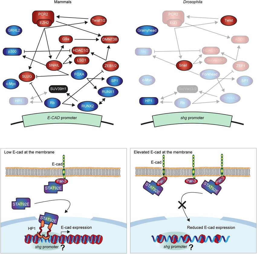
Ramirez Moreno et al.                                                                                             E-Cadherin Transcriptional Regulation

   The strength of adhesion correlates with the number of E-cad              repressive H3K27me3 mark, deactivating E-cad transcription
molecules on cell surfaces (Chu et al., 2004). Consequently, the             (Viré et al., 2006; Cao et al., 2008; Fujii and Ochiai, 2008). The
regulation of E-cad surface levels in epithelia is instrumental              histone methyltransferase G9a places the repressive H3K9me2/3
in many processes, and even mild changes in E-cad levels                     marks on the E-cad promoter (Dong et al., 2012). Histones
profoundly affect many processes such as cell rearrangements,                are subjected to other post-translational modifications that
proliferation, and tissue architecture (Ciesiolka et al., 2004;              contribute to E-cad silencing, such as the removal of the
Lecuit and Yap, 2015; Mohan et al., 2018; Greig and Bulgakova,               H3K4me2 mark by the lysine-specific demethylase 1 (LSD1)
2020). Multiple mechanisms regulate E-cad surface levels                     (Lin et al., 2010), or H3 and H4 deacetylation by the histone
including intracellular trafficking, transcriptional regulation,             deacetylases 1 and 2 (HDAC1/2) (Peinado et al., 2004a). Histone
post-translational modifications, and protein degradation                    phosphorylation and ubiquitination were also suggested to alter
(Palacios et al., 2005; Bertocchi et al., 2012; Serrano-Gomez                transcription of the mammalian E-cad (Surapaneni et al., 2020;
et al., 2016; Brüser and Bogdan, 2017; Cai et al., 2018). In this            Wang et al., 2020). While there is only a low level of DNA
perspective, we discuss transcriptional regulation, as the decrease          methylation in Drosophila (Lyko et al., 2000) other epigenetic
in E-cad protein levels often correlates with reduced mRNA                   mechanisms are conserved (Figure 1B) and might, similarly to
abundance (Bringuier et al., 1999).                                          Snail and Twist, regulate E-cad transcription in invertebrates.
                                                                                 These mechanisms of E-cad silencing closely interact with
                                                                             each other (Figure 1A). For example, SNAIL recruits the
TRANSCRIPTIONAL REPRESSION                                                   repressive complex containing LSD1 and HDAC1/2 (Peinado
                                                                             et al., 2004a; Lin et al., 2014), which may facilitate the subsequent
The best-studied regulation of E-cad transcription in mammalian              recruitment of PRC2 (Ai et al., 2017; Jin et al., 2017, p. 1). SNAIL
cells is silencing by transcription factors (TFs) including SNAIL,           also recruits G9a to the E-cad promoter, whereas both G9a and
SLUG (also known as SNAI1 and SNAI2 in mammals), ZEB1/2,                     PRC2 interact with the DNA methyltransferases DNMT1/3A/3B,
and Twist1/2 (Figure 1A), all of which directly bind conserved               and thus can promote the CpG methylation (Viré et al., 2006;
E-boxes (CANNTG sequences) in the E-cad promoter (Giroldi                    Dong et al., 2012; Lin et al., 2014). Twist1 can also promote
et al., 1997; Comijn et al., 2001; Bolós et al., 2003; Wong                  the recruitment of PRC2 (Cakouros et al., 2012; Malouf et al.,
et al., 2014; Vergara et al., 2016; Russell and Pranjol, 2018).              2013), whereas ZEB1 recruits histone deacetylases, HDAC1/2,
All events of epithelial-to-mesenchymal transition (EMT) during              and possibly DNMT3B (Aghdassi et al., 2012; Zhang et al., 2019).
development involve at least one of these TFs, and they were                 Concurrently, EZH2 facilitates the expression of SNAIL and
all linked to the tumour progression (Gasparotto et al., 2011;               SLUG through an unknown mechanism (Zhang et al., 2017),
Muenst et al., 2013; da Silva et al., 2014; Vergara et al., 2016; Yang       and increases Twist expression by suppressing the miR-361
et al., 2020). Despite the overall similarity in the action of these         (Ihira et al., 2017). Through these interactions, we suggest that
TFs, their different affinities and variable expression allow for a          individual mechanisms act together to ensure the robust silencing
dynamic but tightly regulated expression of E-cad (Bolós et al.,             of the E-cad.
2003; Eger et al., 2005; Schmidt et al., 2005; Vesuna et al., 2008;
Mazda et al., 2011). Regulation of E-cad transcription by these
TFs is extremely conserved across evolution. While Drosophila                TRANSCRIPTIONAL ACTIVATION
and mammalian E-cad differ in their extracellular domains
and are products of independent evolution from a common                      A fluctuation in the activity of a repressive mechanism, for
N-cadherin-like ancestor, transcription of the Drosophila E-cad              example, due to random changes in fundamentally stochastic
(encoded by the shotgun gene) is also inhibited by the Snail and             expression of regulatory genes, could lead to the E-cad
Twist TFs (Figure 1B; Oda et al., 1998, 2005; Oda and Takeichi,              loss (Raj and van Oudenaarden, 2008; Wendt et al., 2011).
2011; Nishiguchi et al., 2016). This could be either due to this             Protein turnover may counteract the resulting short-term
regulation already being present in the ancestor or a result of              E-cad silencing, whereby the balance between recycling and
the parallel evolution due to the same functional requirements.              degradation of endocytosed molecules quickly modulates E-cad
In Drosophila, while Snail represses E-cad during gastrulation in            surface levels (Bryant and Stow, 2004; Huang et al., 2011;
the embryo, it does not inhibit it in the adult midguts (Oda et al.,         Bulgakova et al., 2013; Erami et al., 2015; Bulgakova and Brown,
1998; Campbell et al., 2019), potentially due to the combinatorial           2016). However, this might be insufficient to protect tissue
action of other regulators (Figure 1B).                                      integrity from a transient but excessive drop in the E-cad levels.
    Multiple epigenetic mechanisms also inhibit E-cad expression             Therefore, the robust E-cad expression requires mechanisms for
in mammalian cells. Hypermethylation of the large CpG island                 transcriptional activation.
around the promoter is frequently linked with loss of E-cad                     The traditional view is that E-cad expression is activated by
expression (Yoshiura et al., 1995; van Roy and Berx, 2008).                  constitutive factors in mammalian cells, which are overcome
This DNA hypermethylation is often accompanied by histone                    by repressors (Peinado et al., 2004b). This view was contested
modifications associated with inactive chromatin (Serrano-                   upon discovery of the binding of the TF FOXA/HNF3
Gomez et al., 2016). Most notably, polycomb repressive complex               (Forkhead in Drosophila) to the E-cad promoter, increasing
2 (PRC2) is recruited to the E-cad promoter where its key                    its expression and driving re-epithelization of breast cancer
component, enhancer of zeste homolog 2 (EZH2), places the                    cells (Liu et al., 2005). This activation system interacts with

Frontiers in Cell and Developmental Biology | www.frontiersin.org        2                                       June 2021 | Volume 9 | Article 701175
Ramirez Moreno et al. E-Cadherin Transcriptional Regulation FIGURE 1 | Molecular mechanisms regulating E-cadherin gene expression. (A,B) Summary of the best known negative and positive regulators of E-cad in mammals (A) and Drosophila (B). The expression of E-cad gene (shotgun, shg, in Drosophila) is controlled by transcription factors (ellipses), which inhibit (red) or promote it (green) and interact with the E-cad promoter with varying affinities. Several further proteins modify chromatin at the promoter (squircles) silencing the E-cad expression (red) or increasing it (green). These proteins cooperate, compete, and regulate each other, establishing the intricated network that fine-tune the expression of E-cad. In panel (B) the non-conserved proteins were removed while conserved maintained their relative positions but their names were replaced with those of the Drosophila orthologs. The regulatory roles of transparentized proteins in panels (A,B) were not demonstrated in the respective systems. (C) The proposed model of E-cad modulation of its expression. A subpool of STAT92E translocates into the nucleus independently of the canonical JAK/STAT signalling, where it interacts with heterochromatin protein 1 (HP1). Within euchromatin, HP1 localises at specific loci corresponding to gene promoters, including that of the shg gene. Concurrently, STAT92E is recruited to the cell surface by E-cad and its binding partner Par-3. Therefore, elevated levels of E-cad outcompete STAT92E from the nucleus inhibiting its function in heterochromatin formation and promoter regulation. As the result, elevated E-cad at the cell surface reduces shg expression, ultimately restoring its levels. It is unclear whether this is accompanied by a change in chromatin organisation around shg promoter, indicated by ‘?.’ other E-cad regulatory factors and epigenetic machinery. FOXA the two p300-binding sites in the human E-cad promoter (Liu suppresses the expression of SLUG, combining E-cad activation et al., 2005). This interaction suggests the potential role of with the release of repression (Anzai et al., 2017). FOXA also histone modifications in E-cad transcriptional activation due to interacts with p300, which promotes E-cad expression with the intrinsic histone acetyltransferase (HAT) activity of p300 Frontiers in Cell and Developmental Biology | www.frontiersin.org 3 June 2021 | Volume 9 | Article 701175
Ramirez Moreno et al. E-Cadherin Transcriptional Regulation
(Ogryzko et al., 1996; Chan and Thangue, 2001). While p300 Wnt-activated transcription (Daugherty et al., 2014). Similarly,
can acetylate all histones, it preferentially modifies H3 and p120-catenin antagonises β-catenin-mediated transcriptional
H4 and competes for binding with HDAC1 (Ogryzko et al., activation by interacting with the repressive TF Kaiso (Daniel and
1996; Li et al., 2014), making p300 an excellent candidate to Reynolds, 1999; Kourtidis et al., 2013; Schackmann et al., 2013).
counteract the histone deacetylation at the E-cad promoter Curiously, β-catenin expression is inhibited by the Kaiso–p120-
by HDAC1/2. Finally, FOXA drives E-cad downregulation by catenin complex (Liu et al., 2014), highlighting the reciprocal
interacting with Sp1 and RUNX1/AML1 (Liu et al., 2005; D’Costa interactions between transcriptional mechanisms and adhesion
et al., 2012). Of particular interest for the data presented molecules. In Drosophila, although Kaiso is not present, p120-
below is that in mammalian cells RUNX1/AML1 forms a catenin is nevertheless involved in transcriptional regulation
complex with SUV39H1 [Su(var)3–9 in Drosophila], whereas (Stefanatos et al., 2013). As catenins are recruited by E-cad,
methylation of the H3K9 by SUV39H1 creates a binding site their availability for either performing nuclear functions or
for the heterochromatin protein 1 (HP1) (Lachner et al., 2001; sequestering other TFs outside the nucleus is dependent on
Chakraborty et al., 2003). E-cad levels. Indeed, E-cad levels impact on transcription of
Retinoblastoma (Rb) and c-Myc also specifically activate multiple genes involved in a wide range of processes (Soncin
the expression of E-cad promoter through an interaction with et al., 2011). Furthermore, β-catenin recruits the p300 protein,
the TF AP-2, thus maintaining the epithelial phenotype in which promotes E-cad expression (Mosimann et al., 2009).
mammalian cells (Batsché et al., 1998). Rb interacts with the This raises the questions of whether E-cad contributes to own
TF RUNX2, which acts redundantly with closely related RUNX1 gene expression.
in chondrocyte differentiation (Kimura et al., 2010; Gündüz
et al., 2012; Komori, 2015). This, alongside RUNX2 and RUNX1
cooperation in tumour cells and RUNX1 recruitment to the A FEEDBACK LOOP REGULATING
E-cad promoter, raises a possibility of similar recruitment of E-CAD EXPRESSION THROUGH PAR-3,
RUNX2, and therefore Rb, to the E-cad promoter (Fowler et al., HP1, AND STAT92E
2006). Concurrently, Rb depletion induces expression of SLUG
and ZEB1, suggesting an indirect effect on E-cad expression The polarity determinant Par-3 (Bazooka in Drosophila) is an
(Arima et al., 2008). Additionally to these mechanisms, Rb evolutionarily conserved protein recruited to E-cad adhesion in
is likely to modulate epigenetic mechanisms controlling E-cad both mammals and Drosophila (Harris and Peifer, 2005; Wei
expression, as it targets the HP1 to gene promoters (Nielsen et al., 2005; Iden et al., 2006; Wang et al., 2009; Bulgakova et al.,
et al., 2001). This complexity of E-cad transcriptional regulation 2013; Xue et al., 2013). Par-3 has six protein–protein interaction
is further amplified by the involvement of post-transcriptional domains, making it a versatile scaffold for protein recruitment
mechanisms. For example, c-Myc activates E-cad expression, but (McKinley et al., 2012). In Drosophila, one of the proteins
its substantial overexpression in breast cancer cells leads to post- recruited by Par-3 is the only fly member of the signal transducer
transcriptional repression of E-cad (Batsché et al., 1998; Cowling and activator of transcription (STAT) protein family, STAT92E
and Cole, 2007). While roles of Rb, c-Myc, and Forkhead in the (Hou et al., 1996; Yan et al., 1996). This recruitment of STAT92E
expression of Drosophila E-cad are not known, another TF – is necessary for the efficient Janus kinase (JAK)/STAT signalling
Grhl2 (Grainyhead in Drosophila) – activates E-cad expression in Drosophila epithelial cells (Sotillos et al., 2008). In human
in both mammalian and invertebrate cells. In the former, intron- and mouse cells, the loss of Par-3 activates STAT3 signalling,
bound Grhl2 promotes E-cad transcription through chromatin although it is not known if the loss of STAT3 recruitment to E-cad
looping, playing an essential role in determining an epithelial adhesion sites by Par-3 contributes to this activation (McCaffrey
phenotype (Werth et al., 2010; Xiang et al., 2012). Similarly in et al., 2012; Guyer and Macara, 2015). However, this is very
flies, Grainyhead has four binding sites in the shotgun locus likely, as the loss of E-cad adhesion also leads to STAT3 activation
and its overexpression promotes E-cad transcription, as well as (del Valle et al., 2013).
that of multiple other genes involved in epithelial maturation The canonical signalling pathway comprises STAT
(Yao et al., 2017). translocation into the nucleus after its phosphorylation by
The above mechanisms overlook the potential of the the ligand-activated JAK. However, there is growing evidence
cell adhesion molecules (CAMs) in triggering changes in of non-canonical signalling in both Drosophila and mammals,
gene expression (Robinson and Moberg, 2011; Dasgupta and whereby non-phosphorylated STAT stabilises heterochromatin
McCollum, 2019; Hannezo and Heisenberg, 2019). For example, by binding HP1 (Li, 2008; Hixson et al., 2019). HP1 is an
β-catenin is a TF activated by canonical Wnt signalling (Shang evolutionarily conserved non-histone chromatin protein whose
et al., 2017) and induces expression of SLUG in human carcinoma best-characterised role is the formation and propagation of
cells (Conacci-Sorrell et al., 2003), meaning that the release heterochromatin (Lomberk et al., 2006; Dialynas et al., 2008; Xu
of β-catenin from junctions due to E-cad downregulation can et al., 2014). HP1 is recruited to H3K9me2/3 through SUV39H1,
reinforce this downregulation. Via α-catenin, E-cad regulates and then itself recruits more SUV39H1 molecules, propagating
the Hippo pathway by sequestering the YAP protein outside H3K9 methylation (Lomberk et al., 2006). This is accompanied
the nucleus, thus explaining the antiproliferative properties by DNA methylation in mammalian cells, as both HP1 and
of E-cad (Kim et al., 2011; Hannezo and Heisenberg, 2019; SUV39H1 bind DNMTs (Fuks et al., 2003). HP1 was also linked
Sarpal et al., 2019). α-catenin counteracts β-catenin by inhibiting to other functions including transcriptional regulation, where
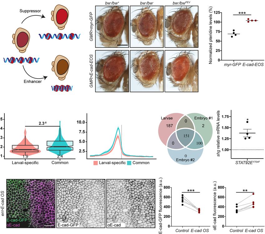
Frontiers in Cell and Developmental Biology | www.frontiersin.org 4 June 2021 | Volume 9 | Article 701175Ramirez Moreno et al. E-Cadherin Transcriptional Regulation HP1 usually represses transcription by facilitating methylation macromolecule metabolic processes (Supplementary Figure 3, of H3K9. However, HP1 also localises at specific loci within terms 7–11); and mitotic cell cycle and cell cycle regulation euchromatin (Dialynas et al., 2008), and there is growing (Supplementary Figure 3, terms 2–6). In contrast, the larval- evidence of its association with transcriptionally active regions specific peaks, which map to promoters of 178 genes, are from C. elegans to Drosophila and humans (Lu et al., 2000; enriched for terms related to epithelial tissue: morphogenesis of Piacentini et al., 2003; Mateescu et al., 2008; McMurchy et al., epithelium, actin filament organisation, epithelium development 2017). In Drosophila, the active euchromatic regions with HP1 and imaginal disc development (Supplementary Figure 3, terms recruitment include developmentally-regulated and heat-shock 19–24). The shotgun promoter overlapped a larval-specific HP1 genes, whose activity correlates with HP1 dosage (Piacentini peak with a signal value of 9.83 (207th promoter rank). This et al., 2003). Non-phosphorylated STAT92E and HP1 co-localise peak is consistent with two potential mechanisms for HP1 at euchromatic regions of Drosophila polytene chromosomes recruitment – via Su(var)3–9 or one of the two fly Rb-family (Shi et al., 2008). HP1 disperses from chromosomes following proteins. Therefore, the modulation of HP1 function downstream hyperactivation of the canonical signalling, suggesting an of E-cad and non-canonical STAT signalling might alter the intricate cross-talk between the two modes of signalling, likely E-cad expression. through the availability of non-phosphorylated STAT92E Indeed, overexpression of the non-phosphorylatable STAT92E (Xu et al., 2014). (STAT92EY704F ), which acts only in the non-canonical signalling As STAT92E promotes heterochromatin gene silencing in a (Karsten et al., 2006; Xu et al., 2014), increased expression dose-dependent manner (Shi et al., 2008), we hypothesised that of the shotgun gene (Figure 2G). Such an increase is an increase of E-cad levels would lead to STAT92E sequestration consistent with the model whereby an unprogrammed drop in at adhesion sites, making it unavailable to bind HP1 and E-cad levels releases STAT92E, which leads to a consequent promote heterochromatin formation alongside other functions increase of E-cad expression and thus restores E-cad levels (Figure 1C). We examined the effects of E-cad overexpression on (Figure 1C). We observed that overexpression of E-cad from heterochromatic gene silencing monitored using position-effect a heterologous UAS promoter leads to downregulation of the variegation (PEV). In this assay, a gene is translocated into a protein expressed from the endogenous locus (Figures 2H– peri-heterochromatic region where its expression is silenced in J). This might be accompanied by simultaneous upregulation a stochastic pattern (Figure 2A). This silencing depends on the of the canonical JAK/STAT signalling as observed for STAT3 spreading of heterochromatin: if a protein normally promotes activation in some human cancers (Loh et al., 2019). We heterochromatin formation, its loss would reduce the amount of postulate that other genes with larval-specific HP1 peaks heterochromatin and suppress the variegation (=loss of silencing behave similarly to E-cad. Then, the activation of non- of the translocated gene, Figure 2A; Elgin and Reuter, 2013). canonical STAT signalling following a stochastic drop in We overexpressed E-cad in the Drosophila eye using the UAS– E-cad levels would reinforce the epithelial nature of the Gal4 system with GMR::Gal4, which expresses Gal4 in all retinal cells as many of these genes are involved in epithelial cells (Hay et al., 1997). We used the variegating allele of the morphogenesis. brown gene (bwPEV ), necessary to produce the red pigment pteridine. Overexpression of E-cad increased the normalised eye pigmentation with pteridine to 104% from 69% in the control OUTLOOK/PERSPECTIVE (Figures 2B,C and Supplementary Figure 1). Therefore, E-cad suppresses PEV in the eye, supporting that elevated E-cad inhibits E-cad levels at the cell surface play an important role in heterochromatin formation. determining cellular properties: cell shape, response to signalling, Next, we sought to determine what genes might be regulated cell division, and neighbour exchange to name just a few through our hypothetical mechanism and examined the HP1 (Lecuit and Lenne, 2007; Rübsam et al., 2017; Mendonsa localisation using publicly available ChIP-seq datasets generated et al., 2018). Not surprisingly, E-cad expression is subjected using third instar larvae (Riddle et al., 2011), which include to complex regulation by multiple interconnected mechanisms. epithelial monolayers of imaginal discs. There were distinct However, to respond to the cell’s needs and modulate E-cad peaks of HP1 overlapping with promoters of 456 genes outside levels accordingly, information about the E-cad levels at the of heterochromatin clusters and non-mappable (non-unique, surface must be transferred to the nucleus. Here, we propose repeatable) regions (Figures 2D,E and Supplementary Figure 2). one such mechanism, whereby surface E-cad feeds back to We compared the larval HP1 peaks with two independent the nucleus through competition for STAT92E binding. We replicates from an earlier developmental stage – embryos at 16– hypothesise that this feedback stabilises E-cad transcription 24 h of development (modENCODE Consortium, Roy et al., enabling robust and resilient cell–cell adhesion. Importantly, 2010). Roughly half of the HP1 peaks in the larval dataset this provides the cell with a tumour-suppressive mechanism, as (155 peaks mapping to 151 loci) were common to all three heterochromatin formation, which is likely to follow a drop in datasets, while another half (187 peaks/loci) were larval-specific E-cad levels, is linked with cellular senescence (Narita et al., (Figure 2F). The common peaks (potentially, developmentally 2003). This contrasts another feedback mechanism whereby conserved) mostly mapped to bi-directional promoters, so that the release of β-catenin from adhesion sites leads to SLUG 155 peaks mapped to a total of 280 genes. These genes are expression and reinforces E-cad silencing in human cells. enriched for housekeeping terms: regulation of primary and Therefore, depending on the context E-cad expression may Frontiers in Cell and Developmental Biology | www.frontiersin.org 5 June 2021 | Volume 9 | Article 701175
Ramirez Moreno et al.                                                                                                               E-Cadherin Transcriptional Regulation

be stabilised or silenced through feedback interactions. It is                          DATA AVAILABILITY STATEMENT
tempting to speculate that further mechanisms of information
transfer from the cell surface to the nucleus exist – not unlike                        The datasets presented in this study can be found in
the multiple and intertwined mechanisms for regulation of                               online repositories. The names of the repository/repositories
E-cad transcription. A comprehensive understanding of E-cad                             and accession number(s) can be found below: http://data.
transcriptional regulation requires the discovery and in-depth                          modencode.org/, 3187; http://data.modencode.org/, 3188; http:
analysis of such mechanisms.                                                            //data.modencode.org/, 3391; http://data.modencode.org/, 3392.

  FIGURE 2 | The crosstalk between E-cadherin, STAT92E and HP1. (A) A diagram of position effect variegation (PEV) in relation to chromatin compaction. (B,C)
  E-cad overexpression suppresses PEV: examples (B) and quantification (C). Scale bar – 0.2 mm, ***p = 0.0001 (unpaired t-test). (D) Overlapping box-and-violin
  plots showing the quantification of rBEADS normalised HP1 signal on larval-specific (red) and common (blue) promoters. The p-value above – the significance
  calculated using U-test. The value below the boxplots – the mean of the signal. (E) The distribution of the rBEADS normalised HP1 signal over transcription start sites
  (TSS) loci of larval-specific (red) and common (blue) genes. The vertical grey line represents the location of TSS, the plots span 1 kb upstream and downstream from
  annotated TSS. (F) A Venn diagram showing the overlaps between peak loci in the larval dataset and two independent embryonic (16–24 h) replicates. All three peak
  sets were filtered from peaks overlapping centromeric heterochromatin clusters and non-mappable, repetitive regions, defined based on GEM mappability analyses.
  modENCODE peak calls have been lifted from dm3 to dm6 genome assembly to match the analyses of larval HP1 peaks. The IDR method with 0.05 p-vale
  threshold was used to collapse the replicates to a common, high confidence peak set. (G) shotgun expression following overexpression of non-phosphorylatable
  STAT92EY704F measured using RT-qPCR and normalised to the expression of house-keeping gene RpL32. Four biological replicates are shown as distinct dots with
  three technical replicates each. *p = 0.02 (one-sample t-test comparing to 1). (H–J) E-cad-GFP expressed from the endogenous promoter is downregulated
  following E-cad overexpression (E-cad OS) from a heterologous UAS promoter. A representative image (H) shows endogenously expressed E-cad-GFP visualised
  with native GFP fluorescence (green, left; grey, middle) and total E-cad visualised with antibody staining (magenta, left; grey, right), scale bar – 10 µm. Levels of
  endogenously expressed E-cad-GFP are quantified in panel (I) and total E-cad levels in panel (J). **p = 0.0018 and ***p = 0.0003 (paired t-test).

Frontiers in Cell and Developmental Biology | www.frontiersin.org                   6                                               June 2021 | Volume 9 | Article 701175
Ramirez Moreno et al.                                                                                                                      E-Cadherin Transcriptional Regulation

AUTHOR CONTRIBUTIONS                                                                         ACKNOWLEDGMENTS
NAB and MRM performed the experiments and wrote the                                          We thank Martin Zeidler for STAT92EY704F flies and the
manuscript. NAB and PAS did the bioinformatics analyses.                                     University of Sheffield’s Wolfson Light Microscopy and
All authors contributed to the article and approved the                                      Drosophila Facillities for their technical support.
submitted version.

                                                                                             SUPPLEMENTARY MATERIAL
FUNDING
                                                                                             The Supplementary Material for this article can be found
This work was supported by a grant from the UKRI BBSRC                                       online at: https://www.frontiersin.org/articles/10.3389/fcell.2021.
(BB/P007503/1) to NAB.                                                                       701175/full#supplementary-material

REFERENCES                                                                                      along the Ink4A/Arf locus in mesenchymal stem cells. Mol. Cell. Biol. 32,
                                                                                                1433–1441. doi: 10.1128/mcb.06315-11
Aghdassi, A., Sendler, M., Guenther, A., Mayerle, J., Behn, C.-O., Heidecke, C.-             Campbell, K., Rossi, F., Adams, J., Pitsidianaki, I., Barriga, F. M., Garcia-Gerique,
   D., et al. (2012). Recruitment of histone deacetylases HDAC1 and HDAC2                       L., et al. (2019). Collective cell migration and metastases induced by an
   by the transcriptional repressor ZEB1 downregulates E-cadherin expression in                 epithelial-to-mesenchymal transition in Drosophila intestinal tumors. Nat.
   pancreatic cancer. Gut 61, 439–448. doi: 10.1136/gutjnl-2011-300060                          Commun. 10:2311.
Ai, S., Peng, Y., Li, C., Gu, F., Yu, X., Yue, Y., et al. (2017). EED orchestration of       Cao, Q., Yu, J., Dhanasekaran, S. M., Kim, J. H., Mani, R.-S., Tomlins,
   heart maturation through interaction with HDACs is H3K27me3-independent.                     S. A., et al. (2008). Repression of E-cadherin by the polycomb group
   eLife 6:e24570.                                                                              protein EZH2 in cancer. Oncogene 27, 7274–7284. doi: 10.1038/onc.20
Anzai, E., Hirata, K., Shibazaki, M., Yamada, C., Morii, M., Honda, T., et al. (2017).          08.333
   FOXA1 induces E-Cadherin expression at the protein level via suppression of               Chakraborty, S., Sinha, K. K., Senyuk, V., and Nucifora, G. (2003). SUV39H1
   slug in epithelial breast cancer cells. Biol. Pharm. Bull. 40, 1483–1489. doi:               interacts with AML1 and abrogates AML1 transactivity. AML1 is methylated
   10.1248/bpb.b17-00307                                                                        in vivo. Oncogene 22, 5229–5237. doi: 10.1038/sj.onc.1206600
Arima, Y., Inoue, Y., Shibata, T., Hayashi, H., Nagano, O., Saya, H., et al. (2008).         Chan, H. M., and Thangue, N. B. L. (2001). p300/CBP proteins: HATs for
   Rb depletion results in deregulation of E-cadherin and induction of cellular                 transcriptional bridges and scaffolds. J. Cell Sci. 114, 2363–2373. doi: 10.1242/
   phenotypic changes that are characteristic of the epithelial-to-mesenchymal                  jcs.114.13.2363
   transition. Cancer Res. 68, 5104–5112. doi: 10.1158/0008-5472.can-07-5680                 Chu, Y.-S., Thomas, W. A., Eder, O., Pincet, F., Perez, E., Thiery, J. P., et al.
Batsché, E., Muchardt, C., Behrens, J., Hurst, H. C., and Crémisi, C. (1998). RB                (2004). Force measurements in E-cadherin–mediated cell doublets reveal rapid
   and c-Myc activate expression of the E-cadherin gene in epithelial cells through             adhesion strengthened by actin cytoskeleton remodeling through Rac and
   interaction with transcription factor AP-2. Mol. Cell. Biol. 18, 3647–3658. doi:             Cdc42. J. Cell Biol. 167, 1183–1194. doi: 10.1083/jcb.200403043
   10.1128/mcb.18.7.3647                                                                     Ciesiolka, M., Delvaeye, M., Imschoot, G. V., Verschuere, V., McCrea, P., Roy, F.,
Bertocchi, C., Vaman Rao, M., and Zaidel-Bar, R. (2012). Regulation of                          et al. (2004). p120 catenin is required for morphogenetic movements involved
   adherens junction dynamics by phosphorylation switches. J. Signal. Transduct.                in the formation of the eyes and the craniofacial skeleton in Xenopus. J. Cell Sci.
   2012:125295.                                                                                 117, 4325–4339. doi: 10.1242/jcs.01298
Bolós, V., Peinado, H., Pérez-Moreno, M. A., Fraga, M. F., Esteller, M., and Cano,           Comijn, J., Berx, G., Vermassen, P., Verschueren, K., van Grunsven, L., Bruyneel,
   A. (2003). The transcription factor Slug represses E-cadherin expression and                 E., et al. (2001). The two-handed e box binding zinc finger protein SIP1
   induces epithelial to mesenchymal transitions: a comparison with Snail and E47               downregulates E-Cadherin and induces invasion. Mol. Cell 7, 1267–1278. doi:
   repressors. J. Cell Sci. 116, 499–511. doi: 10.1242/jcs.00224                                10.1016/s1097-2765(01)00260-x
Bringuier, P. P., Giroldi, L. A., Umbas, R., Shimazui, T., and Schalken, J. A.               Conacci-Sorrell, M., Simcha, I., Ben-Yedidia, T., Blechman, J., Savagner, P., and
   (1999). Mechanisms associated with abnormal E-cadherin immunoreactivity in                   Ben-Ze’ev, A. (2003). Autoregulation of E-cadherin expression by cadherin-
   human bladder tumors. Int. J. Cancer 83, 591–595. doi: 10.1002/(sici)1097-                   cadherin interactions: the roles of beta-catenin signaling. Slug, and MAPK.
   0215(19991126)83:53.0.co;2-6                                                  J. Cell Biol. 163, 847–857. doi: 10.1083/jcb.200308162
Brüser, L., and Bogdan, S. (2017). Adherens junctions on the move—membrane                   Cowling, V. H., and Cole, M. D. (2007). E-cadherin repression contributes to
   trafficking of E-Cadherin. Cold Spring Harb. Perspect. Biol. 9:a029140. doi:                 c-Myc-induced epithelial cell transformation. Oncogene 26, 3582–3586. doi:
   10.1101/cshperspect.a029140                                                                  10.1038/sj.onc.1210132
Bryant, D. M., and Stow, J. L. (2004). The ins and outs of E-cadherin trafficking.           D’Costa, Z. J., Jolly, C., Androphy, E. J., Mercer, A., Matthews, C. M., and
   Trends Cell Biol. 14, 427–434. doi: 10.1016/j.tcb.2004.07.007                                Hibma, M. H. (2012). Transcriptional repression of E-Cadherin by human
Buckley, C. D., Rainger, G. E., Bradfield, P. F., Nash, G. B., and Simmons, D. L.               papillomavirus Type 16 E6. PLoS One 7:e48954. doi: 10.1371/journal.pone.
   (1998). Cell adhesion: more than just glue (Review). Mol. Membr. Biol. 15,                   0048954
   167–176. doi: 10.3109/09687689709044318                                                   da Silva, S. D., Alaoui-Jamali, M. A., Soares, F. A., Carraro, D. M., Brentani, H. P.,
Bulgakova, N. A., and Brown, N. H. (2016). Drosophila p120-catenin is crucial                   Hier, M., et al. (2014). TWIST1 is a molecular marker for a poor prognosis in
   for endocytosis of the dynamic E-cadherin-Bazooka complex. J. Cell Sci. 129,                 oral cancer and represents a potential therapeutic target. Cancer 120, 352–362.
   477–482.                                                                                     doi: 10.1002/cncr.28404
Bulgakova, N. A., Grigoriev, I., Yap, A. S., Akhmanova, A., and Brown, N. H. (2013).         Daniel, J. M., and Reynolds, A. B. (1999). The Catenin p120ctn interacts with Kaiso,
   Dynamic microtubules produce an asymmetric E-cadherin-Bazooka complex                        a Novel BTB/POZ domain zinc finger transcription factor. Mol. Cell Biol. 19,
   to maintain segment boundaries. J. Cell Biol. 201, 887–901. doi: 10.1083/jcb.                3614–3623. doi: 10.1128/mcb.19.5.3614
   201211159                                                                                 Dasgupta, I., and McCollum, D. (2019). Control of cellular responses to mechanical
Cai, J., Culley, M. K., Zhao, Y., and Zhao, J. (2018). The role of ubiquitination               cues through YAP/TAZ regulation. J. Biol. Chem. 294, 17693–17706. doi: 10.
   and deubiquitination in the regulation of cell junctions. Protein Cell 9, 754–769.           1074/jbc.rev119.007963
   doi: 10.1007/s13238-017-0486-3                                                            Daugherty, R. L., Serebryannyy, L., Yemelyanov, A., Flozak, A. S., Yu, H.-J., Kosak,
Cakouros, D., Isenmann, S., Cooper, L., Zannettino, A., Anderson, P., Glackin, C.,              S. T., et al. (2014). α-Catenin is an inhibitor of transcription. Proc. Natl. Acad.
   et al. (2012). Twist-1 Induces Ezh2 recruitment regulating histone methylation               Sci. U. S. A. 111, 5260–5265.

Frontiers in Cell and Developmental Biology | www.frontiersin.org                        7                                                June 2021 | Volume 9 | Article 701175
Ramirez Moreno et al. E-Cadherin Transcriptional Regulation del Valle, I., Rudloff, S., Carles, A., Li, Y., Liszewska, E., Vogt, R., et al. (2013). the brain’s major ion channels and neurotransmitter receptors. BMC Genom. E-cadherin is required for the proper activation of the Lifr/Gp130 signaling 20:677. pathway in mouse embryonic stem cells. Development 140, 1684–1692. doi: Hou, X. S., Melnick, M. B., and Perrimon, N. (1996). Marelle acts downstream 10.1242/dev.088690 of the Drosophila HOP/JAK kinase and encodes a protein similar to the Dialynas, G. K., Vitalini, M. W., and Wallrath, L. L. (2008). Linking mammalian STATs. Cell 84, 411–419. doi: 10.1016/s0092-8674(00)81286-6 heterochromatin protein 1 (HP1) to cancer progression. Mutat. Res. 647, 13–20. Huang, J., Huang, L., Chen, Y.-J., Austin, E., Devor, C. E., Roegiers, F., et al. (2011). doi: 10.1016/j.mrfmmm.2008.09.007 Differential regulation of adherens junction dynamics during apical–basal Dong, C., Wu, Y., Yao, J., Wang, Y., Yu, Y., Rychahou, P. G., et al. (2012). G9a polarization. J. Cell Sci. 124, 4001–4013. doi: 10.1242/jcs.086694 interacts with Snail and is critical for Snail-mediated E-cadherin repression in Iden, S., Rehder, D., August, B., Suzuki, A., Wolburg-Buchholz, K., Wolburg, H., human breast cancer. J. Clin. Invest. 122, 1469–1486. doi: 10.1172/jci57349 et al. (2006). A distinct PAR complex associates physically with VE-cadherin in Eger, A., Aigner, K., Sonderegger, S., Dampier, B., Oehler, S., Schreiber, M., et al. vertebrate endothelial cells. EMBO Rep. 7, 1239–1246. doi: 10.1038/sj.embor. (2005). DeltaEF1 is a transcriptional repressor of E-cadherin and regulates 7400819 epithelial plasticity in breast cancer cells. Oncogene 24, 2375–2385. doi: 10. Ihira, K., Dong, P., Xiong, Y., Watari, H., Konno, Y., Hanley, S. J., et al. (2017). 1038/sj.onc.1208429 EZH2 inhibition suppresses endometrial cancer progression via miR-361/Twist Elgin, S. C. R., and Reuter, G. (2013). Position-effect variegation, heterochromatin axis. Oncotarget 8, 13509–13520. doi: 10.18632/oncotarget.14586 formation, and gene silencing in Drosophila. Cold Spring Harb. Perspect. Biol. Janiszewska, M., Primi, M. C., and Izard, T. (2020). Cell adhesion in cancer: beyond 5:a017780. doi: 10.1101/cshperspect.a017780 the migration of single cells. J. Biol. Chem. 295, 2495–2505. doi: 10.1074/jbc. Erami, Z., Timpson, P., Yao, W., Zaidel-Bar, R., and Anderson, K. I. (2015). There rev119.007759 are four dynamically and functionally distinct populations of E-cadherin in cell Jin, Y., Huo, B., Fu, X., Hao, T., Zhang, Y., Guo, Y., et al. (2017). LSD1 collaborates junctions. Biol. Open 4, 1481–1489. doi: 10.1242/bio.014159 with EZH2 to regulate expression of interferon-stimulated genes. Biomed. Fowler, M., Borazanci, E., McGhee, L., Pylant, S. W., Williams, B. J., Glass, J., Pharmacother. 88, 728–737. doi: 10.1016/j.biopha.2017.01.055 et al. (2006). RUNX1 (AML-1) and RUNX2 (AML-3) cooperate with prostate- Karsten, P., Plischke, I., Perrimon, N., and Zeidler, M. P. (2006). Mutational derived Ets factor to activate transcription from the PSA upstream regulatory analysis reveals separable DNA binding and trans-activation of Drosophila region. J. Cell. Biochem. 97, 1–17. doi: 10.1002/jcb.20664 STAT92E. Cell. Signal. 18, 819–829. doi: 10.1016/j.cellsig.2005.07.006 Fujii, S., and Ochiai, A. (2008). Enhancer of zeste homolog 2 downregulates Kaszak, I., Witkowska-Piłaszewicz, O., Niewiadomska, Z., Dworecka-Kaszak, B., E-cadherin by mediating histone H3 methylation in gastric cancer cells. Cancer Ngosa Toka, F., and Jurka, P. (2020). Role of cadherins in cancer-a review. Int. Sci. 99, 738–746. doi: 10.1111/j.1349-7006.2008.00743.x J. Mol. Sci. 21:7624. doi: 10.3390/ijms21207624 Fuks, F., Hurd, P. J., Deplus, R., and Kouzarides, T. (2003). The DNA Kim, N.-G., Koh, E., Chen, X., and Gumbiner, B. M. (2011). E-cadherin methyltransferases associate with HP1 and the SUV39H1 histone mediates contact inhibition of proliferation through Hippo signaling-pathway methyltransferase. Nucleic Acids Res. 31, 2305–2312. doi: 10.1093/nar/gkg332 components. PNAS 108, 11930–11935. doi: 10.1073/pnas.1103345108 Gasparotto, D., Polesel, J., Marzotto, A., Colladel, R., Piccinin, S., Modena, P., Kimura, A., Inose, H., Yano, F., Fujita, K., Ikeda, T., Sato, S., et al. (2010). Runx1 and et al. (2011). Overexpression of TWIST2 correlates with poor prognosis in head Runx2 cooperate during sternal morphogenesis. Development 137, 1159–1167. and neck squamous cell carcinomas. Oncotarget 2, 1165–1175. doi: 10.18632/ doi: 10.1242/dev.045005 oncotarget.390 Komori, T. (2015). The functions of Runx family transcription factors and Cbfb in Giroldi, L. A., Bringuier, P.-P., de Weijert, M., Jansen, C., van Bokhoven, A., skeletal development. Oral Sci. Int. 12, 83–93. and Schalken, J. A. (1997). Role of E boxes in the repression of E-cadherin Kourtidis, A., Ngok, S. P., and Anastasiadis, P. Z. (2013). p120 catenin: an expression. Biochem. Biophys. Res. Commun. 241, 453–458. doi: 10.1006/bbrc. essential regulator of cadherin stability, adhesion-induced signaling, and cancer 1997.7831 progression. Prog. Mol. Biol. Transl. Sci. 116, 409–432. Greig, J., and Bulgakova, N. A. (2020). Interplay between actomyosin and Lachner, M., O’Carroll, D., Rea, S., Mechtler, K., and Jenuwein, T. (2001). E-cadherin dynamics regulates cell shape in the Drosophila embryonic Methylation of histone H3 lysine 9 creates a binding site for HP1 proteins. epidermis. J. Cell Sci. 133:jcs242321. Nature 410, 116–120. doi: 10.1038/35065132 Gumbiner, B. M. (1996). Cell adhesion: the molecular basis of tissue architecture Lecuit, T., and Lenne, P.-F. (2007). Cell surface mechanics and the control of cell and morphogenesis. Cell 84, 345–357. doi: 10.1016/s0092-8674(00)81279-9 shape, tissue patterns and morphogenesis. Nat. Rev. Mol. Cell Biol. 8, 633–644. Gündüz, V., Kong, E., Bryan, C. D., and Hinds, P. W. (2012). Loss of doi: 10.1038/nrm2222 the retinoblastoma tumor suppressor protein in murine calvaria facilitates Lecuit, T., and Yap, A. S. (2015). E-cadherin junctions as active mechanical immortalization of osteoblast-adipocyte bipotent progenitor cells characterized integrators in tissue dynamics. Nat. Cell Biol. 17, 533–539. doi: 10.1038/ by low expression of N-Cadherin. Mol. Cell Biol. 32, 2561–2569. doi: 10.1128/ ncb3136 mcb.06453-11 Li, W. X. (2008). Canonical and non-canonical JAK–STAT signaling. Trends Cell Guyer, R. A., and Macara, I. G. (2015). Loss of the polarity protein PAR3 activates Biol. 18, 545–551. doi: 10.1016/j.tcb.2008.08.008 STAT3 signaling via an atypical protein Kinase C (aPKC)/NF-κB/Interleukin- Li, X., Yang, H., Huang, S., and Qiu, Y. (2014). Histone deacetylase 1 and p300 6 (IL-6) Axis in mouse mammary cells. J. Biol. Chem. 290, 8457–8468. doi: can directly associate with chromatin and compete for binding in a mutually 10.1074/jbc.m114.621011 exclusive manner. PLoS One 9:e94523. doi: 10.1371/journal.pone.0094523 Hagios, C., Lochter, A., and Bissell, M. J. (1998). Tissue architecture: the ultimate Lin, T., Ponn, A., Hu, X., Law, B. K., and Lu, J. (2010). Requirement of the regulator of epithelial function? Philos. Trans. R Soc. Lond. B Biol. Sci. 353, histone demethylase LSD1 in snai1-mediated transcriptional repression during 857–870. doi: 10.1098/rstb.1998.0250 epithelial-mesenchymal transition. Oncogene 29, 4896–4904. doi: 10.1038/onc. Halbleib, J. M., and Nelson, W. J. (2006). Cadherins in development: cell adhesion, 2010.234 sorting, and tissue morphogenesis. Genes Dev. 20, 3199–3214. doi: 10.1101/gad. Lin, Y., Dong, C., and Zhou, B. P. (2014). Epigenetic regulation of EMT: the snail 1486806 story. Curr. Pharm. Des. 20, 1698–1705. doi: 10.2174/13816128113199990512 Hannezo, E., and Heisenberg, C.-P. (2019). Mechanochemical feedback loops in Liu, Y., Dong, Q.-Z., Wang, S., Xu, H.-T., Miao, Y., Wang, L., et al. (2014). development and disease. Cell 178, 12–25. doi: 10.1016/j.cell.2019.05.052 Kaiso interacts with p120-catenin to regulate β-catenin expression at the Harris, T. J. C., and Peifer, M. (2005). The positioning and segregation of apical cues transcriptional level. PLoS One 9:e87537. doi: 10.1371/journal.pone.0087537 during epithelial polarity establishment in Drosophila. J. Cell Biol. 170, 813–823. Liu, Y.-N., Lee, W.-W., Wang, C.-Y., Chao, T.-H., Chen, Y., and Chen, J. H. doi: 10.1083/jcb.200505127 (2005). Regulatory mechanisms controlling human E-cadherin gene expression. Hay, B. A., Maile, R., and Rubin, G. M. (1997). P element insertion-dependent gene Oncogene 24, 8277–8290. doi: 10.1038/sj.onc.1208991 activation in the Drosophila eye. PNAS 94, 5195–5200. doi: 10.1073/pnas.94.10. Loh, C.-Y., Chai, J. Y., Tang, T. F., Wong, W. F., Sethi, G., Shanmugam, M. K., et al. 5195 (2019). The E-Cadherin and N-Cadherin switch in epithelial-to-mesenchymal Hixson, K. M., Cogswell, M., Brooks-Kayal, A. R., and Russek, S. J. (2019). transition: signaling, therapeutic implications, and challenges. Cells 8:1118. doi: Evidence for a non-canonical JAK/STAT signaling pathway in the synthesis of 10.3390/cells8101118 Frontiers in Cell and Developmental Biology | www.frontiersin.org 8 June 2021 | Volume 9 | Article 701175
Ramirez Moreno et al.                                                                                                                          E-Cadherin Transcriptional Regulation

Lomberk, G., Wallrath, L., and Urrutia, R. (2006). The heterochromatin protein 1                Palacios, F., Tushir, J. S., Fujita, Y., and D’Souza-Schorey, C. (2005). Lysosomal
   family. Genome Biol. 7:228.                                                                     targeting of E-Cadherin: a unique mechanism for the down-regulation of cell-
Lu, B. Y., Emtage, P. C., Duyf, B. J., Hilliker, A. J., and Eissenberg, J. C. (2000).              cell adhesion during epithelial to mesenchymal transitions. Mol. Cell Biol. 25,
   Heterochromatin protein 1 is required for the normal expression of two                          389–402. doi: 10.1128/mcb.25.1.389-402.2005
   heterochromatin genes in Drosophila. Genetics 155, 699–708. doi: 10.1093/                    Paredes, J., Figueiredo, J., Albergaria, A., Oliveira, P., Carvalho, J., Ribeiro, A. S.,
   genetics/155.2.699                                                                              et al. (2012). Epithelial E- and P-cadherins: role and clinical significance in
Lyko, F., Ramsahoye, B. H., and Jaenisch, R. (2000). DNA methylation in                            cancer. Biochim. Biophys. Acta 1826, 297–311.
   Drosophila melanogaster. Nature 408, 538–540. doi: 10.1038/35046205                          Peinado, H., Ballestar, E., Esteller, M., and Cano, A. (2004a). Snail mediates
Malouf, G. G., Taube, J. H., Lu, Y., Roysarkar, T., Panjarian, S., Estecio, M. R., et al.          E-cadherin repression by the recruitment of the Sin3A/histone deacetylase 1
   (2013). Architecture of epigenetic reprogramming following Twist1-mediated                      (HDAC1)/HDAC2 complex. Mol. Cell Biol. 24, 306–319. doi: 10.1128/mcb.24.
   epithelial-mesenchymal transition. Genome Biol. 14:R144.                                        1.306-319.2004
Mateescu, B., Bourachot, B., Rachez, C., Ogryzko, V., and Muchardt, C. (2008).                  Peinado, H., Portillo, F., and Cano, A. (2004b). Transcriptional regulation of
   Regulation of an inducible promoter by an HP1beta-HP1gamma switch. EMBO                         cadherins during development and carcinogenesis. Int. J. Dev. Biol. 48, 365–375.
   Rep. 9, 267–272. doi: 10.1038/embor.2008.1                                                      doi: 10.1387/ijdb.041794hp
Mazda, M., Nishi, K., Naito, Y., and Ui-Tei, K. (2011). E-Cadherin is                           Perez, T. D., and Nelson, W. J. (2004). Cadherin adhesion: mechanisms and
   transcriptionally activated via suppression of ZEB1 transcriptional repressor by                molecular interactions. Handb. Exp. Pharmacol. 2004, 3–21. doi: 10.1007/978-
   small RNA-mediated gene silencing. PLoS One 6:e28688. doi: 10.1371/journal.                     3-540-68170-0_1
   pone.0028688                                                                                 Piacentini, L., Fanti, L., Berloco, M., Perrini, B., and Pimpinelli, S. (2003).
McCaffrey, L. M., Montalbano, J., Mihai, C., and Macara, I. G. (2012). Loss of the                 Heterochromatin protein 1 (HP1) is associated with induced gene expression in
   Par3 polarity protein promotes breast tumorigenesis and metastasis. Cancer Cell                 Drosophila euchromatin. J. Cell Biol. 161, 707–714. doi: 10.1083/jcb.200303012
   22, 601–614. doi: 10.1016/j.ccr.2012.10.003                                                  Raj, A., and van Oudenaarden, A. (2008). Stochastic gene expression and its
McKinley, R. F. A., Yu, C. G., and Harris, T. J. C. (2012). Assembly of Bazooka                    consequences. Cell 135, 216–226. doi: 10.1016/j.cell.2008.09.050
   polarity landmarks through a multifaceted membrane-association mechanism.                    Riddle, N. C., Minoda, A., Kharchenko, P. V., Alekseyenko, A. A., Schwartz, Y. B.,
   J. Cell Sci. 125, 1177–1190. doi: 10.1242/jcs.091884                                            Tolstorukov, M. Y., et al. (2011). Plasticity in patterns of histone modifications
McMurchy, A. N., Stempor, P., Gaarenstroom, T., Wysolmerski, B., Dong, Y.,                         and chromosomal proteins in Drosophila heterochromatin. Genome Res. 21,
   Aussianikava, D., et al. (2017). A team of heterochromatin factors collaborates                 147–163. doi: 10.1101/gr.110098.110
   with small RNA pathways to combat repetitive elements and germline stress.                   Robinson, B. S., and Moberg, K. H. (2011). Cell–cell junctions: α-Catenin and
   eLife 6:e21666.                                                                                 E-Cadherin help fence in Yap1. Curr. Biol. 21, R890–R892.
Mendonsa, A., Na, T.-Y., and Gumbiner, B. M. (2018). E-cadherin in contact                      Rübsam, M., Mertz, A. F., Kubo, A., Marg, S., Jüngst, C., Goranci-Buzhala, G.,
   inhibition and cancer. Oncogene 37, 4769–4780. doi: 10.1038/s41388-018-                         et al. (2017). E-cadherin integrates mechanotransduction and EGFR signaling
   0304-2                                                                                          to control junctional tissue polarization and tight junction positioning. Nat.
modENCODE Consortium, Roy, S., Ernst, J., Kharchenko, P. V., Kheradpour, P.,                       Commun. 8:1250.
   Negre, N., et al. (2010). Identification of functional elements and regulatory               Russell, H., and Pranjol, M. Z. I. (2018). Transcription factors controlling
   circuits by Drosophila modENCODE. Science 330, 1787–1797.                                       E-cadherin down-regulation in ovarian cancer. Biosci. Horizons Int. J. Student
Mohan, A., Schlue, K. T., Kniffin, A. F., Mayer, C. R., Duke, A. A., Narayanan, V.,                Res. 11:hzy010.
   et al. (2018). Spatial proliferation of epithelial cells is regulated by E-Cadherin          Sarpal, R., Yan, V., Kazakova, L., Sheppard, L., Yu, J. C., Fernandez-Gonzalez, R.,
   force. Biophys. J. 115, 853–864. doi: 10.1016/j.bpj.2018.07.030                                 et al. (2019). Role of α-Catenin and its mechanosensing properties in regulating
Mosimann, C., Hausmann, G., and Basler, K. (2009). β-Catenin hits chromatin:                       Hippo/YAP-dependent tissue growth. PLoS Genet. 15:e1008454. doi: 10.1371/
   regulation of Wnt target gene activation. Nat. Rev. Mol. Cell Biol. 10, 276–286.                journal.pgen.1008454
   doi: 10.1038/nrm2654                                                                         Schackmann, R. C. J., Tenhagen, M., Ven, R. A. H., van de, and Derksen, P. W. B.
Muenst, S., Däster, S., Obermann, E. C., Droeser, R. A., Weber, W. P., von                         (2013). p120-catenin in cancer – mechanisms, models and opportunities for
   Holzen, U., et al. (2013). Nuclear expression of snail is an independent negative               intervention. J. Cell Sci. 126, 3515–3525. doi: 10.1242/jcs.134411
   prognostic factor in human breast cancer. Dis. Markers 35, 337–344. doi:                     Schmidt, C. R., Gi, Y. J., Patel, T. A., Coffey, R. J., Beauchamp, R. D., and Pearson,
   10.1155/2013/902042                                                                             A. S. (2005). E-cadherin is regulated by the transcriptional repressor SLUG
Narita, M., Nunez, S., Heard, E., Narita, M., Lin, A. W., Hearn, S. A., et al. (2003).             during Ras-mediated transformation of intestinal epithelial cells. Surgery 138,
   Rb-mediated heterochromatin formation and silencing of E2F target genes                         306–312. doi: 10.1016/j.surg.2005.06.007
   during cellular senescence. Cell 113, 703–716. doi: 10.1016/s0092-8674(03)                   Schock, F., and Perrimon, N. (2002). Molecular mechanisms of epithelial
   00401-x                                                                                         morphogenesis. Annu. Rev. Cell Dev. Biol. 18, 463–493.
Nelson, W. J. (2008). Regulation of cell-cell adhesion by the cadherin-catenin                  Serrano-Gomez, S. J., Maziveyi, M., and Alahari, S. K. (2016). Regulation of
   complex. Biochem. Soc. Trans. 36, 149–155. doi: 10.1042/bst0360149                              epithelial-mesenchymal transition through epigenetic and post-translational
Nielsen, S. J., Schneider, R., Bauer, U. M., Bannister, A. J., Morrison, A., O’Carroll,            modifications. Mol. Cancer 15:18.
   D., et al. (2001). Rb targets histone H3 methylation and HP1 to promoters.                   Shang, S., Hua, F., and Hu, Z.-W. (2017). The regulation of β-catenin activity
   Nature 412, 561–565. doi: 10.1038/35087620                                                      and function in cancer: therapeutic opportunities. Oncotarget 8, 33972–33989.
Nishiguchi, S., Yagi, A., Sakai, N., and Oda, H. (2016). Divergence of structural                  doi: 10.18632/oncotarget.15687
   strategies for homophilic E-cadherin binding among bilaterians. J. Cell Sci. 129,            Shi, S., Larson, K., Guo, D., Lim, S. J., Dutta, P., Yan, S.-J., et al. (2008).
   3309–3319.                                                                                      Drosophila STAT is required for directly maintaining HP1 localization and
Oda, H., and Takeichi, M. (2011). Structural and functional diversity of cadherin at               heterochromatin stability. Nat. Cell Biol. 10, 489–496. doi: 10.1038/ncb1713
   the adherens junction. J. Cell Biol. 193, 1137–1146. doi: 10.1083/jcb.201008173              Soncin, F., Mohamet, L., Ritson, S., Hawkins, K., Bobola, N., Zeef, L., et al. (2011).
Oda, H., Tagawa, K., and Akiyama-Oda, Y. (2005). Diversification of epithelial                     E-cadherin acts as a regulator of transcripts associated with a wide range of
   adherens junctions with independent reductive changes in cadherin form:                         cellular processes in mouse embryonic stem cells. PLoS One 6:e21463. doi:
   identification of potential molecular synapomorphies among bilaterians. Evol.                   10.1371/journal.pone.0021463
   Dev. 7, 376–389. doi: 10.1111/j.1525-142x.2005.05043.x                                       Sotillos, S., Díaz-Meco, M. T., Moscat, J., and Castelli-Gair Hombría, J.
Oda, H., Tsukita, S., and Takeichi, M. (1998). Dynamic behavior of the cadherin-                   (2008). Polarized subcellular localization of Jak/STAT components is required
   based cell-cell adhesion system during Drosophila gastrulation. Dev. Biol. 203,                 for efficient signaling. Curr. Biol. 18, 624–629. doi: 10.1016/j.cub.2008.
   435–450. doi: 10.1006/dbio.1998.9047                                                            03.055
Ogryzko, V. V., Schiltz, R. L., Russanova, V., Howard, B. H., and Nakatani,                     Stefanatos, R. K., Bauer, C., and Vidal, M. (2013). p120 catenin is required for the
   Y. (1996). The transcriptional coactivators p300 and CBP are histone                            stress response in Drosophila. PLoS One 8:e83942. doi: 10.1371/journal.pone.
   acetyltransferases. Cell 87, 953–959. doi: 10.1016/s0092-8674(00)82001-2                        0083942

Frontiers in Cell and Developmental Biology | www.frontiersin.org                           9                                                  June 2021 | Volume 9 | Article 701175
Ramirez Moreno et al.                                                                                                                          E-Cadherin Transcriptional Regulation

Surapaneni, S. K., Bhat, Z. R., and Tikoo, K. (2020). MicroRNA-941 regulates                   Xu, N., Emelyanov, A. V., Fyodorov, D. V., and Skoultchi, A. I. (2014).
   the proliferation of breast cancer cells by altering histone H3 Ser 10                         Drosophila linker histone H1 coordinates STAT-dependent organization of
   phosphorylation. Sci. Rep. 10:17954.                                                           heterochromatin and suppresses tumorigenesis caused by hyperactive JAK-
Takeichi, M. (1977). Functional correlation between cell adhesive properties and                  STAT signaling. Epigenet. Chromatin 7:16. doi: 10.1186/1756-8935-7-16
   some cell surface proteins. J. Cell Biol. 75, 464–474. doi: 10.1083/jcb.75.                 Xue, B., Krishnamurthy, K., Allred, D. C., and Muthuswamy, S. K. (2013). Loss
   2.464                                                                                          of Par3 promotes breast cancer metastasis by compromising cell-cell cohesion.
van Roy, F., and Berx, G. (2008). The cell-cell adhesion molecule                                 Nat. Cell Biol. 15, 189–200. doi: 10.1038/ncb2663
   E-cadherin. Cell Mol. Life Sci. 65, 3756–3788. doi: 10.1007/s00018-008-                     Yan, R., Small, S., Desplan, C., Dearolf, C. R., and Darnell, J. E. (1996). Identification
   8281-1                                                                                         of a stat gene that functions in Drosophila development. Cell 84, 421–430.
Vergara, D., Simeone, P., Franck, J., Trerotola, M., Giudetti, A., Capobianco, L.,                doi: 10.1016/s0092-8674(00)81287-8
   et al. (2016). Translating epithelial mesenchymal transition markers into the               Yang, J., Antin, P., Berx, G., Blanpain, C., Brabletz, T., Bronner, M., et al. (2020).
   clinic: novel insights from proteomics. EuPA Open Proteom 10, 31–41. doi:                      Guidelines and definitions for research on epithelial–mesenchymal transition.
   10.1016/j.euprot.2016.01.003                                                                   Nat. Rev. Mol. Cell Biol. 21, 341–352.
Vesuna, F., van Diest, P., Chen, J. H., and Raman, V. (2008). Twist is a                       Yao, L., Wang, S., Westholm, J. O., Dai, Q., Matsuda, R., Hosono, C., et al.
   transcriptional repressor of E-cadherin gene expression in breast cancer.                      (2017). Genome-wide identification of Grainy head targets in Drosophila
   Biochem. Biophys. Res. Commun. 367, 235–241. doi: 10.1016/j.bbrc.2007.                         reveals regulatory interactions with the POU domain transcription factor Vvl.
   11.151                                                                                         Development 144, 3145–3155.
Viré, E., Brenner, C., Deplus, R., Blanchon, L., Fraga, M., Didelot, C., et al. (2006).        Yoshiura, K., Kanai, Y., Ochiai, A., Shimoyama, Y., Sugimura, T., and Hirohashi,
   The Polycomb group protein EZH2 directly controls DNA methylation. Nature                      S. (1995). Silencing of the E-cadherin invasion-suppressor gene by CpG
   439, 871–874. doi: 10.1038/nature04431                                                         methylation in human carcinomas. PNAS 92, 7416–7419. doi: 10.1073/pnas.
Wang, D., Wang, Y., Wu, X., Kong, X., Li, J., and Dong, C. (2020). RNF20 is critical              92.16.7416
   for snail-mediated E-Cadherin repression in human breast cancer. Front. Oncol.              Zetter, B. R. (1993). Adhesion molecules in tumor metastasis. Semin. Cancer Biol.
   10:613470.                                                                                     4, 219–229. doi: 10.3109/15419069409004440
Wang, Z., Sandiford, S., Wu, C., and Li, S. S.-C. (2009). Numb regulates cell-cell             Zhang, Q., Dong, P., Liu, X., Sakuragi, N., and Guo, S.-W. (2017). Enhancer
   adhesion and polarity in response to tyrosine kinase signalling. EMBO J. 28,                   of Zeste homolog 2 (EZH2) induces epithelial-mesenchymal transition in
   2360–2373. doi: 10.1038/emboj.2009.190                                                         endometriosis. Sci. Rep. 7:6804.
Wei, S.-Y., Escudero, L. M., Yu, F., Chang, L.-H., Chen, L.-Y., Ho, Y.-H., et al.              Zhang, Y., Xu, L., Li, A., and Han, X. (2019). The roles of ZEB1 in tumorigenic
   (2005). Echinoid is a component of adherens junctions that cooperates with                     progression and epigenetic modifications. Biomed. Pharmacother. 110, 400–
   DE-Cadherin to mediate cell adhesion. Dev. Cell 8, 493–504. doi: 10.1016/j.                    408. doi: 10.1016/j.biopha.2018.11.112
   devcel.2005.03.015
Wendt, M. K., Taylor, M. A., Schiemann, B. J., and Schiemann, W. P. (2011). Down-              Conflict of Interest: PAS was a shareholder and the managing director of the
   regulation of epithelial cadherin is required to initiate metastatic outgrowth of           company SmartImmune Ltd.
   breast cancer. Mol. Biol. Cell 22, 2423–2435. doi: 10.1091/mbc.e11-04-0306
Werth, M., Walentin, K., Aue, A., Schönheit, J., Wuebken, A., Pode-Shakked, N.,                The remaining authors declare that the research was conducted in the absence of
   et al. (2010). The transcription factor grainyhead-like 2 regulates the molecular           any commercial or financial relationships that could be construed as a potential
   composition of the epithelial apical junctional complex. Development 137,                   conflict of interest.
   3835–3845. doi: 10.1242/dev.055483
Wong, T.-S., Gao, W., and Chan, J. Y.-W. (2014). Transcription regulation of                   Copyright © 2021 Ramirez Moreno, Stempor and Bulgakova. This is an open-access
   E-Cadherin by Zinc Finger E-Box binding homeobox proteins in solid tumors.                  article distributed under the terms of the Creative Commons Attribution License
   Biomed. Res. Int. 2014:921564.                                                              (CC BY). The use, distribution or reproduction in other forums is permitted, provided
Xiang, X., Deng, Z., Zhuang, X., Ju, S., Mu, J., Jiang, H., et al. (2012). Grhl2               the original author(s) and the copyright owner(s) are credited and that the original
   determines the epithelial phenotype of breast cancers and promotes tumor                    publication in this journal is cited, in accordance with accepted academic practice. No
   progression. PLoS One 7:e50781. doi: 10.1371/journal.pone.0050781                           use, distribution or reproduction is permitted which does not comply with these terms.

Frontiers in Cell and Developmental Biology | www.frontiersin.org                         10                                                   June 2021 | Volume 9 | Article 701175
You can also read