Animal Female Meiosis: The Challenges of Eliminating Centrosomes - MDPI
←
→
Page content transcription
If your browser does not render page correctly, please read the page content below
cells
Review
Animal Female Meiosis: The Challenges of
Eliminating Centrosomes
Oliver J. Gruss      ID

 Institute of Genetics, University of Bonn, 53115 Bonn, Germany; ogruss@uni-bonn.de; Tel.: +49-0228-73-4258;
 Fax: +49-0228-73-4263

 Received: 17 May 2018; Accepted: 3 July 2018; Published: 10 July 2018

 Abstract: Sexual reproduction requires the generation of gametes, which are highly specialised for
 fertilisation. Female reproductive cells, oocytes, grow up to large sizes when they accumulate energy
 stocks and store proteins as well as mRNAs to enable rapid cell divisions after fertilisation. At the
 same time, metazoan oocytes eliminate their centrosomes, i.e., major microtubule-organizing centres
 (MTOCs), during or right after the long growth phases. Centrosome elimination poses two key
 questions: first, how can the centrosome be re-established after fertilisation? In general, metazoan
 oocytes exploit sperm components, i.e., the basal body of the sperm flagellum, as a platform to
 reinitiate centrosome production. Second, how do most metazoan oocytes manage to build up meiotic
 spindles without centrosomes? Oocytes have evolved mechanisms to assemble bipolar spindles solely
 around their chromosomes without the guidance of pre-formed MTOCs. Female animal meiosis
 involves microtubule nucleation and organisation into bipolar microtubule arrays in regulated
 self-assembly under the control of the Ran system and nuclear transport receptors. This review
 summarises our current understanding of the molecular mechanism underlying self-assembly of
 meiotic spindles, its spatio-temporal regulation, and the key players governing this process in
 animal oocytes.

 Keywords:    meiosis; oocyte;                centrosome elimination;    spindle self-assembly;    RanGTP;
 centrosome reassembly

1. Defining the Biological Problem of Centrosome Elimination in Animal Oocytes
     Animal reproductive cells have not only evolved ways to reduce their chromosome content but
also have acquired specialised cell morphologies to cope with the diverse cellular functions required
for fertilisation. Animal oocytes generally grow up to very large sizes. They accumulate energy
stocks and store mRNAs and proteins, which will enable rapid cell divisions without the need for the
duplication of cellular contents right after fertilisation (“cleavage divisions”). Although microtubule
functions are required for oocyte development and maturation, almost all metazoan oocytes eliminate
their centrosomes, i.e., major microtubule-organizing centres (MTOCs), during or right after the long
growth phases of oogenesis [1–3], Figure 1.

Cells 2018, 7, 73; doi:10.3390/cells7070073                                        www.mdpi.com/journal/cells
Cells 2018, 7, 73 2 of 16
Cells 2018, 7, x FOR PEER REVIEW 2 of 16
Figure
Figure1.1. Loss
Loss of
of centrosomes
centrosomes during
during metazoan
metazoanoogenesis.
oogenesis. During
During their
their long
long growth
growth phase,
phase, metazoan
metazoan
oocytes
oocytes eliminate
eliminate centrosomes.
centrosomes. Elimination
Elimination starts from
from the
the pericentriolar
pericentriolar material
material (PCM)
(PCM) and
and ends
ends
with
withdepletion
depletionof ofcentrioles,
centrioles,mostly
mostlybefore
beforeentry
entryinto
intomeiosis.
meiosis.
Generally, an
Generally, an animal
animal centrosome
centrosome comprises
comprises aa pair pair ofofcentrioles
centrioles embedded
embedded into into pericentriolar
pericentriolar
material (PCM). Centrioles consist of barrel-shaped microtubule bundles
material (PCM). Centrioles consist of barrel-shaped microtubule bundles of defined length arranged of defined length arranged
with nine-fold
with nine-fold symmetry,
symmetry, usuallyusuallyininmicrotubule
microtubuletriplets
triplets [4],[4],
around
around which
whichthe the
PCM PCMassembles.
assembles.The
PCM was originally referred to as an “amorphous mass of proteins”
The PCM was originally referred to as an “amorphous mass of proteins” but proved highly but proved highly organised. It
consists of concentric functional layers, which cluster proteins of particular
organised. It consists of concentric functional layers, which cluster proteins of particular functions [5]. functions [5]. The γ-
tubulin
The ring complex
γ-tubulin (γ-TuRC),
ring complex for instance,
(γ-TuRC), concentrates
for instance, in one of
concentrates in the
onePCM-layers that wrap
of the PCM-layers thataround
wrap
centrioles [6,7]. Proteins or protein modules such as the γ-TuRC accumulate
around centrioles [6,7]. Proteins or protein modules such as the γ-TuRC accumulate in the PCM in high in the PCM in high local
concentration building a “selective” phase designed for microtubule assembly
local concentration building a “selective” phase designed for microtubule assembly and organisation, and organisation, as
well
as asas
well other
othercentrosome-associated
centrosome-associatedfunctions functions[8–10].
[8–10].The ThePCM-localised
PCM-localised nucleation
nucleation and minus end
and minus end
capping activities of γ-TuRCs explain the typical microtubule array
capping activities of γ-TuRCs explain the typical microtubule array radiating out from the centrosome radiating out from the
centrosome in animal cells. During the cell cycle, the pair of centrioles
in animal cells. During the cell cycle, the pair of centrioles (referred to as mother centrioles) splits to(referred to as mother
centrioles)
allow splits to allow
the assembly of two the assembly
new daughterof two new daughter
centrioles, centrioles,
separately separately
assembling assembling
on each on each
of the mothers.
of the mothers. When a cell enters mitosis, the two pairs of centrioles build
When a cell enters mitosis, the two pairs of centrioles build up two independent mitotic PCM structures. up two independent
mitotic
These PCM permit
quickly structures. These of
the growth quickly permit the
two separating growth
centres of two
of mitotic separatingarrays
microtubule centresin aofbalanced
mitotic
microtubule arrays in a balanced fashion to initiate spindle formation and
fashion to initiate spindle formation and to maintain robust spindle bipolarity [11–13]. Systematic to maintain robust spindle
bipolarity [11–13].
knockdown approachesSystematic knockdown
in cell culture alongapproaches
with proteomic in cell culture
studies andalong with proteomic
super-resolution studies
microscopy
and super-resolution
have pushed forward our microscopy
molecular have pushed forward
understanding our molecular
of structure, functionunderstanding
and replicationofcapacitystructure,of
function and replication capacity of centrosomes in the last decades. Different
centrosomes in the last decades. Different aspects of centrosome biology have been comprehensively aspects of centrosome
biology have
overviewed inbeen comprehensively
a number overviewed
of excellent reviews, in a Inumber
to which refer foroffurther
excellent reviews,
details (theseto whichbut
include I refer
are
for further details (these include but are not limited to: [14–20]). PCM and
not limited to: [14–20]). PCM and centrioles are surrounded by small proteinaceous structures termed centrioles are surrounded
by small proteinaceous
centriolar structures
satellites. Analysing thetermed
proteincentriolar
compositionsatellites.
and the Analysing
functions theassociated
protein composition
with them has and
the functions
brought associated
centriolar satelliteswith
backthem
intohas
thebrought centriolar satellites
focus of centrosome researchback into the
([21–23], most focus of centrosome
recently discussed
research ([21–23], most recently discussed in Tollenaere et al., in this issue
in Tollenaere et al., in this issue of Cells). Many defining proteins of centrioles, PCM or satellites of Cells). Many defining
are
proteins of centrioles, PCM or satellites are exclusive to these individual
exclusive to these individual centrosome compartments, while other proteins are shared between centrosome compartments,
while
the other proteinsCentrosomal
compartments. are shared between proteins,thealone
compartments.
or regularly Centrosomal proteins,weight
in high molecular alone or regularly
complexes,
in high molecular weight complexes, collectively carry the overall function of centrosomes, in
particular in nucleating and organising microtubules, as well as during the single round of
centrosome replication before cell division.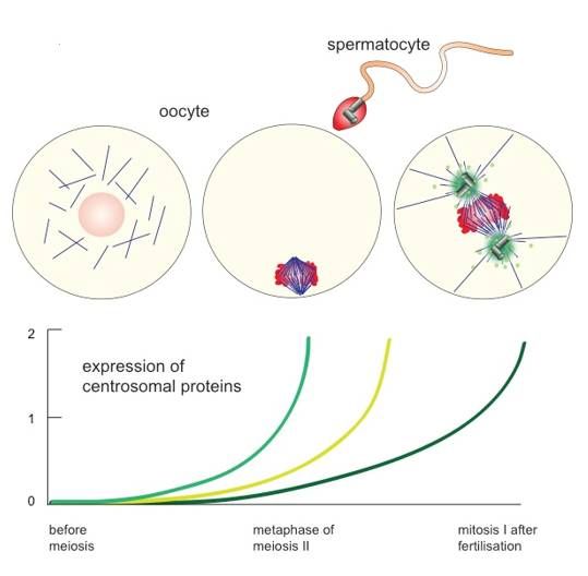
Cells 2018, 7, 73                                                                                   3 of 16

collectively carry the overall function of centrosomes, in particular in nucleating and organising
microtubules, as well as during the single round of centrosome replication before cell division.
      Evidence for centrosome elimination from animal oocytes initially came from electron microscopy
(EM) analysis of oocytes from different species. Here, centrioles are readily visible only during
early or intermediate stages of oogenesis [24–28]. Blocking centrosome loss in different metazoan
model systems revealed its physiological importance: oocytes ought to eliminate centrosomes in
order to avoid a superior number after fertilisation, which would otherwise result in multipolar
spindle formation and chromosome missegregation [29–32]. Elegant studies in sea urchins and flies
documented details about timing and molecular determinants of centrosome elimination in metazoan
oocytes. Starfish oocytes abolish centrosomes late during the asymmetric cell divisions of meiosis
I and II. The starfish uses the two consecutive meiotic divisions not only to dispose of the surplus
chromosomes when gaining a haploid genome, but also dumps 3 out of 4 centrioles into the polar
bodies, which divide from the large oocyte. The last daughter centriole gets degraded soon after
meiosis II in the oocyte cytoplasm. These observations support the idea of an active mechanism to
eliminate centrioles in oocytes [33]. Studies in Drosophila provided further evidence for this hypothesis.
Here, centriole elimination occurs during oogenesis long before meiosis, like in most metazoan oocytes.
It requires the inactivation of Polo1 kinase, proceeds by removing PCM proteins, and ends with the
complete loss of all centrioles [29,34], Figure 1.
      Centrosome elimination during oogenesis seems to be incompatible with the need for microtubule
organisation during female meiotic spindle formation as well as pronuclear migration upon
zygote formation. Moreover, mitotic spindle formation and spindle positioning during the cell
divisions of early embryogenesis necessitate interactions between the spindle and the cell cortex.
The defined orientation of the spindle is a structural basis for cell differentiation in early embryos.
Hence, centrosomes need to establish interactions of the spindle with the cortex and the plasma
membrane via their cytoplasmic (“astral”) microtubules [35–37]. Additionally, spindle formation
ought to be extremely fast during the short cell cycles in early embryos. Fast spindle assembly kinetics
requires active MTOCs [38]. The centrosome is, therefore, generally re-established upon fertilisation, or
right afterwards during early embryogenesis. In most animal species, sperm components (i.e., the basal
body or respective remnants) trigger the reassembly of centrosomes in the cytoplasm of the zygote [39],
Figure 2.
      In general, animal sperm are characterized by extremely compacted chromatin and reduced
cytoplasmic volume as well as an MTOC, which is often stripped down to a single basal body. The basal
body organises the microtubules of the flagellum that drives sperm motion. Although the sperm basal
body and its associated structure (daughter centriole or often only a centriole remnant) generally define
the platform for zygotic centrosome reformation, the process significantly differs from centrosome
duplication in somatic cells, as sperm centrioles undergo various modifications and structural changes
that seem to limit their duplication capacity [40]. The sperm basal body and the daughter centriole
remnant still work as platforms for the first round of centriole duplication in the cytoplasm of the
fertilised ovum. The particular morphology of sperm centrioles and the physiological consequences of
their morphogenesis are reviewed in this issue of Cells by Tomer Avidor-Reiss and colleagues.
Cells 2018, 7, x FOR PEER REVIEW 4 of 16
Cells 2018, 7, 73 4 of 16
Figure 2. Re-assembly of zygotic centrosomes. Metazoan oocytes lose their centrosomes regularly
Figure meiosis
before 2. Re-assembly of zygotic
but rebuild centrosomes.
them only Metazoan
after fertilisation. oocytesuse
Oocytes lose
thetheir centrosomesbasal
sperm-derived regularly
body
before meiosis but rebuild them only after fertilisation. Oocytes use the sperm-derived basal
as a platform for the first centrosome assembly and restart the expression of centrosomal proteins body as
a platform
(see colouredforlines
the first centrosome
in graph) assembly
to allow on-goingandcentrosome
restart the replication
expression afterwards.
of centrosomal proteins
Female (see
meiosis,
coloured lines in graph) to allow on-going centrosome replication
however, occurs in most metazoan species in the absence of centrosomes. afterwards. Female meiosis,
however, occurs in most metazoan species in the absence of centrosomes.
2. Female Meiotic Spindle Formation: Self Assembly of Bipolar Structures
2. Female Meiotic Spindle Formation: Self Assembly of Bipolar Structures
As microtubule organisation remains important for oocyte meiosis as well as for pronuclear
As microtubule
migration organisation
upon fertilisation, remainselimination
centrosome important before
for oocyte
female meiosis as leaves
meiosis well asa for pronuclear
gap, in which
microtubule nucleation and organisation occur in the absence of the canonical animal in
migration upon fertilisation, centrosome elimination before female meiosis leaves a gap, which
MTOCs,
microtubule
Figure nucleation
2. Oocytes of most and organisation
animal species occur in the the
go through absence of the canonical
two meiotic divisionsanimal
without MTOCs, Figure
centrosomes,
2. Oocytes of most animal species
i.e., preformed MTOCs, Figures 2 and 3. go through the two meiotic divisions without centrosomes, i.e.,
preformed MTOCs, Figures 2 and 3.
The progression of oocytes through the meiotic divisions often takes several hours. Compared
to theThe progression
rapid of oocytes
mitotic phases through the which
after fertilisation, meioticregularly
divisionsrun
often takes
in less several
than 60 min,hours. Compared
female meiosis
to the rapid mitotic phases after fertilisation, which regularly run in less than
is slow [38] and more frequently results in chromosome segregation errors than in somatic 60 min, female meiosis
cells,
is slow [38] and more frequently results in chromosome segregation errors than
in particular in mammals [41]. In fact, 80% of human oocytes divide faithfully, but up to 20%
in somatic cells, in
particular in mammals [41]. In fact, 80% of human oocytes divide faithfully, but up to 20% show
show aneuploidy, arising both from defects in meiotic recombination and chromosome segregation
aneuploidy,
errors arising
[42–45]. Goingboth frommeiosis
through defectsIIinnormally
meiotic recombination andinchromosome
involves an arrest metaphase, in segregation
which the errors
ovum
[42–45]. Going through meiosis II normally involves an arrest in metaphase, in which
awaits fertilisation. The centrosome-free bipolar structure of the meiosis II spindle stays with aligned the ovum
awaits fertilisation. The centrosome-free bipolar structure of the meiosis II spindle stays with aligned
chromosomes and stable microtubule-to-kinetochore attachment for many hours, despite the dynamic
chromosomes and stable microtubule-to-kinetochore attachment for many hours, despite the
behaviour of spindle microtubules. These observations pose the question of how microtubules can
dynamic stable
assemble behaviour
meioticofspindles
spindle microtubules.
without a predefinedThese observations
starting pose the
point. In mitosis, question
the pair of how
of centrosomes
microtubules can assemble stable meiotic spindles without a predefined starting point.
mark the two poles of mitotic spindles and define the axis, along which chromosome segregation takes
In mitosis, the
pair of centrosomes mark the two poles of mitotic spindles and define the axis, along which
place. Here, the microtubule nucleating and organising activities densely packed in the PCM of the
chromosome segregation takes place. Here, the microtubule nucleating and organising activities
centrosome pair will readily start the assembly of microtubules at two defined start points (Figure 3).
densely packed in the PCM of the centrosome pair will readily start the assembly of microtubules at
Microtubule assembly in the absence of centrosomes requires an alternative trigger for nucleation
two defined start points (Figure 3). Microtubule assembly in the absence of centrosomes requires an
and organisation. Experiments in amphibian oocytes initially showed that the chromatin takes on
alternative trigger for nucleation and organisation. Experiments in amphibian oocytes initiallyCells 2018, 7, 73 5 of 16
Cells 2018, 7, x FOR PEER REVIEW 5 of 16
the active role in spindle formation [46,47], Figure 3. Of note, chromatin does not provide a platform
showed
for spindlethatformation
the chromatin takes on thebut
like centrosomes active role“instructs”
rather in spindle the formation [46,47],cytoplasm
neighbouring Figure 3. Of to note,
start
chromatin does not provide a platform for spindle formation like centrosomes
microtubule formation [48]. Microtubule nucleation around chromatin proceeds slowly compared but rather “instructs”
thecentrosomal
to neighbouring cytoplasmHowever,
nucleation. to start microtubule
chromatin-inducedformation [48]. Microtubule
microtubule assemblynucleation
results in around
proper
chromatin proceeds slowly compared to centrosomal nucleation.
bipolar spindle formation with time [49]. The main principles and molecular details of centrosomeHowever, chromatin-induced
microtubule
free assembly results
spindle formation in proper
were worked outbipolar
using spindle
cell-freeformation
extracts fromwithamphibian
time [49]. The eggs,main principles
in particular
and molecular details of centrosome free spindle
those of Xenopus laevis, as well as intact metazoan oocytes. formation were worked out using cell-free extracts
fromCell-free
amphibian eggs, in particular those of Xenopus laevis, as well as intact
extracts of Xenopus eggs preserve the natural arrest in metaphase II of meiosis [50]. metazoan oocytes.
Cell-freemeiotic
The formally extractsegg of Xenopus
extractseggs preservegeneral
recapitulate the natural arrest of
principles in metaphase
microtubuleIIassembly
of meiosisseen [50].both
The
formally meiotic egg extracts recapitulate general principles of microtubule
during meiotic spindle formation in oocytes, and mitotic spindle assembly in embyros and somatic assembly seen both
during
cells. meiotic
When spindle
isolated formation
nuclei, regularly in oocytes,
associated and mitotic
with spindle assembly
a centrosome, come intointhe embyros
reaction, and somatic
these will
cells. When isolated nuclei, regularly associated with a centrosome, come
follow the cell cycle, recapitulate centrosome and chromosome duplication at low Cyclin-dependent into the reaction, these will
follow the cell cycle, recapitulate centrosome and chromosome duplication
kinase (Cdk) 1 activity, and build up bipolar spindles when the Cdk1 activity rises [51,52]. Inducing at low Cyclin-dependent
kinase
the (Cdk)reaction
cycling 1 activity,
in and
frogbuild up bipolar
extracts spindles when
in the presence of spermthe Cdk1
results activity rises [51,52].
in chromatin Inducing the
decondensation,
cycling reaction in frog extracts in the presence of sperm results in
its duplication, re-condensation and, finally, its alignment on the metaphase plate of a bipolar chromatin decondensation, its
duplication, re-condensation and, finally, its alignment on the metaphase
spindle. The female cytosol turns back the basal body of the sperm and its daughter centriole plate of a bipolar spindle. The
female
into cytosol turns
functional back theThe
centrosomes. basalfirstbody of the sperm
centrosome and its daughter
pair duplicates centriole
concomitant withinto functional
chromosome
centrosomes. The first centrosome pair duplicates concomitant with chromosome
replication, yielding two centrosomes defining the two poles of the spindle. Remarkably, however, replication, yielding
two centrosomes
these extracts alsodefining
promote the two formation
spindle poles of the spindle.
around Remarkably,
chromatin without however, these as
centrosomes, extracts also
an oocyte
promote spindle formation around chromatin without centrosomes, as an
would do in meiosis. Even the addition of artificial chromatin will lead to the formation of bipolar oocyte would do in meiosis.
Even the addition
microtubule arraysof artificial
that resemble chromatin will leadmeiotic
centrosome-free to the spindles
formation ofof bipolar
intact microtubule
oocytes concerning arrays that
size and
resemble centrosome-free
organisation [53,54]. Although meiotic spindles
oocytes andofeggsintact oocytesvery
represent concerning size cell
specialised andtypes,
organisation [53,54].
the molecular
Although oocytes and eggs represent very specialised cell types, the molecular
analysis of chromatin-driven, meiotic spindle formation in egg extracts strongly influenced our view analysis of chromatin-
driven,
of meiotic spindle
the principles of spindle formation
formation in egg extractsand
in meiosis strongly
mitosisinfluenced our being
(collectively view of the principles
referred to here as of
spindle formation in meiosis and mitosis (collectively being referred to here as M-phase).
M-phase).
Figure 3.3.Oocyte
Figure Oocyte meiosis
meiosis versus
versus mitosis.
mitosis. WhileWhile
spindlespindle
formation formation
occurs in occurs in the
the absence absence of
of centrosomes
centrosomes
in in female
female animal animal
meiosis, mitosismeiosis,
in earlymitosis in early
cell divisions cellfertilisation,
after divisions after fertilisation,
mitosis in somaticmitosis in
cells and
somatic cells and male meiosis ensues in the presence of two centrosomes as microtubule-organizing
male meiosis ensues in the presence of two centrosomes as microtubule-organizing centres (MTOCs).
centres
In (MTOCs). In
both situations, bothconcentration
a high situations, a high concentration
of RanGTP of RanGTP
surrounds surrounds the chromatin.
the chromatin.
3. The Role of RanGTP in Meiotic Spindle Formation
Using these egg extracts, it could be shown that RanGTP plays a key role for chromatin driven
processes in spindle formation. As a member of the Ras-superfamiliy of small GTPases, Ran’s
nucleotide cycle relies on several accessory factors. The highly conserved chromatin-bound proteinCells 2018, 7, 73 6 of 16
3. The Role of RanGTP in Meiotic Spindle Formation
Using these egg extracts, it could be shown that RanGTP plays a key role for chromatin driven
Cells 2018, 7, x FOR PEER REVIEW 6 of 16
processes in spindle formation. As a member of the Ras-superfamiliy of small GTPases, Ran’s
nucleotide cycle relies
RCC1 (Regulator on several accessory
of Chromosome factors. is
Condensation) Thethehighly conserved
only known chromatin-bound
G-nucleotide exchangeprotein
factor for
RCC1 (Regulator of Chromosome Condensation) is the
Ran and converts RanGDP to RanGTP on chromatin (Figure 4). only known G-nucleotide exchange factor for
Ran and converts RanGDP to RanGTP on chromatin (Figure 4).
Figure
Figure 4. Paradigm
4. Paradigm ofmechanism
of the the mechanism
of Ranofin Ran in spindle
spindle formation.
formation. RanGTP numerous
RanGTP activates activates numerous
spindle
assembly factors by release from nuclear import receptors (imp, importin, shown here) or,shown
spindle assembly factors by release from nuclear import receptors (imp, importin, here) or,
alternatively,
alternatively,
association with association withreceptor
nuclear export nuclear CRM1
export (not
receptor CRM1
shown, see (not
text).shown,
Please see
notetext).
that Please
importinnote that
can
importin can be only importin β, or the importin β/importin7, or the importin α/β
be only importin β, or the importin β/importin7, or the importin α/β heterodimer. The production heterodimer. The
production of RanGTP depends on the activity of the chromatin protein Rcc1,
of RanGTP depends on the activity of the chromatin protein Rcc1, the G-nucleotide exchange factor the G-nucleotide
exchange factor of Ran.
of Ran.
The active, GTP-bound form of Ran requires Ran binding proteins 1 or 2 (RanBP1/2) and the Ran
The active, GTP-bound form of Ran requires Ran binding proteins 1 or 2 (RanBP1/2) and the Ran
GTPase activating protein (RanGAP) for GTP hydrolysis [55]. Directly adding recombinant Ran and
GTPase activating protein (RanGAP) for GTP hydrolysis [55]. Directly adding recombinant Ran and its
its associated proteins to Xenopus egg extracts allows manipulating endogenous RanGTP levels.
associated proteins to Xenopus egg extracts allows manipulating endogenous RanGTP levels. Blocking
Blocking RanGTP production completely abolishes spindle formation around artificial chromosomes
RanGTP production completely abolishes spindle formation around artificial chromosomes [56],
[56], while it has no influence on microtubule nucleation from isolated centrosomes [56]. The
while it has no influence on microtubule nucleation from isolated centrosomes [56]. The “opposite”
“opposite” experiment, i.e., increasing RanGTP levels, stimulates the nucleation of a radial
experiment, i.e., increasing RanGTP levels, stimulates the nucleation of a radial microtubule array
microtubule array (“aster”) formed around both isolated centrosomes [57] and sperm centrioles that
(“aster”) formed around both isolated centrosomes [57] and sperm centrioles that stay in the vicinity
stay in the vicinity of chromatin [58–61]. The addition of recombinant RanGTP to Xenopus cell-free
of chromatin [58–61]. The addition of recombinant RanGTP to Xenopus cell-free extracts influences
extracts influences dynamics of microtubules nucleated from centrosomes [60–62] but also leads to
dynamics of microtubules nucleated from centrosomes [60–62] but also leads to the formation of
the formation of spindle-like structures in the absence of chromatin or centrosomes [56,58,60,63,64].
spindle-like structures in the absence of chromatin or centrosomes [56,58,60,63,64]. The activity of
The activity of RanGTP in inducing the formation of microtubule assemblies in M-phase
RanGTP in inducing the formation of microtubule assemblies in M-phase independently of any
independently of any particular source shows that RanGTP, directly or indirectly, mediates a variety
particular source shows that RanGTP, directly or indirectly, mediates a variety of different processes
of different processes in spindle formation during the M-phase. The analogy to Ran’s main function
in spindle formation during the M-phase. The analogy to Ran’s main function in interphase nuclei
in interphase nuclei has helped to explain how Ran functions in mitosis. Nuclear import works via a
has helped to explain how Ran functions in mitosis. Nuclear import works via a family of related
family of related nuclear import receptors (importins), which bind nuclear proteins in the cytoplasm
nuclear import receptors (importins), which bind nuclear proteins in the cytoplasm and promote
and promote their entry into the nucleus. Inside the nucleoplasm, RanGTP directly binds to these
importins, induces a conformational switch, and liberates the bound nuclear protein inside the
nucleus. In turn, nuclear RanGTP can pick up a protein destined for nuclear export together with an
export receptor (exportin) to mediate protein export. Indeed, it could be demonstrated that RanGTP
activates numerous microtubule regulators using the same conformational switches in M-phase on
two nuclear transport receptors—importin β and exportin1/CRM1—to locally activate (importin β),Cells 2018, 7, 73 7 of 16
their entry into the nucleus. Inside the nucleoplasm, RanGTP directly binds to these importins,
induces a conformational switch, and liberates the bound nuclear protein inside the nucleus. In turn,
nuclear RanGTP can pick up a protein destined for nuclear export together with an export receptor
(exportin) to mediate protein export. Indeed, it could be demonstrated that RanGTP activates numerous
microtubule regulators using the same conformational switches in M-phase on two nuclear transport
receptors—importin β and exportin1/CRM1—to locally activate (importin β), or to specifically target
(exportin 1) downstream activities in spindle formation. Among these mechanisms, the release from
importin β is the predominant mechanism of target activation in the M-phase (Figure 4). Proteins can
interact with importin β directly or via adaptor proteins, such as importin α. In either case, RanGTP
binds to importin β and releases the bound factors upon inducing a conformational switch in importin
β. Most factors identified as Ran targets in the M-phase show direct importin α binding via a nuclear
localisation signal (NLS). The microtubule associated proteins TPX2 (targeting protein for XKlp2) [65]
and NuMA (nuclear protein of the mitotic apparatus) [66,67], for instance, are activated by RanGTP
when released from importins and localize to the spindle in M-phase in a Ran-GTP dependent manner.
In interphase, when the nucleus is intact, the NLS confers importin binding in the same manner.
Here, importin mediates nuclear import, and RanGTP releases the nuclear protein from importin in
the nucleus [68]. The interaction of TPX2 with importin α, therefore, has two consequences: first,
it mediates nuclear import of the protein in interphase and, second, it regulates its activity in mitosis.
The interaction is special as the key NLS in TPX2 contacts the minor binding site in importin α, while
most other NLS proteins interact with importin α’s major binding side. This unusual binding mode
may enable competitive sequestration of TPX2 even in the presence of many free NLS-containing
proteins and may ensure proper spatial regulation of TPX2 in response to RanGTP [69], Figure 4.
Beyond TPX2 and NuMA, numerous proteins have been identified in Xenopus egg extracts, which are
under the control of RanGTP and importins, including the microtubule associated proteins HURP
(hepatoma upregulated protein) and NuSAP (nucleolar spindle associated protein), the chromatin
remodelling proteins ISWI and KANSL1/3, the RNA binding protein Rae1 and the nuclear pore
complex (NPC) component Mel-28/ELYS. Consistently, all these proteins remain nuclear in interphase
but perform key functions in M-phase in microtubule nucleation, microtubule organisation, spindle
pole formation and anaphase spindle stability [70–73].
4. The Mechanism of Centrosome-Independent Microtubule Nucleation
Most mechanistic insights of spindle self-assembly once more came from Xenopus egg extracts
due to the biochemical accessibility of the system. In egg extracts, new microtubules are generated by
the activity of RanGTP regulating TPX2 and communicating with Mel-28 and γ-TuRC. TPX2, released
from importins via RanGTP, associates with and activates Aurora A kinase [74], a mechanism that also
stimulates Aurora A in intact human somatic cells [75]. Biochemical and structural analysis showed
that the very N-terminus of TPX2 drives a conformational change in the activation segment of the
kinase, which makes the t-loop phosphorylation less accessible for dephosphorylation and kinase
inactivation [76–78]. Aurora A, in turn, modifies microtubule-associated and free centrosomal proteins
to modulate their functions in spindle pole organisation. These include TPX2 itself, Kinesin 5/Eg5,
TACC (transformed acidic coiled-coil protein) and the NEDD (Neddylation) 1 protein [79,80]. Aurora
A-mediated phosphorylation of NEDD1 on S405, as well as recruitment of the microtubule-associated
proteins X-RHAMM (Xenopus receptor for hyaluron-mediated mobility) and TPX2 help to activate
the γ-TuRC in Xenopus egg extracts [81]. Of note, TPX2 also nucleates microtubule directly and
differently than the γ-TuRC by promoting longitudinal and lateral interactions between tubulin
dimers. This stabilises the formation of microtubule seeds as the rate limiting step in microtubule
assembly [82]. Initially assembled microtubules generate further polymers: the Augmin complex,
recently reconstituted from 8 subunits in vitro [83], enables microtubule nucleation on preexisting
microtubules. It recruits γ-TuRC to their lattice, where TPX2 is required to assemble in a stable
way the new microtubules branching out from the initial polymers [84,85]. Augmin can, thus,Cells 2018, 7, 73 8 of 16
catalyze additional nucleation of microtubules by γ-TuRC recruitment and activation on a single
microtubule filament [86]. These molecular insights explain the previously observed non-linear
increase in RanGTP-induced microtubule formation [87]. They also show how the main nucleating
activities of TPX2 and γ-TuRC converge in the assembly of additional microtubules from pre-existing
polymers. In Xenopus egg extracts, immunodepletion of Augmin leads to diminished microtubule
density and spindle pole fragmentation [88], similar to what has been observed upon partial depletion
of the γ-TuRC from egg extracts [89]. In turn, the addition of recombinant Augmin initiates free
microtubule assembly, which is further stimulated by RanGTP [86]. Recently, γ-TuRC-activation
domains have been proposed to reside in the C-terminal part of Xenopus TPX2 [90]. These activation
domains, commonly called γ-TuNA (γ-TuRC nucleation activator), are only visible for γ-TuRC once
TPX2 has been released from importins, providing the most direct link between RanGTP and γ-TuRC
as the main activity for templating new microtubules.
Additional experiments in human, mouse and fly oocytes showed that reducing RanGTP
levels does not completely inhibit but delays or impairs meiotic spindle formation [91–93].
Although the remaining spindle assembly activity in these oocytes may have come from residual
RanGTP, the observations are in line with the idea of additional pathways for centrosome-free
microtubule formation. Experiments in Xenopus cell-free extracts [94–96] and fly oocytes [97,98]
have, for instance, demonstrated that the chromatin-bound chromosomal passenger complex
(CPC) contributes to microtubule nucleation from chromosomes in meiotic spindle formation in
a RanGTP-independent fashion.
5. The Mechanism of Spindle Organization without Centrosomes
Spindle bipolarity requires balanced and robust microtubule nucleation at and around the
two spindle poles. The early phase of spindle formation without centrosomes involves undirected
microtubule nucleation next to chromatin. Newly formed microtubules will be stabilised by additional
Ran targets including Rae1 [99] and shortly sorted and focused at their common minus ends by the
minus-end directed motor proteins Dynein—with the help of NuMA—and the inverted Kinesin
14 [100,101]. The organisation and bundling of microtubule minus ends, which leads to pole
formation, leaves free plus ends growing towards the chromatin. Once these plus ends get into
contact with plus ends from a second microtubule array, they will recruit motor proteins and other
microtubule-associated proteins specifically recognising the antiparallel order of these microtubules.
In particular, the tetrameric kinesin Kinesin-5/Eg5 can sort anti-parallel polymers to establish a stable
bipolar structure. Its inhibition leads to failure in spindle bipolarisation in cell-free egg extracts and in
living mouse oocytes [101–104]. In egg extracts, Eg5 is directly activated by RanGTP in the vicinity
of chromatin and indirectly via the activation of HURP, which helps Eg5 to function in anti-parallel
sorting [62,105,106]. Female mouse Hurp k.o. animals are viable but sterile as their oocytes fail to
establish a robust central spindle [107]. Preformed, centrosome-free MTOCs in mouse oocytes require
Eg5 to get fragmented upon meiotic spindle formation. This fragmentation has to precede sorting
along existing microtubules and is a prerequisite for achieving bipolarity [104,108,109]. At least in
cell-free Xenopus egg extracts, Ran then works beyond spindle assembly. It activates ISWI, which
stabilises microtubules only after the metaphase-to-anaphase-transition to allow proper chromosome
segregation [110]. ISWI helps microtubules to remain stable during anaphase [70], while two specific
subunits of the chromatin remodelling complex NSL (non specific lethal), KANSL1 and 3, dissociate
from chromatin already before metaphase-to-anaphase-transition to stabilise microtubule minus
ends [73].
The concept of RanGTP acting on importins and exportins in M-phase has been further
validated in C.elegans and Drosophila embryos as well as in plant cells [111–114]. In all systems,
the prevailing model is based on the chromatin-binding of Ran’s exchange factor RCC1 in M-phase.
After nuclear envelope breakdown (NEB), RanGTP freely diffuses within the mitotic cytoplasm along
with the hydrolysis promoting activities, RanBP1/2 and RanGAP. Localised RanGTP productionCells 2018, 7, 73 9 of 16
on chromatin together with delocalised Ran’s GTP hydrolysis in the mitotic cytoplasm result in a
soluble gradient of RanGTP with the highest concentration next to chromatin. This explains how
Ran downstream activities involved in spindle formation in M-phase become activated only in the
vicinity of chromatin [70,115,116], Figure 4. Experiments in Xenopus egg extracts documented how this
gradient could be further sharpened: Cytosolic RanBP1, phosphorylated in M-phase on a single Serine
residue (S60), forms a complex with Ran and RCC1, which inhibits the cytosolic activity of the latter
and restrains cytosolic RanGTP production [117].
6. Communication between Chromatin and Newly Formed Centrosomes
Fast kinetics and robustness of spindle formation require the cooperation between the
preformed MTOCs and the chromatin-associated activities on microtubule nucleation and organisation.
Re-established MTOCs quickly assemble new microtubules. They dominate over chromatin-induced
microtubule formation in early development [38] and take the lead later in differentiated somatic
cells. However, the activity of chromatin remains essential [38]. In Xenopus egg extracts,
the immunodepletion of the Ran target TPX2 completely abolishes chromatin-induced microtubule
assembly [65]. Although its depletion does not inhibit basic microtubule nucleation from isolated
centrosomes, it strongly impairs bipolar spindle formation driven by sperm nuclei, which comprise
chromatin as well as centrosomes [57]. Consistently, RanGTP, produced from chromatin, stimulates
centrosomal microtubule nucleation (Figure 4). Further biochemical experiments have enabled the
identification and characterisation of Cdk11 as the key effector in Ran-regulated increase in microtubule
nucleation from centrosomes [118]. Additional evidence from cell-free extracts and somatic cells
underlines that the centrosome with its associated functions “sees” the activity of RanGTP in the mitotic
cytoplasm after NEB. The increase in centrosomal nucleation observed at mitotic onset (“centrosome
maturation”) accelerates right at NEB when RanGTP is released into the mitotic cytoplasm [119].
Centrosomal maturation involves the recruitment of additional PCM components [9] and restructuring
centriolar satellites [21–23], which converge to increase mitotic microtubule nucleation and organisation
together with centrosomal functions in signalling and cell cycle control [120]. This goes along with
the release of numerous proteins from chromosomes. These turn from a function on chromatin to a
Ran-regulated function on microtubules in the mitotic cytoplasm [121].
The interplay between NEB and spindle formation goes beyond rising activities of RanGTP in
the mitotic cytoplasm. The NPC, consisting of some 30 protein components in copy numbers of
8 to 64 [122] dissociates into defined subcomplexes at mitotic onset concomitant with NEB [123].
The NPC protein Mel-28/ELYS was initially characterised as a pioneering component for nuclear
envelope reassembly after mitosis. Both in human cells and in C.elegans embryos, Mel-28 rebinds to
chromatin in late mitosis to seed an assembly platform for post-mitotic NPC formation [123]. In mitosis,
however, Mel-28 serves additional functions that depend on the disassembly of the NPC. Released
from the NPC, distinct populations of Mel-28 stay in the Nup107 subcomplex but also find new
interaction partners including the γ-TuRC [124]. While the Nup107 subcomplex together with Mel-28
binds to and stabilises microtubule-kinetochore interactions, Mel-28 released from the subcomplex
becomes essential for γ-TuRC-mediated microtubule nucleation. Consistently, its depletion from egg
extracts abolishes spindle formation both in the absence and in the presence of centrosomes [124,125].
Thus, NPC disassembly is not merely a necessary evil of open mitosis but it is a necessary prerequisite
for spindle formation.
7. Self-Assembly of Centrioles in Oocytes
Meiotic spindle self-assembly initiated by Ran-GTP and possibly other chromatin-localised
activities highlights an intriguing feature of female reproductive cells: the ovum cytoplasm accumulates
moieties required for spindle formation to an extent and to a concentration that allows, once the right
trigger is provided, assembly of a functional bipolar spindle without preformed MTOCs.Cells 2018, 7, 73 10 of 16
Similarly, the egg cytoplasm readily self-assembles centrioles de novo even in the absence of basal
bodies or basal body remnants. Although most metazoan organisms arise from sexual reproduction,
a variety of organisms, in particular within the class of insects, can develop upon parthenogenesis.
As oocytes of these species lose their centrioles before or during female meiosis, their female egg
cytoplasm has to assemble centrosomes, including centrioles, de novo. De novo centrosome formation
can also be recapitulated in Xenopus egg extracts. As in somatic cells, Xenopus Plk4 (XPlk4) initiates
centriole assembly, and centrioles are readily detected by EM after the addition or overexpression of
XPlk4 to egg extracts [126]. Sea-urchin eggs or egg extracts, forced to start parthenogenesis, produce
huge amounts of centrioles de novo [127,128]. The egg cytoplasm holds the capacity to assemble the
stereotypic centriole structure as building blocks of centrioles accumulate in eggs before fertilisation
(Figure 2). This suggests that unfertilised eggs maintain a signal to inhibit premature centriole
formation prior to fertilisation. Although oocytes initially block centriole de novo assembly in meiosis,
the proteins and protein complexes of the PCM form centriole-free spindle poles by self-assembly
in meiosis. Mouse zygotes follow this strategy even after fertilisation: two spindle poles reform
from a number of centriole-free MTOCs during the first cell divisions. Acentriolar spindle formation
during mouse meiosis and the early mitotic divisions require the help of the actin cytoskeleton to
ensure robust spindle formation and chromosome segregation [129–133]. Of note, the centrosome-free
pathway uses microtubule assembly promoting factors, which will later become true centrosome
components. Centrosomal proteins Cep152, Cep192 and Pericentrin act in concert with Plk4 to
assemble centriole-free spindle poles in meiosis and in early mitoses in mice [134–138]. Centrosomal
as well as non-centrosomal spindle formation pathways commonly use the activity of γ-TuRC, which
carries essential functions in microtubule nucleation during spindle formation irrespective of the
absence or presence of centrosomes [7]. These examples underline that the two pathways share many
proteins and protein modules with functions in microtubule nucleation and organisation.
8. Concluding Remarks
Loss and rebirth of the animal MTOC, the centrosome, accompanies meiosis and fertilisation in
metazoan organisms. In most metazoans, centrosome elimination precedes meiosis, which necessitates
a pathway for meiotic spindle formation in the absence of centrosomes. The mechanisms of spindle
self-assembly in meiosis have evolved using mostly the same molecular determinants that are found to
nucleate and organise microtubules in the PCM of centrosomes. Centrosome-free microtubule assembly
requires RanGTP produced around chromatin, the nucleation promoting activity of kinetochores,
as well as positive feedback loops of microtubule nucleation from pre-existing polymers. Analysing
spindle assembly in the absence of centrosomes has not only helped to comprehend the biological
challenges of meiosis, fertilisation and fertility, it has also highlighted the idea of a cooperation
of different microtubule assembly pathways, both centrosomal and non-centrosomal, for spindle
assembly in all our somatic cells.
Funding: This research was funded by the German Research Foundation (DFG), grant number GR1737/9-1.
Conflicts of Interest: The author declares no conflict of interest. The founding sponsors had no role in the design
of the study; in the collection, analyses, or interpretation of data; in the writing of the manuscript, and in the
decision to publish the results.
References
1. Severson, A.F.; von Dassow, G.; Bowerman, B. Oocyte Meiotic Spindle Assembly and Function. Curr. Top.
Dev. Biol. 2016, 116, 65–98. [CrossRef] [PubMed]
2. Schatten, G. The centrosome and its mode of inheritance: The reduction of the centrosome during
Gametogenesis and its restoration during fertilization. Dev. Biol. 1994, 165, 299–335. [CrossRef] [PubMed]
3. Clift, D.; Schuh, M. Restarting life: Fertilization and the transition from meiosis to mitosis. Nat. Rev. Mol.
Cell Biol. 2013, 14, 549–562. [CrossRef] [PubMed]Cells 2018, 7, 73 11 of 16
4. Loncarek, J.; Bettencourt-Dias, M. Building the right centriole for each cell type. J. Cell Biol. 2018, 217, 823–835.
[CrossRef] [PubMed]
5. Luders, J. The amorphous pericentriolar cloud takes shape. Nat. Cell Biol. 2012, 14, 1126–1128. [CrossRef]
[PubMed]
6. Kollman, J.M.; Merdes, A.; Mourey, L.; Agard, D.A. Microtubule nucleation by gamma-tubulin complexes.
Nat. Rev. Mol. Cell Biol. 2011, 12, 709–721. [CrossRef] [PubMed]
7. Lin, T.C.; Neuner, A.; Schiebel, E. Targeting of gamma-tubulin complexes to microtubule organizing centers:
Conservation and divergence. Trends Cell Biol. 2015, 25, 296–307. [CrossRef] [PubMed]
8. Woodruff, J.B.; Wueseke, O.; Hyman, A.A. Pericentriolar material structure and dynamics. Philos. Trans. R.
Soc. Lond. Ser. B Biol. Sci. 2014, 369. [CrossRef] [PubMed]
9. Fry, A.M.; Sampson, J.; Shak, C.; Shackleton, S. Recent advances in pericentriolar material organization:
Ordered layers and scaffolding gels. F1000Research 2017, 6, 1622. [CrossRef] [PubMed]
10. Woodruff, J.B.; Ferreira Gomes, B.; Widlund, P.O.; Mahamid, J.; Honigmann, A.; Hyman, A.A.
The Centrosome Is a Selective Condensate that Nucleates Microtubules by Concentrating Tubulin. Cell 2017,
169, 1066–1077. [CrossRef] [PubMed]
11. Petry, S. Mechanisms of Mitotic Spindle Assembly. Annu. Rev. Biochem. 2016, 85, 659–683. [CrossRef]
[PubMed]
12. Prosser, S.L.; Pelletier, L. Mitotic spindle assembly in animal cells: A fine balancing act. Nat. Rev. Mol.
Cell Biol. 2017, 18, 187–201. [CrossRef] [PubMed]
13. McIntosh, J.R. Mitosis. Cold Spring Harbor Perspect. Biol. 2016, 8. [CrossRef] [PubMed]
14. Nigg, E.A.; Cajanek, L.; Arquint, C. The centrosome duplication cycle in health and disease. FEBS Lett. 2014,
588, 2366–2372. [CrossRef] [PubMed]
15. Sluder, G. One to only two: A short history of the centrosome and its duplication. Philos. Trans. R. Soc. Lond.
Ser. B Biol. Sci. 2014, 369. [CrossRef] [PubMed]
16. Firat-Karalar, E.N.; Stearns, T. The centriole duplication cycle. Philos. Trans. R. Soc. Lond. Ser. B Biol. Sci.
2014, 369. [CrossRef]
17. Hinchcliffe, E.H. Centrosomes and the art of mitotic spindle maintenance. Int. Rev. Cell Mol. Biol. 2014,
313, 179–217. [CrossRef] [PubMed]
18. Fu, J.; Hagan, I.M.; Glover, D.M. The centrosome and its duplication cycle. Cold Spring Harbor Perspect. Biol.
2015, 7, a015800. [CrossRef] [PubMed]
19. Paz, J.; Luders, J. Microtubule-Organizing Centers: Towards a Minimal Parts List. Trends Cell Biol. 2018,
28, 176–187. [CrossRef] [PubMed]
20. Nigg, E.A.; Holland, A.J. Once and only once: Mechanisms of centriole duplication and their deregulation in
disease. Nat. Rev. Mol. Cell Biol. 2018. [CrossRef] [PubMed]
21. Bärenz, F.; Mayilo, D.; Gruss, O.J. Centriolar Satellites: Busy Orbits around the Centrosome. Eur. J. Cell Biol.
2011, 90, 983–989. [CrossRef] [PubMed]
22. Tollenaere, M.A.; Mailand, N.; Bekker-Jensen, S. Centriolar satellites: Key mediators of centrosome functions.
Cell. Mol. Life Sci. 2015, 72, 11–23. [CrossRef] [PubMed]
23. Hori, A.; Toda, T. Regulation of centriolar satellite integrity and its physiology. Cell. Mol. Life Sci. 2017,
74, 213–229. [CrossRef] [PubMed]
24. Szollosi, D.; Calarco, P.; Donahue, R.P. Absence of centrioles in the first and second meiotic spindles of mouse
oocytes. J. Cell Sci. 1972, 11, 521–541. [PubMed]
25. Sluder, G.; Miller, F.J.; Lewis, K.; Davison, E.D.; Rieder, C.L. Centrosome inheritance in starfish zygotes:
Selective loss of the maternal centrosome after fertilization. Dev. Biol. 1989, 131, 567–579. [CrossRef]
26. Nakashima, S.; Kato, K.H. Centriole behavior during meiosis in oocytes of the sea urchin Hemicentrotus
pulcherrimus. Dev. Growth Differ. 2001, 43, 437–445. [CrossRef] [PubMed]
27. Shirato, Y.; Tamura, M.; Yoneda, M.; Nemoto, S. Centrosome destined to decay in starfish oocytes.
Development 2006, 133, 343–350. [CrossRef] [PubMed]
28. Sathananthan, A.H.; Selvaraj, K.; Girijashankar, M.L.; Ganesh, V.; Selvaraj, P.; Trounson, A.O. From oogonia
to mature oocytes: Inactivation of the maternal centrosome in humans. Microsc. Res. Tech. 2006, 69, 396–407.
[CrossRef] [PubMed]Cells 2018, 7, 73 12 of 16
29. Pimenta-Marques, A.; Bento, I.; Lopes, C.A.; Duarte, P.; Jana, S.C.; Bettencourt-Dias, M. A mechanism for
the elimination of the female gamete centrosome in Drosophila melanogaster. Science 2016, 353, aaf4866.
[CrossRef] [PubMed]
30. Washitani-Nemoto, S.; Saitoh, C.; Nemoto, S. Artificial parthenogenesis in starfish eggs: Behavior of nuclei
and chromosomes resulting in tetraploidy of parthenogenotes produced by the suppression of polar body
extrusion. Dev. Biol. 1994, 163, 293–301. [CrossRef] [PubMed]
31. Manandhar, G.; Schatten, H.; Sutovsky, P. Centrosome reduction during gametogenesis and its significance.
Biol. Reprod. 2005, 72, 2–13. [CrossRef] [PubMed]
32. Kim, D.Y.; Roy, R. Cell cycle regulators control centrosome elimination during oogenesis in Caenorhabditis
elegans. J. Cell Biol. 2006, 174, 751–757. [CrossRef] [PubMed]
33. Borrego-Pinto, J.; Somogyi, K.; Karreman, M.A.; Konig, J.; Muller-Reichert, T.; Bettencourt-Dias, M.;
Gonczy, P.; Schwab, Y.; Lenart, P. Distinct mechanisms eliminate mother and daughter centrioles in meiosis
of starfish oocytes. J. Cell Biol. 2016, 212, 815–827. [CrossRef] [PubMed]
34. Schoborg, T.A.; Rusan, N.M. Taking Centrioles to the Elimination Round. Dev. Cell 2016, 38, 10–12. [CrossRef]
[PubMed]
35. Moorhouse, K.S.; Burgess, D.R. How to be at the right place at the right time: The importance of spindle
positioning in embryos. Mol. Reprod. Dev. 2014, 81, 884–895. [CrossRef] [PubMed]
36. Chaigne, A.; Terret, M.E.; Verlhac, M.H. Asymmetries and Symmetries in the Mouse Oocyte and Zygote.
Results Probl. Cell Differ. 2017, 61, 285–299. [CrossRef] [PubMed]
37. Hasley, A.; Chavez, S.; Danilchik, M.; Wuhr, M.; Pelegri, F. Vertebrate Embryonic Cleavage Pattern
Determination. Adv. Exp. Med. Biol. 2017, 953, 117–171. [CrossRef] [PubMed]
38. Cavazza, T.; Peset, I.; Vernos, I. From meiosis to mitosis—The sperm centrosome defines the kinetics of
spindle assembly after fertilization in Xenopus. J. Cell Sci. 2016, 129, 2538–2547. [CrossRef] [PubMed]
39. Inoue, D.; Wittbrodt, J.; Gruss, O.J. Loss and Rebirth of the Animal Microtubule Organizing Center: How
Maternal Expression of Centrosomal Proteins Cooperates with the Sperm Centriole in Zygotic Centrosome
Reformation. Bioessays 2018, 40, e1700135. [CrossRef] [PubMed]
40. Avidor-Reiss, T.; Gopalakrishnan, J. Building a centriole. Curr. Opin. Cell Biol. 2013, 25, 72–77. [CrossRef]
[PubMed]
41. Verlhac, M.H.; Terret, M.E. Oocyte Maturation and Development. F1000Research 2016, 5. [CrossRef] [PubMed]
42. Hunt, P.A. Meiosis in mammals: Recombination, non-disjunction and the environment. Biochem. Soc. Trans.
2006, 34, 574–577. [CrossRef] [PubMed]
43. Pacchierotti, F.; Adler, I.D.; Eichenlaub-Ritter, U.; Mailhes, J.B. Gender effects on the incidence of aneuploidy
in mammalian germ cells. Environ. Res. 2007, 104, 46–69. [CrossRef] [PubMed]
44. Webster, A.; Schuh, M. Mechanisms of Aneuploidy in Human Eggs. Trends Cell Biol. 2017, 27, 55–68.
[CrossRef] [PubMed]
45. Greaney, J.; Wei, Z.; Homer, H. Regulation of chromosome segregation in oocytes and the cellular basis for
female meiotic errors. Hum. Reprod. Update 2017. [CrossRef] [PubMed]
46. Karsenti, E.; Newport, J.; Hubble, R.; Kirschner, M. Interconversion of metaphase and interphase microtubule
arrays, as studied by the injection of centrosomes and nuclei into Xenopus eggs. J. Cell Biol. 1984,
98, 1730–1745. [CrossRef] [PubMed]
47. Karsenti, E.; Newport, J.; Kirschner, M. The respective roles of centrosomes and chromatin in the conversion
of microtubule arrays from interphase to metaphase. J. Cell Biol. 1984, 99, 47s–54s. [CrossRef] [PubMed]
48. Karsenti, E.; Vernos, I. The mitotic spindle: A self-made machine. Science 2001, 294, 543–547. [CrossRef]
[PubMed]
49. Hallen, M.A.; Endow, S.A. Anastral spindle assembly: A mathematical model. Biophys. J. 2009, 97, 2191–2201.
[CrossRef] [PubMed]
50. Masui, Y.; Markert, C.L. Cytoplasmic control of nuclear behavior during meiotic maturation of frog oocytes.
J. Exp. Zool. 1971, 177, 129–145. [CrossRef] [PubMed]
51. Murray, A. Xenopus Laevis: Practical Uses in Cell and Molecular Biology; Kay, B.K., Peng, H.B., Eds.; Academic
Press, Inc.: San Diego, CA, USA, 1991; Volume 36, pp. 581–605.
52. Sawin, K.E.; Mitchison, T.J. Mitotic spindle assembly by two different pathways in vitro. J. Cell Biol. 1991,
112, 925–940. [CrossRef] [PubMed]Cells 2018, 7, 73 13 of 16
53. Heald, R.; Tournebize, R.; Blank, T.; Sandaltzopoulos, R.; Becker, P.; Hyman, A.; Karsenti, E. Self-organization
of microtubules into bipolar spindles around artificial chromosomes in Xenopus egg extracts. Nature 1996,
382, 420–425. [CrossRef] [PubMed]
54. Heald, R.; Tournebize, R.; Habermann, A.; Karsenti, E.; Hyman, A. Spindle assembly in Xenopus egg extracts:
Respective roles of centrosomes and microtubule self-organization. J. Cell Biol. 1997, 138, 615–628. [CrossRef]
[PubMed]
55. Cavazza, T.; Vernos, I. The RanGTP Pathway: From Nucleo-Cytoplasmic Transport to Spindle Assembly and
Beyond. Front. Cell Dev. Biol. 2015, 3, 82. [CrossRef] [PubMed]
56. Carazo-Salas, R.E.; Guarguaglini, G.; Gruss, O.J.; Segref, A.; Karsenti, E.; Mattaj, I.W. Generation of
GTP-bound Ran by RCC1 is required for chromatin-induced mitotic spindle formation. Nature 1999,
400, 178–181. [CrossRef] [PubMed]
57. Gruss, O.J.; Wittmann, M.; Yokoyama, H.; Pepperkok, R.; Kufer, T.; Silljé, H.; Karsenti, E.; Mattaj, I.W.;
Vernos, I. Chromosome-induced microtubule assembly mediated by TPX2 is required for spindle formation
in HeLa cells. Nat. Cell Biol. 2002, 4, 871–879. [CrossRef] [PubMed]
58. Wilde, A.; Zheng, Y. Stimulation of microtubule aster formation and spindle assembly by the small GTPase
Ran. Science 1999, 284, 1359–1362. [CrossRef] [PubMed]
59. Zhang, Y.; Heidebrecht, H.; Rott, A.; Schlegelberger, B.; Parwaresch, R. Assignment of human
proliferation associated p100 gene (C20orf1) to human chromosome band 20q11.2 by in situ hybridization.
Cytogenet. Cell Genet. 1999, 84, 182–183. [CrossRef] [PubMed]
60. Kalab, P.; Pu, R.T.; Dasso, M. The ran GTPase regulates mitotic spindle assembly. Curr. Biol. 1999, 9, 481–484.
[CrossRef]
61. Carazo-Salas, R.E.; Gruss, O.J.; Mattaj, I.W.; Karsenti, E. Ran-GTP coordinates regulation of microtubule
nucleation and dynamics during mitotic-spindle assembly. Nat. Cell Biol. 2001, 3, 228–234. [CrossRef]
[PubMed]
62. Wilde, A.; Lizarraga, S.B.; Zhang, L.; Wiese, C.; Gliksman, N.R.; Walczak, C.E.; Zheng, Y. Ran stimulates
spindle assembly by altering microtubule dynamics and the balance of motor activities. Nat. Cell Biol. 2001,
3, 221–227. [CrossRef] [PubMed]
63. Ohba, T.; Nakamura, M.; Nishitani, H.; Nishimoto, T. Self-organization of microtubule asters induced in
Xenopus egg extracts by GTP-bound Ran. Science 1999, 284, 1356–1358. [CrossRef] [PubMed]
64. Zhang, C.; Hughes, M.; Clarke, P.R. Ran-GTP stabilises microtubule asters and inhibits nuclear assembly in
Xenopus egg extracts. J. Cell Sci. 1999, 112, 2453–2461. [PubMed]
65. Gruss, O.J.; Carazo-Salas, R.E.; Schatz, C.A.; Guarguaglini, G.; Kast, J.; Wilm, M.; Le Bot, N.; Vernos, I.;
Karsenti, E.; Mattaj, I.W. Ran Induces Spindle Assembly by Reversing the Inhibitory Effect of Importin alpha
on TPX2 Activity. Cell 2001, 104, 83–93. [CrossRef]
66. Nachury, M.V.; Maresca, T.J.; Salmon, W.C.; Waterman-Storer, C.M.; Heald, R.; Weis, K. Importin beta Is a
Mitotic Target of the Small GTPase Ran in Spindle Assembly. Cell 2001, 104, 95–106. [CrossRef]
67. Wiese, C.; Wilde, A.; Moore, M.S.; Adam, S.A.; Merdes, A.; Zheng, Y. Role of Importin b in Coupling Ran to
Downstream Targets in Microtubule Assembly. Science 2001, 4, 4–8. [CrossRef]
68. Hetzer, M.; Gruss, O.J.; Mattaj, I.W. The Ran GTPase as a marker for chromosome position in spindle
formation and nuclear envelope assembly. Nat. Cell Biol. 2002, 4, E177–E184. [CrossRef] [PubMed]
69. Giesecke, A.; Stewart, M. Novel binding of the mitotic regulator TPX2 (target protein for Xenopus kinesin-like
protein 2) to importin-alpha. J. Biol. Chem. 2010, 285, 17628–17635. [CrossRef] [PubMed]
70. Gruss, O.J. Ras Superfamily Small G Proteins: Biology and Mechanisms; Wittinghofer, A., Ed.; Springer: Basel,
Switzerland, 2014; Volume 2, pp. 125–150.
71. Okada, N.; Sato, M. Spatiotemporal Regulation of Nuclear Transport Machinery and Microtubule
Organization. Cells 2015, 4, 406–426. [CrossRef] [PubMed]
72. Forbes, D.J.; Travesa, A.; Nord, M.S.; Bernis, C. Nuclear transport factors: Global regulation of mitosis.
Curr. Opin. Cell Biol. 2015, 35, 78–90. [CrossRef] [PubMed]
73. Meunier, S.; Shvedunova, M.; Van Nguyen, N.; Avila, L.; Vernos, I.; Akhtar, A. An epigenetic regulator
emerges as microtubule minus-end binding and stabilizing factor in mitosis. Nat. Commun. 2015, 6, 7889.
[CrossRef] [PubMed]You can also read