ATF5, a putative therapeutic target for the mitochondrial DNA 3243A G mutation-related disease
←
→
Page content transcription
If your browser does not render page correctly, please read the page content below
www.nature.com/cddis

ARTICLE                  OPEN

ATF5, a putative therapeutic target for the mitochondrial DNA
3243A > G mutation-related disease
                                                                                                                      ✉
Xinpei Gao1,7, Zhixin Jiang2,7, Xinfeng Yan3, Jiping Liu1, Fengwen Li2, Peng Liu2, Jialu Li1, Yuehua Wei4, Yi Eve Sun1 ,
             5✉                        6✉
Yinan Zhang     and Congrong Wang

© The Author(s) 2021

    The mitochondrial DNA m.3243A > G mutation is well-known to cause a variety of clinical phenotypes, including diabetes, deafness,
    and osteoporosis. Here, we report isolation and expansion of urine-derived stem cells (USCs) from patients carrying the m.3243A >
    G mutation, which demonstrate bimodal heteroplasmy. USCs with high levels of m.3243A > G mutation displayed abnormal
    mitochondrial morphology and function, as well as elevated ATF5-dependent mitochondrial unfolded protein response (UPRmt),
    together with reduced Wnt/β-catenin signaling and osteogenic potentials. Knockdown of ATF5 in mutant USCs suppressed UPRmt,
    improved mitochondrial function, restored expression of GSK3B and WNT7B, and rescued osteogenic potentials. These results
    suggest that ATF5-dependent UPRmt could be a core disease mechanism underlying mitochondrial dysfunction and osteoporosis
    related to the m.3243A > G mutation, and therefore could be a novel putative therapeutic target for this genetic disorder.

    Cell Death and Disease (2021)12:701 ; https://doi.org/10.1038/s41419-021-03993-1

INTRODUCTION                                                                                mutation in blood leukocytes were significantly associated with
Mitochondrion is the center for energy production and its                                   lower bone mineral density [12].
dysregulation has been linked to various human diseases [1–3].                                 Such a broad spectrum of clinical manifestations could be
By metabolizing glucose and lipids through TCA cycle and                                    attributed to the heteroplasmic nature of the m.3243A > G
β-oxidation, respectively, mitochondria store biological energy in                          mutation, where mutant transcripts could present in different
adenosine triphosphate (ATP). This process requires the coordina-                           ratio to wild-type transcripts in different cell types or tissues [13–
tion of more than 80 proteins that form 5 major respiratory chain                           15]. Biochemical analysis revealed that high heteroplasmy levels of
complexes, thirteen of which are encoded by the mitochondrial                               the m.3243A > G mutation reduced mitochondrial tRNALeu (UUR)
genome [4, 5]. In addition to the 13 respiratory chain subunits, the                        abundance, decreased its aminoacylation, and thus inhibited
mitochondrial genome also encodes 2 rRNAs and 22 tRNAs,                                     normal post-transcriptional modifications [16–20]. Cells with high
mutations of which have been reported to be involved in certain                             levels of the m.3243A > G mutation have impaired protein
diseases, such as Leigh syndrome, Kearns-Sayre syndrome, Lever                              synthesis and respiratory activity [21, 22]. However, the molecular
hereditary optic neuropathy (LHON), etc. [6, 7]. The mitochondrial                          mechanism underlying the pathology of osteogenesis remains
DNA A3243G (m.3243A > G) mutation in the tRNALeu (UUR) gene is                              unclear.
one of the most common mutations in the mitochondrial genome,                                  Recently, the mitochondrial unfolded protein response (UPRmt)
which is linked to a clinical syndrome termed MELAS (mitochon-                              has been found to protect mitochondria from damages caused by
drial encephalo-myopathy, lactic acidosis, and stroke-like epi-                             misfolded or mutated proteins [4, 23–26]. Loss of mitochondrial
sodes) [8, 9]. The m.3243A > G mutation is also associated with                             DNA or imbalance between mitochondria-nuclear protein synth-
other clinical features, including maternally inherited sensori-                            esis activates UPRmt, which in turn improves mitochondrial
neural hearing impairment, as well as diabetes, accompanied by                              function and promotes survival in C. elegans [27, 28]. ATF5 is a
other phenotypes, such as, cardiomyopathy, ataxia, basal ganglia                            transcription factor functions in the UPRmt pathway. It targets
calcification, and macular retinal dystrophy [10]. Interestingly, a                          genes involved in mitochondrial protein homeostasis [29, 30],
previous case-control study indicated that the m.3243A > G                                  including mtHSP70, HSP60 and LONP1. In contrast to this positive
mutation is associated with premature bone aging, characterized                             role, in C. elegans, constitutively active UPRmt has been reported to
by reduced bone mass, impaired structure and strength [11]. Our                             increase mutated mitochondrial DNA, leading to mitochondrial
recent study further suggested that high levels of the m.3243A > G                          dysfunction [31, 32].

1
 Shanghai Institute of Stem Cell Research and Clinical Translation, Shanghai East Hospital, Tongji University, School of Medicine, Shanghai, China. 2Shanghai Jiao Tong University
Affiliated Sixth People’s Hospital, Shanghai Key Laboratory of Diabetes, Department of Endocrinology and Metabolism, Shanghai, China. 3Shanghai East Hospital, Tongji
University School of Medicine, Department of Endocrinology, Shanghai, China. 4Department of Oncology, Shanghai Ninth People’s Hospital, Shanghai Jiao Tong University School
of Medicine, Shanghai, People’s Republic of China. 5Shanghai Jiao Tong University Affiliated Sixth People’s Hospital, The Metabolic Disease Biobank, Shanghai, China.
6
 Department of Endocrinology & Metabolism, Shanghai Fourth People’s Hospital, School of Medicine, Tongji University, Shanghai, China. 7These authors contributed equally:
Xinpei Gao, Zhixin Jiang. ✉email: yi.eve.sun@gmail.com; zhyn@sjtu.edu.cn; crwang@tongji.edu.cn
Edited by P. Pinton

Received: 19 March 2021 Revised: 29 June 2021 Accepted: 1 July 2021

Official journal of CDDpress
X. Gao et al.
                  2
                         To examine the molecular etiology underlying the mitochon-            either high (>90%) or low ( G contents, with
                      drial disorder caused by the m.3243A > G mutation, we generated          only a few clones containing medium levels of the mutation (Fig.
                      urine-derived stem cells (USCs) from patients with the m.3243A >         1E, F). We termed USC clones with high mutation rates (>90%) as
                      G mutation. We reported that high levels of m.3243A > G                  “Mutant-high (Mut-H)”, and low mutation rates ( G mutation              carrying the genetic mutation, were uniformly spindle-like,
                      simultaneously inhibited the Wnt/β-catenin pathway, which is             characteristic of MSCs. These USCs retained robust proliferation
                      known to be essential for osteogenesis [33]. Downregulation of           capabilities after several passages (Fig. 1G). The heteroplasmy
                      UPRmt by ATF5 knockdown rescued the defects in mitochondrial             levels of USCs remained the same for at least 7 passages,
                      function, disinhibited Wnt/β-catenin, and consequently improved          suggesting a stable transmission of the mutation (Table S2 and
                      osteogenesis from mutant USCs. Our study provided the first               S3). The m.3243A > G mutation did not appear to alter the
                      connection between UPRmt and osteogenesis, suggesting a                  karyotype of the USCs (SI Fig. S2). Flow cytometry revealed that all
                      possible mechanism underlying the pathology of m.3243A > G               USC clones from affected patients and controls showed stable
                      related osteoporosis, and identified ATF5 as a potential therapeu-        expression of SSEA-4 (SI Fig. S3), as well as other MSC cell surface
                      tic target for this disease.                                             markers (CD29, CD73, CD90), renal epithelial marker (CD13), and
                                                                                               epithelial basal cell marker (CD44). As expected, the USCs were
                                                                                               negative for hematopoietic stem cell markers (CD34, CD45),
                      RESULTS                                                                  endothelial lineage markers (CD31), and human leukocyte antigen
                      Clinical characteristics of patients with the m.3243A > G                (HLA-DR). These data indicated that these cells were originated
                      mutation                                                                 from renal tissues, rather than from hematopoietic or endothelial
                      A total of 13 patients (8 males and 5 females) with the m.3243A > G      lineages.
                      mutation as well as 13 age, sex, BMI-matched healthy controls were
                      enrolled in this study. Clinical characteristics of all subjects were    Impaired mitochondrial morphology and function in mutant-
                      shown in supplementary Table S1. Compared to controls, the               high USCs
                      median levels for HbA1c were significantly higher in the m.3243A >        The bimodal segregation and stable transmission of the mtDNA
1234567890();,:

                      G mutation group (7.8% vs. 5.7%, P < 0.01) (Fig. 1A and Table S1).       mutation heteroplasmy provided us an ideal isogenic setting for
                      Twelve out of 13 m.3243A > G carriers were diagnosed with                further analyses. We examined the mitochondrial morphology of
                      diabetes mellitus (92.3%). The mean age at diagnosis was                 patient-specific USCs with high and low mutations as well as
                      40.75 ± 14.50 years, and mean diabetes duration was 6.67 ± 7.62          controls by using transmission electron microscopy (TEM). As
                      years, which were typical for patients with this genetic disorder. As    shown in Fig. 2, mitochondria of control and Mut-L USCs were
                      expected, another typical feature of this disease, bilateral             enriched with normal cristae (Fig. 2A–a, b; a’, b’; a”, b”). However,
                      sensorineural hearing loss was observed in 8 out of 13 patients          mitochondria of Mut-H USCs displayed abnormal cristae structures
                      (61.54%), In addition to these two well-known clinical manifesta-        (Fig. 2A–c, c’, c”, 2B). Healthy mitochondria displayed dynamically
                      tions, a high proportion (53.85%) of m.3243A > G patients were           connected tubular structures, which were elongated and sausage-
                      diagnosed with osteoporosis/osteopenia. Bone mineral densities           like (Fig. 2A–a), while Mut-H USCs contained much less elongated
                      (BMD) of total hip and femoral stem in mutation carriers were            and sausage-like mitochondria (Fig. 2C). Instead, mitochondria of
                      significantly lower than those of controls (total hip: 0.87 ± 0.11 vs.    Mut-H USCs appeared swollen and with decreased matrix density.
                      0.99 ± 0.12 g/cm2, femoral stem: 1.04 ± 0.13 vs. 1.18 ± 0.16 g/cm2;      From TEM images, we also observed connected mitochondria,
                      Ps < 0.05) (Fig. 1B and Table S1), which was consistent to previous      indicative of fission or fusion events (Fig. 2A, arrow heads). The
                      reports [11] demonstrating osteoporosis being a novel disease            percentage of mitochondria undergoing fission/fusion in Mut-H
                      related phenotype. Among many other possibilities, the decreased         USCs was significantly lower than those in Mut-L and control USCs
                      BMD was most likely resulted from deficits in osteogenic                  (Fig. 2D). Together, these results demonstrated that mitochondrial
                      differentiation from mesenchymal stem cells (MSCs).                      morphology was significantly impaired by high but not low levels
                                                                                               of m.3243A > G mutation.
                      Isolation and characterization of USCs from controls and                    Mitochondrial functions were further examined by reactive
                      patients with the m.3243A > G mutation                                   oxygen species (ROS) and mitochondrial membrane potentials
                      The average heteroplasmy levels of the m.3243A > G mutation in           (ΔΨm). By staining cells with DCFH-DA, a well-established marker
                      leukocytes, saliva, and urine sediment were 21.15 ± 11.15%,              for intracellular ROS, we showed that Mut-H USCs had higher
                      28.06 ± 12.93% and 64.24 ± 18.81%, respectively (Table S1 and            levels of ROS, as compared to Mut-L and control USCs (Fig. 2E, F).
                      Fig. 1C), indicating that urine sediment was enriched for the            In addition, by staining USCs with JC-1, we found that Mut-H USCs
                      m.3243A > G mutation. USCs are mainly composed of MSC-like               had much lower ΔΨm than Mut-L and control USCs (Fig. 2G, H).
                      cells, which can self-renew and differentiate to osteocytes,
                      chondrocytes, as well as adipocytes [34]. Patients-specific USCs          High M.3243A > G heteroplasmy levels activated UPRmt and
                      could be an ideal source for studying the cellular and molecular         reduced Wnt/β-catenin signaling in USCs
                      mechanisms underlying m.3243A > G mutation-related diseases,             RNA sequencing was performed to determine the molecular basis
                      particularly, osteoporosis.                                              of the morphological and physiological changes between Mut-H
                         We derived USCs from three healthy individuals and three              and Mut-L USCs (Fig. 3A). Gene ontology (GO) analysis exhibited
                      patients with the m.3243A > G mutation, whose detailed clinical          enriched expression of several gene families in Mut-H versus Mut-
                      information was presented in Table 1. USCs were isolated from            L USCs (Fig. 3B, C). Genes in NAD+/NADH metabolic pathway as
                      urine samples by minimal processing as shown in Fig. 1D. After           well as genes in response to oxygen levels, which typically
                      5–7 days of the initial plating, small, compact cell clusters derived    regulate mitochondrial functions, were affected (Fig. 3B, C).
                      from individual cells were observed (Passage 0, P0). These cells         Interestingly, a potent Wnt ligand, WNT7B as well as a core Wnt
                      began to form larger colonies after 7 days of additional culturing       signaling component, GSK3B, were down-regulated in Mut-H USCs
                      (P1, Fig. 1D). We were able to isolate and expand 11, 12 and 13          (Fig. 3D), consistent with the notion that Mut-H USCs had reduced
                      USC clones from Patient 1, 2, and 3, respectively. The levels of         osteogenic potentials. Since impaired mitochondrial function
                      heteroplasmy in these patient-specific USC clones were evaluated          could induce UPRmt, we also examined a panel of 10 UPRmt
                      by pyrosequencing at P3 (Fig. 1D, E and S1). The USC clones              genes (ATF5, HSPA4, HSPD1, LONP1, DDIT3, SPG7, HSPA9, DNAJA3,
                      showed a bimodal distribution of mutation heteroplasmy, e.g.             CLPP, ATF4), and found out that, the average expression level of

                                                                                                                    Cell Death and Disease (2021)12:701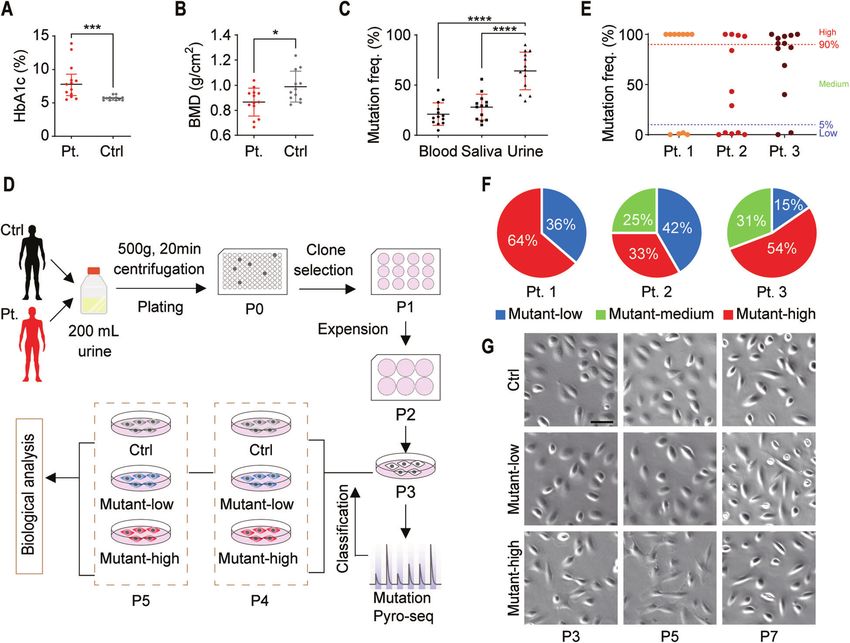
X. Gao et al.
                                                                                                                                                 3

Fig. 1 Isolation and proliferation of urine-derived mesenchymal stem cells (USCs) from patients with mitochondrial DNA 3243A > G
mutation and health controls. Box plots of HbA1c levels (A) and BMD T-score at total hip (B) in 13 m.3243A > G mutation carriers and 13 age,
sex, BMI-matched healthy controls. C Box plot of m.3243A > G mutation frequency in leukocytes, saliva and urine sediment of 13 m.3243A > G
mutation carriers. D A schematic workflow of isolation urine-derived stem cells from urine. Urine samples were collected from patients with
mt.3243A > G mutation and healthy controls and then centrifuged. Cells in the urine sample were resuspended in fresh USC medium, plated
on 96-well plated coated with gelatin for 7 days. Colonies were identified and sub-cultured in 12-well plate, 6-well plate and 10 cm plate to
expand. The levels of heteroplasmy in the derived USC clones were evaluated by pyrosequencing at passage 3 and marked for further
analysis. E The USCs showed a bimodal degree of mutation heteroplasmy, e.g., either high (>90%) or low ( G contents, with
only few clones containing medium level of mutation from three m.3243A > G mutation carriers. F The distribution of mutant-low, mutant-
medium and mutant-high USC clones from three m.3243A > G mutation carriers. G USCs retained a robust proliferation capability after several
passages. No differences in cell morphology were found in USCs with different heteroplasmy levels. Scale bar = 50 µm.

these genes was elevated in Mut-H USCs (Fig. 3E). We confirmed            improve mitochondrial functions that are impaired by high level
this result by RT-qPCR (Fig. 4A). Western blot further validated that    of the m.3243A > G mutation.
the levels of these UPRmt-related proteins were also elevated in
Mut-H USCs (Fig. 4B–G). In addition, decreased expression of             UPRmt inhibition alleviated deficits in osteogenesis from
WNT7B (Fig. 4H), as well as decreased levels of p-GSK3β in Mut-H         m.3243A > G USCs
USCs were also confirmed (Fig. 4I, J).                                    Since inhibiting UPRmt improved mitochondrial functions in Mut-H
                                                                         USCs (Fig. 5), given that ATF5 has been reported to play a
UPRmt inhibition rescued defective mitochondrial functions in            regulatory role during osteogenesis [33], we examined whether
Mut-H USCs                                                               inhibition of ATF5 could rescue the osteogenesis defect. Upon
ATF5 is a critical transcription factor mediating UPRmt in human         ATF5 knockdown, the levels of GSK3B and WNT7B were increased
cells [30]. In our study, upon ATF5 knockdown by siRNA, the              in all USCs, and restored to wild-type levels in Mut-H USCs (Fig. 6A,
expression levels of mtHSP70, HSP60 and LONP1 were greatly               B). RUNX2, OCN and BMP2 are classic markers for osteogenesis. The
reduced in all USCs (Fig. 5A), consistent with a critical role of ATF5   osteogenesis defect was shown by immunocytochemical analyses
in regulating UPRmt. To further investigate the role of UPRmt in         as well as Alkaline Phosphatase and Alizarin Red S staining (Fig.
m.3243A > G induced mitochondrial deficits, we examined ΔΨm               S4). Knockdown of ATF5 increased the expression of RUNX2, OCN
and ROS after ATF5 knockdown. ATF5 deficiency increased ΔΨm               and BMP2 (Fig. 6C–E), suggesting enhanced osteogenic potentials.
(Fig. 5B, C), and reduced ROS levels (Fig. 5D, E) in all USCs.           Further, ATF5 knockdown corrected the osteogenesis defect in
Importantly, the abnormally high ROS levels in Mut-H USCs were           m.3243A > G Mut-H USCs (Fig. 6F), suggesting this gene may serve
profoundly reduced (Fig. 5D, E), reaching wild-type levels. These        as a potential therapeutic target for treatment of osteoporosis
results suggest that targeting ATF5-mediated UPRmt could                 related to m.3243A > G mutation.

Cell Death and Disease (2021)12:701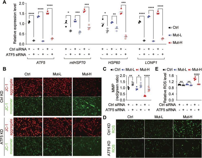
X. Gao et al.
4
Insulin 30 U/day; acarbose 50 mg/day
DISCUSSION
Insulin 60 U/d; acarbose 300 mg/day
In this study, we reported the first case of generating urine-
derived mesenchymal stem cells (USCs) from m.3243A > G
patients. We discovered that ATF5-mediated UPRmt could be an
essential mechanism underlying the impaired mitochondrial
function and poor osteogenic potentials in Mut-H USCs. Our
study represented one of many efforts to circumvent the hurdles
in establishing appropriate in vitro models for mitochondrial
Insulin 34 U/day
mutation research. The lack of effective in vitro and in vivo models
of mitochondrial DNA mutation has hampered mechanistic
Medication
studies [35]. The traditional cytoplasmic hybrid (cybrid) cell lines
[36] or human induced pluripotent stem (hiPS) cells were either
artificial or laborious to generate [15, 37, 38]. HiPS cells often carry
/
/
/
additional genetic and mitochrondral mutations created during
the induction process [39, 40]. Recently, we and others have
successfully derived USCs from human subjects through a simple,
HbA1c (%)
low-cost, and non-invasive method [41, 42]. One interesting
feature of our USCs from patients with the m.3243A > G mutation
was the bimodal distribution of heteroplasmy. Some clones were
5.4
5.8
5.2
8.0
5.9
13
nearly homoplasmic wild-type while the majorities were nearly
homoplasmic mutant-type. The mutant-high USCs could be an
ideal in vitro model for studying the cellular and molecular
BMD of total hip (g/cm2)
alterations resulted from the m.3243A > G mutation, while
mutant-low USCs could be potential cell sources for autologous
cell-replacement therapy. Medium heteroplasmy is potentially
linked to a growth disadvantage, which remains to be determined.
Our results provided supports to the emerging role of m.3243A > G
in bone mineralization deficiency. We and another group recently
reported that m.3243A > G mutation was associated with loss of bone
0.978
0.888
0.861
0.918
0.843
0.726
density in affected patients [11, 12]. In the current study, we further
confirmed this newly characterized pathological phenotype (Table 1),
and started to unveil the underlying signaling pathways linking the
Information on patient donors with m.3243A > G and healthy controls for generation of USCs.
m.3243A > G mutation to the loss of bone density. By taking
Diabetes mellitus; SNHL;
Typical clinical features
advantages of the in vitro USCs model, we showed that activation
Diabetes mellitus; SNHL
Diabetes mellitus; SNHL
of the mitochondrial stress response UPRmt and the decline in the
Wnt/β-catenin pathway could result in poor osteogenic potentials of
Mut-H USCs. Inhibition of UPRmt by knocking down of ATF5 reversed
the expression of GSK3B and WNT7B, resulting in increased
mineralization of in vitro cultured Mut-H USCs. Our study has
therefore suggested that ATF5 could be a novel and potential
therapeutic target for treating osteoporosis due to m.3243A > G. The
improved osteogenesis from ATF5-depleted USCs might be linked to
/
/
/
improved mitochondrial functions. It remains to be determined
whether other m.3243A > G-associated pathological phenotypes such
BMI (kg/m2)
as diabetes, deafness, and other symptoms in MELAS could also be
ameliorated by ATF5 targeted inhibition.
The upstream signaling turning on UPRmt in mutant-high
18.5
17.9
20.1
21.6
20.4
19.0
m.3243A > G remains unclear. UPRmt can be activated by an
imbalance between mitochondrial versus nuclear protein synth-
esis. Therefore, one possibility is that the m.3243A > G mutation to
tRNA gene reduces protein synthesis in the mitochondria,
Male
Male
Male
Male
Male
Male
triggering the mitonuclear imbalance, hence UPRmt activation.
Sex
Indeed, decreased mitochondrial translation in m.3243A > G cells
has been reported in several studies [21, 22], and increased
expression of mitochondrial stress-responsive genes including
Age (years)
heat shock protein (HSPs) have also been reported [16].
Interestingly, mutant-low USCs did not induce UPRmt, nor did
they have altered osteogenesis or mitochondrial morphology or
31
30
24
30
39
27
functions. These results were somewhat expected, because the
mutant-low USCs in this study have mutation rate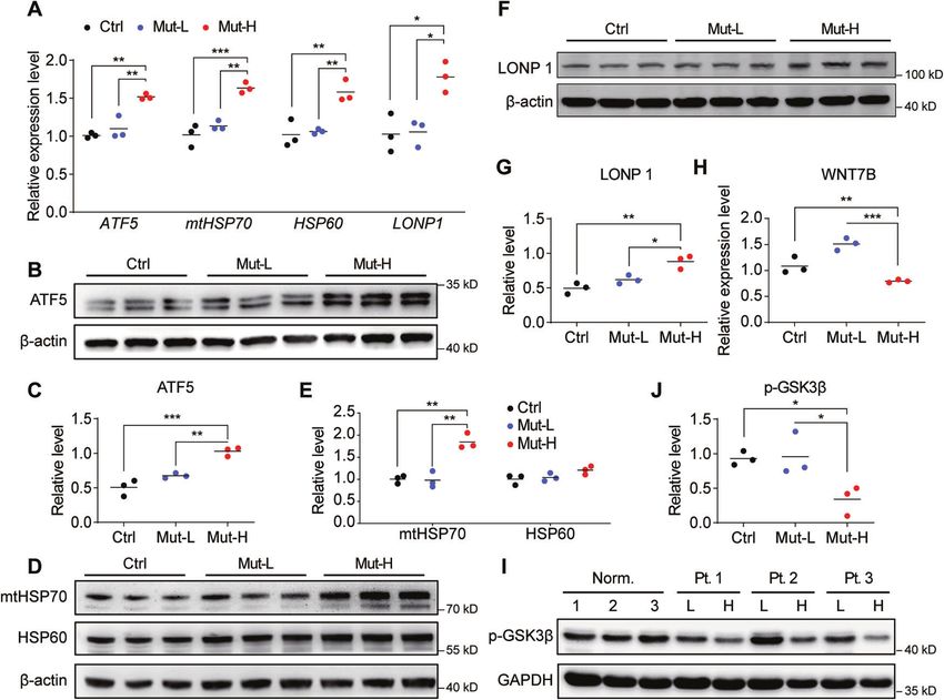
X. Gao et al.
                                                                                                                                                  5

Fig. 2 Impaired mitochondrial morphology and function in m.3243A > G mutant-high USCs. A Mitochondrial morphology as examined by
transmission electric microscopy (TEM). Mitochondrial morphology in Ctr USCs (a–a”: a. Cross-section of Ctr USCs showed round, sausage-like
and elongated mitochondria; a’. Enlarged mitochondrial with rich cristae; a”. Mitochondrial junction indicating fission/fusion events (purple
arrow)), Mut-L USCs (b–b”, similar to those in controls), and Mut-H USCs (c–c”: c. cross-section of Mut-H USCs showed mostly abnormally round,
little sausage-like and no elongated mitochondria; c’. Enlarged mitochondria lacking normal cristae; and c”. abnormal mitochondrial junctions).
B Quantification plot showed Mut-H USCs contained high levels of mitochondria with impaired cristae structure. C Quantification plot showed
Mut-H USCs contained less elongated and sausage-like mitochondria. D Quantification plot showed Mut-H USCs contained less fission/fusion
junctions. E Intracellular reactive oxygen species (ROS) generation was measured by flow cytometry detecting DCF fluorescence intensity. F
Quantification plot for E. The ROS level was increased in Mut-H USCs. G The percentage of abnormal-MMP cells was measured by flow
cytometry detecting the percentage of green fluorescence of JC-1 dye. H Quantification plot for G. The mitochondrial membrane potential
was decreased in Mut-H USCs USCs. Scale bar: 6 µm. Mitochondria with indicated morphologies were quantified from TEM images. *P < 0.05,
**P < 0.01, ***P < 0.001.

Cell Death and Disease (2021)12:701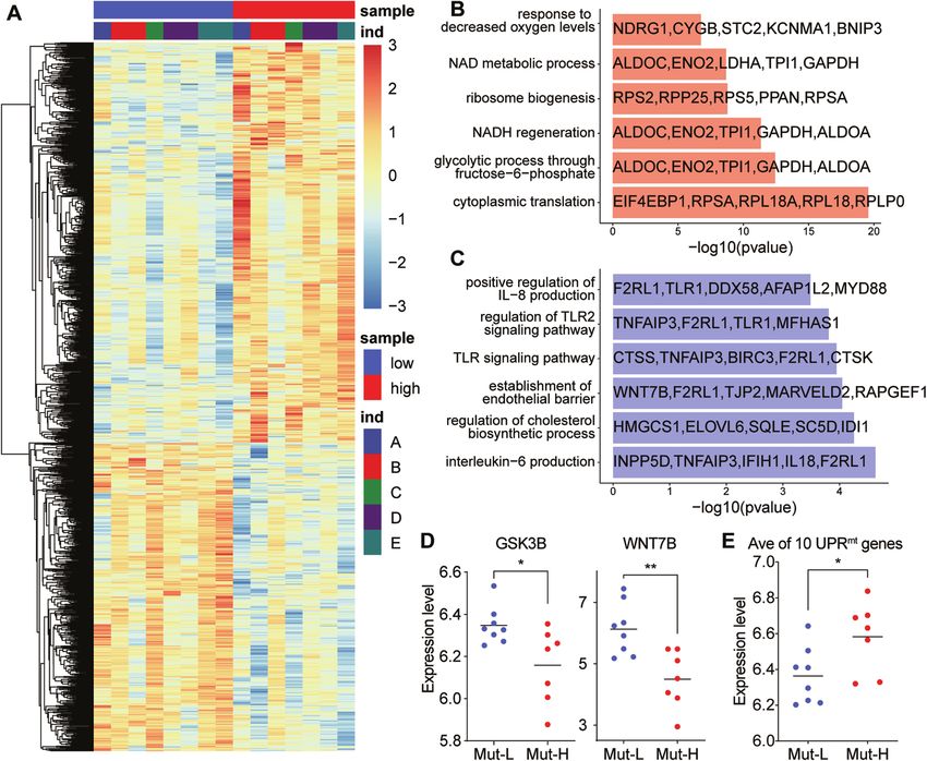
X. Gao et al.
6

    Fig. 3 Different transcriptional profiles between Mut-H and Mut-L USCs. A Heatmap of whole transcriptome data showing different gene
    transcription from Mut-H to Mut-L USCs. B Gene ontology (GO) analysis exhibit different enrichment of gene families in Mut-H USCs. C Gene
    ontology (GO) analysis exhibit different enrichment of gene families in Mut-L USCs. D GSK3B and WNT7B mRNA expression decreased in Mut-
    H USCs. E A panel of ten genes related to UPRmt was found to express higher in Mut-H USCs. *P < 0.05, **P < 0.01.

    UPRmt could also function to promote the expansion of defective             Sixth People’s Hospital. A total of 13 participants carrying the m.3243A > G
    mtDNA, leading to accumulation of defective mitochondria,                   mutation were found, including 12 carriers with diabetes and 1 carrier with
    whereas UPRmt inhibition preferentially depleted defective mtDNA            normal glucose tolerance. In addition, thirteen sex- and age-matched
    [31, 32]. The UPRmt could function similarly in our study, where            healthy controls with no m.3243A > G mutation, no history of diabetes, no
    UPRmt activation in USCs kept high levels of m.3243A > G                    hearing difficulties and no osteoporosis were also enrolled. The general
                                                                                clinical characteristics of each participant (e.g., history of diabetes and
    mutation, leading to compromised mitochondria and reduced                   complications, treatment, as well as mitochondria-associated symptoms)
    osteogenesis. On the contrary, inhibition of UPRmt by ATF5                  were obtained through standard questionnaires and comprehensive
    knockdown improved mitochondrial function and promoted                      clinical examinations.
    osteogenesis, through mechanisms involving amplified Wnt
    signaling. It will be important to study at the molecular level,
                                                                                m.3243A > G mutation analysis
    whether and how UPRmt regulates m.3243A > G heteroplasmy in                 Peripheral blood leukocytes, saliva, and urine samples were obtained from
    the disease contexts.                                                       all subjects. DNA was extracted from samples using an automated nucleic
       Taken together, by establishing human USCs as an in vitro                acid extraction instrument (Lab-Aid 820; BioV, China). High-resolution
    model to study mitochondria DNA mutations, we revealed an                   melting analysis was used for rapid m.3243A > G mutation scanning. The
    important role of ATF5-dependent UPRmt in maintaining mito-                 accurate quantification of the heteroplasmy levels of the m.3243A > G
    chondrial functions and osteogenic potentials. Our study provided           mutation in different samples was determined by pyrosequencing as
    new insights for better understanding osteoporosis in patients              previously described [43].
    with m.3243A > G mutation and identified ATF5 being a potential
    therapeutic target for this pathological condition.                         Cell culture
                                                                                Urine samples from three m.3243A > G participants and three healthy
                                                                                individuals were collected and cultured. Isolation and amplification of USCs
    MATERIALS AND METHODS                                                       has been described before [34, 44]. Fresh urine samples (200 ml) from
    Human subjects                                                              patients were processed immediately by adding penicillin (10 kU/ml) and
    This study was approved by the Institutional Review Board of Shanghai       streptomycin (10 mg/ml) to prevent contamination, then centrifuged and
    Jiao Tong University Affiliated Sixth People’s Hospital and was conducted    washed with phosphate-buffered saline (PBS). The sediment containing live
    in accordance with the Declaration of Helsinki. Written informed consent    cells was resuspended in Dulbecco’s modified Eagle medium (DMEM)
    was obtained from each subject. Participants were recruited to screen for   supplemented with 2 % (vol/vol) fetal bovine serum (FBS; Gibco, USA), 10 ng/
    the m.3243A > G mutation at the Shanghai Jiao Tong University Affiliated     ml of human epidermal growth factor (hEGF), 2 ng/ml of platelet-derived

                                                                                                       Cell Death and Disease (2021)12:701
X. Gao et al.
7
Fig. 4 Mut-H USCs had elevated UPRmt and reduced GSK3B and WNT7B. A The mRNA levels of UPRmt target genes (ATF5, HSP70, HSP60 and
LONP1) were elevated in Mut-H USCs. Total RNAs were extracted from Ctr, Mut-L and Mut-H USCs, and expression of UPRmt target genes were
quantified through real-time quantitative PCR (RT-qPCR). B–G The protein levels of UPRmt target genes (ATF5, HSP70, HSP60 and LONP1) were
elevated in mutant-high USCs. Total protein lysates were extracted from USCs and protein levels were analyzed by western blot. β-actin served as
internal loading control. C, E, G were quantification plot for B, D, F, respectively. H mRNA levels of WNT7B were reduced in Mut-H USCs. I Protein
levels of p-GSK3β were decreased in Mut-H USCs. GADPH serves as internal control. J Quantification plot for I. *P < 0.05, **P < 0.01, ***P < 0.001.
growth factor (PDGF), 1 ng/ml of transforming growth factor (TGF)-β, 2 ng/ml About 60 mg of tissues were ground into powder by liquid nitrogen in a
of basic fibroblast growth factor (bFGF), 0.5 μM hydrocortisone, 25 μg/ml of 2ml tube, followed by being homogenized for 2 min and rested
insulin, 20 μg/ml of transferrin, 549 ng/ml of epinephrine, 50 ng/ml of horizontally for 5 min. The mix was centrifuged for 5 min at 12,000 × g at
triiodothyronine (T3), L-glu and antibiotics. The cell suspension was plated 4 °C, then the supernatant was transferred into anew EP tube with 0.3 ml
into gelatin-coated 96-well plates and incubated at 37 °C in a humidified chloroform/isoamyl alcohol (24:1). The mix was shacked vigorously for 15 s,
atmosphere with 5 % CO2. After 7 days, nonadherent cells were removed by and then centrifuged at 12,000 × g for 10 min at 4 °C. After centrifugation,
washing with PBS and colonies derived from single cells were obtained. The the upperaqueous phase where RNA remained was transferred into a new
cells were passaged using 0.25% trypsin before confluence. tube with equal volume ofsupernatant of isopropyl alcohol, then
centrifuged at 13,600 rpm for 20 min at 4 °C. Afterdeserting the super-
natant, the RNA pellet was washed twice with 1 ml 75% ethanol, then the
Identification of USC surface markers by flow cytometry
mix was centrifuged at 13,600 rpm for 3 min at 4 °C to collect residual
The protocol to characterize cell surface markers by flow cytometry was
ethanol, followed by thepellet air dry for 5-10 min in the biosafety cabinet.
modified [44]. Briefly, cells were blocked with cold PBS containing 1% bovine
Finally, 25–100 µl of DEPC-treatedwater was added to dissolve the RNA.
serum albumin (BSA) for 30 min, then incubated with the following
Subsequently, total RNA was qualified and quantified using a Nano Drop
fluorescence-conjugated antibodies (Becton Dickinson, USA) for 1 h: CD29-
and Agilent 2100 bioanalyzer (Thermo Fisher Scientific, MA, USA).
PE, CD73-PE, CD90-PE, CD44-FITC, CD13-FITC, SSEA4-PE, CD31-FITC, CD45-FITC,
CD34-PE and HLA-DR-PE. Isotype-matched monoclonal antibodies were used
as controls (BD Biosciences). Cells were washed to remove unbound antibodies mRNA library construction. Oligo(dT)-attached magnetic beads were used to
and analyzed by using Guava easyCyte™ (Millipore, Billerica, MA, USA). purified mRNA. Purified mRNA was fragmentedinto small pieces with fragment
buffer at appropriate temperature. Then First-strand cDNA wasgenerated using
random hexamer-primed reverse transcription, followed by a second-strand
Karyotype analysis cDNAsynthesis. afterwards, A-Tailing Mix and RNA Index Adapters were added
Karyotype analysis was used to test chromosomal stability of USCs at by incubating to endrepair. The cDNA fragments obtained from previous step
passage 9. Cells were incubated in 20 µg/ml colchicines for 4 h at room were amplified by PCR, and productswere purified by Ampure XP Beads, then
temperature and then treated with 0.075 mM potassium chloride for dissolved in EB solution. The product was validated on the Agilent
15 min, finally fixed with methanol-to-acetic acid solution (3:1). Geimsa Technologies 2100 bioanalyzer for quality control. The double stranded PCR
staining were performed to visualize G-banding. Images were captured by productsfrom previous step were heated denatured and circularized by the
microscope (BX51; Olympus, Japan). splint oligo sequence to get the final library. The single strand circle DNA (ssCir
DNA) was formatted as the final library. The final library was amplified with
RNA sequencing and library preparation phi29 to make DNA nanoball (DNB), which had >300 copies of one molecular,
Total RNA extraction. Total RNA was extracted from the tissues using DNBs were loaded into the patterned nanoarray and single end 50 basesreads
Trizol (Invitrogen, Carlsbad, CA, USA) according to manual instruction. were generated on BGIseq500 platform (BGI-Shenzhen, China)
Cell Death and Disease (2021)12:701X. Gao et al.
8
Fig. 5 ATF5 knockdown reversed mitochondrial function in Mut-H USCs. A ATF5 knockdown decreased UPRmt related gene expression.
USCs were transfected with siRNA specific to ATF5 for 48 h and the mRNA levels of UPRmt related genes (ATF5, HSP70, HSP60 and LONP1)
expression were quantified by RT-qPCR. B Mitochondrial membrane potential was increased by ATF5 knockdown. USCs were transfected with
siRNA specific to ATF5 for 48 h and mitochondrial membrane potential was measured using fluorescence probe JC-1 assay system. Red color
indicates cells with normal mitochondrial membrane potential while green indicates cells with loss of mitochondrial membrane potential. C
Quantification plot for B. D Intracellular ROS was decreased by ATF5 knockdown. USCs were transfected with siRNA specific to ATF5 for 48 h
and stained with DCFH-DA. Green color indicate ROS-positive cell population. E Quantification plot for D. *P < 0.05, **P < 0.01, ***P < 0.001,
****P < 0.0001. Scale bar = 50 um.
Transmission electron microscopy (TEM)
Cells were fixed in 2.5% glutaraldehyde, and then loaded to copper grids JC-1 aggregates showing red fluorescence. The ratio of red fluorescence
coated with Formvar. After dehydration through a graded series of ethanol, and green fluorescence represented the ΔΨm of USCs. Fluorescence
the cell samples were embedded in Epon. Ultrathin sections were stained intensities were measured by ImageJ software [45]. The samples were also
with 2% uranyl acetate and lead citrate, then examined by TEM (H-7650, analyzed with the Cytomix FC500 flow cytometer [47]. Mitochondrial
HITACHI, Japan) membrane potential abnormality was expressed as “increases in the
percentage of abnormal-MMP cells”.
ROS measurement Real-time quantitative PCR (RT-qPCR). Total RNA was isolated by using
DCFH-DA probe was used for intracellular ROS measurement. Briefly, cells Trizol and reverse transcribed by using HiScript II Q RT SuperMix for qPCR
were washed with PBS and harvested, followed by incubation with 10 µM (Vazyme, China). RT-qPCR was performed by using AceQ Universal SYBR
DCFH-DA at 37 °C for 30 min in the dark. Cells were then washed with PBS qPCR Master Mix (Vazyme, China) with corresponding primer sets, which
and resuspended in DMEM. Fluorescence intensity was detected by a can be found in the supplemental Table S4. Gene expression levels were
Fluorescence Microscope (Nikon Ti-U, Tokyo, Japan) with excitation (Ex) calculated using the 2−ΔΔCt method (Livak and Schmittgen 2001). All
and emission (Em) wavelengths of 488 and 525 nm and the images were assays were repeated at least three times.
obtained with a Nikon Digital Sight DS-Fi2 camera. All fluorescence
intensities were measured by ImageJ. Cellular ROS contents were also
measured using a Cytomix FC500 flow cytometer. Western blotting
The total proteins were extracted from USCs using RIPA Lysis Buffer
Mitochondrial membrane potential measurement. Tetra-ethyl-benz-imida- (Beyotime, Shanghai, China). Total cell lysates were separated on 10%
zolyl-carbocyanine iodide (JC-1) was used to measure mitochondrial sodium dodecyl sulfate polyacrylamide gel electrophoresis and trans-
membrane potential as described before [45, 46]. USCs in DMEM were ferred onto polyvinylidene fluoride membranes. After blocking with 5%
incubated with an equal volume of staining solution containing 5 μg/ml nonfat dried milk in TBST, blots were probed with primary antibodies to
JC-1 at 37 °C for 20 min. Cells were washed three times with PBS and ATF5, mtHSP70, HSP60, LONP1, β-actin (Abcam, USA. ATF5: ab184923,
resuspended in DMEM. The samples were observed under a Fluorescence mtHSP70: ab171089, HSP60: ab190828, LONP1: ab103809, β-actin:
Microscope with Ex/Em wavelengths of 490/530 nm for JC-1 monomers ab8226) overnight at 4 °C and secondary antibody (Abcam, Cambridge,
showing green fluorescence and Ex/Em wavelengths of 525/590 nm for MA, USA) incubation at 37 °C for 1 h according to standard protocols.
Cell Death and Disease (2021)12:701X. Gao et al.
9
Fig. 6 Osteogenesis was impaired in Mut-H USCs but could be alleviated by ATF5 knockdown. A, B ATF5 knockdown increased WNT7B and
GSK3B gene expression. ATF5 was knockdown by siRNA for 48 h and the mRNA levels of WNT7B and GSK3B were quantified through RT-qPCR.
C–E Expressions of osteogenesis-related markers were increased by ATF5 knockdown in all USCs. USCs were transfected with siRNA specific to
ATF5 for 48 h and cultured in osteogenic induction medium for 48 h. mRNA levels of RUNX2, OCN and BMP2 were quantified through RT-qPCR.
F Alizarin Red S staining of calcium levels in Ctr, Mut-L, and Mut-H USCs. USCs were cultured in osteogenic induction medium with or without
siRNA transfection specific to ATF5 for 48 h and cultured in osteogenic induction medium for 21 days. Cells were stained with Alizarin Red S
and imaged with dissecting microscope under bright field. *P < 0.05, **P < 0.01, ***P < 0.001, ****P < 0.0001. Scale bar = 50 µm.
Immunoreactive proteins were visualized using an electrochemilumines- 10 min, then stained with 1% Alizarin Red S for 5 min. Cells were washed
cence system (Tinon, China). with PBS thoroughly to remove unbound dye and microscopic imaged.
siRNA knockdown Statistical analysis
The siRNAs used for downregulation of the capsid genes as well as the Data were expressed as the mean ± STD or as median (interquartile range
negative control siRNA were obtained from GenePharma, Shanghai, 25–75%) as appropriate. Differences between m.3243A > G carriers and
China (ATF5 siRNA: GCGAGUUUGAUUUCAAGCUTT, AGCUGUGAAAUCAAC controls were determined using the Student’s t-test or the Mann–Whitney
UCGCTT). The transient transfection of siRNA was performed with the U-test. Differences among mutant-high, mutant-low and control were
siRNA-Mate transfection reagent (GenePharma) according to manufac- analyzed by one-way ANOVA with Tukey’s correction for multiple compar-
turer’s instruction. isons. All P-values were two-sided, and values of P < 0.05 were considered
statistically significant (*P < 0.05, **P < 0.01, ***P < 0.001, ****P < 0.0001). All
statistics were performed with GraphPad Prism 8.
Osteogenic differentiation and identification
When USCs reached 80% confluence, cells were induced to differentiated
into osteogenic lineage cells by osteogenic induction media (Cyagen
Biosciences, China). To investigate the effect of inductive osteogenesis by REFERENCES
gene transfection only, ATF5 siRNA transduced USCs were cultured in 1. Wallace DC. A mitochondrial bioenergetic etiology of disease. J Clin Invest.
osteogenic induction media at 37 °C and 5% CO2. The medium was 2013;123:1405–12.
replaced every two to three days. After 14 days induction, alkaline 2. Gorman GS, Chinnery PF, DiMauro S, Hirano M, Koga Y, McFarland R, et al.
phosphatase staining was performed by an Alkaline Phosphatase Kit Mitochondrial diseases. Nat Rev Dis Prim. 2016;2:16080.
(Sigma-Aldrich, St. Louis, MO, USA) according to the manufacturer’s 3. Suomalainen A, Battersby BJ. Mitochondrial diseases: the contribution of orga-
instructions. After 21 days induction, Alizarin Red S staining was utilized to nelle stress responses to pathology. Nat Rev Mol Cell Biol. 2018;19:77–92.
detect calcified matrix deposition as described before [48]. Briefly, cells 4. Quiros PM, Mottis A, Auwerx J. Mitonuclear communication in homeostasis and
were washed with PBS and fixed with 4% formaldehyde solution for stress. Nat Rev Mol Cell Biol. 2016;17:213–26.
Cell Death and Disease (2021)12:701X. Gao et al.
10
5. Latorre-Pellicer A, Moreno-Loshuertos R, Lechuga-Vieco AV, Sanchez-Cabo F, 29. Nargund AM, Fiorese CJ, Pellegrino MW, Deng P, Haynes CM. Mitochondrial and
Torroja C, Acin-Perez R, et al. Mitochondrial and nuclear DNA matching shapes nuclear accumulation of the transcription factor ATFS-1 promotes OXPHOS
metabolism and healthy ageing. Nature. 2016;535:561–5. recovery during the UPR(mt). Mol Cell. 2015;58:123–33.
6. Suzuki T, Nagao A, Suzuki T. Human mitochondrial tRNAs: biogenesis, function, 30. Fiorese CJ, Schulz AM, Lin YF, Rosin N, Pellegrino MW, Haynes CM. The tran-
structural aspects, and diseases. Annu Rev Genet. 2011;45:299–329. scription factor ATF5 mediates a mammalian mitochondrial UPR. Curr Biol.
7. Yarham JW, Elson JL, Blakely EL, McFarland R, Taylor RW. Mitochondrial tRNA 2016;26:2037–43.
mutations and disease. Wiley Interdiscip Rev RNA. 2010;1:304–24. 31. Lin YF, Schulz AM, Pellegrino MW, Lu Y, Shaham S, Haynes CM. Maintenance and
8. Goto Y, Nonaka I, Horai S. A mutation in the tRNA(Leu)(UUR) gene associated with propagation of a deleterious mitochondrial genome by the mitochondrial
the MELAS subgroup of mitochondrial encephalomyopathies. Nature. unfolded protein response. Nature. 2016;533:416–9.
1990;348:651–3. 32. Gitschlag BL, Kirby CS, Samuels DC, Gangula RD, Mallal SA, Patel MR. Homeostatic
9. Morten KJ, Poulton J, Sykes B. Multiple independent occurrence of the 3243 responses regulate selfish mitochondrial genome dynamics in C. elegans. Cell
mutation in mitochondrial tRNA(leuUUR) in patients with the MELAS phenotype. Metab. 2016;24:91–103.
Hum Mol Genet. 1995;4:1689–91. 33. Guan J, Zhang J, Guo S, Zhu H, Zhu Z, Li H, et al. Human urine-derived stem cells
10. Murphy R, Turnbull DM, Walker M, Hattersley AT. Clinical features, diagnosis and can be induced into osteogenic lineage by silicate bioceramics via activation of
management of maternally inherited diabetes and deafness (MIDD) associated the Wnt/beta-catenin signaling pathway. Biomaterials. 2015;55:1–11.
with the 3243A>G mitochondrial point mutation. Diabet Med. 2008;25:383–99. 34. Guan JJ, Niu X, Gong FX, Hu B, Guo SC, Lou YL, et al. Biological characteristics of
11. Langdahl JH, Frederiksen AL, Hansen SJ, Andersen PH, Yderstraede KB, Duno M, human-urine-derived stem cells: potential for cell-based therapy in neurology.
et al. Mitochondrial point mutation m.3243A>G associates with lower bone Tissue Eng Part A. 2014;20:1794–806.
mineral density, thinner cortices, and reduced bone strength: a case-control 35. van den Ouweland JM, Lemkes HH, Ruitenbeek W, Sandkuijl LA, de Vijlder MF,
study. J Bone Min Res. 2017;32:2041–8. Struyvenberg PA, et al. Mutation in mitochondrial tRNA(Leu)(UUR) gene in a large
12. Geng X, Zhang Y, Yan J, Chu C, Gao F, Jiang Z, et al. Mitochondrial DNA mutation pedigree with maternally transmitted type II diabetes mellitus and deafness. Nat
m.3243A>G is associated with altered mitochondrial function in peripheral blood Genet. 1992;1:368–71.
mononuclear cells, with heteroplasmy levels and with clinical phenotypes. Diabet 36. Wilkins HM, Carl SM, Swerdlow RH. Cytoplasmic hybrid (cybrid) cell lines as a
Med. 2019;36:776–83. practical model for mitochondriopathies. Redox Biol. 2014;2:619–31.
13. Lightowlers RN, Taylor RW, Turnbull DM. Mutations causing mitochondrial dis- 37. Fujikura J, Nakao K, Sone M, Noguchi M, Mori E, Naito M, et al. Induced plur-
ease: What is new and what challenges remain? Science. 2015;349:1494–9. ipotent stem cells generated from diabetic patients with mitochondrial DNA
14. Grady JP, Pickett SJ, Ng YS, Alston CL, Blakely EL, Hardy SA. et al. mtDNA het- A3243G mutation. Diabetologia. 2012;55:1689–98.
eroplasmy level and copy number indicate disease burden in m.3243A>G 38. Zou Y, Wang A, Huang L, Zhu X, Hu Q, Zhang Y, et al. Illuminating NAD(+)
mitochondrial disease. EMBO Mol Med. 2018;10:e8262. https://doi.org/10.15252/ metabolism in live cells and in vivo using a genetically encoded fluorescent
emmm.201708262. sensor. Dev Cell. 2020;53:240–52. e247
15. Hamalainen RH, Manninen T, Koivumaki H, Kislin M, Otonkoski T, Suomalainen A. 39. Gore A, Li Z, Fung HL, Young JE, Agarwal S, Antosiewicz-Bourget J, et al. Somatic
Tissue- and cell-type-specific manifestations of heteroplasmic mtDNA 3243A>G coding mutations in human induced pluripotent stem cells. Nature. 2011;471:63–7.
mutation in human induced pluripotent stem cell-derived disease model. Proc 40. Park J, Lee Y, Shin J, Lee HJ, Son YB, Park BW, et al. Mitochondrial genome
Natl Acad Sci USA. 2013;110:E3622–30. mutations in mesenchymal stem cells derived from human dental induced
16. Picard M, Zhang J, Hancock S, Derbeneva O, Golhar R, Golik P, et al. Progressive pluripotent stem cells. BMB Rep. 2019;52:689–94.
increase in mtDNA 3243A>G heteroplasmy causes abrupt transcriptional repro- 41. Zhao T, Luo D, Sun Y, Niu X, Wang Y, Wang C, et al. Human urine-derived stem
gramming. Proc Natl Acad Sci USA. 2014;111:E4033–42. cells play a novel role in the treatment of STZ-induced diabetic mice. J Mol Histol.
17. Park H, Davidson E, King MP. The pathogenic A3243G mutation in human 2018;49:419–28.
mitochondrial tRNALeu(UUR) decreases the efficiency of aminoacylation. Bio- 42. Zhang D, Wei G, Li P, Zhou X, Zhang Y. Urine-derived stem cells: a novel and
chemistry. 2003;42:958–64. versatile progenitor source for cell-based therapy and regenerative medicine.
18. Borner GV, Zeviani M, Tiranti V, Carrara F, Hoffmann S, Gerbitz KD, et al. Genes Dis. 2014;1:8–17.
Decreased aminoacylation of mutant tRNAs in MELAS but not in MERRF patients. 43. Yan JB, Zhang R, Xiong C, Hu C, Lv Y, Wang CR, et al. Pyrosequencing is an accurate
Hum Mol Genet. 2000;9:467–75. and reliable method for the analysis of heteroplasmy of the A3243G mutation in
19. Yasukawa T, Suzuki T, Ueda T, Ohta S, Watanabe K. Modification defect at patients with mitochondrial diabetes. J Mol Diagnostics JMD. 2014;16:431–9.
anticodon wobble nucleotide of mitochondrial tRNAs(Leu)(UUR) with pathogenic 44. Jiang ZZ, Liu YM, Niu X, Yin JY, Hu B, Guo SC, et al. Exosomes secreted by human
mutations of mitochondrial myopathy, encephalopathy, lactic acidosis, and urine-derived stem cells could prevent kidney complications from type I diabetes
stroke-like episodes. J Biol Chem. 2000;275:4251–7. in rats. Stem Cell Res Ther. 2016;7:24.
20. Helm M, Florentz C, Chomyn A, Attardi G. Search for differences in post- 45. Widlansky ME, Wang J, Shenouda SM, Hagen TM, Smith AR, Kizhakekuttu TJ, et al.
transcriptional modification patterns of mitochondrial DNA-encoded wild-type Altered mitochondrial membrane potential, mass, and morphology in the mono-
and mutant human tRNALys and tRNALeu(UUR). Nucleic Acids Res. nuclear cells of humans with type 2 diabetes. Transl Res: J Lab Clin Med.
1999;27:756–63. 2010;156:15–25.
21. King MP, Koga Y, Davidson M, Schon EA. Defects in mitochondrial protein 46. Doughan AK, Harrison DG, Dikalov SI. Molecular mechanisms of angiotensin II-
synthesis and respiratory chain activity segregate with the tRNA(Leu(UUR)) mediated mitochondrial dysfunction: linking mitochondrial oxidative damage
mutation associated with mitochondrial myopathy, encephalopathy, lactic and vascular endothelial dysfunction. Circulation Res. 2008;102:488–96.
acidosis, and strokelike episodes. Mol Cell Biol. 1992;12:480–90. 47. Gravance CG, Garner DL, Baumber J, Ball BA. Assessment of equine sperm
22. Janssen GM, Maassen JA, van Den Ouweland JM. The diabetes-associated 3243 mitochondrial function using JC-1. Theriogenology. 2000;53:1691–703.
mutation in the mitochondrial tRNA(Leu(UUR)) gene causes severe mitochondrial 48. Qin H, Zhu C, An Z, Jiang Y, Zhao Y, Wang J, et al. Silver nanoparticles promote
dysfunction without a strong decrease in protein synthesis rate. J Biol Chem. osteogenic differentiation of human urine-derived stem cells at noncytotoxic
1999;274:29744–8. concentrations. Int J Nanomed. 2014;9:2469–78.
23. Shpilka T, Haynes CM. The mitochondrial UPR: mechanisms, physiological func-
tions and implications in ageing. Nat Rev Mol Cell Biol. 2018;19:109–20.
24. Jovaisaite V, Mouchiroud L, Auwerx J. The mitochondrial unfolded protein
response, a conserved stress response pathway with implications in health and
ACKNOWLEDGEMENTS
disease. J Exp Biol. 2014;217:137–43. Pt 1
We thank the supports of Biobank of Shanghai Jiao Tong University Affiliated Sixth
25. Mottis A, Herzig S, Auwerx J. Mitocellular communication: Shaping health and
People’s Hospital.
disease. Science. 2019;366:827–32.
26. Jovaisaite V, Auwerx J. The mitochondrial unfolded protein response-
synchronizing genomes. Curr Opin Cell Biol. 2015;33:74–81.
27. Houtkooper RH, Mouchiroud L, Ryu D, Moullan N, Katsyuba E, Knott G, et al.
Mitonuclear protein imbalance as a conserved longevity mechanism. Nature. AUTHOR CONTRIBUTIONS
2013;497:451–7. CW, YZ, and YES designed the project. XG, XY, and J Li performed RT-PCT and western
28. Mouchiroud L, Houtkooper RH, Moullan N, Katsyuba E, Ryu D, Canto C, et al. The experiments. ZJ, XY, FL and PL performed USC isolation, expansion and osteogenic
NAD(+)/sirtuin pathway modulates longevity through activation of mitochon- induction experiments. J Liu, YZ, CW, YES, and YW performed data analysis. XG, CW,
drial UPR and FOXO signaling. Cell. 2013;154:430–41. YZ, and YES wrote the manuscript.
Cell Death and Disease (2021)12:701X. Gao et al.
11
FUNDING Reprints and permission information is available at http://www.nature.com/
This work was partly supported by National Natural Science Foundation of China (NSFC reprints
81770883, 82070913), Research start-up fund from Shanghai Fourth People’s Hospital
(sykyqd01801). Shanghai Science and Technology Development Fund (20ZR1446000), Publisher’s note Springer Nature remains neutral with regard to jurisdictional claims
The National Key Research and Development Program (2020YFC2002800). in published maps and institutional affiliations.
COMPETING INTERESTS
The authors declare no competing interests.
Open Access This article is licensed under a Creative Commons
Attribution 4.0 International License, which permits use, sharing,
ETHICS STATEMENT adaptation, distribution and reproduction in any medium or format, as long as you give
This study was approved by the Institutional Review Board of Shanghai Jiao Tong appropriate credit to the original author(s) and the source, provide a link to the Creative
University Affiliated Sixth People’s Hospital and was conducted in accordance with Commons license, and indicate if changes were made. The images or other third party
the Declaration of Helsinki. Written informed consent was obtained from each material in this article are included in the article’s Creative Commons license, unless
subject. indicated otherwise in a credit line to the material. If material is not included in the
article’s Creative Commons license and your intended use is not permitted by statutory
regulation or exceeds the permitted use, you will need to obtain permission directly
ADDITIONAL INFORMATION from the copyright holder. To view a copy of this license, visit http://creativecommons.
Supplementary information The online version contains supplementary material org/licenses/by/4.0/.
available at https://doi.org/10.1038/s41419-021-03993-1.
Correspondence and requests for materials should be addressed to Y.E.S., Y.Z. or C.W. © The Author(s) 2021
Cell Death and Disease (2021)12:701You can also read