Preliminary preclinical study of Chol-DsiRNA polyplexes formed with PLL 30 -PEG 5K for the RNAi-based therapy of breast cancer
←
→
Page content transcription
If your browser does not render page correctly, please read the page content below
NANO-0000102363; No of Pages 14

                                                Nanomedicine: Nanotechnology, Biology, and Medicine
                                                                33 (2021) 102363

                                                                                                                               nanomedjournal.com

  Preliminary preclinical study of Chol-DsiRNA polyplexes formed with
      PLL[30]-PEG[5K] for the RNAi-based therapy of breast cancer
    Zhen Ye, PhD b , Mai Mohamed Abdelmoaty, MSc b, c , Vishakha V. Ambardekar, PhD b, 1 ,
           Stephen M. Curran, MSc b , Shetty Ravi Dyavar, PhD d , Lora L. Arnold f ,
        Samuel M. Cohen, MD, PhD e, f , Devendra Kumar, PhD b , Yazen Alnouti, PhD b ,
         Don W. Coulter, MD g , Rakesh K. Singh, PhD a, f , Joseph A. Vetro, PhD a, b,⁎
             a
             Center for Drug Delivery and Nanomedicine, College of Pharmacy, University of Nebraska Medical Center, Omaha, NE, USA
               b
                 Department of Pharmaceutical Sciences, College of Pharmacy, University of Nebraska Medical Center, Omaha, NE, USA
          c
           Therapeutic Chemistry Department, Pharmaceutical and Drug Industries Research Division, National Research Centre, Giza, Egypt
                  d
                    Department of Pharmacy Practice, College of Pharmacy, University of Nebraska Medical Center, Omaha, NE, USA
                             e
                              Havlik-Wall Professor of Oncology, University of Nebraska Medical Center, Omaha, NE, USA
                        f
                          Department of Pathology and Microbiology, University of Nebraska Medical Center, Omaha, NE, USA
    g
      Department of Pediatrics, Division of Pediatric Hematology/Oncology, Department of Radiation Oncology, J. Bruce Henriksen Cancer Research
                                         Laboratories, University of Nebraska Medical Center, Omaha, NE, USA
                                                               Revised 28 December 2020

Abstract

    RNA interference molecules have tremendous potential for cancer therapy but are limited by insufficient potency after i.v. administration.
We previously found that Chol-DsiRNA polyplexes formed between cholesterol-modified dicer-substrate siRNA (Chol-DsiRNA) and the
cationic diblock copolymer PLL[30]-PEG[5K] greatly increase the activity of Chol-DsiRNA against a stably expressed reporter mRNA in
primary murine syngeneic breast tumors after daily i.v. dosing. Here, we provide a more thorough preliminary preclinical study of Chol-
DsiRNA polyplexes against the therapeutically relevant target protein, STAT3. We found that Chol-DsiSTAT3 polyplexes greatly increase
plasma exposure, distribution, potency, and therapeutic activity of Chol-DsiSTAT3 in primary murine syngeneic 4T1 breast tumors after i.v.
administration. Furthermore, inactive Chol-DsiCTRL polyplexes are well tolerated by healthy female BALB/c mice after chronic i.v.
administration at 50 mg Chol-DsiCTRL/kg over 28 days. Thus, Chol-DsiRNA polyplexes may be a good candidate for Phase I clinical trials
to improve the treatment of breast cancer and other solid tumors.
© 2021 Elsevier Inc. All rights reserved.
Key words: RNA interference; DsiRNA; Drug delivery; Chol-DsiRNA polymer micelles; Chol-siRNA polymer micelles

   Abbreviations: 4T1, syngeneic murine breast cancer epithelial cells/         RNA interference (RNAi) is a natural, intracellular process
   TNBC model cells; 4T1-Luc, 4T1 cells that stably express luciferase;     that selectively decreases the expression of any specific protein
   Chol-DsiRNA, DsiRNA modified with 3′-cholesterol (sense strand);         at the mRNA level through the complementary binding of gene-
   Chol-*siRNA, nuclease-resistant siRNA modified with 3′-cholesterol       specific dsRNA molecules including microRNA (miRNA);
   (sense strand); DsiRNA, dicer-substrate siRNA; miRNA, microRNA; N/       small, interfering RNA (siRNA); or longer dicer-substrate
   P Ratio, charge molar ratio of positively charged amines from the
   polymer to negatively charged phosphates from the RNAi molecule; PLL
                                                                            siRNA (DsiRNA) (“RNAi molecules”). 1 Several proteins have
   [n]-PEG[5K], diblock copolymer of n poly-L-lysine residues and 5 kDa     been identified in tumor or tumor-associated cells where
   polyethylene glycol; Polyplexes, polymer complexes; RNAi, RNA            suppression can produce a therapeutic effect and/or increase
   interference; siRNA, small, interfering RNA; STAT3, signal transducer    the efficacy of current cancer treatments. 2 Thus, RNAi
   and activator of transcription factor 3.                                 molecules have tremendous potential in the treatment of cancer.
   Declaration of competing interests: JAV is cofounder of a company that       Many cancer therapy applications require i.v. administration
   licenses the described technology.
 ⁎ Corresponding author at: 986025 Nebraska Medical Center, WSH             to achieve a therapeutic effect. The potencies of RNAi molecules
   3026A, Omaha, Nebraska.
                                                                            after i.v. administration, however, are extremely low or
   E-mail address: jvetro@unmc.edu (J.A. Vetro).                            undetectable. 1 Several nanoscale dosage forms have been
 1
   Current address: Lupin Ltd., 46/47, A, Village Nande, Taluka Mulshi      developed to improve the potency of RNAi molecules after i.v.
   Dist. Pune, India.                                                       administration using a wide range of nanomaterials and/or
https://doi.org/10.1016/j.nano.2021.102363
1549-9634/© 2021 Elsevier Inc. All rights reserved.
2 Z. Ye et al / Nanomedicine: Nanotechnology, Biology, and Medicine 33 (2021) 102363
3,4
modified RNAi molecules. Identifying nanomaterials and/or *siLUC polyplexes under the same dosage regimen. The
RNAi molecule modifications that increase the potency of increased duration of RNAi activity, however, is likely due to
mRNA suppression in primary tumors and metastases and are differences in the activities of Chol-DsiRNA polyplexes and
suitable for clinical application, however, remains a critical Chol-siRNA polyplexes in primary 4T1-Luc tumors after i.v.
barrier to developing nanoscale dosage forms for RNAi-based administration and not differences in the activities of DsiRNA
therapies. and siRNA in 4T1 cells as electroporation of 4T1-Luc cells with
Conjugating 3′-cholesterol to nuclease-resistant siRNA equimolar amounts of DsiLUC or siLUC suppresses luciferase
(Chol-*siRNA) increases siRNA suppression of an endogenous activity to the same extent over 72 h. 11 Chol-DsiRNA
mRNA in the liver and jejunum after i.v. administration but with polyplexes also have higher loading (50 wt% Chol-DsiRNA
relatively low potency (50 mg Chol-*siRNA/kg). 5 We previ- vs. 25 wt% Chol-siRNA) and almost fully protect Chol-DsiRNA
ously found that Chol-siRNA polymer complexes (Chol-siRNA from degradation in 90% murine serum for 24 h at 37 °C without
polyplexes) formed with a block copolymer of 10 poly-L-lysine expensive nuclease-resistance modifications to DsiRNA. 11
residues and 5 kDa polyethylene glycol (PLL[10]-PEG[5K]) These results suggest Chol-DsiRNA polyplexes are a more
increase Chol-siRNA suppression of a native mRNA (CYPB) in promising lead formulation than Chol-siRNA polyplexes to
murine mammary MVEC (~90% suppression) to a much greater increase the potency and sustain the activity of RNAi molecules
extent than Chol-siRNA polyplexes formed with a block in solid breast tumors after i.v. administration.
copolymer of 50 poly-L-lysine residues and 5 kDa polyethylene In this study, we provide a more thorough preliminary
glycol (PLL[50]-PEG[5K]) (30% suppression), biodegradable preclinical assessment of Chol-DsiRNA polyplexes formed with
nanogels (40% suppression), or a graft copolymer of 2 kDa PLL[30]-PEG[5K] against the therapeutically relevant target
branched PEI and 10 kDa PEG (30% suppression) without gene STAT3 in primary murine syngeneic breast tumors after i.v.
affecting cell viability. 6 Given that PLL block length affects administration by determining (i) Chol-DsiSTAT3 pharmacoki-
Chol-siRNA polyplex activity and nuclease protection in vitro, netics, distribution, half-maximal ED50, and kinetics of STAT3
we next directly compared Chol-*siRNA polyplexes formed mRNA suppression in primary 4T1 breast tumors; (ii) rate-based
with PLL[10]-PEG[5K], PLL[30]-PEG[5K], or PLL[50]-PEG T/C ratio of Chol-DsiSTAT3 polyplex activity against primary
[5K] and found that polyplexes formed with PLL[30]-PEG[5K] 4T1 breast tumor growth; and (iii) toxicity of inactive Chol-
increase the potency of Chol-*siRNA to the greatest extent DsiCTRL polyplexes in healthy male and female BALB/c mice
against a stably expressed reporter gene (luciferase) in primary after dose escalation to 50 mg Chol-DsiCTRL/kg and in healthy
murine syngeneic 4T1 breast tumors (4T1-Luc) after daily i.v. female BALB/c mice after chronic administration at 50 mg
administration at 2.5 mg Chol-*siLUC/kg over three days Chol-DsiCTRL/kg over 28 days.
without affecting body weight. 7 Similar increases in RNAi
potency using PEGylated diblock copolymers and hydrophobi-
cally modified siRNA were later independently reported in Methods
primary ectopic human xenogeneic HeLa cervical tumors with
targeted, core-crosslinked “Chol-siRNA micelles” composed of Polymer
conventional Chol-siRNA (5′-cholesterol conjugated to the Methoxy-poly(ethylene glycol)-b-poly(L-lysine hydrochlo-
antisense strand) and integrin-targeted PLL[45]-b-PEG[12 K]- ride) with a 5 kDa polyethylene glycol block (PEG[5K]) and a
cRGD diblock copolymers 8 and in primary human xenogeneic 30 poly-L-lysine block (PLL [30]) [PLL[30]-PEG[5K]; avg. MW
MDA-MB-231 breast tumors with polyplexes composed of 9900 Da] was obtained from Alamanda Polymers (Huntsville,
palmitic acid-modified siRNA (PA-siRNA) and more hydro- AL). The number of Lys residues within the PLL [30] block was
p h o b i c p ( D M A E M A - c o - B M A ) - b- P E G [ 5 K ] d i b l o c k ±10% (PLL [27] to PLL [33]) and the polydispersity index of the
copolymers. 9 Thus, nanoscale dosage forms composed of entire polymer was between 1 and 1.1.
hydrophobically-modified RNAi molecules complexed with
PEGylated diblock copolymers have the potential to increase
RNAi molecules
the potency of RNAi molecules in solid tumors and metastases
after i.v. administration. DsiRNA was HPLC-purified, asymmetric, 25-mer dsRNA with
Although Chol-siRNA polyplexes formed with PLL[30]- 2-nucleotide UU overhangs on the 3′ end of the antisense strand: (1)
PEG[5K] increase the potency of Chol-*siLUC in primary DsiCTRL (GE Dharmacon) sense: 5′-CGU UAA UCG CGU AUA
murine 4T1-Luc breast tumors after i.v. administration, LUC AUA CGC GUA U-3′, antisense: 5′-AUA CGC GUA UUA UAC
mRNA suppression decreases 24 h after the last treatment. 7 GCG AUU AAC G(UU)-3′; MW: 16,523.02 g/mol; sequence
Given that polyplexes of dicer substrate siRNA (DsiRNA) and based on scrambled siRNA (IDT). (2) DsiSTAT3 (GE Dharmacon)
Lipofectamine® 2000 have 100-fold higher potency and longer sense: 5′-GGU CAA AUU UCC UGA GUU GAA UUA U-3′,
duration of mRNA suppression in HEK293 cells in vitro than antisense: 5′-AUA AUU CAA CUC AGG AAA UUU GAC C
polyplexes of siRNA and Lipofectamine® 2000 10, we next (UU)-3′; MW: 16,493.0 g/mol; sequence based on siGENOME
compared the activities of Chol-*siRNA polyplexes and Chol- Mouse STAT3 (GE Dharmacon). (3) Chol-DsiCTRL (IDT)
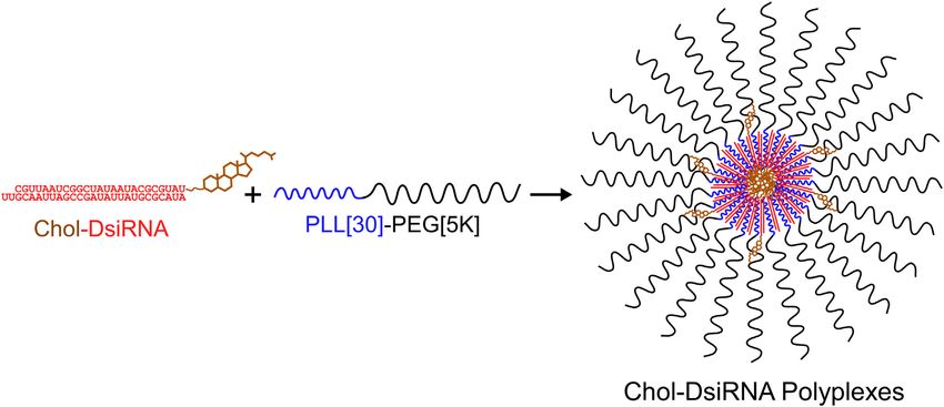
DsiRNA polyplexes formed with PLL[30]-PEG[5K] (Figure DsiCTRL with 3′-cholesterol conjugated to the sense strand; MW:
1) 11 and found that Chol-DsiLUC polyplexes increase the 17,278.9 g/mol. (4) Chol-DsiSTAT3 (GE Dharmacon) DsiSTAT3
duration of LUC mRNA suppression in primary murine 4T1-Luc modified with 3′-cholesterol as described for Chol-DsiCTRL; MW:
breast tumors after i.v. administration ~48 h longer than Chol- 17,197.92 g/mol.Z. Ye et al / Nanomedicine: Nanotechnology, Biology, and Medicine 33 (2021) 102363 3
Figure 1. Idealized electrostatic self-assembly of Chol-DsiRNA polyplexes formed with PLL[30]-PEG[5K]. A solution of negatively-charged DsiRNA (red)
modified with 3′-cholesterol (brown) on the sense strand (Chol-DsiRNA) is added to a solution of positively-charged diblock copolymers composed of 30 poly-
L-lysine residue blocks (blue) and 5 kDa polyethylene glycol blocks (black) (PLL[30]-PEG[5K]) at an N/P molar charge ratio of 1 (moles of positively-charged
primary amines (N)/moles of negatively charged phosphates). The negatively-charged phosphate backbones of Chol-DsiRNA then electrostatically bind and
neutralize the positively-charged PLL [30] blocks, converting PLL[30]-PEG[5K] unimers into amphiphilic diblock copolymers that spontaneously self-assemble
into Chol-DsiRNA polymer complexes (polyplexes) that are further stabilized by hydrophobic interactions between 3′-cholesterol groups. A similar idealized
self-assembly at slightly higher N/P ratio is envisioned for Chol-siRNA polyplexes formed with PLL[30]-PEG[5K].
For in vitro studies, lyophilized RNAi molecules were normalized to electroporated 4T1 cells alone at the indicated time
resuspended in sterile, nuclease-free ddH20 [100 μM] as directed points, and compared by two-tailed, unpaired t test.
(GE Dharmacon) and stored in aliquots (10 μL) at −80 °C. For in
Formation of Chol-DsiRNA polyplexes
vivo studies, lyophilized RNAi molecules were resuspended in
the indicated buffer on the day of injection. The concentration of a 2× Polymer Complexation Solution at
an N/P charge molar ratio of one mole positively-charged
Cell culture primary amines (N) from the PLL [30] block of PLL[30]-PEG
Murine 4T1 breast cancer epithelial cells (CRL-2539, ATCC) [5K] to one mole negatively-charged phosphates (P) from the
were cultured in Complete RPMI-1640 Media composed of phosphate backbone of Chol-DsiRNA (N/P 1) 11 was calculated
RPMI 1640/L-glutamine [1.1481 mM] (GE Healthcare Life based on the concentration of the 2× Chol-DsiRNA Complex-
Sciences) containing heat-inactivated FBS [10% v/v; endotoxin ation Solution4 Z. Ye et al / Nanomedicine: Nanotechnology, Biology, and Medicine 33 (2021) 102363
Solution plus 10 μL for experimental loss by diluting a thawed viscosity: 0.95 cP [viscosity of water at 22 °C]); remaining
frozen stock of Chol-DsiRNA with sterilized (0.2 μm filter) parameters: auto). The average estimated concentration of Chol-
HEPES Buffer [0.1 M HEPES in deionized H2O, pH 7.4] to DsiRNA polyplexes for each 1 nm bin from three independent
twice the desired concentration of Chol-DsiRNA in the final analyses was normalized as a percentage of the total average
Concentrated Stock Solution; (iii) preparing a 2× Polymer estimated concentration of Chol-DsiRNA polyplexes. A plot of
Complexation Solution plus 10 μL for experimental loss by accumulated percent of total Chol-DsiRNA polyplexes at each
dissolving sterilized PLL[30]-PEG[5K] in HEPES buffer at diameter (y-axis) vs. ln diameter (x-axis) was then fit against a
1 mg polymer/mL, incubating at r.t. for 30 min, and diluting cumulative Gaussian (percent) model using GraphPad Prism 9 to
with HEPES Buffer to the calculated 2× concentration; (iv) determine a best-fit mean and standard deviation from the
forming a Concentrated Stock Solution of Chol-DsiRNA lognormal curve.
polyplexes by adding the 2× Chol-DsiRNA Complexation The average zeta potential of Chol-DsiCTRL polyplexes (N/P
Solution dropwise to the 2× Polymer Complexation Solution at 1) at 25 °C in 0.1 M HEPES, pH 7.4 [1.5 mg Chol-DsiCTRL/
1/1 (v/v), mixing the solution by pipette aspiration/dispensation mL] was determined (n = 3 independent samples from the same
(30 s), and incubating (RT, 30 min). batch with 12 Zeta runs per sample) using a ZetaSizer Nano ZA
For in vivo studies, endotoxin-free solutions/reagents were (Malvern Instruments, Malvern, UK) equipped with a He-Ne
used where possible. A Concentrated Stock Solution of Chol- laser (λ = 633 nm) as the incident beam.
DsiRNA alone or Chol-DsiRNA polyplexes at N/P 1 was
prepared on the day of injection by (i) determining the total Endotoxin levels
volume of Concentrated Chol-DsiRNA Stock Solution (Chol- Chol-DsiCTRL endotoxin levels were quantitated by the
DsiRNA alone and Chol-DsiRNA polyplexes) required for all manufacturer (IDT). PLL[30]-PEG[5K] [0.2 mg/mL] and Chol-
mice in the study based on the injected dose (mg/kg), individual DsiRNA polyplexes [0.05 mg Chol-DsiSTAT3/mL] endotoxin
mass of each mouse, and total prepared i.v. injection volume levels were quantitated using an Endochrome-K Limulus
(125 μL for dose response/kinetics studies or 225 μL for toxicity Amebocyte Lysate Kit (Charles River Labs) as directed with
studies per mouse) plus 50 μL for experimental loss; (ii) endotoxin-free reagents and disposable labware under aseptic
preparing a 2× Chol-DsiRNA Complexation Solution (1/2 total conditions.
required volume of Concentrated Stock Solution of Chol-
DsiRNA) plus 25 μL for experimental loss by resuspending Transfection of murine 4T1 cells
lyophilized Chol-DsiRNA as directed by the manufacturer in
sterilized HEPES buffer to twice the desired concentration of Murine 4T1 cells were seeded in 6-well plates [0.5 × 10 6
Chol-DsiRNA in the Concentrated Stock Solution; (iii) steriliz- cells/well] in RPMI-1640 Media (3 mL) and incubated 14 to
ing PLL[30]-PEG[5K] and preparing 2× Polymer Complexation 16 h before transfection. On the day of transfection, a
Solution (same volume as 2× Chol-DsiRNA Complexation concentrated Stock Solution of Chol-DsiRNA polyplexes
Solution) as described for transfection studies above; (iv) [2 μM Chol-DsiRNA] was prepared as described (Section 2.5)
preparing the required volume of Concentrated Stock Solution then diluted to 200 nM Chol-DsiRNA in Complete RPMI-1640
of Chol-DsiRNA polyplexes as described above and (v) lacking FBS and antibiotics. Old growth media were aspirated
preparing the required volume of Concentrated Stock Solution and diluted polyplexes or Complete RPMI-1640 lacking FBS
of Chol-DsiRNA alone by diluting the 2× Chol-DsiRNA and antibiotics (1.5 mL) were added for 4 h; then an equal
Complexation Solution 1/1 (v/v) with sterilized HEPES Buffer. volume of 20% FBS Complete RPMI-1640 (1.5 mL) was added
The required i.v. injection volume (100 μL or 200 μL) of Chol- before further incubation for 20 h (24 h from the start of
DsiRNA alone or Chol-DsiRNA polyplexes plus 25 μL for each transfection). Average murine STAT3 mRNA copy numbers per
mouse was then prepared by combining (i) volume of ng total RNA ± propagated SD (n = 3 replicates from two
Concentrated Chol-DsiRNA or Chol-DsiRNA Polyplex Stock independent treatments) were then determined by RT-ddPCR
Solution adjusted to deliver the required dose of Chol-DsiRNA (Section 2.9), normalized to untreated cells, and compared by
based on mouse bodyweight, (ii) volume of sterilized (0.2 μm two-tailed, unpaired t test.
filter) HEPES buffer plus 1.5 M NaCl at 1/10 dilution [0.15 M
Quantitation of STAT3 mRNA and Chol-DsiSTAT3 by Reverse
NaCl final conc.], and (iii) volume of HEPES buffer adjusted to
Transcription-Droplet Digital™ PCR (RT-ddPCR)
final volume.
For mRNA, total purified RNA was diluted in sterile,
Hydrodynamic diameter and zeta potential of Chol-DsiCTRL nuclease-free ddH20 and converted to cDNA [~1 μg total]
polyplexes (Superscript™ VILO™ cDNA Synthesis Kit, Life Technolo-
gies). The number of STAT3 mRNA copies was determined by
The average hydrodynamic diameter of Chol-DsiCTRL droplet-digital PCR (ddPCR) (QX200 Droplet Digital PCR
polyplexes (N/P 1) was determined by nanoparticle tracking System, BioRad) from 10 ng of template cDNA using
analysis (NanoSight LM10 and NTA 2.3 analytical software, PrimeTime® Taqman assays (IDT, Coralville, USA) for murine
Malvern Instruments, UK). Chol-DsiRNA polyplexes [0.25 mg STAT3 mRNA and BioRad ddPCR reagents/consumables as
Chol-DsiSTAT3/mL] were prepared as described for Transfec- directed. STAT3 mRNA copies were then normalized to the
tion (Section 2.5) and recorded (shutter speed: 500, Gain: 680) mass of total RNA (template cDNA) [ng] in the ddPCR reaction
for video analysis (screen gain: 6; solution temperature: 22 °C; (assumes 1:1 RT conversion of total RNA to cDNA).Z. Ye et al / Nanomedicine: Nanotechnology, Biology, and Medicine 33 (2021) 102363 5
For Chol-DsiRNA, total purified RNA was diluted in sterile, Rad) and transferred (Mini Trans-Blot® Cell, BioRad) to PVDF
nuclease-free ddH20 and converted to cDNA [~1 μg total] membranes as directed. PVDF membranes were incubated with
(TaqMan™ MicroRNA Reverse Transcription Kit, Invitro- rocking in blocking buffer [5% (w/v) nonfat dry milk in TBST
gen™, ThermoFisher). Copy numbers of Chol-DsiSTAT3 buffer] (r.t., 1 h), rinsed with TBST (r.t., 5 s), incubated with
were quantitated by ddPCR using two different amounts of primary antibodies (β-actin [sc-47778] and Stat3 [sc-8019],
template cDNA [tumor: 6.67, 33.5 ng; lungs: 6.67, 33.35 ng Santa Cruz Biotechnology) [1:200 in TBST] (4 °C, overnight),
(Chol-DsiSTAT3 alone), 1.334, 13.4 ng (Chol-DsiSTAT3 rinsed 3× with TBST (r.t., 5 min), and incubated with secondary
polyplexes); liver: 1.334, 13.34 ng (Chol-DsiSTAT3 alone), antibody (horseradish peroxidase-conjugated mouse IgGκ bind-
0.1334, 1.334 (Chol-DsiRNA polyplexes); spleen, kidneys: ing protein; m-IgGκ BP-HRP [sc-516102], Santa Cruz Biotech-
0.2668, 1.334 ng; heart, brain: 13.34, 133.4 ng], stem-loop nology) [1:5000 in TBST] (r.t., 1 h), and rinsed 3× with TBST as
quantitative Custom TaqMan Small RNA Assays (Invitrogen™, above. The blot was incubated with HRP substrate (Luminata
ThermoFisher) for the antisense strand of Chol-DsiSTAT3, and Classico Western HRP, Sigma) as directed, visualized (MyECL
BioRad ddPCR reagents/consumables as directed. imager, Thermo Scientific), and the average ratios of Stat3/β-
Chol-DsiSTAT3 antisense copies/μL/ng template cDNA Actin protein band intensities ±SD (n = 2 measurements) were
were calculated as the slope of Chol-DsiSTAT3 antisense strand determined by imaging densitometry (ImageJ software).
copies/μL (y-axis) vs. mass of template cDNA (x-axis) (2
amounts). Mass of Chol-DsiSTAT3 (ng)/mg tissue was then
Pharmacokinetics of Chol-DsiSTAT3 after i.v. administration
calculated by multiplying Chol-DsiSTAT3 antisense copies/μL/
ng template cDNA with the slope from the standard curve of ng All procedures were approved by the University of Nebraska
Chol-DsiSTAT3 (y-axis) vs. Chol-DsiSTAT3 antisense copies/ Medical Center Institutional Animal Care and Use Committee
μL (x-axis) and dividing by the volume-normalized mass of the including guidelines for the humane treatment of animals. Chol-
tissue sample (10 mg). DsiSTAT3 alone and Chol-DsiSTAT3 polyplexes were prepared
for in vivo studies (Section 2.5) and injected (0.1 mL) into the tail
Tumor growth delay of primary murine 4T1 breast tumors vein of female BALB/c mice (n = 5 mice) at 2.5 mg Chol-
DsiSTAT3/kg (Section 2.10). Blood (~0.1 mL) from treated and
All procedures were approved by the University of Nebraska
untreated mice (plasma background) was collected into Li-
Medical Center Institutional Animal Care and Use Committee
heparinized tubes (0.3 mL Microvette Tubes, Sarstedt) from a
including guidelines for the humane treatment of animals.
submandibular bleed (5 mm Goldenrod lancet, Braintree Scien-
Primary tumors were prepared in female BALB/c mice as
tific) and plasma supernatants (2500 RCF, 15 min) (~50 μL) were
described 7 but hair was first removed from the region above the
stored at −80 °C. A standard curve for Chol-DsiSTAT3 was
tumor cell inoculation site (mammary fat pad #4) by shaving/
prepared by spiking Chol-DiSTAT3 or Chol-DsiSTAT3 poly-
applying Nair cream (removed with warm water and gauze after
plexes [450, 225, 112.5, 56.3, 28.1, and 14.1 ng Chol-DsiSTAT3]
30 s then rinsed with 70% ethanol) and 10-fold fewer 4T1 cells
into untreated plasma (5 μL) (n = 2 per standard).
[5 × 10 5 4T1 cells total] were injected SQ to facilitate tumor
Total RNA was isolated from plasma (miRNeasy Mini,
volume measurements by 3D surface scanning (TumorImager,
Qiagen) with modifications by adding QIAzol Lysis Reagent at
Biopticon). Tumor volumes and body weights were measured
3:1 plasma (v:v), vortexing, incubating (r.t., 5 min), adding
every other day before treatment, then daily until 48 h after the
water-soluble cholesterol (Cholesterol-Water Soluble, Sigma)
last treatment.
[1 mg/mL in deionized H2O] at 1:2 plasma (v/v) then SDS [10%]
When tumor volumes reached 30 to 50 mm 3 (day 0), vehicle
at 3:1 plasma (v/v), heating (95 °C, 5 min) and cooling (2 min)
alone [0.1 M HEPES, 0.15 M NaCl, pH 7.4, filter-sterilized] or
(4 cycles heating/cooling), adding chloroform at 10:1 plasma (v/
vehicle containing the indicated Chol-DsiRNA polyplexes was
v), vortexing, incubating (r.t., 2 min), pelleting (13,000 RCF,
injected (0.1 mL) into the tail-vein [2.5 mg Chol-DsiRNA/kg]
15 min, 2 times), adding 100% EtOH at 5:1 supernatant (v/v),
on days 0, 2, 4, and 6. Average daily tumor volumes ±SD (n = 5
and column purification as described (MiRNeasy Mini, Qiagen)
mice) were compared at each time point by multiple t tests. A
then quantitated by ABS260 (NanoDrop spectrophotometer,
rate-based T/C ratio and associated P value 12 were calculated as
ThermoFisher Scientific).
directed using the author-provided excel spreadsheet. On day 8
Total RNA isolated from plasma (2 μL) and Kit Diluent
(48 h after final dose), tumors were isolated and stored at −80 °C,
(98 μL) (n = 3 wells) was pipetted into a 96-well black (clear
and Stat3 protein levels were determined by Western blot
bottom) plate. Fluorophore (Quant-iT™ RiboGreen® RNA
(Section 2.11).
Reagent and Kit, Invitrogen) [1/1000 dilution in Kit Diluent]
Quantitation of Stat3 protein in primary 4T1 tumors by Western (100 μL) was added to each well, the plate was incubated (r.t., 2–
blot 5 min), and fluorescence (Ex480/Em520) was measured (Molec-
ular Devices SpectraMax ID3, top read). Samples and standards
Total protein was extracted from thawed tumors using Cell were diluted as necessary to fall within the linear range of
Extraction Buffer (Invitrogen) containing 1 mM PMSF, 1× fluorescence measurements. The average plasma concentration
protease inhibitor cocktail, and 1× EDTA (Thermo Scientific) of Chol-DsiSTAT3 at each time point ± propagated SD (n = 5
and quantified using Pierce BCA protein assay (Thermo mice) was then calculated as the difference between the plasma
Scientific) as directed. Protein lysates [40 μg] were resolved by concentration of total RNA in treated vs. untreated plasma.
SDS-PAGE (Mini-PROTEAN TGX Precast Protein Gels, Bio- Pharmacokinetic parameters were determined by non-6 Z. Ye et al / Nanomedicine: Nanotechnology, Biology, and Medicine 33 (2021) 102363
compartmental analysis (Phoenix WinNonlin version 8.2, (0.2 mL) biweekly into the tail vein of male (n = 6 mice) and
Certara, Princeton, NJ, USA). Area under the curve (AUC) female BALB/c mice (n = 6 mice) at an initial dose of 3.12 mg
was calculated using the linear log trapezoidal rule. Chol-DsiCTRL/kg and bodyweight was measured daily until
4 days after the final dose. The dose for each consecutive
Distribution of Chol-DsiSTAT3 in tumors and organs after i.v. injection was increased following a modified Fibonacci scheme
administration (100%, 65%, 52%, 40%, 29%, 33%, 33% increase over previous
dose) to a maximum of 50 mg Chol-DsiCTRL/kg (8 doses over
All procedures were approved by the University of Nebraska
28 days). Mice were euthanized at the end of the study by an
Medical Center Institutional Animal Care and Use Committee
overdose of Fatal-Plus® (Vortech Pharmaceuticals) (150 mg/kg
including guidelines for the humane treatment of animals. Chol-
body weight). Average daily body weights ±SEM (n = 6 mice)
DsiSTAT3 alone and Chol-DsiSTAT3 polyplexes were prepared
within each sex were compared to vehicle by 2-way ANOVA
for in vivo studies (Section 2.5) and injected (0.1 mL) into
female BALB/c mice (n = 5 mice) at 2.5 mg Chol-DsiSTAT3/
Chronic dosing of Chol-DsiCTRL polyplexes
kg (Section 2.10). After 15 min, mice were euthanized
(isoflurane drop jar/cervical dislocation), blood was removed/ All procedures were approved by the University of Nebraska
organs were perfused (sterile PBS) by cardiac puncture, organs Medical Center Institutional Animal Care and Use Committee
and tumors were isolated, weighed, suspended in RNAprotect including guidelines for the humane treatment of animals. Female
Tissue Reagent (Qiagen) as directed, and stored at −80 °C. BALB/c mice, Chol-DsiCTRL alone, and Chol-DsiCTRL poly-
To isolate total RNA, two pieces of isolated organs [lungs plexes were prepared as described (Section 2.14). On day 0,
~30 mg; liver ~50 mg; spleen ~30 mg; kidneys ~40 mg; heart vehicle alone or vehicle containing Chol-DsiCTRL or Chol-
~15 mg; brain ~30 mg] or tumors [~10 mg] were thawed and DsiCTRL polyplexes was injected (0.2 mL) biweekly into the tail
independently homogenized (Precellys Evolution technology, vein of male (n = 6 mice) and female BALB/c mice (n = 10 mice)
Bertin Instruments) in QIAzol Lysis Reagent (Qiagen) [10 μL at the indicated dose of Chol-DsiCTRL/kg. Bodyweight was
QIAzol Lysis Reagent:1 mg tissue] on “Hard” mode. A standard measured on each day of injection and 3 days after the final dose (9
curve for Chol-DsiSTAT3 and Chol-DsiSTAT3 polyplexes was doses over 28 days) and euthanized as described (Section 2.14).
prepared for each organ or tumor by spiking in Chol-DsiSTAT3 Toxicity was assessed by weight gain and terminal bodyweight,
or Chol-DsiSTAT3 polyplexes [tumor, lungs, liver, spleen, food/water consumption, histopathological evaluation of the
kidneys: 0.6, 3, 15, 75, 150, 300 ng Chol-DsiSTAT3; heart, thymus, lungs, liver, spleen, kidneys, and bone marrow, and a
brain: 0.15, 0.3, 0.6, 3, 15, 75 ng Chol-DsiSTAT3] into the complete blood count (CBC) including platelet count, serum liver
initial “Hard” mode homogenates of the respective organs or enzymes, creatinine, and BUN. Average values ±SEM (n = 10
tumors isolated from untreated mice. SDS [5% (w/v) in mice or less as indicated) of treatment groups were compared to
deionized H2O] [20 μL:1 mg tissue] and water-soluble choles- vehicle by 2-way ANOVA where appropriate.
terol (Sigma) [10 mg/mL in deionized H2O] [0.2 μL:1 mg
tissue] were added to the initial “Hard” mode homogenates and
further homogenized on “Soft” mode. A portion of the final Results
homogenate (300 μL; normalizes to 10 mg of tissue sample for
RNA extraction) was heated (92 °C, 5 min), cooled (r.t., 10 s), Activity of Chol-DsiSTAT3 polyplexes in murine syngeneic 4T1
and vortexed (4 cycles of heating, cooling, vortexing). 13 Total breast cancer epithelial cells
RNA was extracted by column purification after preparing the
Triple negative breast cancer (TNBC) is a subtype of breast
final homogenate as directed (miRNeasy Mini Kit, Qiagen,
cancer characterized by the absence of receptors for estrogen
Hilden, Germany) and quantitated by ABS260 (NanoDrop
(ER), progesterone (PR), and human epidermal growth factor
spectrophotometer, ThermoFisher Scientific). The average
(HER2) that has the highest rates of growth, metastasis, and
mass of Chol-DsiSTAT3 (ng)/mass of tissue (mg) ± propagated
recurrence, and the worst stage-dependent prognosis. 14 Al-
SD (n = 2 tissue/tumor pieces from n = 5 mice) was quantitated
though TNBC represents only 15% of new breast cancer
by RT-ddPCR (Section 2.9) and compared in each tissue by
diagnoses, it accounts for ~30% of breast cancer-related deaths
nonparametric two-tailed t tests with Holm–Sidak correction.
(~150,000 deaths worldwide) and has fewer treatment options
Dose escalation of Chol-DsiCTRL polyplexes due to the absence of ER, PR, and HER2. 15
Several therapeutically relevant proteins have been identified
All procedures were approved by the University of Nebraska in TNBC where suppression could potentially improve treatment
Medical Center Institutional Animal Care and Use Committee outcomes. 16 One such protein, signal transducer and activator of
including guidelines for the humane treatment of animals. Mice transcription factor 3 (STAT3), is a receptor tyrosine kinase
were acclimated to the animal room for approximately 7 days to (RTK)-associated signaling protein that is activated and
determine that they were healthy based on observed behavior and upregulated by cytokines and growth factors found in the
weight gain. Mice were randomized into treatment cohorts by tumor microenvironment. 17 Receptor activation of STAT3
weight and sex. Chol-DsiCTRL alone and Chol-DsiCTRL results in STAT3 dimerization, nuclear translocation, binding
polyplexes were prepared for in vivo studies as described to STAT3-specific DNA binding elements, and subsequent
(Section 2.5). On day 0, vehicle alone or vehicle containing transcription of genes involved in the regulation of cell
Chol-DsiCTRL or Chol-DsiCTRL polyplexes was injected differentiation, proliferation, apoptosis, angiogenesis, metastasis,Z. Ye et al / Nanomedicine: Nanotechnology, Biology, and Medicine 33 (2021) 102363 7
Figure 2. Suppression of murine STAT3 mRNA by DsiSTAT3 and Chol-DsiSTAT3 polyplexes in murine 4T1 breast cancer epithelial cells 24 h after treatment.
4T1 cells were (A) electroporated alone or in the presence of inactive DsiCTRL (white bar) or active DsiSTAT3 (gray bar) [300 nM] then incubated at 37 °C for
24 h or (B) incubated 4 h with serum-free media alone or containing inactive Chol-DsiCTRL (white bar) or Chol-DsiSTAT3 (black bar) [200 nM] complexed
with PLL[30]-PEG[5K] at an N/P ratio of 1 (50 wt% Chol-DsiRNA) before adding media containing 20% FBS at 1/1 (v/v) and incubating at 37 °C for 20 h.
Average murine STAT3 mRNA copy numbers per ng total RNA ± propagated SD (n = (A) 2 or (B) 3 replicates from two independent treatments) were then
determined by RT-ddPCR, normalized to (A) electroporation-only 4T1 cells or (B) untreated 4T1 cells, respectively, and compared by two-tailed, unpaired t test.
Results are representative of at least two independent experiments.
and immune responses. 18 Furthermore, STAT3 is one of the normalized murine STAT3 mRNA copy numbers vs. electropo-
main transcription factors involved in the immunomodulation of ration-only 4T1 cells 24 h after treatment by RT-ddPCR (Figure
cancer 19 and elevated expression of activated STAT3 is 2, A). Electroporation with DsiSTAT3 decreased STAT3 mRNA
associated with poor prognosis for breast cancer and many copy numbers 81% below electroporation-only 4T1 cells (Figure
other solid tumors. 18 , 20 Thus, given the role of STAT3 in breast 2, A, gray bar), whereas electroporation with DsiCTRL had no
and other solid tumors and presence of similar STAT proteins effect (Figure 2, A, white bar). Thus, the DsiSTAT3 sequence is
(STAT 1, 2, 4, 5, and 6) 21, there is great interest in specifically sufficiently active and the DsiCTRL sequence is sufficiently
inhibiting the expression of STAT3. inactive against murine STAT3 mRNA expression in murine
The syngeneic murine breast cancer epithelial cell line, 4T1, is a syngeneic breast cancer epithelial cells.
good model for TNBC because it lacks ER, PR, and HER2, shares To determine if Chol-DsiRNA polyplexes increase the potency
substantial molecular features with human TNBC 22, can be grown in of Chol-DsiRNA against a therapeutically relevant target gene in
immune competent female BALB/c mice, is poorly immunogenic, murine syngeneic breast cancer epithelial cells, we treated 4T1
has rates of growth and patterns of metastasis that resemble human cells with Chol-DsiCTRL or Chol-DsiSTAT3 complexed with
breast cancer, and the extent of late stage disease is comparable to PLL[30]-PEG[5K] at N/P 1 (50 wt% Chol-DsiRNA) and
stage IV breast cancer. 23,24 STAT3 is also important in the 4T1 compared normalized murine STAT3 mRNA copy numbers to
breast tumor model as inhibition of STAT3 by shRNA inhibits untreated 4T1 cells 24 h after treatment by RT-ddPCR (Figure 2,
tumor formation and the frequency of spontaneous metastases. 25 B). Transfection with Chol-DsiSTAT3 polyplexes decreased
Delivery of siSTAT3 to 4T1 cells using membrane-penetrating STAT3 mRNA copy numbers 64% below untreated 4T1 cells
peptides also inhibits 4T1 invasion and migration in vitro 26 and (Figure 2, B, black bar), whereas transfection with Chol-DsiCTRL
intratumoral administration of shSTAT3-expressing plasmids polyplexes decreased STAT3 mRNA copy numbers by 6% (Figure
inhibits primary tumor growth and spontaneous lung metastases. 27 2, B, white bar). These results indicate that STAT3 suppression in
We previously found that Chol-DsiRNA polyplexes increase 4T1 cells is due primarily to the activity of complexed Chol-
the potency of Chol-DsiLUC against stably expressed luciferase DsiSTAT3 and not potential non-specific effects of Chol-DsiRNA
in 4T1-Luc cells in vitro and as part of primary 4T1-Luc breast polyplexes. Thus, Chol-DsiRNA polyplexes increase the potency
tumors after i.v. administration. 11 Thus, we hypothesized that of Chol-DsiRNA against a therapeutically relevant target gene in
Chol-DsiRNA polyplexes can increase the potency of Chol- murine syngeneic breast cancer epithelial cells.
DsiRNA against therapeutically relevant genes expressed in 4T1
cells such as STAT3. Potency of Chol-DsiSTAT3 polyplex activity in primary murine
To identify an active DsiSTAT3 sequence and an inactive syngeneic 4T1 breast tumors after i.v. administration
DsiCTRL sequence against murine STAT3 mRNA in murine
syngeneic breast cancer epithelial cells, we electroporated 4T1 We previously found that Chol-DsiLUC polyplexes formed
cells with inactive DsiCTRL or murine DsiSTAT3 based on with PLL[30]-PEG[5K] at N/P 1 (50 wt% Chol-DsiLUC)
commercially available siSTAT3 sequences and compared suppress the luminescence of stably expressed luciferase in8 Z. Ye et al / Nanomedicine: Nanotechnology, Biology, and Medicine 33 (2021) 102363
Figure 3. Dose response and kinetics of STAT3 mRNA suppression in primary murine 4T1 breast tumors after i.v. administration of Chol-DsiSTAT3
polyplexes. (A) Vehicle alone (HEPES/0.15 M NaCl; 0.1 mL) or vehicle containing increasing doses of Chol-DsiSTAT3 (black circles) or a high dose of
inactive Chol-DsiCTRL (white circle) complexed with PLL[30]-PEG[5K] at N/P ratio 1 (~50 wt% Chol-DsiRNA) was injected into the tail veins of female
BALB/c mice bearing a single subcutaneous 4T1 tumor (~30 to 50 mm 3) in the mammary fat pad. After 48 h, average ratios of murine STAT3 to murine HPRT1
mRNA copies in primary 4T1 breast tumors ± propagated SD (n = 5 mice) were determined by RT-ddPCR then normalized and compared to vehicle alone
(0 mg Chol-DsiSTAT3/kg) by nonparametric Kruskal–Wallis one-way ANOVA with Dunn's post-test. A half maximal ED50 was calculated from a nonlinear fit
of the data. (B) Vehicle alone (HEPES/0.15 M NaCl; 0.1 mL) or vehicle containing Chol-DsiSTAT3 (0.5 mg/kg) complexed with PLL[30]-PEG[5K] at N/P
ratio 1 (~50 wt% Chol-DsiRNA) was injected into the tail veins of female BALB/c mice bearing a single subcutaneous 4T1 tumor (~30 to 50 mm 3) in the
mammary fat pad. At the indicated time point after injection, average ratios of murine STAT3 mRNA copies to murine HPRT1 mRNA copies in primary 4T1
breast tumors ± propagated SD (n = 5 mice) were determined by RT-ddPCR then normalized and compared to vehicle alone at the same time point by
nonparametric Mann–Whitney two-tailed t test. Results are representative of at least two independent experiments.
early-stage primary murine syngeneic 4T1-Luc breast tumors by a therapeutically relevant target gene in primary murine
~78% after daily i.v. injections of 2.5 mg Chol-DsiLuc/kg over syngeneic breast tumors after i.v. administration.
three days, whereas uncomplexed Chol-DsiLuc under the same
dosage regimen has no effect. 11 Daily injections at a constant
Kinetics of Chol-DsiSTAT3 polyplex activity in primary murine
dose, however, precluded determining the actual potency of
syngeneic breast tumors after i.v. administration
Chol-DsiRNA polyplexes in primary murine syngeneic breast
tumors. We previously found that Chol-DsiLUC polyplexes maxi-
To determine the potency of Chol-DsiRNA polyplexes mally suppress the luminescence of stably expressed luciferase
against a therapeutically relevant target gene in primary murine in primary murine syngeneic 4T1-Luc breast tumors 48 h after
syngeneic breast tumors after i.v. administration, we intrave- the first daily i.v. injection of a three-day regimen at 2.5 mg
nously administered increasing doses of Chol-DsiSTAT3 (up to Chol-DsiLuc/kg and maintain near-maximal suppression at least
5 mg/kg) or a single maximum dose of inactive Chol-DsiCTRL 48 h after the last injection. 11 Multiple daily i.v. injections and
(5 mg/kg) complexed with PLL[30]-PEG[5K] at N/P 1 (50 wt% the possibility of dose depot effects in the primary tumor by
Chol-DsiRNA) and compared normalized murine STAT3 potential tumor dose saturation at 2.5 mg Chol-DsiLuc/kg,
mRNA copy numbers in early-stage primary 4T1 breast tumors however, precluded determining the actual kinetics of Chol-
to treatment with vehicle alone by RT-ddPCR (Figure 3, A). We DsiRNA Polyplex activity in primary murine syngeneic breast
chose a 48-h timepoint given that (i) Chol-DsiLUC polyplexes tumors.
maximally suppress the luminescence of stably expressed To determine the kinetics of Chol-DsiSTAT3 Polyplex
luciferase in primary murine 4T1 breast tumors 48 h after the activity in primary murine syngeneic breast tumors after i.v.
first daily i.v. injection of a three-day regimen 11 and (ii) administration, we intravenously injected Chol-DsiSTAT3
luciferase activity closely matches luciferase mRNA and protein complexed with PLL[30]-PEG[5K] at N/P 1 (50 wt% Chol-
levels due the short intracellular half-life of luciferase protein DsiSTAT3) and compared normalized STAT3 mRNA copy
(~2 h). 28 numbers in early-stage primary murine 4T1 breast tumors to
Chol-DsiSTAT3 polyplexes (Figure 3, A, black circles) vehicle alone every 24 h over 96 h (four days) by RT-ddPCR
suppressed STAT3 mRNA copy numbers in primary 4T1 breast (Figure 3, B). We chose an i.v. dose of Chol-DsiSTAT3
tumors to a maximum of 54% vs. vehicle alone at the highest polyplexes that did not maximally suppress STAT3 mRNA
dose (5 mg Chol-DsiSTAT3/kg) with a half-maximal ED50 of expression in primary 4T1 breast tumors (0.5 mg Chol-
0.3 ± 0.1 mg/kg, whereas inactive Chol-DsiCTRL polyplexes DsiSTAT3/kg) (Figure 3, A, black circles) to exclude possible
had no effect at the highest dose of Chol-DsiSTAT3 polyplexes tumor dose saturation effects on the kinetics of Chol-DsiSTAT3
(5 mg Chol-DsiCTRL/kg) (Figure 3, A, white circle). These activity in the primary tumor.
results indicate that the suppression of STAT3 mRNA Chol-DsiSTAT3 polyplexes (Figure 3, B, black circles)
expression in primary 4T1 tumors is due to the activity of suppressed STAT3 mRNA copy numbers in primary 4T1 breast
complexed Chol-DsiSTAT3 and not potential non-specific tumors to a maximum of 43% vs. vehicle alone after 48 h but
effects of the Chol-DsiRNA polyplexes. Thus, Chol-DsiRNA suppression decreased to 11% after 24 h (72 h post-injection).
polyplexes greatly increase the potency of Chol-DsiRNA against Thus, similar to Chol-DsiLuc Polyplex activity against theZ. Ye et al / Nanomedicine: Nanotechnology, Biology, and Medicine 33 (2021) 102363 9
Figure 4. Effect of Chol-DsiSTAT3 polyplexes on primary murine 4T1 breast tumor growth and murine STAT3 protein expression after multiple i.v. treatments.
(A) Vehicle alone (white triangles) or vehicle containing Chol-DsiSTAT3 (black circles) or inactive Chol-DsiCTRL (Figure S2, white circles) complexed with
PLL[30]-PEG[5K] at N/P ratio 1 (50 wt% Chol-DsiRNA) was injected at 2.5 mg Chol-DsiRNA/kg into the tail veins of female BALB/c mice (black arrows)
bearing a single subcutaneous 4T1 breast tumor. Average daily tumor volumes ±SD (n = 5 mice) were then determined by 3D surface scanning and compared at
each time point by multiple t tests. A rate-based T/C ratio and associated P value [11] were calculated as described. (B) On day 8 (48 h after final i.v. injection),
steady-state murine Stat3 protein levels normalized to steady-state murine β-Actin protein levels in primary 4T1 tumors were determined by Western Blot and
(C) average ratios of Stat3/β-Actin protein band intensities ±SD (n = 2 measurements) were determined by imaging densitometry. Protein bands from images of
the same Western blot were used to generate (B). Results are representative of at least two independent experiments.
luminescence of stably expressed luciferase in primary 4T1-Luc Chol-DsiSTAT3 polyplexes suppressed 50% of total murine
breast tumors after the last daily i.v. injection of 2.5 mg Chol- Stat3 protein in primary 4T1 breast tumors vs. vehicle alone 48 h
DsiRNA/kg 11, a single dose of Chol-DsiSTAT3 polyplexes that after the final i.v. dose (day 8) (Figure 4, B & C). Furthermore,
is unlikely to saturate the primary tumor is maximally active in average body weights of mice from all treatment groups
primary murine breast tumors 48 h after i.v. administration but gradually increased over the course of the study (not shown),
maintains maximal mRNA suppression less than 24 h. indicating that (i) tumor growth inhibition is due to the activity of
complexed Chol-DsiSTAT3 and not animal weight loss and (ii)
Activity of Chol-DsiSTAT3 polyplexes against the growth of Chol-DsiSTAT3 polyplexes are not toxic under the current
primary murine syngeneic breast tumors after i.v. administra- dosage regimen. Thus, given that the National Cancer Institute at
tion NIH defines high anti-tumor activity as T/C ratios ≤10% at non-
toxic doses 12, Chol-DsiSTAT3 polyplexes have high anti-tumor
STAT3 is critical for the growth of 4T1 murine breast activity against primary murine syngeneic breast tumors after i.v.
tumors. 25 , 27 , 29 Thus, given that Chol-DsiSTAT3 polyplexes administration.
suppressed the expression of STAT3 mRNA in primary 4T1
tumors after i.v. administration (Figure 3), we expected that Pharmacokinetics of Chol-DsiSTAT3 and Chol-DsiSTAT3
Chol-DsiSTAT3 polyplexes would also inhibit 4T1 tumor polyplexes in healthy female BALB/c mice after i.v. adminis-
growth. tration
To determine if Chol-DsiSTAT3 polyplexes are therapeuti-
cally active against primary murine syngeneic breast tumors after DsiRNA must enter the cytosol of target cells to be cleaved by
i.v. administration, we intravenously injected Chol-DsiSTAT3 or Dicer into siRNA and incorporated into RISC complexes before
inactive Chol-DsiCTRL complexed with PLL[30]-PEG[5K] at suppressing complementary mRNA. 30 Thus, Chol-DsiRNA
N/P 1 (50 wt% Chol-DsiRNA) [2.5 mg Chol-DsiRNA/kg] every polyplexes are expected to increase the potency of Chol-DsiRNA
other day to early-stage 4T1 breast tumor-bearing mice and in primary syngeneic breast tumors after i.v. administration, in
compared tumor volumes to vehicle alone over 8 days by 3D part, by affecting the pharmacokinetics and distribution of Chol-
surface scanning (Figure 4, A, Figure S2). We administered DsiRNA.
2.5 mg Chol-DsiSTAT3/kg to ensure polyplexes maximal To first determine if Chol-DsiRNA polyplexes formed with
suppression of STAT3 mRNA expression in primary 4T1 breast PLL[30]-PEG[5K] affect the pharmacokinetics of Chol-Dsi-
tumors (Figure 3, A, black circles). STAT3, we intravenously injected Chol-DsiSTAT3 alone or
Chol-DsiSTAT3 polyplexes (Figure 4, A, black circles) complexed with PLL[30]-PEG[5K] at N/P 1 (50 wt% Chol-
inhibited the growth of primary murine 4T1 breast tumors at a DsiSTAT3) [2.5 mg Chol-DsiSTAT3/kg] into the tail veins of
rate-based T/C ratio of 8.6% 12 vs. vehicle alone (Figure 4, A, healthy female BALB/c mice. We then indirectly determined
white triangles), whereas inactive Chol-DsiCTRL polyplexes plasma concentrations of Chol-DsiSTAT3 over time as an
(Figure S1, white circles) had no effect on the growth of primary increase in total RNA extracted from the plasma of treated vs.
murine 4T1 breast tumors vs. vehicle alone (Figure S1, white untreated mice (Figure 5) given that total RNA in plasma from
triangles) under the same dosage regimen. Consistent with untreated mice is relatively constant (not shown). The high
STAT3 mRNA suppression (Figure 3, A) and the inhibition of variability of plasma concentrations and limited number of time
primary 4T1 breast tumor growth (Figure 4, A, black circles), points (Figure 5, A & B) prevented complete and accurate10 Z. Ye et al / Nanomedicine: Nanotechnology, Biology, and Medicine 33 (2021) 102363
Figure 5. Effect of Chol-DsiRNA polyplexes on the pharmacokinetics of Chol-DsiSTAT3 healthy female BALB/c mice after i.v. administration. (A) Chol-
DsiSTAT3 alone or (B) complexed with PLL[30]-PEG[5K] at N/P ratio 1 (50 wt% Chol-DsiSTAT3) [2.5 mg Chol-DsiSTAT3/kg] was injected into the tail
veins of healthy female BALB/c mice and average plasma concentrations of Chol-DsiSTAT3 ± SD (n = 5 mice) were determined indirectly at each time point
as differences in total extracted RNA from the plasma of treated vs. untreated mice by fluorescence assay. Error bars are present in (A) but indistinguishable at the
current scale.
DsiSTAT3/mg tumor]. Consistent with increased tumor distri-
Table 1 bution, Chol-DsiRNA polyplexes (Figure 6, black bars) also
Pharmacokinetic parameters of Chol-DsiSTAT3 and Chol-DsiSTAT3
polyplexes in healthy female BALB/c mice after IV administration.
increased the distribution of Chol-DsiSTAT3 4-fold to the
kidneys [4 ± 2 (SD) vs. 1.0 ± 0.4 ng Chol-DsiSTAT3/mg
PK parameter Chol-DsiSTAT3 Chol-DsiSTAT3 polyplexes organ], 11.2-fold to the lungs [4.6 ± 0.6 (SD) vs. 0.41 ±
(±SD) (±SD)
0.05 ng Chol-DsiSTAT3/mg organ], 9-fold to the liver [8.1 ±
C0 56 (64) 110 (7) 0.9 (SD) vs. 0.9 ± 0.2 ng Chol-DsiSTAT3/mg organ], and 4.3-
AUC0-∞ (h*μg/mL) 3 (1) 39 (13) fold to the spleen [10 ± 2 (SD) vs. 2.3 ± 0.4 ng Chol-
MRT0-∞ (h) 0.8 (0.9) 0.9 (0.4)
DsiSTAT3/mg organ] vs. uncomplexed Chol-DsiSTAT3 (Figure
Average plasma concentrations at time point = 0 (C0) and mean residence 6, white bars). Furthermore, although much lower overall levels
time (MRT0-∞) of Chol-DsiSTAT3 were determined by non-compartmental were observed than in other tissues, Chol-DsiRNA polyplexes
analysis (Phoenix WinNonlin) and average area under the curve (AUC0-∞) of
Chol-DsiSTAT3 (±SD from 5 mice) was determined by the linear log
increased the distribution of Chol-DsiSTAT3 4.6-fold to the
trapezoidal rule of the respective PK profiles (Figure 5). brain [0.41 ± 0.05 ng (SD) vs. 0.088 ± 0.007 ng Chol-Dsi-
STAT3/mg tumor] and 9.3-fold to the heart [0.27 ± 0.04 (SD)
vs. 0.029 ± 0.005 ng Chol-DsiSTAT3/mg tumor]. Thus, Chol-
compartmental analysis of the PK profiles. Despite the limited DsiRNA polyplexes may increase the potency of Chol-
analysis, Chol-DsiRNA polyplexes increased the AUC of Chol- DsiSTAT3 in primary syngeneic breast tumors after i.v.
DsiRNA 13-fold [39 ± 13 (SD) vs. 3 ± 1 h*μg/mL] (Table 1) administration, in part, by increasing the distribution of Chol-
and decreased clearance 8.3-fold [0.0012 ± 0.0003 (SD) vs. DsiSTAT3 to the primary tumor.
0.01 ± 0.01 L/min/kg] (not shown). Thus, Chol-DsiRNA poly-
plexes may increase the potency of Chol-DsiSTAT3 in primary Dose escalation of Chol-DsiCTRL polyplexes by i.v. adminis-
syngeneic breast tumors after i.v. administration, in part, by tration
increasing plasma exposure to Chol-DsiSTAT3. Chol-DsiRNA polyplexes formed with PLL[30]-PEG[5K]
Distribution of Chol-DsiSTAT3 and Chol-DsiSTAT3 polyplexes significantly increase the potency of Chol-DsiLUC 11 and Chol-
in 4T1 breast tumor-bearing female BALB/c mice DsiSTAT3 in primary murine syngeneic breast tumors after
multiple daily i.v. injections at 2.5 mg/kg without affecting body
To next determine if Chol-DsiSTAT3 polyplexes affect the weight gain over the course of the study. This suggests that Chol-
distribution of Chol-DsiSTAT3 to primary murine syngeneic DsiRNA polyplexes are well tolerated after i.v. administration.
breast tumors and normal tissues after i.v. administration, we To determine a maximally tolerated dose (MTD) of Chol-
intravenously injected Chol-DsiSTAT3 alone or complexed with DsiRNA polyplexes in mice after i.v. administration, we
PLL[30]-PEG[5K] at N/P 1 (50 wt% Chol-DsiSTAT3) [2.5 mg intravenously injected healthy male and female BALB/c mice
Chol-DsiSTAT3/kg] into the tail vein of primary 4T1 breast with vehicle alone or vehicle containing increasing doses of
tumor-bearing female BALB/c mice and compared the distribu- Chol-DsiCTRL alone or complexed with PLL[30]-PEG[5K] at
tion of Chol-DsiSTAT3 in perfused organs and tumors fifteen N/P 1 (50 wt% Chol-DsiCTRL) up to 50 mg Chol-DsiCTRL/kg
minutes post-injection by RT-ddPCR of the anti-sense strand of two times a week over 28 days (8 injections total) and compared
Chol-DsiSTAT3 (Figure 6). Chol-DiSTAT3 polyplexes (Figure body weights (Figure 7, A, Table S1), terminal body and tissue
6, black bars) increased the distribution of Chol-DsiSTAT3 2.7- weights (Table S2), the presence of liver cysts (Table S3), and
fold to primary 4T1 tumors vs. uncomplexed Chol-DsiSTAT3 histopathology of the liver, spleen, kidneys, and femurs after
(Figure 6, white bars) [1.6 ± 0.4 (SD) vs. 0.6 ± 0.1 ng Chol- decalcification (Table S4). We focused on inactive Chol-Z. Ye et al / Nanomedicine: Nanotechnology, Biology, and Medicine 33 (2021) 102363 11
Figure 6. Effect of Chol-DsiRNA polyplexes on the distribution of Chol-DsiSTAT3 in 4T1 breast tumor-bearing mice after i.v. administration. Chol-DsiSTAT3
alone [2.5 mg/kg] or complexed with PLL[30]-PEG[5K] at N/P ratio 1 (50 wt% Chol-DsiSTAT3) was injected into the tail veins of female BALB/c mice (black
arrows) bearing single, orthotopic (mammary fat pad) 4T1 tumors (~30 to 50 mm 3). After 15 min, the average ng of Chol-DsiSTAT3/mg tissue ± SD (n = 5
mice) was determined by RT-ddPCR of the DsiSTAT3 antisense strand and compared in each tissue by nonparametric two-tailed t tests with Holm–Sidak
correction.
Figure 7. Dose escalation and chronic dosing of Chol-DsiCTRL or Chol-DsiCTRL polyplexes in healthy mice by i.v. administration. (A) Vehicle alone
(endotoxin-free PBS, 0.2 mL) or vehicle containing uncomplexed Chol-DsiCTRL or Chol-DsiCTRL complexed with PLL[30]-PEG[5K] at N/P 1 (50 wt%
Chol-DsiCTRL) was injected biweekly into the tail veins of healthy male and female BALB/c mice at the indicated dose (black arrows) and average daily body
weights ±SEM (n = 6 mice) (Table S1) within each sex were compared to vehicle by 2-way ANOVA. Deaths: avehicle (day 18, one male, one female); bChol-
DsiCTRL (day 8, one male). (B) Vehicle alone (endotoxin-free PBS, 0.2 mL) or vehicle containing uncomplexed Chol-DsiCTRL or Chol-DsiCTRL complexed
with PLL[30]-PEG[5K] at N/P 1 (50 wt% Chol-DsiCTRL) was injected into the tail veins of healthy female BALB/c mice biweekly at the indicated dose (black
arrows). Average daily body weights ±SEM (n = 10 mice) were compared to vehicle by 2-way ANOVA. Deaths: aChol-DsiCTRL (day 14, one female); bChol-
DsiCTRL polyplexes [50 mg Chol-DsiCTRL/kg] (day 3, one female, day 14 two females, day 21 one female).
DsiCTRL polyplexes (Table 2) to separate the possibility of The liver did not show any histopathologic changes and the
additional toxicity by the suppression of STAT3 by Chol- spleens only showed differences in blood congestion but without
DsiSTAT3 polyplexes. abnormalities in the lymphoid structures.
Male (Figure 7, A, black symbols) and female mice (Figure 7, Chol-DsiCTRL polyplexes had the highest number of
A, white symbols) treated with vehicle alone (Figure 7, A, surviving male and female mice with liver cysts (4 out of 6),
triangles), Chol-DsiCTRL alone (Figure 7, A, circles), or Chol- but the number was similar to female mice treated with Chol-
DsiCTRL polyplexes (Figure 7, A, circles) continued to gain DsiCTRL (3 out of 6) and both male (3 out of 5) and female mice
weight up to four days after the highest dose [50 mg Chol- (2 out of 5) treated with vehicle alone (Table S3). These cysts,
DsiCTRL/kg] despite deaths with vehicle alone (day 18: one however, commonly occurred in BALB/c mice and were of
male and one female) and Chol-DsiCTRL alone (day 8: one similar size and frequency between treatment groups. Further-
male). Male and female mice from all treatment groups also had more, most liver cysts were microscopic in size and differences
similar terminal body weights (Table S2). in numbers between treatment groups may have been due to
Dose escalation with Chol-DsiCTRL increased relative liver sampling. As such, cystic changes in the liver are not considered
weights by 10% in male mice and decreased total and relative to be treatment related.
liver weights by ~8% in female mice compared to vehicle alone Liver, spleen, kidney, and femur histology was normal in
but was not associated with histopathologic changes. Chol- male and female mice from all treatment groups with occasional
DsiCTRL polyplexes also increased total and relative spleen mice having changes that are common in BALB/c mice but
weights by ~37% in male mice and decreased total and relative without differences between groups and thus are not considered
liver weights by 5 to 7% compared to vehicle alone (Table S2). treatment related (Table S4). Two deaths occurred with vehicle12 Z. Ye et al / Nanomedicine: Nanotechnology, Biology, and Medicine 33 (2021) 102363
Table 2
Representative characteristics of Chol-DsiCTRL, PLL[30]-PEG[5K], and Chol-DsiCTRL polyplexes.
Sample Endotoxin EU/mg (±SD) Loading Hydrodynamic Diameter Zeta potential
Chol-DsiCTRL wt% nm (±SD) mV (±SD)
Chol-DsiCTRL 0.104 (n.r.) a N/A N/A N/A
PLL[30]-PEG[5K] 0.0171 (0.0008) b N/A N/A N/A
Chol-DsiCTRL polyplexes (N/P 1) 0.023 (0.002) b 50 33 (2) c 5.2 (0.7) d
a
Determined by manufacturer (IDT).
b
Determined by Endochrome-K™ kit.
c
Calculated from a nonlinear fit of the lognormal distribution of Chol-DsiCTRL polyplex diameters in 0.1 M HEPES, pH 7.4 (Figure S2).
d
Determined in 0.1 M HEPES, pH 7.4.
alone (Figure 7, A, day 18: one male and one female) and one ulceration with acute inflammation, thus related to the repeated
death occurred with Chol-DsiCTRL (Figure 7, A, day 8, one injections and not the administered material (Table S8).
male), whereas no deaths occurred with Chol-DsiCTRL WBC, hemoglobin, hematocrit, lymphocytes, and platelets
polyplexes, suggesting that deaths were likely caused by the from all treatment groups were also within normal values in
stress of multiple i.v. injections. Thus, i.v. administration of BALB/c mice (Charles River Laboratories) (Table S9) and
Chol-DsiCTRL alone or complexed with PLL[30]-PEG[5K] at trauma observed around tail injection sites was consistent with
N/P 1 appears to be well tolerated by healthy male and female damage from the injection itself (not shown). Furthermore, Chol-
mice to at least 50 mg Chol-DsiCTRL/kg. DsiRNA polyplex endotoxin levels at 50 mg Chol-DsiCTRL/kg
(0.023 EU/mg Chol-DsiCTRL polyplexes, Table 2) were below
maximum FDA limits for i.v. administration to a 20 g mouse per
Chronic dosing with Chol-DsiCTRL polyplexes by i.v. admin- hour (0.1 EU/mg drug) 31, indicating that deaths were likely
istration caused by the stress of multiple i.v. injections and not the
To assess the toxicity of Chol-DsiRNA polyplexes in mice administered material or levels of endotoxin. Thus, chronic i.v.
after chronic dosing by i.v. administration, we intravenously administration of Chol-DsiRNA alone or complexed with PLL
injected healthy female BALB/c mice with vehicle alone or [30]-PEG[5K] is well tolerated by healthy female mice up to at
vehicle containing uncomplexed Chol-DsiCTRL at 50 mg/kg or least 50 mg Chol-DsiCTRL/kg.
Chol-DsiCTRL complexed with PLL[30]-PEG[5K] at N/P 1
(50 wt% Chol-DsiCTRL) at 25 and 50 mg Chol-DsiCTRL/kg
biweekly over 28 days (9 injections total) and compared body Discussion
weights (Figure 7, B, Table S5), food and water consumption
(Table S6), terminal body and tissue weights (Table S7), Our study provides evidence that Chol-DsiRNA polyplexes
histopathology of major organs and tail vein injection sites formed with PLL[30]-PEG[5K] are highly potent in solid primary
(Table S8), and hematology (Table S9). murine syngeneic breast tumors and well tolerated after i.v.
Mice chronically treated with vehicle alone (Figure 7, B, administration. We found that Chol-DsiRNA polyplexes (i) greatly
white triangles), uncomplexed Chol-DsiCTRL (Figure 7, B, increase the potency (half-maximal ED50 of 0.3 ± 0.1 mg Chol-
white circles), or Chol-DsiCTRL polyplexes at 25 mg Chol- DsiRNA/kg) (Figure 3, A) and therapeutic activity (rate-based T/C
DsiCTRL/kg (Figure 7, B, black/white squares) or 50 mg Chol- ratio of 8.6%) (Figure 4, A) of Chol-DsiRNA against an
DsiCTRL/kg (Figure 7, B, black squares) continued to gain endogenous, therapeutically relevant target gene (STAT3) in
weight up to four days after the last dose despite deaths with early-stage primary murine 4T1 breast tumors after i.v. adminis-
50 mg/kg Chol-DsiCTRL alone (day 14) and 50 mg/kg Chol- tration and (ii) are well tolerated as an inactive dosage form (i.e.,
DsiCTRL polyplexes (days 3, 14 [2 mice], 21). Mice from all not targeting a specific mRNA) to at least 50 mg Chol-DsiCTRL/
treatment groups also had similar rates of food and water kg by healthy male and female BALB/c after i.v. dose escalation
consumption (Table S6) and terminal body weights (Table S7). over 28 days (8 injections total, 2 per week) (Figure 7, A, Tables
Chronic i.v. administration of Chol-DsiCTRL polyplexes S1-S4) and by healthy female BALB/c after chronic i.v.
increased total and relative spleen weights by 22% at 25 mg administration at 50 mg Chol-DsiCTRL/kg over 28 days (9
Chol-DsiCTRL/kg and 29% at 50 mg/Chol-DsiCTRL/kg and injections total, 2 per week) (Figure 7, B, Tables S5-S9).
increased total and relative liver weights by 10% at 50 mg Chol- Our study also provides evidence that Chol-DsiRNA
DsiCTRL/kg compared to vehicle alone (Table S7) although no polyplexes formed with PLL[30]-PEG[5K] may increase the
histological abnormalities were observed in the lungs, liver, potency of Chol-DsiRNA in primary murine syngeneic tumors,
kidneys, spleen, brain, thymus, or femurs of any treatment group in part, by increasing plasma exposure and subsequent
compared to vehicle alone. There were occasional animals in localization to the tumor. We found that Chol-DsiRNA
each group with minimal changes commonly seen in BALB/c polyplexes increase the AUC0-∞ of Chol-DsiSTAT3 in plasma
mice and were, consequently, not considered treatment related. 13-fold (Figure 5, Table 1) and distribution to primary 4T1 breast
Changes in the tails at the injection sites were similar between tumors 2.7-fold (Figure 6) vs. uncomplexed Chol-DsiSTAT3
groups with changes ranging from mild chronic inflammation to after i.v. administration.You can also read


