The Spitzoid lesion: rethinking Spitz tumors, atypical variants, 'Spitzoid melanoma' and risk assessment
←
→
Page content transcription
If your browser does not render page correctly, please read the page content below
Modern Pathology (2006) 19, S21–S33
                                                              & 2006 USCAP, Inc All rights reserved 0893-3952/06 $30.00
                                                              www.modernpathology.org

The Spitzoid lesion: rethinking Spitz tumors,
atypical variants, ‘Spitzoid melanoma’
and risk assessment
Raymond L Barnhill
Departments of Dermatology and Pathology, University of Miami Miller School of Medicine, Miami, FL, USA

  Although much remains to be learned about Spitzoid lesions, there is increasing evidence that these tumors
  may be a type of melanocytic neoplasm distinct from conventional melanocytic nevi and malignant melanoma.
  In the current communication, the author has attempted to describe accurately the state-of-the-art surrounding
  these lesions, their nomenclature, and assessment of risk. Acknowledging the peculiar nature of Spitzoid
  lesions, the author prefers the term Spitz tumor rather than ‘Spitz nevus’ (except perhaps for the most typical
  lesions) and argues against using the term ‘Spitzoid melanoma’ until more information is available to justify
  such a term. The author also believes that patients are best served by the comprehensive evaluation of Spitzoid
  lesions and their classification into three categories: (1) Spitz tumor without significant abnormality, (2) Spitz
  tumor with one or more atypical features (atypical Spitz tumor), including those judged to have indeterminate
  biological potential, and (3) malignant melanoma, rather than the two categories of ‘Spitz nevus’ and melanoma.
  Only rigorous characterization of sufficient numbers of Spitzoid lesions and long-term follow-up of patients will
  provide truly objective information for the formulation of optimal guidelines for the management of patients
  with these lesions.
  Modern Pathology (2006) 19, S21–S33. doi:10.1038/modpathol.3800519

  Keywords: Spitz nevus, Spitz tumor, melanoma

In 1910, Darier and Civatte described in some detail          proportion of these lesions remain a daunting
an unusual (Spitzoid) melanocytic tumor develop-              challenge to histopathological interpretation and
ing rapidly on the nose of a young child, and they            the diagnosis rendered is in many instances tenta-
were completely thwarted in their efforts to deci-            tive, probabilistic, and often arbitrary.
pher whether the lesion was benign or malignant.1                The aim of this paper is not to review exhaustively
One century later, one can question whether                   the evolution of knowledge and thinking about
pathologists have made any real progress in resolv-           Spitzoid lesions over the past century but rather to
ing this very same conundrum: the inability to                make certain observations and to offer a philosophi-
accurately interpret many such Spitzoid lesions (see          cally practical approach to the difficult Spitzoid
Note) histologically and to know their biological             tumor.
potential! Of course, over these ensuing 95 years a              It is useful to revisit the historic paper published
certain amount of information has been collected              by Sophie Spitz2 in 1948. Her article was entitled
about these lesions and guidelines have been                  ‘Melanomas of childhood’ indicating that Spitz did
proposed for distinguishing ‘Spitz nevi’ from malig-          not consider these lesions as a group to be ‘unequi-
nant melanoma (Tables 1 and 2).2–24 Because of these          vocally benign nev(i) of childhood’ nor malignant
guidelines for diagnosis, many dermatopathologists            melanomas of adults. Spitz also concluded that
may, on the surface, appear to be at ease or to have          ‘differentiation histologically between the juvenile
greater confidence in dealing with this problem.              and adult melanomas could not be made with
However, the reality of the situation is that a large         certainty in most cases’, and this was evidenced by
                                                              fatal metastases developing in one of 13 cases
                                                              reported by Spitz. The latter point is reinforced by
Correspondence: Dr RL Barnhill, MD, Departments of Dermato-   recent experience. In 1999, we described one case
logy and Pathology, University of Miami Miller School of      (among thirty lesions studied) thought by consensus
Medicine, and Global Pathology Laboratory Services, 16250
N.W. 59th Avenue, Suite 201, Miami Lakes, FL 33014, USA.
                                                              to be a ‘typical Spitz nevus’ that ultimately resulted
E-mail: rbarnhill@mad.miami.edu                               in death of the patient.25 Furthermore, Spitz thought
Received and accepted 7 October 2005                          that the onset of puberty was a singularly important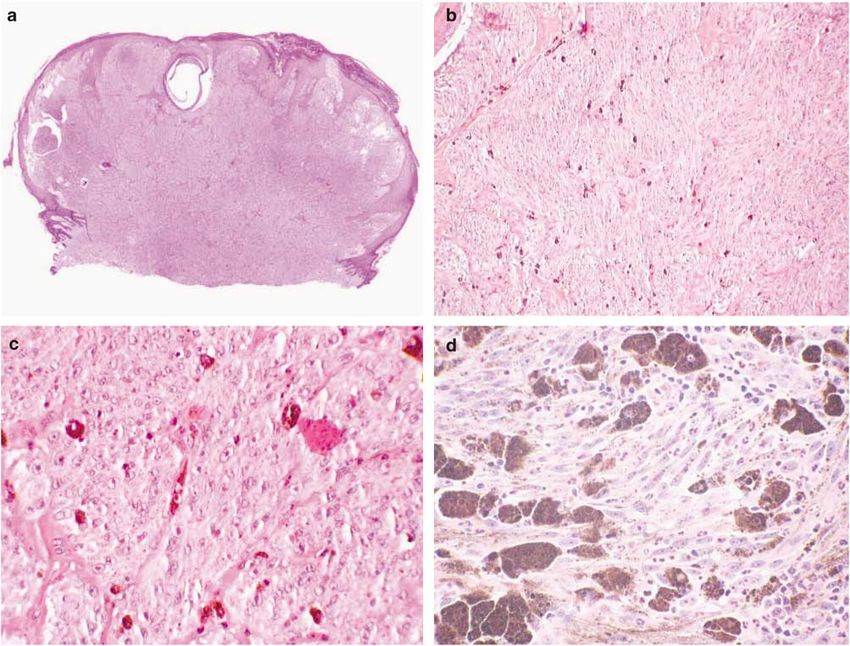
Spitzoid lesion
                                                                     RL Barnhill
S22
          factor in the rise in mortality from melanoma noted                       that both Spitz’s and our fatal cases developed in 12-
          with age and the extreme rarity of death from                             year-old female subjects who undoubtedly were in
          melanocytic lesions before puberty. It is noteworthy                      puberty or were postpubertal. One is left with the
                                                                                    distinct impression that Sophie Spitz remained
          Table 1 Characteristics of conventional Spitz tumors                      unsettled by this group of lesions and did not at
                                                                                    all consider them a variant of ordinary nevus.
          Clinical features                                                            Over the past half century there has been the
            Configuration: papule or nodule, often dome-shaped, or plaque
            Size: small (usually o1 cm)a
                                                                                    progressive trend to consider the Spitz tumor as
            Profile: often smooth surface topography                                simply a variant of benign melanocytic nevi quite
            Color: pink/red; pigment variants occur                                 distinct from melanoma and to utilize the terms
            Age: majority in children and adolescents, but at any age               ‘Spitz nevus, Spitz’s nevus, or spindle and epithe-
            Location: face and extremities, most common                             lioid cell nevus’ to describe this lesion.3–24 This
            Number: usually solitary; rarely multiple forms occur
            Symptoms: commonly asymptomatic; rarely pruritic                        approach has come about from the reports of series
            History of growth: months; usually less than a year                     of Spitzoid lesions and the promulgation of histolo-
                                                                                    gical criteria from these series. As we have described
          Histopathological criteria                                                various authors have reviewed their own series of
            Architectural features
              Symmetrya
                                                                                    ‘Spitz nevi’ and accordingly formulated their ‘criter-
              Sharp lateral demarcationa                                            ia’ from these self-selected (and consequently
              Regular pattern of epidermal hyperplasia                              biased) series of lesions (Table 2).7,11,12,26 The
              Zonation with depth (eg, ‘maturation’)a                               inherent flaws resulting from this exercise involve
                Side to side uniformity, that is, nests and fascicles of            circular reasoning, the cases in general have not
                melanocytes with fairly uniform size and shape and regular
                spacing                                                             been population-based, the number of cases has
                Diminished cellular density with depth                              often not been adequate, and the cases have not had
                Nests diminish in size and show transition to single cells          long-term follow-up (of 8–10 years) to know the
                with depth                                                          outcomes.22,25,26 Following up on the observations
                Diminished cellular and nuclear sizes with depth
              Often wedge-shaped configuration in dermis
                                                                                    that the vast majority of these Spitzoid lesions do
              Orderly nondisruptive infiltration of collagen by melanocytes         not seem to recur or to metastasize, they have been
                                                                                    lumped as a group in benign melanocytic nevi, even
               Cytological features                                                 though some proportion of lesions seem impossible
                 Spindle and/or epithelioid cell typea                              to distinguish from melanoma and uncommon or
                 Overall monomorphous population of cellsa
                 Low nuclear-to-cytoplasmic ratio                                   rare lesions behave aggressively. Thus, the term
                 Opaque or ground-glass cytoplasms                                  ‘Spitz nevus’ has become firmly entrenched in the
                 Nuclei with open, delicate chromatin patterns                      medical literature. The author acknowledges with-
                 Uniform nucleoli                                                   out question that the most typical or banal Spitz
                 Occasional striking pleomorphism in a minority of cells
                                                                                    tumors share particular features with conventional
               Other helpful diagnostic features                                    acquired melanocytic nevi. One can argue that this
                 Mitotic rate o2/mm2                                                group of Spitz lesions (that do not appreciably
                 Absent or rare, but not atypical, mitoses in deep partsa           deviate from this characterization of a benign
                 Mononuclear and multinucleate giant cells                          melanocytic lesion) should be termed Spitz nevi.
                 Irregular contours of growth at deep margina
                 Dull pink (Kamino) bodiesa                                         However, the problem with the indiscriminate use
                 Paucity or absence of single-cell upward spread (in central        of ‘nevus’ is that it connotes a lesion that is
                 part of lesion if present)                                         completely benign and a priori presents no risk to
                 Junctional clefts                                                  the patient. Obviously, this is not the case for some
                 Loss of cohesion between cells (retraction spaces)
                 Perivascular or diffuse inflammatory infiltrate
                                                                                    subset of Spitzoid lesions as has clearly been shown
                 Superficial distribution of pigmentation                           by Sophie Spitz herself (and many others)!
                 Telangiectasia and edema                                              It is neither rational nor practical and it is
                                                                                    potentially harmful to patients and physicians alike
          a
              Most helpful features.                                                (see below) to label prospectively virtually all

          Table 2 Contrasting criteria for Spitz tumors from reports in the literature30

          Criterion                        Kernen and Ackerman7           Reed et al26             Weedon and Little12   Paniago-Pereira et al11

          Pagetoid spread                  Not stated                     Not stated               Occasional            Sparse
          Nuclear atypia                   Absent                         Minimal                  Present               Not important
          Giant cells                      Occasional                     Present                  Present               Not helpful
          Epidermis                        Thinned                        Hyperplastic             Hyperplastic          Hyperplastic
          Cell type                        Mostly spindled                No comment               Mostly spindled       Mostly spindled
          Dermal mitoses                   Rare                           Variable (rarely high)   Common                Occasional
          Atypical mitoses                 Rare                           Not stated               Uncommon              Rare
          Maturation with descent          Mostly absent                  Present                  Mostly absent         Prominent
          Deep dermal margin               Pushing                        Infiltrative             Infiltrative          Pushing

Modern Pathology (2006) 19, S21–S33
Spitzoid lesion
                                                          RL Barnhill
                                                                                                                                 S23
Spitzoid lesions as nevi and then to have to redress      Table 3 Protocol for Spitz tumors
the issue at some later date. This usually involves
reclassifying some lesions as malignant or melano-        1. Examination of the entire lesion
                                                          2. Application of all histopathological, clinical, and other
ma some time later after the development of                  attributes for assessing abnormalities present
metastases or death. It is obvious that this practice     3. Seek consultation
also leads to the self-correcting behavior of lowering    4. Placement into risk category (Table 4)
the threshold for diagnosing melanoma in order to         5. Management of patient
avoid a disastrous outcome of missing melanomas
and metastasizing lesions. For the latter reasons, the
author favors the term ‘Spitz or Spitz-like tumor’
(equally acceptable terms might include Spitz,            Table 4 Categorization of Spitzoid lesions according to risk
Spitz-like, or Spitzoid lesion, melanocytoma or           stratification
neoplasm) and the use of an intermediate or third
                                                          Spitz tumor without atypicality
category lesions that are difficult to classify as        Atypical Spitz tumor (Spitz tumor with one or more atypical
clearly benign or malignant, that is, Spitz tumors        features)
that are ‘atypical, controversial, or biologically          Lesions with indeterminate biological potential (lesions
indeterminate’ (see below).27                             difficult to classify as unquivocably benign or malignant)
                                                          Malignant melanoma
   It is apparent that one message stemming from the
published work on Spitzoid lesions is that if one
utilizes the ‘established criteria’ one can very easily
interpret a Spitzoid neoplasm as either: (1) Spitz’s
nevus or (2) malignant melanoma.28 Another trend          rather arbitrary interpretation of all Spitzoid lesions
has been to interpret almost all Spitzoid lesions in      as either ‘Spitz’s nevus’ or malignant melanoma is
children as benign ‘Spitz nevi’ and many Spitzoid         too simple, not realistic, and may be harmful to
lesions in adults (particularly beyond the age of 30–     patients.
40 years) as malignant melanoma, almost without              As the author has previously suggested, a more
any attention to the histopathological features (RL       realistic solution is to employ a well-defined
Barnhill, 2005, personal observations) As mentioned       protocol (Table 3) for the systematic and rigorous
above, a corollary of the latter practice, a literal      evaluation of Spitzoid lesions utilizing all histo-
‘dumbing-down of criteria’, has been to designate         pathological, clinical, and ancillary information
virtually all Spitzoid lesions as melanoma. While         available.25,27,29 Having collected this information,
medical–legal concerns have understandably influ-         one can then assign a given lesion to one of three
enced the behavior of pathologists in this area, the      categories (Table 4): (1) Spitz tumors without
latter practices often offer pathologists no better       appreciable abnormality (Figure 1), (2) Spitz tumors
solution than that of ‘flipping a coin’, or literally     with one or more atypical features (atypical Spitz
abandoning the practice of histopathology.                tumor) (Figure 2) including those with indetermi-
   The author does not believe that this overly           nate biological or malignant potential (Figure 3), and
simplistic attempt to assign an often arbitrary           (3) malignant melanoma. In effect, this involves risk
diagnosis of either ‘Spitz’s nevus’ or malignant          stratification of lesions, open and honest communi-
melanoma to every Spitzoid lesion28 serves patients       cation with patients, and rational decision-making
well nor provides a realistic methodology for deal-       for patient management, that is neither harmful nor
ing prospectively with an often profoundly difficult      negligent. Given the imperfect state of current
problem. Such an exercise undoubtedly results in          information, the author acknowledges that this
both the overdiagnosis and underdiagnosis of              exercise still remains largely subjective and is
melanoma in a certain number of cases. Patients           dependent on the knowledge, experience, and
may unfairly suffer the psychological burden of a         common sense of the pathologist and other physi-
grave diagnosis and may undergo overly aggressive         cians involved in the care of the patient.
and potentially harmful therapies. At the same time,         The remainder of the paper will elaborate on this
some proportion of patients will have the false and       process of evaluating Spitz tumors with particular
unjustified assurances of a benign diagnosis and          emphasis on the characteristics of atypical Spitz
may not receive appropriate treatment and follow-         tumors. Finally, the controversial group of melano-
up. Rather, we must be honest with patients and           mas closely resembling Spitz tumors, or ‘Spitzoid’
above all try to do no harm. We must accept that          melanomas, will be discussed and whether their
currently we have a rather poor understanding of the      distinction from atypical Spitz tumors is possible at
biological nature of Spitzoid lesions and that current    present.
criteria for their diagnosis are wanting. We must
adopt a pragmatic approach that attempts to avoid
both the overdiagnosis of melanoma and inappropri-        Spitz tumors with atypical features
ate management of patients. Einstein’s philosophy
that one should try to ‘make things simpler but not       The obvious goal of developing criteria for any
too simple’ is especially applicable here. Thus, the      tumor system is to be able to discriminate lesions

                                                                                                  Modern Pathology (2006) 19, S21–S33
Spitzoid lesion
RL Barnhill
S24
Figure 1 Compound Spitz tumor without significant atypicality of a 22-year-old male with lesion involving the left upper arm. The
lesion measures less than 10 mm in diameter and 0.61 mm in Breslow thickness. Other attributes: no asymmetry, sharp circumscription,
no ulceration, only minimal focal pagetoid spread, and there are no mitoses in the small dermal component. This lesion requires
complete excision and the patient follow-up examinations at least once a year for example. (a) Scanning magnification show a slightly
raised tumor with general symmetry. (b) The lesion demonstrates orderly appearance with regular junctional nesting and small dermal
component. (c) The lesion shows focal pagetoid spread and fairly regular junctional nesting of melanocytes. (d) Note uniformity of
spindle and epithelioid cells in the dermis.
that are ‘malignant, behave aggressively, or have concluded that even among experts the published
other adverse prognostic properties’ from those that guidelines for the dichotomous distinction of Spitz
are biologically ‘benign’ or pose no threat to the nevi from melanoma were inadequate. The latter
patient. We set out to test the hypothesis as to work has been the incentive for the ongoing study
whether the generally accepted ‘criteria’ for Spitz and formulation of criteria for and risk stratification
nevi truly discriminate ‘benign’ lesions from aggres- of Spitzoid lesions (Tables 5 and 6).30
sive or malignant lesions in a paper published in
1999.25 The latter study involved a blinded review of
30 melanocytic lesions exhibiting features of Spitz Background of the atypical Spitz tumor
nevus (the cases included ‘typical Spitz nevi’, Spitz
tumors with atypical features, biologically indeter- The term ‘atypical Spitz tumor’ was first used in
minate Spitz-like lesions, and unequivocal melano- the English language literature (to the author’s
mas) by 10 pathologists. The outcome of the project knowledge) in 1975 by Reed et al26 in illustrating
showed that this panel of pathologists could not a Spitz tumor (in a photomicrograph) that differed
reach consensus about diagnosing such Spitz le- from conventional Spitz nevi by exhibiting con-
sions or distinguishing them from melanoma using fluent and densely cellular fascicles of spindle
the conventional criteria from the literature.25 As cells that ‘crowded and compressed their stroma’.
has already been mentioned one of the cases thought In a subsequent report of 32 cases, Smith et al31
by consensus to be a typical ‘Spitz nevus’ eventually introduced the term ‘spindle and epithelioid
resulted in death of the patient. Therefore, we cell nevus with atypia and metastasis (malignant
Modern Pathology (2006) 19, S21–S33Spitzoid lesion
RL Barnhill
S25
Figure 2 Compound Spitz tumor with atypical features of a 36-year-old woman with lesion from the right thigh. The tumor measures less
than 10 mm in diameter and at least 2.1 mm in Breslow thickness. Other attributes: slight asymmetry, reasonable circumscription, no
ulceration, no pagetoid melanocytosis, lack of maturation, high cellular density and confluence of melanocytes in dermis, 2/mm2 mitotic
rate, deep mitosis, and prominent nuclear pleomorphism. This lesion lacks sufficient atypicality in the author’s experience for
conventional melanoma. Such a lesion requires re-excision with margins of about 1 cm and careful follow-up at least every 6 months.
SLN biopsy may be considered at some institutions but has no proven benefit. (a) Scanning magnification shows a raised dome-shaped
tumor with the general appearance of Spitz tumor and slight asymmetry. (b) Note lack of maturation of dermal component. (c) The lesion
exhibits junctional nesting of melanocytes but no pagetoid spread. The nests of melanocytes display prominent cellularity and
confluence. (d) There is no maturation at the base of the tumor.
Spitz nevus)’ (see below) to describe a series however, long-term follow-up of these patients has
of Spitz-like melanocytic tumors characterized by not been reported. There were no distinctive
large size (41 cm), frequent ulceration, deep exten- features that predicted the development of metas-
sion into subcutaneous fat with bulbous ‘pushing’ tases.
margins, prominent cellular density, lack of matura- The author has reported a series of 12 atypical
tion, cytological atypia greater than that expected Spitz-like melanocytic tumors in children and
for a ‘Spitz nevus’ (large and pleomorphic cells, adolescents.29 There were three tumors (classified
prominent nucleoli), significant numbers of mitoses as melanoma) among the 12 cases that were
(up to 5 in a single high-power field), and focal associated with metastases and the death of one
necrosis. These tumors developed in relatively patient. Two of the latter patients had only single
young individuals (41% o14 years and 82% o29 lymph node metastases and have been disease-free
years of age), and virtually all were located on with long-term follow-up. The latter two tumors also
the head and neck, and extremities (71%). Six potentially could be described as ‘metastasizing
patients were also observed to have regional lymph Spitz tumors’. In general, all 12 tumors were
node metastases with involvement of the sinuses characterized by many of the same features as
and parenchyma by tumor identical to the primary reported by Smith et al,31 that is, large size,
cutaneous lesion. According to the authors, there ulceration, significant depth, prominent cellular
was no subsequent progression of the disease; density, lack of maturation, a pushing border,
Modern Pathology (2006) 19, S21–S33Spitzoid lesion
RL Barnhill
S26
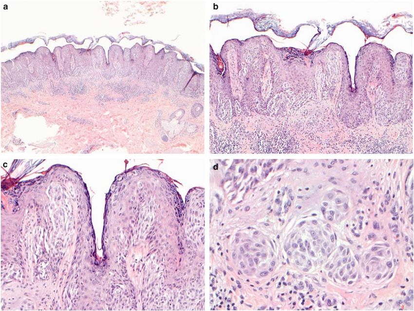
Figure 3 Malignant melanoma (or metastasizing Spitz tumor?) of a 7-year-old boy with lesion from the left back initially diagnosed as
atypical Spitz nevus at an another institution. The tumor measures 9 mm in diameter and at least 4.4 mm in Breslow thickness. Other
attributes: asymmetry, reasonable circumscription, no ulceration, no pagetoid melanocytosis, no maturation, high cellular density and
confluence of melanocytes throughout dermis, 9/mm2 mitotic rate, deep mitoses, and prominent cytological atypia. The lesion recurred
in a matter of months and two SLNs contained large deposits of an atypical melanocytic tumor. Long-term follow-up will be needed to
determine whether there is disease progression or not. (a) Scanning magnification shows a large raised polypoid tumor with slight
asymmetry. (b) The superficial portion of the tumor is characterized by confluent sheets of melanocytes replacing dermis. (c) Note
striking density of melanocytes at base with cytological atypia. Many nuclei contain prominent nucleoli. (d) SLN containing large
tumoral deposit replacing large part of node. Note pronounced cytological atypia of spindled melanocytes.
variable but often frequent and deep mitosis, and Criteria for the assessment of Spitz
prominent cellular pleomorphism. tumors
From the reports cited above, it is apparent that
there is a not only an experiential but also a factual A Spitzoid lesion should be initially screened
basis for formulating criteria for Spitzoid lesions histopathologically to determine whether an obv-
with abnormal features and that these attributes ious melanoma is present. If clearcut melanoma can
potentially may have predictive value for risk be excluded, Spitz-like tumors should be system-
stratification (and the rational for an intermediate atically assessed with the following histopathologi-
category of Spitzoid lesions) (Table 5).29 Based on cal, clinical, and ancillary parameters30 (Table 5)
the information from the cases referred to we have (Figures 1–3).
developed the grading protocol outlined in Table 6
to aid in the assessment of metastatic risk of Spitz Histopathology
tumors with abnormal features in children and
adolescents.30 In an evaluation of 30 atypical Organizational attributes
Spitz-like melanocytic tumors, an increasing score Size (diameter in mm): The diameter of the lesion is
resulted in greater risk for the development of recorded in millimeters. Most typical Spitz tumors
metastases. measure under 10 mm and frequently o5–6 mm.
Modern Pathology (2006) 19, S21–S33Spitzoid lesion
RL Barnhill
S27
Table 5 Histopathological criteria for atypical Spitz tumors Ulceration: The ulceration is abnormal.30 How-
ever, since ulceration is induced by trauma in
Organizational criteria almost all instances, its significance will vary.
Diameter in mm (Z10 mm considered abnormal)
Depth in mm (involvement of subcutaneous fat considered Poor circumscription: The most banal Spitz
abnormal) tumors tend to be sharply circumscribed at their
Ulceration peripheries whereas atypical lesions are often less
Poor circumscription well-circumscribed.20 This parameter obviously cor-
Pagetoid melanocytosis over a large front
Prominent confluence of melanocytes
relates with asymmetry and other organizational
High cellular density attributes.
Lack of zonation and maturation Pagetoid melanocytosis: Pagetoid spread may be
Asymmetry observed not infrequently in Spitz tumors.21 How-
Few or no dull pink (Kamino) bodies ever, such pagetoid spread should be limited to the
Proliferational criteria lower half of epidermis, should not extend periph-
Significant mitotic rate—42–6/mm2 erally, should be only focal, and sparsely cellular
Deep/marginal mitoses (Figure 1). More extensive pagetoid spread invol-
Proliferation index, that is, Ki-67 expression ving the upper half of the epidermis, a large zone of
Between 2 and 10% (Vollmer)
Z10% (Kapor et al38)
the lesion (at least one high-power field), and in a
single-cell or small nested pattern is distinctly
Cytological criteria abnormal. External trauma may be a factor leading
Granular vs ground glass cytoplasm to excessive pagetoid spread in benign lesions.
High nuclear to cytoplasmic ratios
Loss of delicate or dispersed chromatin patterns
Thickening of nuclear membranes Prominent confluence of melanocytes. High cellu-
Hyperchromatism lar density: These two parameters are closely
Large nucleoli correlated and are among the most important criteria
for assessing melanocytic lesions.27 Unfortunately,
these characteristics are subjective and therefore
difficult to recognize reliably and reproducibly.
These two parameters are also often closely linked
Table 6 Assessment of Spitz tumors in children and adolescents
for risk for metastasis30 to diminished or absent maturation (see below).
Confluent cellular aggregates or nodules of consid-
Parameter Scorea erable size and with crowded appearance in the
dermal component, particularly replacing the der-
Age (years) mis, and extending deep without maturation are
0–10 0 also decidedly atypical (Figures 2 and 3). Breslow
11–17 1
thickness obviously may capture significance of
Diameter (mm) such expansile dermal nodules. One caveat is the
0–10 0 occurrence of such nodules in Spitz tumors of young
410 1 children, which may take on less importance in the
latter context.29
Involvement of subcutaneous fat
Absent 0
Present 2 Lack of zonation and maturation. Zonation refers
to the side-to-side homogeneity often observed in
Ulceration typical Spitz rumors (Figure 1).20,29 Whereas matura-
Absent 0
Present 2
tion is the progressively diminished sizes of nests of
melanocytes and gradual dispersion of melanocytes
Mitotic activity (mm2) to smaller nests and single cells with depth. The
0–5 0 latter phenomenon, in its most developed state,
6–8 2 involves a nondisruptive infiltration of melanocytes
49 5
among collagen bundles and involution of melano-
a
Total score indicates increasing risk for metastasis.
cytes to smaller cells with smaller nuclei with
depth. Therefore, the nonuniformity (heterogeneity
of organization) of a lesion when scanned from side
to side and the continued presence of nests and
Size beyond 10 mm is generally considered abnor- fascicles of similar sizes deep indicate potentially
mal.29–31 This is a continuous variable and there are aggressive properties (Figures 2 and 3).
obviously exceptions to this criterion.
Tumor thickness (measured in mm): Significant Few or no dull pink (Kamino) bodies. Although
(Breslow) depth and involvement of the subcuta- this feature may be seen in both Spitz tumors and
neous fat are considered abnormal.30 melanomas, the presence of clearcut aggregates of
Asymmetry: Increasing asymmetry is abnormal as Kamino bodies may be a marker suggesting a more
in all melanocytic lesions.20 conventional Spitz tumor.32
Modern Pathology (2006) 19, S21–S33Spitzoid lesion
RL Barnhill
S28
Table 7 Immunohistochemical evaluation of Spitz tumors and equivocal (in the author’s judgement such indices
atypical variants as compared to conventional nevi and malignant may thus suggest an atypical Spitz tumor and
melanoma (from Kapur et al38)
potentially one that is biologically indeterminate].
Ki-67% p21% Fatty acid synthase The qualitative loss of Ki-67 expression vs contin-
ued labeling with depth as with markers also
Banal nevi (19) 0.53 0.21 0.21 correlates with maturation and less atypical lesion.
Spitz tumors (26) 5.04 46.50 0.46
Atypical Spitz tumors (10) 10.0 49.25 1.56 Cytological attributes
Malignant melan (18) 36.83 27.50 2.28
The assessment of cytological attributes is of great
importance but remains highly subjective and lacks
reproducibility.27 Cytological features suggesting a
more atypical Spitzoid lesion and possibly melano-
Proliferational attributes ma include heterogeneity of cell type throughout the
Mitotic rate per mm2. The mitotic rate of the lesion, particularly in an asymmetrical or haphazard
dermal component is one of the most important pattern; high-nuclear-to-cytoplasmic ratios; granular
parameters for evaluating Spitzoid lesions as in- or ‘dusty’ cytoplasm vs the ground glass cytoplasm
creasing proliferative rate seems to correlate with of Spitz melanocytes; absence of delicate or dis-
likelihood of aggressive behavior or malignancy.30,33 persed chromatin patterns with thickening of nucle-
Furthermore, this parameter is quantifiable. Mitoses ar membranes; a large proportion of melanocytes
observed in the deepest parts of the dermal or with hyperchromatic nuclei; and the presence of
subcutaneous component, that is, near the deep large eosinophilic nucleoli.
margin, also seem to have greater significance than
more superficially located mitoses.33 There are no
absolute thresholds for the mitotic rate being Clinical Attributes
indicative of malignancy, and the author cautions
Age
against using mitotic rate alone (or any single
The vast majority of Spitz tumors occur in young
criterion alone) for the interpretation of a lesion as
individuals particularly under the age of 10–20
being malignant. It is important in almost all
years.2–24 The older the patient, especially indivi-
instances to have several distinctly abnormal para-
duals beyond the age of 20–30 years, the greater the
meters present to confirm malignancy. Every lesion
likelihood of malignancy. As a general rule, one’s
must be systematically assessed on a case-by-case
threshold for diagnosing melanoma in such lesions
basis. In any case, high mitotic rates, particularly
should correlate inversely with the age of the patient,
beyond six mitoses per mm2 raise considerable
that is, a high threshold for very young individuals
concern for malignancy.30 Unusual circumstances
and a lower threshold for elderly individuals.
that may confound the importance of high mitotic
Recently, Vollmer39 has provided guidelines for the
rate include: (1) developing Spitz tumors may be in
diagnosis of Spitzoid lesions vs melanoma.
a growth phase and mitotic rate may have less
The author has nonetheless noted that Spitz
significance (RL Barnhill, 2005, personal observa-
tumors may occur with greater frequency in older
tions), (2) Spitz tumors in very young individuals
adults than has been appreciated owing to the
may have somewhat higher mitotic rates, and (3)
propensity of many pathologists to a priori interpret
external trauma and significant inflammation may
them as melanoma (RL Barnhill, 2005, personal
be factors leading to higher mitotic rates
observations).
Proliferation index, that is, Ki-67 expression. The
Other clinical attributes
expression of proliferation markers such as Ki-67
Other clinical factors such as the location of the
has taken on increasing importance for the assess-
ment of malignant melanoma.34 As a result, the use tumor, clinical appearance, history of recent
changes in a long-standing stable lesion, and family
of Ki-67 and closely related indicators may provide
history of melanoma should be considered care-
information beyond that of mitotic rate on the
fully.27 Spitz tumors commonly involve the extre-
proliferation status of Spitzoid lesions.34–38
Although requiring much more study for standardi- mities and face. The location of atypical tumors on
sites less commonly involved by Spitz tumor, such
zation, Ki-67 expression may be useful in the risk
as the back, is also another factor suggesting careful
stratification of Spitz tumors. For example, Kapor et
al38 have recently shown that atypical Spitz tumors scrutiny of the lesion for melanoma.
have a mean Ki-67 labeling index of 10% relative to
0.53% in ordinary nevi, 5.04% in conventional Special Techniques
Spitz tumors, and 36.83% in conventional melano-
mas (Table 7). Vollmer’s37 data suggest that a Ki-67 Immunohistochemistry
proliferation index less than 2% favors a conven- Spitzoid lesions have been evaluated with a variety
tional Spitz tumor, one greater than 10% suggests of melanocytic markers.27,34,40–43 S100 protein and
melanoma, and indices between 2 and 10% are Mart-1 show diffuse expression throughout both
Modern Pathology (2006) 19, S21–S33Spitzoid lesion
RL Barnhill
S29
Spitz tumors and melanoma in contrast to the 80%) in conventional melanomas49 and (70–90%) in
characteristics diminished expression of HMB-45, melanocytic nevi50 and suggests a different and
tyrosinase, and other markers toward the base of perhaps yet to be characterized developmental
Spitz tumors.40,41 In addition, there is a gradient of pathway for Spitzoid lesions.
diminished proliferation with increasing depth of
the dermal component paralleling mitotic rate and
cyclin D1 expression in Spitz tumors.34,35,42,43 Spitz Sentinel lymph node biopsy
tumors also appear to exhibit lower rates of p53, bcl-
The recent application of the sentinel lymph node
2, and fatty acid synthase expression compared to
(SLN) biopsy to assess atypical Spitz-like tumors
melanoma.42,38 Fatty acid synthase may also aid in
provides a means of obtaining more information
distinguishing atypical Spitz tumors from melano-
about the biological characteristics of such le-
ma. For example, according to Kapor et al38 ordinary
sions.51–53 Atypical Spitz-like tumors may have
nevi show 0.21%, conventional Spitz tumors 0.46%,
metastatic deposits in the peripheral sinuses or
atypical Spitz tumors 1.56%, and melanomas 2.28%
parenchyma of SLNs.51 Although in general the
labeling with fatty synthase. Although many of the
finding of tumor deposits in SLN is de facto
latter markers are of interest, they require rigorous
evidence for melanoma, only the study of sufficient
assessment with greater numbers of cases and long-
numbers of cases with long-term follow-up will
term follow-up in order determine whether they
provide the data to know the biological significance
have any predictive value in the evaluation of
of such SLN deposits associated with atypical Spitz
Spitzoid lesions.
tumors, if they are inherently different from con-
ventional melanomas, and if they potentially have
Comparative genomic hybridization and fluorescence
better prognoses. Although large lymph node depos-
in situ hybridization
its demonstrating pronounced cytological atypia,
The majority of Spitz tumors studied by comparative
prominent mitotic rates, and necrosis are almost
genomic hybridization (CGH) have had no chromo-
certainly indicative of malignancy, the significance
somal abnormalities.44,45 On the other hand, ampli-
of small bland microscopic deposits is unclear and
fication of chromosome 11p has been observed in a
requires more detailed analysis. Almost all comple-
subset of cases and was found to correlate with Spitz
tion lymphadenectomies for atypical Spitz tumors
tumors that were often larger in size, dermal-based,
with positive SLNs have failed to show any residual
desmoplastic, had vesicular nuclei in melanocytes,
tumor.52,53 In addition, the latter patients have had
and exhibited dermal infiltrating features. Further
no further disease recurrence with continued fol-
study of Spitz tumors with gains of 11p has shown
low-up of 2–3 years.53 Atypical Spitz tumor deposits
an increase in copy number of and mutations in the
in SLNs may possibly have a different biology or
HRAS gene by fluorescence in situ hybridization.
significance than metastases from conventional
The latter abnormalities do not usually occur in
melanoma. Many questions remain to be answered
melanoma and consequently the authors have
about the significance of SLN involvement by
suggested that such findings (and the absence of
atypical Spitz tumors, particularly microscopic
chromosomal aberrations in most Spitz tumors) may
metastases. These findings may have relevance to
aid in the distinction of Spitz tumors from melano-
the entity of the so-called ‘metastasizing Spitz
ma. Other conclusions from the latter studies were
tumor’ as some of these patients have remained
that Spitz tumors are probably clonal proliferations,
disease-free with long-term follow-up of up to 15
the majority of melanocytes are diploid with some
years or more.29,31 Among the limited number of
large nuclei being polyploid, and Spitz tumors may
cases studied thus far by SLN biopsy, such Spitzoid
be cytogenetically distinct from melanoma.45
tumors have not necessarily had the degree of
abnormality, that is, size 41 cm, ulceration, invol-
Loss of heterozygosity
vement of subcutaneous fat, high mitotic rate, as
Two independent studies have recently demon-
such tumors resulting in macroscopic metastases.51
strated loss of heterozygosity on chromosome 9p
with DNA polymorphic markers in two of 27 and
five of five ‘Spitz nevi’.46,47 The latter findings Metastasizing Spitz tumor
provide additional evidence for the close relation-
ship between Spitz tumors and melanoma. As already mentioned above, some melanocytic
lesions classified as ‘Spitz nevi’ have spread to
Analysis of gene mutations regional lymph nodes without subsequent disease
Recent work has shown that a series of conventional progression.3,8,29,31,54,55 The concept of localized,
Spitz tumors and the so-called Spitzoid melanomas presumably ‘benign’ metastases has been proposed
(? metastasizing Spitz tumors) in prepubescent to explain this rare phenomenon. However, some of
children failed to show any hotspot activating these metastasizing melanocytic lesions, albeit re-
mutations in the B-raf, N-ras, or H-ras genes.48 The sembling in many ways ‘Spitz nevi’, have tended to
general absence of B-raf mutations in Spitzoid be unusually large (ie, 41 cm), often deep with
lesions contrasts with a high rate of mutation (53– involvement of subcutaneous fat, ulcerated, and to
Modern Pathology (2006) 19, S21–S33Spitzoid lesion
RL Barnhill
S30
have high mitotic rates or showed other uncommon Spitzoid lesion. Since we lack objective data and
features (see above).25,29,30,31 An alternative explana- sufficient follow-up, the significance or weighting of
tion is that these tumors are unusual melanomas the various features already mentioned has not been
with Spitzoid features, which may or may not have a established. However, at present the final interpreta-
less aggressive potential for spread beyond regional tion of a Spitzoid lesion remains almost entirely
lymph nodes. More definitive characterization is histopathological with important consideration gi-
needed before any conclusions can be drawn about ven to clinical information. Almost all other para-
the latter group of tumors. meters have not yet been sufficiently studied as to
have any significant impact on the final interpreta-
tion. However, some indices such as the Ki-67
Spitzoid melanoma labelling, the gradient of expression of markers such
as HMB45 (gp100) or fatty acid synthase, and CGH
Although it can justifiably be argued that some
may provide useful information on the final delib-
proportion of melanomas (perhaps large) resemble
eration about a lesion. In the future, such ancillary
Spitz tumors and the converse, the term Spitzoid
data may take on much greater importance.
melanoma, if used at all, should be reserved for
Some may point out that such an approach seems
melanomas that truly have a striking morphological
to render many Spitz tumors atypical. In the process
resemblance to Spitz tumors.20,27,33,56 The term prob-
of evaluating Spitz tumors common sense must
ably best describes a rare group of tumors often
prevail, and one must keep in mind that one is most
developing in young individuals who are only
likely dealing with a biological continuum with
diagnosed as melanoma in retrospect, that is, after
many Spitz tumors at the ‘benign’ or ‘less aggressive’
the development of metastases and an aggressive
end of the spectrum. There is little question that as
course. Given the profound difficulty of distinguishing
these various parameters progressively accumulate
some Spitz tumors from melanoma, the author
in number and severity, the probability of an
discourages the use of term Spitzoid melanoma since
it may result in the indiscriminate labeling of a aggressive phenotype or melanoma increases.30
It is apparent that certain parameters take on more
heterogeneous group of lesions including benign Spitz
significance than others (* represents most helpful
tumors, lesions that are biologically indeterminant,
features).30 Potentially aggressive tumors or melano-
conventional melanomas, and also the rare controver-
mas thus often have large size* (45–6 mm, often
sial group of tumors mentioned above: ‘metastasizing
410 mm*); may have significant depth*; demon-
Spitz tumor’. The latter group of lesions includes some
strate distinct asymmetry*; poor circumscription;
that have given rise to single lymph node metastases
heterogeneity of cellular populations*; more dis-
without subsequent recurrence on long-term follow-
ordered intraepidermal proliferative patterns of
up. It cannot be overemphasized that as a group all of
melanocytes without clefting; extensive pagetoid
these unusual Spitz-like tumors require more detailed
spread; irregular epidermal alterations including
study as to their biological nature.
thinning and effacement; significant melanocytic
In general, there are no distinctive clinical
density and confluence*; and the lack of or zonation
features.27 Such Spitzoid melanomas often have
or diminished cellular density with depth (matura-
abnormal clinical attributes such as size 45–6 mm,
tion)*. The lack of uniformity or homogeneity of cell
asymmetry, and irregular coloration suggesting an
type along comparable strata (from side to side) of
atypical nevus or melanoma. Some such lesions may
the tumor cannot be overemphasized as a major
suggest a Spitz tumor clinically but it must be
criterion favoring melanoma. Similarly, the failure
recalled that the clinical diagnosis of a Spitz tumor
of a tumor to show progressive dispersion of
is rather imprecise.
melanocytes to smaller aggregates and particularly
The diagnosis of a melanoma as ‘Spitzoid’ as
to single melanocytes (among apparently unaffected
mentioned above is based on the striking architec-
collagen bundles) in the deepest part of the lesion
tural and cytological resemblance to a Spitz tumor.27
also suggests melanoma. Usually concurrent with
Thus, such features potentially include any of the
depth is the uniform diminution of cellular and
following: dome-shaped, plaque-like, or wedge-
nuclear sizes and regular spacing of melanocytes in
shaped morphology; little or no asymmetry; epider-
a Spitz tumor; the failure to observe the latter feature
mal hyperplasia; clefting about intraepidermal nests
should prompt consideration of melanoma.
of melanocytes; presence of dull pink or Kamino
Cytological features favoring melanoma include
bodies; some evidence of zonation or maturation;
alterations that are a distinct departure from what is
and especially a population of enlarged epithelioid
considered acceptable for a Spitz tumor*: hetero-
and/or spindled melanocytes with abundant opaque
geneity of cell type throughout the lesion, parti-
or ‘ground glass’ cytoplasms.
cularly in an asymmetrical or haphazard pattern;
high-nuclear-to-cytoplasmic ratios; granular or
Final analysis for diagnostic interpretation ‘dusty’ cytoplasm vs the ground glass cytoplasm of
Spitz melanocytes; absence of delicate or dispersed
All histopathological, clinical, and ancillary criteria chromatin patterns with thickening of nuclear
must be weighed in the final interpretation of a membranes; a large proportion of melanocytes with
Modern Pathology (2006) 19, S21–S33Spitzoid lesion
RL Barnhill
S31
hyperchromatic nuclei; and large eosinophilic nu- distinguish from melanoma.25,36 Some of the latter
cleoli. tumors have resulted in metastases(Barnhill et al25
As discussed above, the greater the absolute rate and RL Barnhill, 2005, personal observations). It is
(per mm2)* and number of deeply located (dermal) the author’s opinion that Spitz-like melanocytic
mitoses*, the more evidence one has favoring tumors assigned an indeterminate biological poten-
melanoma. Atypical mitoses and necrotic cells tial require surgical margins of approximately 1 cm
suggest melanoma but are not absolute. since this is considered the minimum standard of
Finally, among the clinical factors age* is of care for melanoma. The author acknowledges that
paramount importance. It is obvious that a lesion there are currently no definitive data available on the
with the gross morphological features suggesting issue of surgical margins for melanocytic lesions in
melanoma warrants special attention. A persistently general. Although of unproven benefit, SLN biopsy
changing lesion over weeks or months significantly may be considered for selected lesions (general-
raises concern for melanoma; whereas a lesion that is ly41 mm in thickness). Patients should be carefully
unchanged over many years argues against melanoma. monitored by regular examinations for recurrence
Acknowledging that this differential diagnosis is (and metastasis in the case of atypical Spitz tumors).
the most difficult one in melanoma pathology, there All patients should be managed on an individual
are circumstances that make it even more exasperat- basis and efforts made to avoid both overly aggres-
ing if not impossible. In particular, trauma and sive and suboptimal management strategies.
significant host response often introduce abnormal
features such as asymmetry, heterogeneity, and
cytological abnormality suggesting the greater like- Conclusions
lihood of melanoma. It must be kept in mind that the
nuclei in Spitz tumors are delicate and that any The author has attempted to portray accurately the
artefact such as tissue compression or overstaining current state of the art surrounding Spitzoid lesions,
or significant host response may introduce altera- their nomenclature, and assessment of risk.
tions suggesting greater cytological atypicality. In Although much remains to be learned about these
the latter circumstances, the pathologist must con- lesions, there is increasing evidence that they may be
sider carefully all of the criteria available before a type of melanocytic neoplasm distinct from
rendering an interpretation. conventional melanocytic nevi and malignant mela-
When entertaining the possibility of melanoma, noma. Acknowledging the peculiar nature of Spit-
one must always consider a Spitz tumor with zoid lesions, the author prefers the term Spitz tumor
overlapping features of pigmented spindle cell rather than ‘Spitz nevus’ and argues against using
tumor and one with phenotypic heterogeneity the term ‘Spitzoid melanoma’ until more information
(‘combined nevus’). Pigmented spindle cell tumors is available to justify such a term. The author has
show considerable overlap with Spitz tumors and also provided rational arguments that patients are
may introduce features suggesting melanoma such as best served by the comprehensive evaluation of
greater pagetoid spread, expansile papillary dermal Spitzoid lesions and their classification into three
nests, and the absence ground glass cytoplasm. Spitz categories: (1) Spitz tumor without significant
tumors with phenotypic heterogeneity (‘combined abnormality, (2) Spitz tumors with one or more
nevus’) may exhibit asymmetry and heterogeneity, atypical features (atypical Spitz tumor), including
two attributes suggesting melanoma. One must those judged to have indeterminate biological po-
assess each component of such a lesion individually tential, and (3) malignant melanoma, rather than the
with the criteria already mentioned (Table 5), and it two categories of ‘Spitz nevus’ and melanoma. Such
will usually be possible to resolve the issue. an approach honestly recognizes our current lack of
knowledge about many of these lsions and avoids
Management overdiagnosis of melanoma and under recognition of
potentially aggressive neoplasms. The author has
The author recommends that all Spitz tumors be outlined a protocol for this assessment of histo-
fully resected in order to facilitate complete histo- pathological, clinical, and ancillary data and sub-
pathological examination and also to diminish the sequent risk stratification. In conclusion, only
risk of recurrence. Atypical Spitz tumors obviously through rigorous characterization of sufficient num-
require comparable excision for the same reasons but bers of Spitzoid lesions and long-term follow-up of
with greater clearance (up to 1 cm) in order to patients will we be able to have truly objective
provide even greater assurance that they are ‘wholly information about these lesions. Such data would
out’. The reasons for recommending excision with allow us to finally formulate optimal guidelines for
margins free of the tumor are that (1) I have observed the care of patients with these lesions.
at least one Spitz tumor not completely excised that
persisted (recurred) at the same site and eventuated Note
in metastases and death,25 and (2) some persistent/
recurrent Spitz tumors may be more atypical than Herein the terms Spitzoid or Spitz-like melanocytic
the original lesions and even more difficult to lesion, tumor, or neoplasm are used generically
Modern Pathology (2006) 19, S21–S33Spitzoid lesion
RL Barnhill
S32
and interchangeably to refer to the broad spectrum Murphy G (eds). Melanocytic Tumors of the Skin.
of lesions from the most conventional or typical Armed Forces Institute of Pathology: Washington,
Spitz tumor to biologically indeterminate and even 1990, pp 40–57.
aggressive or malignant lesions closely resembling 18 Mooi W, Krausz T. Spitz naevus, desmoplastic Spitz
naevus and pigmented spindle cell naevus. In:
Spitz tumors. Spitz tumor indicates conventional Mooi W, Krausz T (eds). Biopsy: Pathology of Melano-
lesions (‘Spitz nevi’) without clearly observable cytic Disorders. Chapman & Hall: London, 1992,
abnormalities; atypical Spitz tumor refers to Spitz pp 156–185.
tumors with one or more and often several distinctly 19 Binder S, Asnog C, Paul E, et al. The histology and
abnormal characteristics generally not present in differential diagnosis of Spitz nevus. Semin Diagn
typical Spitz tumors, and including biologically Pathol 1993;10:36–46.
indeterminate lesions. 20 Barnhill RL, Piepkorn M, Busam KJ. The Pathology of
Melanocytic Nevi and Malignant Melanoma, 2nd edn.
Springer-Verlag: New York, 2004.
References 21 Busam KJ, Barnhill RL. Pagetoid Spitz nevus. Intra-
epidermal Spitz tumor with prominent pagetoid
1 Darier J, Civatte A. Naevus ou naevo-carcinoma chez spread. Am J Surg Pathol 1995;19:1061–1067.
un nourisson. Bull Soc Franc Derm Syph 1910;21: 22 Piepkorn M. On the nature of histologic observations:
61–63. the case of the Spitz nevus. J Am Acad Dermatol
2 Spitz S. Melanomas of childhood. Am J Pathol 1995;32:248–254.
1948;24:591–609. 23 Cramer SF. The melanocyte differentiation pathway in
3 Allen A, Spitz S. Malignant melanoma: a clinico- Spitz nevi. Am J Dermatopathol 1998;20:555–570.
pathological analysis of the criteria for diagnosis and 24 Mooi WJ. Spitz nevus and its histologic simulators.
prognosis. Cancer 1953;6:1–45. Adv Anat Pathol 2002;9:209–221.
4 McWorther H, Woolner L. Pigmented nevi, juvenile 25 Barnhill RL, Argenyi ZB, From L, et al. Atypical Spitz
melanomas, and malignant melanomas in children. nevi/tumors: lack of consensus for diagnosis, discri-
Cancer 1954;7:564–585. mination from melanoma, and prediction of outcome.
5 Helwig E. Seminar on skin neoplasms and dermatoses. Hum Pathol 1999;30:513–520.
In: Proceedings of the Twentieth Seminar of the 26 Reed R, Ichinose H, Clark W, et al. Common and
American Society of Clinical Pathologists; September uncommon melanocytic nevi and borderline melano-
11, 1954; Am Soc Clin Pathol 1955;63–67. mas. Semin Oncol 1975;2:119–147.
6 Allen A. Juvenile melanomas of children and adults 27 Barnhill RL, Piepkorn M, Busam KJ. The Pathology of
and melanocarcinomas of children. Arch Dermatol Melanocytic Nevi and Malignant Melanoma, 2nd edn.
1960;82:325–335. Springer-Verlag: New York, 2004.
7 Kernen J, Ackerman L. Spindle cell nevi and epithe- 28 Mones JM, Ackerman AB. Atypical. Spitz’s nevus,
lioid cell nevi (so-called juvenile melanomas) in ‘malignant’ Spitz’s nevus, and ‘metastasizing’ Spitz’s
children and adults: a clinicopathological study of 27 nevus: a critique in historical perspective of three
cases. Cancer 1960;13:612–625. concepts flawed fatally. Am J Dermatopathol 2004;26:
8 Kopf A, Andrade R. Benign juvenile melanoma. In: 310–333.
Kopf A, Andrade R (eds). Yearbook of Dermatology 29 Barnhill RL, Flotte T, Fleischli M, et al. Childhood
1965–1966. Yearbook Publishing: Chicago, IL, 1966, melanoma and atypical Spitz-tumors. Cancer 1995;76:
pp 7–52. 1833–1845.
9 Coskey R, Mehregan A. Spindle cell nevi in adults and 30 Spatz A, Calonje E, Handfield-Jones S, et al. Spitz
children. Arch Dermatol 1973;108:535–536. tumors in children: a grading system for risk stratifica-
10 Echevarria R, Ackerman L. Spindle and epithelioid tion. Arch Dermatol 1999;135:282–285.
nevi in the adult. Clinicopathologic report of 26 cases. 31 Smith K, Skelton H, Lupton G, et al. Spindle cell and
Cancer 1967;20:175–189. epithelioid cell nevi with atypia and metastasis
11 Paniago-Pereira C, Maize J, Ackerman A. Nevus of (malignant Spitz nevus). Am J Surg Pathol 1989;13:
large spindle and/or epithelioid cells (Spitz’ nevus). 931–939.
Arch Dermatol 1978;114:1811–1823. 32 Kamino H, Misheloff E, Ackerman A, et al. Eosino-
12 Weedon D, Little J. Spindle and epithelioid cell nevi in philic globules in Spitz’s nevi. New findings and a
children and adults. A review of 211 cases of the Spitz diagnostic sign. Am J Surg Pathol 1979;1:319–324.
nevus. Cancer 1977;40:217–225. 33 Walsh N, Crotty K, Palmer A, et al. Spitz nevus versus
13 Weedon D. The Spitz nevus. Clin Oncol 1984;3: spitzoid malignant melanoma: an evaluation of the
493–507. current distinguishing histopathologic criteria. Hum
14 Gartmann H, Ganser M. Der Spitz-Naevus. Spindelzel- Pathol 1998;29:1105–1112.
len und/oder Epithelienzellennaevus. Eine klinische 34 Li LX, Crotty KA, McCarthy SW, et al. A zonal
Analyse von 652 Tumoren. Z Hautkr 1985;60:22–28. comparison of MIB1-Ki67 immunoreactivity in benign
15 Peters M, Goellner J. Spitz nevi and malignant and malignant melanocytic lesions. Am J Dermato-
melanomas of childhood and adolescence. Histo- pathol 2000;22:489–495.
pathology 1986;10:1289–1302. 35 Bergman R, Malkin L, Sabo E, et al. MIB-1 monoclonal
16 Merot Y, Frenk E. Spitz nevus (large spindle and/or antibody to determine proliferative activity of Ki-67
epithelioid cell nevus). Age-related involvement of the antigen as an adjunct to histopathologic differential
suprabasal epidermis. Virchows Arch A (Pathol Anat) diagnosis of Spitz nevi. J Am Acad Dermatol
1989;415:97–101. 2001;44:500–504.
17 Elder D, Murphy G. Spindle and epithelioid cell nevus 36 Harvell JD, Bastian BC, LeBoit PE. Persistent (recur-
(Spitz nevus). Atlas of tumor pathology. In: Elder D, rent) Spitz Nevi: A histopathologic, immunohisto-
Modern Pathology (2006) 19, S21–S33Spitzoid lesion
RL Barnhill
S33
chemical, and molecular pathologic study of 22 cases. 46 Healy E, Belgaid C, Takata M, et al. Allelotypes of
Am J Surg Pathol 2002;26:654–661. primary cutaneous melanoma and benign melanocytic
37 Vollmer RT. Use of Bayes rule and MIB-1 proliferation nevi. Cancer Res 1996;56:589–593.
index to discriminate Spitz nevus from malignant 47 Bogdan I, Burg G, Boni R. Spitz nevi display allelic
melanoma. Am J Clin Pathol 2004;122:499–505. deletions. Arch Dermatol 2001;137:1417–1420.
38 Kapor P, Selim MA, Roy LC, et al. Spitz nevi and 48 Gill M, Cohen J, Renwick N, et al. Genetic similarities
atypical Spitz nevi/tumors: a histologic and immuno- between Spitz nevus and Spitzoid melanoma in
histochemical analysis. Mod Pathol 2005;18:197–204. children. Cancer 2004;101:2636–2640.
39 Vollmer RT. Patient age in Spitz nevus and malignant 49 Davies H, Bignell GH, Cox C, et al. Mutations of the
melanoma: implication of Bayes rule for differential BRAF gene in human cancer. Nature 2002;417:949–954.
diagnosis. Am J Clin Pathol 2004;121:872–877. 50 Pollock PM, Harper UL, Hansen KS, et al. High
40 Rode J, Williams R, Jarvis L, et al. S-100 protein, frequency of BRAF mutations in nevi. Nat Genet 2003;
neuron-specific enolase, and nuclear DNA content in 33:19–20.
Spitz nevus. J Pathol 1990;161:41–45. 51 Lohman CM, Coit DG, Brady MS, et al. Sentinel lymph
41 Bergman R, Dromi R, Trau H, et al. The pattern of node biopsy in paitents with diagnositcally controver-
HMB-45 antibody staining in compound Spitz nevi. sial spitzoid melanocytic tumors. Am J Surg Pathol
Am J Dermatopathol 1995;17:542–546. 2002;26:47–55.
42 Kanter-Lewensohn L, Hedblad MA, Wejde J, et al. 52 Su LD, Fullen DR, Sondak VK, et al. Sentinel lymph node
Immunohistochemical markers for distinguishing biopsy for patients with problematic spitzoid melanocytic
Spitz nevi from malignant melanomas. Mod Pathol lesions: a report on 18 patients. Cancer 2003;97:499–507.
1997;10:917–920. 53 Roaten JB, Partrick DA, Pearlman N, et al. Sentinel
43 Ewanowich C, Brynes R K, Medeiros J, et al. Cyclin D1 lymph node biopsy for melanoma and other melano-
expression in dysplastic nevi. Arch Pathol Lab Med cytic tumors in adolescents. J Pediatr Surg 2005;40:
2001;125:208–210. 232–235.
44 Bastian BC, Wesselman U, Pinkel D. LeBoit Molecular 54 Reed R. Malignant Spitz nevus Case presented at the
cytogenetic analysis of Spitz nevis shows clear Clinicopathology Conference, American Academy of
differences to melanoma. J Invest Dermatol 1999;113: Pathology Meeting; New York, December 1980.
1065–1069. 55 Delacretaz J. Melanoma juvenile (melanome de Spitz) a
45 Bastian BC, LeBoit PE, Pinkel D. Mutations and copy evolution maligne. Dermatologica 1969;139:79–83.
number increase of HRAS in Spitz nevi with distinctive 56 Okun MR. Melanoma resembling spindle and epithe-
histopathologic features. Am J Pathol 2000;157:967–972. lioid cell nevus. Arch Dermatol 1979;115:1416–1420.
Modern Pathology (2006) 19, S21–S33You can also read