Convergence of heteromodal lexical retrieval in the lateral prefrontal cortex - Nature
←
→
Page content transcription
If your browser does not render page correctly, please read the page content below
www.nature.com/scientificreports
OPEN Convergence of heteromodal
lexical retrieval in the lateral
prefrontal cortex
Alexander A. Aabedi1, Sofia Kakaizada1, Jacob S. Young1, Jasleen Kaur1, Olivia Wiese2,
Claudia Valdivia1, Saritha Krishna1, Christina Weyer‑Jamora1,3, Mitchel S. Berger1,
Daniel H. Weissman2, David Brang2,4 & Shawn L. Hervey‑Jumper1,4*
Lexical retrieval requires selecting and retrieving the most appropriate word from the lexicon to
express a desired concept. Few studies have probed lexical retrieval with tasks other than picture
naming, and when non-picture naming lexical retrieval tasks have been applied, both convergent
and divergent results emerged. The presence of a single construct for auditory and visual processes of
lexical retrieval would influence cognitive rehabilitation strategies for patients with aphasia. In this
study, we perform support vector regression lesion-symptom mapping using a brain tumor model to
test the hypothesis that brain regions specifically involved in lexical retrieval from visual and auditory
stimuli represent overlapping neural systems. We find that principal components analysis of language
tasks revealed multicollinearity between picture naming, auditory naming, and a validated measure of
word finding, implying the existence of redundant cognitive constructs. Nonparametric, multivariate
lesion-symptom mapping across participants was used to model accuracies on each of the four
language tasks. Lesions within overlapping clusters of 8,333 voxels and 21,512 voxels in the left lateral
prefrontal cortex (PFC) were predictive of impaired picture naming and auditory naming, respectively.
These data indicate a convergence of heteromodal lexical retrieval within the PFC.
Lexical retrieval, the process by which a word that best conveys a given concept is selected from the lexicon, is
universally required for natural language and impaired in nearly all forms of aphasia1,2. Models of lexical retrieval
during speech production describe two main processing steps. First, the meaning of a word is accessed (i.e., lexi-
cal semantics). Next, the sound code is accessed (i.e., lexical phonology)3–8. Incongruencies exist in current lexical
retrieval models in part because the process has been predominantly examined using visual confrontation (i.e.,
picture naming) tasks6,9. Specifically, various authors have proposed that picture naming begins with visual object
recognition, followed by retrieval of nonverbal conceptual knowledge of the s timulus10. These visuoperceptual
processes then facilitate access to lexical semantics and lexical phonology. To provide a more ecologically valid
and comprehensive representation of lexical retrieval, a number of studies have since incorporated non-visual,
auditory-only naming t asks11. However, rather than generate a unified cognitive model of lexical retrieval that
encompasses distinct sensory modalities, studies employing visual and auditory naming tasks have presented
conflicting results.
Existing convergent mechanisms of lexical retrieval describe overlapping visual and auditory neural sys-
tems within the dominant frontotemporal region. For example, Hamberger et al. (2001) used direct electrical
stimulation (DES) to show that both visual and auditory naming sites converge within the posterior temporal
cortex12. Similarly, Hamberger et al. (2014) used functional magnetic resonance imaging (fMRI)13 to show that
frontotemporal regions involved in auditory naming overlapped with those in picture naming. Finally, Forseth
et al. (2018) used a combination of fMRI, DES, and electrocorticography to demonstrate broad overlapping
networks encompassing picture and auditory n aming14.
Alternatively, divergent mechanisms support distinct anatomic and network correlates of lexical retrieval
in response to visual and auditory stimuli. Malow et al. (1996) found “auditory only” naming sites primarily in
posterior temporal regions using DES15. Hamberger et al. (2001)12 and Hamberger and Seidel (2009)16 reported
1
Department of Neurological Surgery, University of California San Francisco, San Francisco, CA 94143,
USA. 2Department of Psychology, University of Michigan At Ann Arbor, Ann Arbor, MI 48109, USA. 3Department of
Psychiatry, Zuckerberg San Francisco General Hospital, San Francisco, CA 94110, USA. 4These authors contributed
equally: David Brang and Shawn L. Hervey-Jumper. *email: shawn.hervey-jumper@ucsf.edu
Scientific Reports | (2021) 11:6305 | https://doi.org/10.1038/s41598-021-85802-5 1
Vol.:(0123456789)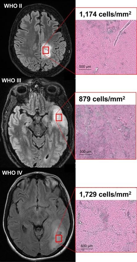
www.nature.com/scientificreports/
that patients with lesions in the same area had impairments in visual but not auditory naming, and that the
anterior temporal lobe conferred specificity for auditory naming, but not picture naming.
Anomic/dynsomic aphasia is a common finding in adult brain tumor patients and the presence or absence
of overlapping neural systems for lexical retrieval have tangible consequences for p atients17. For example, cog-
nitive rehabilitation strategies for patients with aphasia may focus on compensatory strategy training such as
paced speech, associative cuing, and verbal circumlocution if separate neural systems exist for visual and audi-
tory stimuli18–23. If, however, distinct sensory stimuli converge on a single brain region, cognitive rehabilitation
strategies may be better served by environmental interventions, such as supportive communication s trategies24,25.
Incongruent results among existing studies may reflect limitations of the methodologies used to study het-
eromodal lexical retrieval. Indeed, DES is restricted to regions exposed during brain mapping surgery, may be
affected by the administration of anesthetics, and induces non-physiologic, backward propagation of action
potentials, thereby limiting its spatial s pecificity26–28. Furthermore, studies employing DES often do not assess
subcortical tissue, differentiate speech arrest (i.e., a transient dysfunction in general speech production) from
true anomia, or match stimuli on content category. Finally, while functional imaging modalities such as elec-
trocorticography, fMRI, and PET offer correlational insights with varying temporal and spatial precisions, they
cannot generate conclusions about requisite brain areas with causal c ertainty29.
Lesion symptom mapping (LSM) is poised to identify cortical and subcortical regions that are necessary for a
given task, either directly or indirectly via involvement of tissues connected to distant brain regions (diaschisis)30.
Traditionally, LSM was implemented using a chronic stroke lesion model, thereby offering a view of language
processing based on vascular territory. However, LSM has since been expanded to include lesions formed by
intrinsic brain tumors, cytoreduction surgery, traumatic brain injury, and neurodegenerative disease31–34. Because
dominant-hemisphere intrinsic brain tumors in particular lead to substantial rates of dysnomic aphasia35 and
can encompass broad cortical and subcortical regions without confinement to stereotyped vascular distributions,
they may serve as optimal lesion models to study lexical retrieval in response to visual and auditory stimuli.
Furthermore, ensuing results may inform disease-specific therapeutic strategies during cognitive rehabilitation.
In this study, we used a permutation-based, multivariate approach to LSM to identify cortical and subcortical
regions necessary for heteromodal lexical retrieval. Because recent data using a combination of functional imag-
ing and DES offer conflicting models of lexical retrieval, it remains unknown whether separate auditory- and
visual-only regions exist, or whether the neural systems subserving lexical retrieval exist through distributed
convergence zones. Thus, our aims were twofold: (1) to determine whether lesions in frontotemporal regions
impair lexical retrieval in a sensory-specific manner and (2) to identify causal convergence zones with a lesion-
symptom mapping framework.
The left lateral prefrontal cortex (PFC) in particular is an excellent anatomic candidate for convergence
of lexical retrieval given its dual role in (a) processing lexical semantics (specifically within the pars orbitalis
and triangularis) and (b) serving as an interface for working memory and executive control in the presence of
competing alternatives36,37. Considering the reliance of picture naming and auditory naming on these cognitive
functions, and evidence of heteromodal convergence hubs in non-lexical d omains38, we hypothesize that tumor
infiltration of the left lateral PFC will be associated with selective impairments in lexical retrieval regardless of
input sensory modality.
Materials and methods
Participants. Eighty-nine adult patients presenting with World Health Organization (WHO) II-IV gliomas
and brain metastasis at the University of California San Francisco (UCSF) between 2017 and 2020 were recruited
in a longitudinal prospective clinical trial of language and neurocognitive outcomes (trial registration: NCI-
2020-02,286, 11/30/2017). This included fifty-three adult patients (participants) with dominant hemisphere
tumors and a disease- and age-matched control cohort of thirty-six patients (controls) with non-dominant hem-
isphere tumors. No patients had pre-existing neurological impairment prior to tumor diagnosis. Hemisphere
of language dominance was established via magnetoencephalography (MEG)39. Tumor histology was classified
according to the WHO 2016 classification of CNS t umors40. Fresh tissue samples were imaged using stimulated
Raman histology with cell counting of core specimens using previously established p rotocols41. Participants
and controls were on a standard regimen of dexamethasone and levetiracetam for control of brain edema and
seizures, respectively. All patients provided written informed consent for study enrollment, which was approved
by UCSF’s institutional review board (CHR-17-23,215). The study was performed in accordance with the Dec-
laration of Helsinki.
Experimental design and statistical analysis. Preoperative language assessments. Each participant
and control underwent a language assessment one day prior to cytoreduction surgery using the validated Quick
Aphasia Battery (QAB) which provides weighted scores for each of its seven predefined language domains (“sub-
tests”): word comprehension, sentence comprehension, word finding, grammatical construction, motor speech,
repetition, and reading42. The QAB was used in this study given its prior use in adult brain tumor patients and
ability to provide rapid (yet comprehensive) assessments of language functions. Furthermore, the QAB was
particularly valuable for the purposes of this study as it provides a validated measure of lexical retrieval through
its word finding subtest. Nine participants did not undergo QAB testing and were excluded from components of
the analysis that required QAB scores. QAB scores between participants and controls were compared using the
two-tailed Wilcoxon rank-sum test after confirming non-normality of scoring distributions both visually and
via the Shapiro–Wilk test. This comparison was 80% powered to identify a medium-to-large effect size (Cohen’s
d of 0.67) using a significance level of 0.05. To identify sources of potential confounding in language scores, cat-
Scientific Reports | (2021) 11:6305 | https://doi.org/10.1038/s41598-021-85802-5 2
Vol:.(1234567890)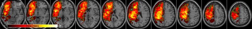
www.nature.com/scientificreports/
egorical comparisons of demographics and baseline clinical data between study participants and controls were
performed with Pearson’s chi-squared test.
In addition to completing the QAB, each of the fifty-three participants performed four additional language
tasks for use in lesion-symptom mapping. These tasks consisted of naming pictorial representations of common
objects and animals (Picture Naming, PN), reading two-syllable text (Text Reading, TR), naming common objects
and animals via auditory descriptions (Auditory Naming, AN) and describing line drawings with intact syntax
(Syntax, Syn)28,43. The correct answers for all four tasks were matched on word frequency (i.e., commonality
within the English language) using S UBTLEXWF scores provided by the Elixcon project (http://elexicon.wustl.
edu/) and on content category44.
All of the tasks were delivered on a laptop with a 15-inch monitor (60 Hz refresh rate) that was positioned
two feet away from the seated patient in a quiet clinical setting. Task stimuli were randomized and presented
using PsychToolbox45,46. Slides were manually advanced by the research coordinator either immediately after
the participant provided a response or after six seconds if no response was given. All of the tasks were scored
on a scale from 0 to 4 by a trained clinical research coordinator who was initially blinded to all clinical data
(including imaging studies) using the guidelines provided by the QAB. No participants had uncorrectable visual
or hearing loss.
To identify which language tasks were most strongly associated with the validated measure of lexical retrieval
provided by the QAB (i.e., the word finding subtest), generalized linear models were fitted between the word
finding subtest and each of the four language tasks. Model significance was determined using a two-tailed F-test.
Principal components analysis (PCA) was performed to collapse the five language measures into a smaller set of
dimensions (i.e., principal components) that account for a majority of the variance in the dataset. By removing
redundancies in the behavioral data, PCA is able to identify common cognitive constructs. In this study, the
largest resulting principal component (i.e., defined by a weighted combination of our five language measures)
was used in lesion-symptom mapping to identify brain regions subserving a common cognitive construct. All
statistical analyses were performed on R version 3.6.2. Corrections for multiple comparisons among the language
data were made by controlling the family-wise error rate with the Holm-Bonferroni method47.
Magnetic resonance imaging and pre‑processing. Each participant underwent a standard preoperative imag-
ing protocol on a 3 T scanner including a) pre- and post-contrast T1-weighted imaging with gadolinium and
b) T2-weighted fluid attenuation inversion recovery (FLAIR) imaging with slice thicknesses between 1 and 1.5
mm43. All imaging was performed within three days of the language assessments.
Lesions were segmented either manually or semi-automatically using ITK-SNAP 3.8.0 (http://www.itksnap.
org/) by a trained co-author blinded to language outcomes scores (AAA)48. The borders of contrast enhancement
on T1-weighted post-gadolinium sequences were used to identify lesion boundaries in patients with WHO grade
IV contrast enhancing tumors (n = 43 patients). Abnormal FLAIR signal was used to identify lesion boundaries
in patients with WHO grades II and III non-enhancing tumors (n = 10)32,49. Accuracy of the lesion masks was
independently confirmed by a separate examiner also blinded to language outcomes scores (SHJ) and disagree-
ments were resolved by reaching a consensus.
Using Clinical Toolbox on SPM12 (https://www.fil.ion.ucl.ac.uk/spm/software/spm12/), each anatomical
T1-weighted image was normalized to the standardized Montreal Neurological Institute template (MNI152)50,51.
The resulting transformation matrix was applied to the participant’s corresponding lesion mask to facilitate
comparisons across participants. For participants with non-enhancing lesions, the T2-weighted FLAIR derived
lesion mask was registered to the anatomical image prior to normalization.
Lesion‑Symptom and Statistical Analysis. Support vector regression lesion-symptom mapping (SVR-LSM) was
performed to identify the precise anatomic locations associated with lower scores on each of the four language
tasks (PN, TR, AN, and Syn) and the first principal component derived from PCA52. Analyses were conducted
using the SVRLSMgui package (https://github.com/atdemarco/svrlsmgui/) for MATLAB which provides a
permutation-based multivariate approach to lesion-symptom mapping with significant advantages over mass-
univariate analyses53. As opposed to traditional voxel lesion-symptom mapping (VLSM), by including every
lesion voxel as a simultaneous covariate, SVR-LSM accounts for the inherent spatial autocorrelation of brain
tumors54,55. Additionally, by performing random permutations of the behavioral data to create null distributions,
it is robust to the Type I and II errors that often arise from false discovery rate and Bonferroni methods for mul-
tiple comparisons corrections, respectively56.
Total lesion volume was linearly regressed out of both the lesion masks and language scores to limit the biasing
impact of large lesions on language impairment, while retaining sensitivity to fluctuations in task performance.
For inclusion in each task-based analysis, each voxel was required to include at least two participants with over-
lapping lesions57. A threshold of P < 0.005 was used to determine statistical significance for individual voxels while
a threshold of P < 0.05 was used for cluster-level (i.e., groups of contiguous voxels) corrections after performing
10,000 permutations of the language data across participants. Hyperparameters were left at their default settings
(gamma of 5 and cost of 30). Analyses were fully parallelized on a high-performance computer with 32 cores at
4.2 GHz and 256 GB of random-access memory. SPM12 was used to generate statistical parametric maps and
BrainNet Viewer (https://www.nitrc.org/projects/bnv/) to generate three-dimensional representations of the
resulting statistical maps58.
Scientific Reports | (2021) 11:6305 | https://doi.org/10.1038/s41598-021-85802-5 3
Vol.:(0123456789)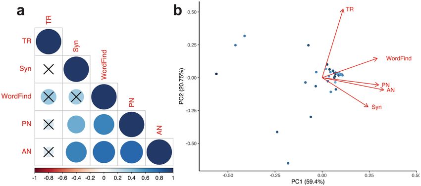
www.nature.com/scientificreports/
Characteristic Participants Controls P-value
n 53 36
Sex 17 F, 36 M 22 F, 14 M 0.013
Mean Age (SD) 51.2 (17.3) 49.4 (15.0) 0.54
Handedness 3 L, 47 R, 3 unk 6 L, 27 R, 3 unk 0.19
Education 0.35
High school 10 6
Some college 11 7
Bachelors 11 13
Graduate 6 4
Unk 15 6
Tumor Laterality 49 L, 4 R 1 L, 35 R 3.6 × 1 0–16
Tumor Grade/Pathology 0.18
WHO Grade I—II 11 9
WHO Grade III 15 4
WHO Grade IV 24 23
Metastasis 3 0
Table 1. Demographics and clinical summary. Comparisons between categorical variables were made with the
chi-squared test while continuous variables were compared with the Wilcoxon rank-sum test for participants
with dominant hemisphere lesions and controls with non-dominant hemisphere lesions. Handedness was
determined using preoperative magnetoencephalography. Education was self-reported in patients who
completed the Neuro-QoL assessment. Pathologic diagnoses were made by board-certified pathologists using
the World Health Organization (WHO) Revised Classification of Tumors in the CNS. Unk.-Unknown.
Results
Demographics and clinical data are summarized in Table 1. There were no significant differences in mean age,
handedness, education, or oncologic features between participants and controls. Participants were significantly
more likely to be male and to have left-sided tumors. Tissues sampled from regions of FLAIR or T1-weighted
post-gadolinium signal abnormality from three different participants with WHO grade II, III and IV gliomas are
presented in Fig. 1. All participants and controls had WHO II-IV gliomas as defined by WHO 2016 molecular
subclassifications and their imaged tissues demonstrated cellular neoplasms with extensive disruption of normal
cytoarchitecture.
Our main goal was to identify the neuroanatomical regions that are necessary for heteromodal lexical retrieval
using a lesion-symptom model. As such, first, we compared language domain performance between participants
and controls across all seven QAB subtests to establish whether or not participants had significant impairments
in lexical retrieval. Compared to controls, participants exhibited statistically significantly lower performance on
word finding (P = 0.0096), but not on any of the six remaining subtests of the QAB (Fig. 2). Second, we sought
to identify associations between each of the four language tasks (Table 2) and the word finding subtest using
generalized linear models computed across all participants (Fig. 3a). Word finding was predicted by picture
naming (r = 0.66, P = 0.000029) and auditory naming (r = 0.76, P = 0.0000001) but not by text reading (r = 0.37,
P = 0.093) or Syn (r = 0.36, P = 0.093). Significant associations were also found between PN and AN (r = 0.80,
P = 0.000000008), Syn and PN (r = 0.49, P = 0.0088), and Syn and AN (r = 0.68, P = 0.000016).
Given these results, we next performed PCA to a) reveal and remove redundancies in the behavioral data
and b) identify a set of weighted combinations of the language measures that subsequently represent a single,
common cognitive construct. PCA revealed two principal components (PC), PC1 and PC2, that were responsi-
ble for 59.4% and 20.75% of the variance in the five language measures, respectively (Fig. 3b). Picture naming,
auditory naming, and word finding demonstrate the greatest collinearity with PC1. Text reading, on the other
hand, is roughly collinear with PC2.
Having established the importance of picture naming and auditory naming in explaining the variance in the
word finding subtest, we then used these two tasks in conjunction with SVR-LSM to uncover the anatomical
regions necessary for visually- and auditorily-prompted lexical retrieval, respectively. Of the 53 participants,
48 had lesions with at least one overlapping voxel with another participant and were therefore included in the
lesion-symptom mapping analysis. All five excluded participants had right-sided, dominant-hemispheric tumors
(confirmed by MEG) with non-overlapping lesions. The final lesion overlap map used for SVR-LSM is outlined
in green in Fig. 4.
Using this lesion map, SVR-LSM was then performed for each of the four language tasks testing the hypothesis
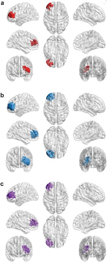
that lesioned voxels are associated with lower task accuracies. For picture naming, one of eight clusters survived
cluster-level corrections (8,333 voxels, P = 0.045). The resulting cluster for picture naming is centered in the left
lateral PFC and encompasses Brodmann areas 10 and 45–48 (Fig. 5a). For auditory naming, one of ten clusters
survived thresholding in an analogous region of the lateral PFC (Fig. 5b), except with greater involvement of
subcortical areas (21,512 voxels, P = 0.0034). Notably, no significant clusters for auditory naming were observed
in the temporal areas. The significant cluster in picture naming overlaps completely with, and accounts for 38.7%
Scientific Reports | (2021) 11:6305 | https://doi.org/10.1038/s41598-021-85802-5 4
Vol:.(1234567890)www.nature.com/scientificreports/
Figure 1. T2-weighted fluid attenuation inversion recovery (FLAIR) images of three participants with World
Health Organization (WHO) grade II, III, and IV glioma (left). Red boxes indicate regions of FLAIR signal
abnormality biopsied for pathologic examination and cell-counting with Raman scattering microscopy (right).
Pseudo-H&E images of fresh biopsy specimens demonstrate ablation of normal cytoarchitecture across all
WHO tumor grades. Here, lesion cellularity and necrotic features escalate with increasing WHO grade.
Figure 2. Pairwise Wilcoxon rank-sum tests between 44 patients with dominant-hemisphere intraparenchymal
tumors (participants) and 36 patients with non-dominant tumors (controls) on each of the seven predefined
Quick Aphasia Battery (QAB) subtests. **P = 0.0096, corrected for multiple comparisons with the Holm-
Bonferroni method. NS-not statistically significant (P > 0.05). Word Comp.-word comprehension. Sentence
Comp.-sentence comprehension. Grammar-grammatical construction. Error bars represent standard error.
of, auditory naming’s significant cluster. Finally, SVR-LSM was performed using PC1 from the PCA. Only one
of fourteen clusters survived cluster-level corrections (11,944 voxels, P = 0.019). This cluster overlaps with the
resulting clusters from picture naming and auditory naming (Fig. 5c).
Neither TR nor Syn survived cluster-level corrections. For TR, no significant voxels were found after voxel-
based thresholding. Syn, on the other hand, yielded smaller clusters of statistically significant voxels interspersed
throughout the left anterior frontal lobe. However, the size of these clusters fell short of the critical threshold size
that was derived from performing SVR-LSM on random permutations of the scores.
Scientific Reports | (2021) 11:6305 | https://doi.org/10.1038/s41598-021-85802-5 5
Vol.:(0123456789)www.nature.com/scientificreports/
Task (n trials) Mean Score SE Mean
Picture Naming (48) 3.84 0.051
Text Reading (27) 3.97 0.014
Auditory Naming (32) 3.62 0.074
Syntax (28) 3.80 0.058
Table 2. Language testing. Tasks were delivered in a quiet clinical setting and matched on word frequency.
Each stimulus was scored from 0 to 4 using guidelines provided by the Quick Aphasia Battery.
Figure 3. Operationalization of the word finding subtest of the QAB (WordFind) using four computerized
language tasks: picture naming (PN), text reading (TR), auditory naming (AN), and syntax formation (Syn). a
correlogram summarizing the results of univariate generalized linear models fitted to each language task and
WordFind. Color bar represents each correlation coefficient and crosses indicate non-significant associations
(P > 0.05) after corrections for multiple comparisons. b biplot between principal component 1 and 2 (PC1
and PC2) derived from principal components analysis (PCA) on WordFind and the four language tasks.
Corresponding loadings are represented by the red arrows.
Figure 4. Map depicting regions in which at least two participants had overlapping lesions, projected on
the standard-space Montreal Neurological Institute template (MNI152), total n = 48. Regions in which actual
overlap occurred and where the analysis was restricted to is outlined in green. Accordingly, five participants with
non-overlapping right-sided tumors were excluded from lesion-symptom mapping.
Discussion
Cognitive models of lexical retrieval have been heavily influenced by visual confrontation (picture naming) tasks.
Further, existing studies have implemented methods that either lack causal certainty or precise spatial localiza-
tion. Therefore, published results are mixed with respect to whether visual and auditory stimuli represent overlap-
ping (convergent) or non-overlapping (divergent) neural systems. This unresolved conflict has direct implications
on cognitive rehabilitation strategies for brain tumor patients with dysnomic aphasia. In this study, we propose
a convergent model of lexical retrieval within a lesion-symptom framework for both visual and auditory inputs.
First, we established that intrinsic brain tumors cause extensive disruption of normal cytoarchitecture (Fig. 1),
leading to selective impairments in word finding (Fig. 2). This finding is in line with previous reports of the
prevalence of selective dysnomia in patients with dominant hemisphere intrinsic brain t umors59. Furthermore,
it supports the use of our brain tumor lesion model in particular to study lexical retrieval, since participants in
other clinical populations tend to have additional confounding language i mpairments60,61.
Scientific Reports | (2021) 11:6305 | https://doi.org/10.1038/s41598-021-85802-5 6
Vol:.(1234567890)www.nature.com/scientificreports/
Figure 5. Results of support vector regression lesion-symptom mapping (SVR-LSM) after voxels thresholded
at P < 0.005 underwent cluster-level corrections with 10,000 permutations (cluster threshold = P < 0.05).
3-Dimensional models of resulting clusters were generated using the Montreal Neurological Institute template
via BrainNet Viewer (https://www.nitrc.org/projects/bnv/). a Lesions in a cluster of 8,333 voxels in the left lateral
PFC were predictive of impaired picture naming (P = 0.045). b Lesions in a larger cluster of 21,512 were voxels
predictive of impaired auditory naming (P = 0.0034). Clusters in a and b overlap completely with the cluster in
a extending deeper into subcortical regions. c Results of SVR-LSM using the principal component scores for
each participant on the first principal component (PC1). This cluster consists of 11,944 voxels (P = 0.019) and
demonstrates overlap with the clusters in a and b.
Scientific Reports | (2021) 11:6305 | https://doi.org/10.1038/s41598-021-85802-5 7
Vol.:(0123456789)www.nature.com/scientificreports/
Next, we demonstrated strong correlations between accuracy on picture naming and auditory naming, provid-
ing initial evidence of the existence of lexical retrieval pathways that are agnostic to the sensory modality of the
input (Fig. 3). From a behavioral perspective, this finding replicates those in lesion studies performed in several
clinical populations. For instance, Hamberger and Seidel (2003) found that patients with left temporal lobe
epilepsy had co-occurring impairments in visual and auditory naming compared to a) healthy controls and b)
patients with right temporal lobe e pilepsy62. Miller et al. (2010) and Hirsch et al. (2016) also reported significant
associations between picture naming and auditory naming in lesion studies of patients with dementia63,64. Hirsch
et al. in particular used PCA in a cohort of 458 patients to reveal a unique redundancy between picture naming
and auditory naming that was not shared with any of their other twenty-five cognitive and linguistic measures.
In the present study, PCA led to analogous results: the similarity in loadings between PN, AN, and word finding
argue for a single linguistic construct (i.e., lexical retrieval) that may be differentiated from constructs contribut-
ing to other linguistic measures such as TR and Syn.
As predicted by the results of our language assessments, multivariate lesion-symptom mapping revealed
overlapping clusters for picture naming, auditory naming, and PC1 in the left lateral prefrontal cortex (Fig. 5A-
C). The significance of this brain region in particular is that it is capable of processing the critical components of
lexical retrieval, namely semantic access and executive control (or more specifically, selection between competing
items). Indeed, various authors have proposed that the lateral PFC serves as a generic region for multimodal
integration given its preferential activation during demanding tasks that require selection between competing
alternatives which may include both semantic and phonological i tems65,66. Future investigation will be required
to dissociate the contributions of the lateral PFC to these interdependent components.
Notably, in contrast to prior DES studies12,67, a separate cluster specific to auditory naming was not identified
in the anterior temporal lobe, a well-represented region in our lesion overlap mask. Our results instead align
with a number of more recent, mixed-method studies that argue for a convergence hub in the lateral PFC for
both visual and auditory i nputs14,68. Specifically, by timing the evolution of cortical responses to the onset of task
stimuli using electrocorticography, Forseth et al. (2018) showed that the inferior frontal gyrus (a component
of the lateral PFC) serves as an interface between lexical and phonological pathways during both picture and
auditory naming14. Taken with the results of the present lesion study, these findings provide compelling and
causal evidence that lexical retrieval indeed represents a unified cognitive construct that is agnostic to the input
modality. The lack of convergence of TR and Syn in these regions, despite sharing the same input modality as
PN, further argues that lexical retrieval predominantly relies on downstream language networks that are ana-
tomically distinct from those engaged during orthographic (TR) and syntactic (Syn) processing. For example,
in contrast to our cohort of patients with anomic aphasia, individuals with acquired phonological alexia (i.e.,
impaired text reading) demonstrate global impairments in phonological processing that generalize to non-visual
sensory modalities, but typically have intact lexical-semantic p rocessing69.
Notably, case reports and small clinical series of patients with modality-specific naming impairments such
as visual (i.e. optic aphasia), auditory, and tactical anomia from brain lesions may seem to argue against the
existence of a single construct subserving lexical r etrieval70–72. However, these studies have not been widely
validated in large clinical cohorts using rigorous image-based methods and thus cannot be adequately evaluated
for confounding variables. Specifically, concurrent damage to primary sensory processing or association areas
cannot be ruled out without the granularity provided by voxel-based analyses. For instance, Hamberger and
Seidel (2009) reported that a cohort of fourteen patients with anterior temporal lobe lesions (defined as < 5 cm
from the temporal pole) had impaired auditory, but not picture n aming16. However, of those fourteen patients,
only five had structural lesions. The remaining “anterior lesion” patients were defined by the presence of seizure
foci on subdural or scalp EEG. Given a) the absence of lesion overlap maps for examination and b) the ability
of epileptic foci to transiently impair remote brain areas, we cannot rule out the possibility that the observed
differences in auditory and picture naming performance were due to confounding damage to auditory sensory
processing areas (i.e. A1-A3). Indeed, the “auditory-only” naming sites identified by Malow et al. (1996) via DES
(i.e. transient induction of iatrogenic lesions) are chiefly located in the superior temporal gyrus where various
features of auditory stimuli are e ncoded73–75. Analogous concerns have also been raised about other modality-
specific anomia syndromes76.
Since the late nineteenth century, it has been known that the brain, at its most fundamental level, is made of
individual cells which together form discrete networks that influence cognition and behavior. Efforts to uncover
the role of neuroanatomic structures and underlying network dynamics have established both causal and correla-
tive cognitive frameworks. However, these physiological models may lack disease-specific relevance. Therefore,
the discovery of overlapping neural systems for lexical retrieval in a brain tumor lesion-symptom mapping study
has implications beyond causal adjudication between established scientific models. One salient example comes
from competing rehabilitative strategies for patients with aphasia. On the one hand, compensatory training
relies on the use of alternative stimuli through strategies such as paced speech, associative cuing, and verbal
circumlocution to leverage distant yet overlapping neural systems for a given sensory domain. Compensatory
training may therefore be considered most promising in the setting of divergent visual and auditory lexical
retrieval systems18–23. If, however, distinct sensory stimuli converge on a single brain region and/or network, it
may be best if cognitive rehabilitation strategies focus on environmental interventions, such as supportive com-
munication strategies following a lesion to that r egion24,25. The optimal rehabilitation strategy for patients with
brain tumors and subsequent aphasia continues to be an area of active investigation. Therefore, causal evidence
using disease-specific models of physiology will contribute to a nuanced understanding of therapeutic options.
Limitations of the Present Study. It is first worth discussing the typical limitations that apply to any
lesion-symptom analysis, namely that conclusions about behavioral contributions of brain areas lying outside
Scientific Reports | (2021) 11:6305 | https://doi.org/10.1038/s41598-021-85802-5 8
Vol:.(1234567890)www.nature.com/scientificreports/
our lesion masks cannot be made. For this reason, we expanded our analysis to include voxels in which at
least two patients overlapped, rather than more restrictive cutoffs of five or more found in previous studies.
By performing multivariate comparisons on a high-performance computer that can support these increasingly
memory- and computationally intensive analyses, we were able to avoid the reductions in statistical power that
typically constrain studies employing mass-univariate tests with post-hoc corrections for multiple comparisons.
While lowering the lesion overlap threshold may increase exposure to outlier effects, our implementation of
non-parametric statistics for lesion-symptom mapping mitigate this concern by calculating exact p-values with-
out any a priori assumptions about the underlying distribution. In our study, as in others, the impact of outliers
was greatly attenuated by performing 10,000 random permutations of the input behavioral data to generate a
null distribution for each test s tatistic77. Despite these measures, however, our lesion mask was not able to cap-
ture posteroinferior regions of the perisylvian network, thereby restricting our conclusions to the frontal cortex,
anterior temporal cortex, and angular gyrus.
Second, the expansive nature of the lesions under study (which inherently facilitates group-level analyses)
simultaneously limits our ability to precisely localize the neuronal subpopulations involved in each of the two
convergent tasks or determine how these subpopulations may causally interact to produce correct responses.
Furthermore, while there is some debate on whether brain tumors can serve as reliable clinical models for
lesion-symptom analyses, this position can be extended to stroke for which VLSM was developed and is most
commonly implemented. Furthermore, brain tumor histology confirmed disordered neural structures, increased
cellularity, and altered cytoarchitecture (Fig. 1). Because perfect clinical models for brain lesions likely do not
exist, the literature may be best served by an increase, rather than a decrease, in the diversity of the clinical
populations under s tudy78.
Conclusion
To summarize, generalized linear modeling and principal components analysis revealed multicollinearity between
picture naming, auditory naming, and word finding tasks. Support vector regression lesion-symptom mapping
across participants was used to uncover associations between lower task accuracies on each of the four language
tasks and lesioned voxels. Picture naming and auditory naming lesions demonstrated overlapping clusters within
the left lateral PFC. This study demonstrates that cortical and subcortical brain regions involved in lexical
retrieval from visual and auditory stimuli represent overlapping neural systems.
Received: 5 December 2020; Accepted: 3 March 2021
References
1. Andreetta, S., Cantagallo, A. & Marini, A. Narrative discourse in anomic aphasia. Neuropsychologia 50, 1787–1793 (2012).
2. Kemmerer, D. Cognitive Neuroscience of Language. (Psychology Press, 2014).
3. Caramazza, A. How many levels of processing are there in lexical access?. Cogn. Neuropsychol. 14, 177–208 (1997).
4. Dell, G. S., Schwartz, M. F., Martin, N., Saffran, E. M. & Gagnon, D. A. Lexical access in aphasic and nonaphasic speakers. Psychol.
Rev. 104, 801–838 (1997).
5. Griffin, Z. M. & Bock, K. Constraint, word frequency, and the relationship between lexical processing levels in spoken word
production. J. Mem. Lang. 38, 313–338 (1998).
6. Levelt, W. J. M., Roelofs, A. & Meyer, A. S. A theory of lexical access in speech production. Behav. Brain Sci. 22, 1–38 (1999).
7. Rapp, B. & Goldrick, M. Speaking words: contributions of cognitive neuropsychological research. Cogn. Neuropsychol. 23, 39–73
(2006).
8. Graves, W. W., Grabowski, T. J., Mehta, S. & Gordon, J. K. A neural signature of phonological access: distinguishing the effects of
word frequency from familiarity and length in overt picture naming. J. Cogn. Neurosci. 19, 617–631 (2007).
9. Damasio, H., Grabowski, T. J., Tranel, D., Hichwa, R. D. & Damasio, A. R. A neural basis for lexical retrieval. Nature 380, 499–505
(1996).
10. Ungrady, M. B., Flurie, M., Zuckerman, B. M., Mirman, D. & Reilly, J. Naming and knowing revisited: eyetracking correlates of
anomia in progressive aphasia. Front. Hum. Neurosci. 13, 57 (2019).
11. Bell, B. D., Seidenberg, M., Hermann, B. P. & Douville, K. Visual and auditory naming in patients with left or bilateral temporal
lobe epilepsy. Epilepsy. Res. 55, 29–37 (2003).
12. Hamberger, M. J., Goodman, R. R., Perrine, K. & Tamny, T. Anatomic dissociation of auditory and visual naming in the lateral
temporal cortex. Neurology 56, 56–61 (2001).
13. Hamberger, M. J., Habeck, C. G., Pantazatos, S. P., Williams, A. C. & Hirsch, J. Shared space, separate processes: Neural activation
patterns for auditory description and visual object naming in healthy adults. Hum. Brain Mapp. 35, 2507–2520 (2014).
14. Forseth, K. J. et al. A lexical semantic hub for heteromodal naming in middle fusiform gyrus. Brain 141, 2112–2126 (2018).
15. Malow, B. A. et al. Cortical stimulation elicits regional distinctions in auditory and visual naming. Epilepsia 37, 245–252 (1996).
16. Hamberger, M. J. & Seidel, W. T. Localization of cortical dysfunction based on auditory and visual naming performance. J. Int.
Neuropsychol. Soc. 15, 529–535 (2009).
17. Santini, B. et al. Cognitive outcome after awake surgery for tumors in language areas. J. Neurooncol. 108, 319–326 (2012).
18. Cicerone, K. D. et al. Evidence-based cognitive rehabilitation: updated review of the literature from 2003 through 2008. Arch. Phys.
Med. Rehabil. 92, 519–530 (2011).
19. Francis, D. R., Clark, N. & Humphreys, G. W. Circumlocution-induced naming (CIN): A treatment for effecting generalisation in
anomia?. Aphasiology 16, 243–259 (2002).
20. Maddy, K. M., Capilouto, G. J. & McComas, K. L. The effectiveness of semantic feature analysis: an evidence-based systematic
review. Ann. Phys. Rehabil. Med. 57, 254–267 (2014).
21. Johnstone, B. & Stonnington, H. H. Rehabilitation of neuropsychological disorders : a practical guide for rehabilitation professionals.
(Psychology Press, 2011). https://doi.org/10.4324/9780203837931.
22. Winans-Mitrik, R. L. et al. Description of an intensive residential aphasia treatment program: rationale, clinical processes, and
outcomes. Am. J. Speech Lang. Pathol. 23, S330-342 (2014).
23. Yu, Z.-Z., Jiang, S.-J., Jia, Z.-S., Xiao, H.-Y. & Zhou, M.-Q. Study on language rehabilitation for aphasia. Chin. Med. J. (Engl) 130,
1491–1497 (2017).
Scientific Reports | (2021) 11:6305 | https://doi.org/10.1038/s41598-021-85802-5 9
Vol.:(0123456789)www.nature.com/scientificreports/
24. Turner, S. & Whitworth, A. Conversational partner training programmes in aphasia: a review of key themes and participants’ roles.
Aphasiology 20, 483–510 (2006).
25. Simmons-Mackie, N., Raymer, A., Armstrong, E., Holland, A. & Cherney, L. R. Communication partner training in aphasia: a
systematic review. Arch. Phys. Med. Rehabil. 91, 1814–1837 (2010).
26. Mandonnet, E., Winkler, P. A. & Duffau, H. Direct electrical stimulation as an input gate into brain functional networks: principles,
advantages and limitations. Acta Neurochir. 152, 185–193 (2010).
27. Ritaccio, A. L., Brunner, P. & Schalk, G. Electrical stimulation mapping of the brain: basic principles and emerging alternatives. J.
Clin. Neurophysiol. 35, 86–97 (2018).
28. Aabedi, A. A. et al. Assessment of wakefulness during awake craniotomy to predict intraoperative language performance. J. Neu‑
rosurg. 1, 1–8 (2019).
29. Weber, M. J. & Thompson-Schill, S. L. Functional neuroimaging can support causal claims about brain function. J. Cogn. Neurosci.
22, 15 (2010).
30. Bates, E. et al. Voxel-based lesion–symptom mapping. Nat. Neurosci. 6, 448–450 (2003).
31. Mesulam, M. et al. Neurology of anomia in the semantic variant of primary progressive aphasia. Brain 132, 2553–2565 (2009).
32. Campanella, F., Shallice, T., Ius, T., Fabbro, F. & Skrap, M. Impact of brain tumour location on emotion and personality: a voxel-
based lesion–symptom mapping study on mentalization processes. Brain 137, 2532–2545 (2014).
33. Knutson, K. M. et al. Neural correlates of apathy revealed by lesion mapping in participants with traumatic brain injuries. Hum.
Brain Mapp. 35, 943–953 (2014).
34. Foulon, C. et al. Advanced lesion symptom mapping analyses and implementation as BCBtoolkit. Gigascience 7, 21 (2018).
35. Davie, G. L., Hutcheson, K. A., Barringer, D. A., Weinberg, J. S. & Lewin, J. S. Aphasia in patients after brain tumour resection.
Aphasiology 23, 1196–1206 (2009).
36. Hoshi, Y. et al. Spatiotemporal characteristics of hemodynamic changes in the human lateral prefrontal cortex during working
memory tasks. Neuroimage 20, 1493–1504 (2003).
37. De Pisapia, N., Slomski, J. A. & Braver, T. S. Functional specializations in lateral prefrontal cortex associated with the integration
and segregation of information in working memory. Cereb. Cortex 17, 993–1006 (2007).
38. Liuzzi, A. G. et al. Cross-modal representation of spoken and written word meaning in left pars triangularis. Neuroimage 150,
292–307 (2017).
39. Findlay, A. M. et al. Dynamics of hemispheric dominance for language assessed by magnetoencephalographic imaging. Ann.
Neurol. 71, 668–686 (2012).
40. Louis, D. N. et al. The 2016 World Health Organization Classification of Tumors of the Central Nervous System: a summary. Acta
Neuropathol. 131, 803–820 (2016).
41. Ji, M. et al. Detection of human brain tumor infiltration with quantitative stimulated Raman scattering microscopy. Sci. Transl.
Med. 7, 309 (2015).
42. Wilson, S. M., Eriksson, D. K., Schneck, S. M. & Lucanie, J. M. A quick aphasia battery for efficient, reliable, and multidimensional
assessment of language function. PLoS ONE 13, e0192773 (2018).
43. Lee, A. T. et al. The impact of high functional connectivity network hub resection on language task performance in adult low- and
high-grade glioma. J. Neurosurg. https://doi.org/10.3171/2020.1.JNS192267 (2020).
44. Brysbaert, M. & New, B. Moving beyond Kučera and Francis: a critical evaluation of current word frequency norms and the
introduction of a new and improved word frequency measure for American English. Behav. Res. Methods 41, 977–990 (2009).
45. Brainard, D. H. The psychophysics toolbox. Spat. Vis. 10, 433–436 (1997).
46. Pelli, D. G. The VideoToolbox software for visual psychophysics: transforming numbers into movies. Spat. Vis. 10, 437–442 (1997).
47. Blakesley, R. E. et al. Comparisons of methods for multiple hypothesis testing in neuropsychological research. Neuropsychology
23, 255–264 (2009).
48. Yushkevich, P. A. et al. User-guided 3D active contour segmentation of anatomical structures: significantly improved efficiency
and reliability. Neuroimage 31, 1116–1128 (2006).
49. Faulkner, J. W. & Wilshire, C. E. Mapping eloquent cortex: a voxel-based lesion-symptom mapping study of core speech production
capacities in brain tumour patients. Brain Lang. 200, 104710 (2020).
50. Fonov, V., Evans, A., McKinstry, R., Almli, C. & Collins, D. Unbiased nonlinear average age-appropriate brain templates from birth
to adulthood. Neuroimage 47, S102 (2009).
51. Fonov, V. et al. Unbiased average age-appropriate atlases for pediatric studies. Neuroimage 54, 313–327 (2011).
52. Zhang, Y., Kimberg, D. Y., Coslett, H. B., Schwartz, M. F. & Wang, Z. Multivariate lesion-symptom mapping using support vector
regression. Hum. Brain Mapp. 35, 5861–5876 (2014).
53. DeMarco, A. T. & Turkeltaub, P. E. A multivariate lesion symptom mapping toolbox and examination of lesion-volume biases and
correction methods in lesion-symptom mapping. Hum. Brain Mapp. 39, 4169–4182 (2018).
54. Mah, Y.-H., Husain, M., Rees, G. & Nachev, P. Human brain lesion-deficit inference remapped. Brain 137, 2522–2531 (2014).
55. Ghaleh, M. et al. Dissociable mechanisms of verbal working memory revealed through multivariate lesion mapping. Cereb Cortex
30, 2542–2554 (2020).
56. Mirman, D. et al. Corrections for multiple comparisons in voxel-based lesion-symptom mapping. Neuropsychologia 115, 112–123
(2018).
57. Benavides-Varela, S. et al. Zero in the brain: A voxel-based lesion–symptom mapping study in right hemisphere damaged patients.
Cortex 77, 38–53 (2016).
58. Xia, M., Wang, J. & He, Y. BrainNet viewer: a network visualization tool for human brain connectomics. PLoS ONE 8, e68910
(2013).
59. Noll, K. R., Sullaway, C., Ziu, M., Weinberg, J. S. & Wefel, J. S. Relationships between tumor grade and neurocognitive functioning
in patients with glioma of the left temporal lobe prior to surgical resection. Neuro. Oncol. 17, 580–587 (2015).
60. Godefroy, O., Dubois, C., Debachy, B., Leclerc, M. & Kreisler, A. Vascular aphasias. Stroke 33, 702–705 (2002).
61. Rogalski, E. et al. Anatomy of language impairments in primary progressive aphasia. J. Neurosci. 31, 3344–3350 (2011).
62. Hamberger, M. J. & Seidel, W. T. Auditory and visual naming tests: Normative and patient data for accuracy, response time, and
tip-of-the-tongue. J. Int. Neuropsychol. Soc. 9, 479–489 (2003).
63. Miller, K. M., Finney, G. R., Meador, K. J. & Loring, D. W. Auditory responsive naming versus visual confrontation naming in
Dementia. Clin. Neuropsychol. 24, 103–118 (2010).
64. Hirsch, J. A., Cuesta, G. M., Jordan, B. D., Fonzetti, P. & Levin, L. The auditory naming test improves diagnosis of naming deficits
in Dementia. SAGE Open 6, 2158244016665693 (2016).
65. Friederici, A. D., Opitz, B. & von Cramon, D. Y. Segregating semantic and syntactic aspects of processing in the human brain: an
fMRI investigation of different word types. Cereb. Cortex 10, 698–705 (2000).
66. Zhang, J. X., Feng, C.-M., Fox, P. T., Gao, J.-H. & Tan, L. H. Is left inferior frontal gyrus a general mechanism for selection?. Neu‑
roimage 23, 596–603 (2004).
67. Hamberger, M. J., McClelland, S., McKhann, G. M., Williams, A. C. & Goodman, R. R. Distribution of auditory and visual naming
sites in nonlesional temporal lobe epilepsy patients and patients with space-occupying temporal lobe lesions. Epilepsia 48, 531–538
(2007).
Scientific Reports | (2021) 11:6305 | https://doi.org/10.1038/s41598-021-85802-5 10
Vol:.(1234567890)www.nature.com/scientificreports/
68. Rauschecker, J. P. & Scott, S. K. Maps and streams in the auditory cortex: nonhuman primates illuminate human speech processing.
Nat. Neurosci. 12, 718–724 (2009).
69. Beeson, P. M., Rising, K., Kim, E. S. & Rapcsak, S. Z. A treatment sequence for phonological alexia/agraphia. J. Speech Lang. Hear
Res. 53, 450–468 (2010).
70. Beauvois, M. F., Saillant, B., Meininger, V. & Lhermitte, F. Bilateral tactile aphasia: a tacto-verbal dysfunction. Brain 101, 381–401
(1978).
71. Beauvois, M. F. Optic aphasia: a process of interaction between vision and language. Philos. Trans. R. Soc. Lond. B Biol. Sci. 298,
35–47 (1982).
72. Endo, K., Miyasaka, M., Makishita, H., Yanagisawa, N. & Sugishita, M. Tactile agnosia and tactile aphasia: symptomatological and
anatomical differences. Cortex 28, 445–469 (1992).
73. Mesgarani, N., Cheung, C., Johnson, K. & Chang, E. F. Phonetic feature encoding in human superior temporal gyrus. Science 343,
1006–1010 (2014).
74. Tang, C., Hamilton, L. S. & Chang, E. F. Intonational speech prosody encoding in the human auditory cortex. Science 357, 797–801
(2017).
75. Yi, H. G., Leonard, M. K. & Chang, E. F. The encoding of speech sounds in the superior temporal gyrus. Neuron 102, 1096–1110
(2019).
76. Lebrun, Y. Tactile aphasia: a hundred-year-old controversy. Adv. Speech Lang. Pathol. 2, 1–7 (2000).
77. Rorden, C., Bonilha, L. & Nichols, T. E. Rank-order versus mean based statistics for neuroimaging. Neuroimage 35, 1531–1537
(2007).
78. Duffau, H. Do brain tumours allow valid conclusions on the localisation of human brain functions?. Cortex 47, 1016–1017 (2011).
Acknowledgements
We deeply appreciate insights from Sofia Jimenez and Matthew Soleiman within their respective fields of psy-
cholinguistics and cognitive neuroscience.
Author contributions
A.A.A.: data analysis, interpretation, and drafting the manuscript. S.K.: data collection and interpretation. J.S.Y.:
reviewed the manuscript for intellectual content. O.W.: data collection. J.K.: data collection. C.V.: data collec-
tion and reviewed the manuscript for intellectual content. S.K.: reviewed the manuscript for intellectual con-
tent. C.W.-J.: reviewed the manuscript for intellectual content. M.S.B.: reviewed the manuscript for intellectual
content. D.H.W.: reviewed the manuscript for intellectual content. D.B.: data interpretation and reviewed the
manuscript for intellectual content. S.L.H.-J.: study design, interpretation, drafting the manuscript and reviewed
the manuscript for intellectual content.
Competing interests
The authors declare no competing interests.
Additional information
Correspondence and requests for materials should be addressed to S.L.H.-J.
Reprints and permissions information is available at www.nature.com/reprints.
Publisher’s note Springer Nature remains neutral with regard to jurisdictional claims in published maps and
institutional affiliations.
Open Access This article is licensed under a Creative Commons Attribution 4.0 International
License, which permits use, sharing, adaptation, distribution and reproduction in any medium or
format, as long as you give appropriate credit to the original author(s) and the source, provide a link to the
Creative Commons licence, and indicate if changes were made. The images or other third party material in this
article are included in the article’s Creative Commons licence, unless indicated otherwise in a credit line to the
material. If material is not included in the article’s Creative Commons licence and your intended use is not
permitted by statutory regulation or exceeds the permitted use, you will need to obtain permission directly from
the copyright holder. To view a copy of this licence, visit http://creativecommons.org/licenses/by/4.0/.
© The Author(s) 2021
Scientific Reports | (2021) 11:6305 | https://doi.org/10.1038/s41598-021-85802-5 11
Vol.:(0123456789)You can also read