The Evolution of White Matter Microstructural Changes After Mild Traumatic Brain Injury: A Longitudinal DTI and NODDI Study - bioRxiv
←
→
Page content transcription
If your browser does not render page correctly, please read the page content below
bioRxiv preprint first posted online Jun. 14, 2018; doi: http://dx.doi.org/10.1101/345629. The copyright holder for this preprint
       (which was not peer-reviewed) is the author/funder. All rights reserved. No reuse allowed without permission.

                      The Evolution of White Matter Microstructural Changes
                               After Mild Traumatic Brain Injury:
                              A Longitudinal DTI and NODDI Study

   Eva M. Palacios1, Julia P Owen2, Esther L. Yuh1,3, Maxwell B. Wang1, Mary J. Vassar3,4, Adam
    R. Ferguson3,4,5, Ramon Diaz-Arrastia6, Joseph T. Giacino7,8, David O. Okonkwo9, Claudia S.
   Robertson10, Murray B. Stein11,12, Nancy Temkin13, Sonia Jain12, Michael McCrea14, Christine L.
   Mac Donald13, Geoffrey T. Manley3,4, Pratik Mukherjee1,3,15* and the TRACK-TBI Investigators

              1. Department of Radiology & Biomedical Imaging, UCSF, San Francisco, CA, USA;
                    2. Department of Radiology, University of Washington, Seattle, WA, USA;
    3. Brain and Spinal Cord Injury Center, Zuckerberg San Francisco General Hospital and Trauma Center,
                                            San Francisco, CA, USA;
                     4. Department of Neurological Surgery, UCSF, San Francisco, CA, USA;
                   5. San Francisco Veterans Affairs Medical Center, San Francisco, California
                6. Department of Neurology, University of Pennsylvania, Philadelphia, PA, USA;
           7. Department of Physical Medicine and Rehabilitation, Spaulding Rehabilitation Hospital,
                                            Charlestown, MA, USA;
     8. Department of Physical Medicine and Rehabilitation, Harvard Medical School, Boston, MA 02115
    9. Department of Neurological Surgery, University of Pittsburgh Medical Center, Pittsburgh, PA, USA;
                10. Department of Neurosurgery, Baylor College of Medicine, Houston, TX, USA;
             11. Department of Psychiatry, University of California, San Diego, La Jolla, CA, USA;
           12. Department of Family Medicine & Public Health, University of California, San Diego,
                                              La Jolla, CA, USA;
            13. Department of Neurological Surgery, University of Washington, Seattle, WA, USA;
    14. Departments of Neurosurgery and Neurology, Medical College of Wisconsin, Milwaukee, WI, USA;
   15. Department of Bioengineering & Therapeutic Sciences, University of California, San Francisco, USA.

   * Corresponding author: Pratik Mukherjee, MD, PhD.
   Department of Radiology & Biomedical Imaging, Bioengineering & Therapeutic Sciences
   University of California, San Francisco
   Box 0946, 185 Berry Street
   San Francisco, CA, 94143-0946, USA
   Electronic address: pratik.mukherjee@ucsf.edu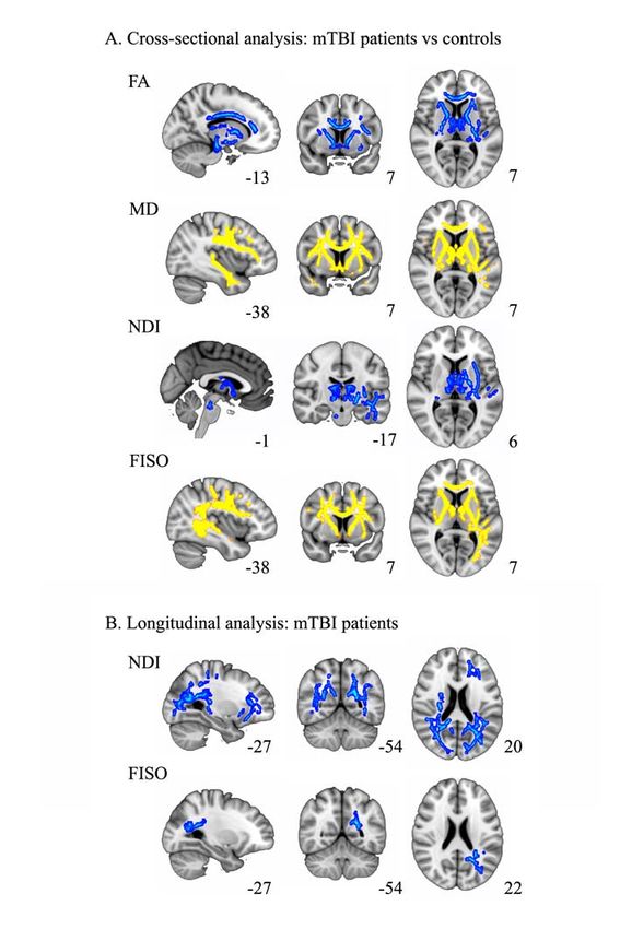
bioRxiv preprint first posted online Jun. 14, 2018; doi: http://dx.doi.org/10.1101/345629. The copyright holder for this preprint
       (which was not peer-reviewed) is the author/funder. All rights reserved. No reuse allowed without permission.

   ABSTRACT

   Neuroimaging biomarkers show promise for improving precision diagnosis and prognosis after
   mild traumatic brain injury (mTBI), but none has yet been adopted in routine clinical practice.
   Biophysical modeling of multishell diffusion MRI, using the neurite orientation dispersion and
   density imaging (NODDI) framework, may improve upon conventional diffusion tensor imaging
   (DTI) in revealing subtle patterns of underlying white matter microstructural pathology, such as
   diffuse axonal injury (DAI) and neuroinflammation, that are important for detecting mTBI and
   determining patient outcome. With a cross-sectional and longitudinal design, we assessed
   structural MRI, DTI and NODDI in 40 mTBI patients at 2 weeks and 6 months after injury and
   14 matched control participants with orthopedic trauma but not suffering from mTBI at 2
   weeks. Self-reported and performance-based cognitive measures assessing postconcussive
   symptoms, memory, executive functions and processing speed were investigated in post-acute
   and chronic phase after injury for the mTBI subjects. Machine learning analysis was used to
   identify mTBI patients with the best neuropsychological improvement over time and relate this
   outcome to DTI and NODDI biomarkers. In the cross-sectional comparison with the trauma
   control group at 2 weeks post-injury, mTBI patients showed decreased fractional anisotropy
   (FA) and increased mean diffusivity (MD) on DTI mainly in anterior tracts that corresponded to
   white matter regions of elevated free water fraction (FISO) on NODDI, signifying vasogenic
   edema. Patients showed decreases from 2 weeks to 6 months in white matter neurite density on
   NODDI, predominantly in posterior tracts. No significant longitudinal changes in DTI metrics
   were observed. The machine learning analysis divided the mTBI patients into two groups based
   on their recovery. Voxel-wise group comparison revealed associations between white matter
   orientation dispersion index (ODI) and FISO with degree and trajectory of improvement within
   the mTBI group. In conclusion, white matter FA and MD alterations early after mTBI might
   reflect vasogenic edema, as shown by elevated free water on NODDI. Longer-term declines in
   neurite density on NODDI suggest progressive axonal degeneration due to DAI, especially in
   tracts known to be integral to the structural connectome. Overall, these results show that the
   NODDI parameters appear to be more sensitive to longitudinal changes than DTI metrics. Thus,
   NODDI merits further study in larger cohorts for mTBI diagnosis, prognosis and treatment
   monitoring.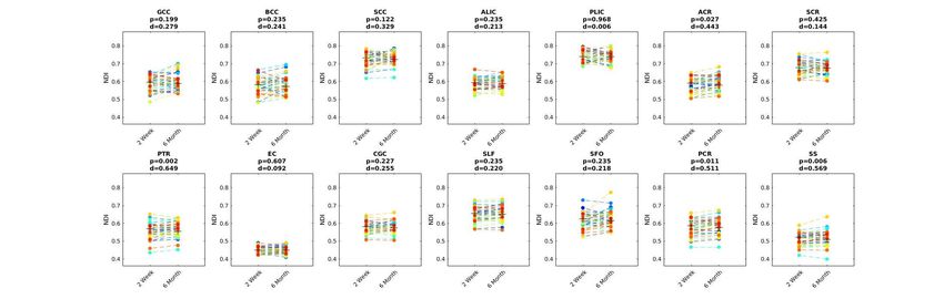
bioRxiv preprint first posted online Jun. 14, 2018; doi: http://dx.doi.org/10.1101/345629. The copyright holder for this preprint
       (which was not peer-reviewed) is the author/funder. All rights reserved. No reuse allowed without permission.

   INTRODUCTION

   Despite increasing evidence from preclinical and human studies that mild traumatic brain injury
   (mTBI) causes axonal shearing injury of white matter microstructure that can affect the long-
   term cognitive, neuropsychiatric, and social domains of function, the lack of reliable objective
   tools to measure such pathology is a barrier to clinical translation. (Manley & Mass, 2013
   JAMA; Bigler a al.2013, Levin & Diaz-Arrastia, 2015 Lancet; Radhakrishnan et al., 2016
   Lancet). The common assumption, even among healthcare professionals, those mTBI patients
   will return to premorbid levels of function shortly after the traumatic event often results in these
   patients not receiving appropriate follow-up care after the acute injury. Sensitive tests that can
   detect white matter microstructural alterations early after injury and inform the likely path to
   recovery are needed to improve care and to develop future therapies.

   Diffusion tensor imaging (DTI) is the most extensively used technique worldwide to study the
   microstructural properties of white matter in vivo (Basser et al., 1994; Mori et al., 2006;
   Mukherjee et al., 2008). DTI studies of mTBI have shown microstructural white matter
   disruption that can lead to neurocognitive and behavioral deficits after mTBI (Yuh EL et al.,
   2014 JN, Croall ID et al., 2014 Neurology; Oehr, et al., 2017 Meta-analysis). However,
   traditional DTI metrics such as mean diffusivity (MD) and fractional anisotropy (FA) represent
   basic statistical descriptions of diffusion that do not directly correspond to biophysically
   meaningful parameters of the underlying tissue. Furthermore, DTI assumes Gaussian diffusion
   within a single microstructural compartment and is therefore insensitive to the complexity of
   white matter microstructure, which requires a non-Gaussian model with multiple compartments
   (Jones et al, 2010). Perhaps as a result, prior DTI studies have produced conflicting results with
   some papers reporting abnormally reduced white matter FA in mTBI and others reporting
   elevations or no change in FA (Eirud et al., 2014). Other contributing factors to this discordance
   in the literature include small effect sizes of DTI changes due to mTBI, small sample sizes and
   the dynamic nature of microstructural white matter alterations after mTBI.

   In this investigation, we overcome the limitations of DTI by applying a more advanced multi-
   compartment diffusion model known as neurite orientation dispersion and density imaging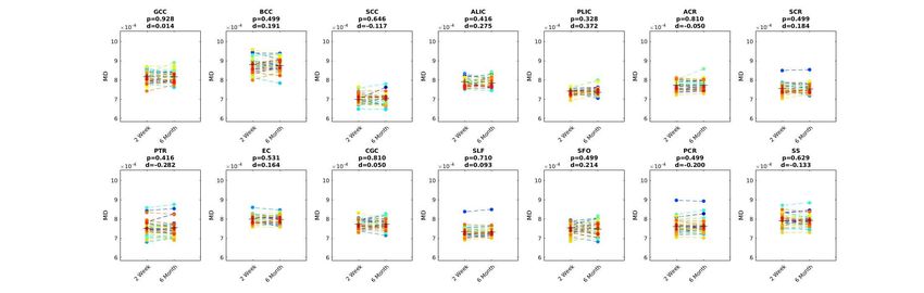
bioRxiv preprint first posted online Jun. 14, 2018; doi: http://dx.doi.org/10.1101/345629. The copyright holder for this preprint
       (which was not peer-reviewed) is the author/funder. All rights reserved. No reuse allowed without permission.

   (NODDI) (Zhang et al., 2012; Ojelescu et al., 2017 for review). NODDI leverages recent
   progress in high-performance magnetic field gradients for MRI scanners that can achieve
   diffusion-weighting factors much higher than the standard b=1000 s/mm2 for DTI and therefore
   probe more complex non-Gaussian properties of white matter diffusion. The NODDI biophysical
   model uses this richer diffusion imaging data to measure properties of three microstructural
   environments: intracellular, extracellular, and free water. One such metric is the intracellular
   volume fraction, referred to as the neurite density index (NDI) and which primarily represents
   axonal density within white matter. Another is orientation dispersion index (ODI) of neurites,
   which is higher in loosely organized white matter and lower in tracts with largely parallel fiber
   bundles such as the corpus callosum. The volume fraction of the isotropic diffusion compartment
   (FISO) estimates the free water content (Zhang et al.,2012). These NODDI parameters have been
   validated in histopathological studies of animal and human brains (Sepehrband, et al., 2015;
   Sato et al., 2016), been used to study brain development (Yi Shi Chang et al., 2015; Mah et al.,
   2017) and to detect subtle brain damage in other disorders (Billiet et al., 2015; Timmers et al.,
   2015; Kamagata et al., 2016; Caverzasi E, et al., 2016; Schneider T et al., 2017).

   In this study, we employ NODDI to: i) investigate early white matter changes at two weeks post-
   injury in mTBI patients versus trauma controls; ii) determine the evolution of white matter
   changes of the mTBI patients from two weeks to six months after injury; and iii) explore the
   prognostic significance of these white matter microstructural changes for symptomatic and
   cognitive outcome after mTBI. Comparing DTI to NODDI serially after mTBI, we hypothesize
   that the early microstructural white matter changes of mTBI are driven by increases in free
   water, such as from vasogenic edema due to neuroinflammation, whereas longer-term changes
   reflect decreases in axonal density due to evolving white matter degeneration.

   MATERIALS AND METHODS

   Participants

   All participants were enrolled at the Zuckerberg San Francisco General Hospital and Trauma
   Center as part of the prospective Transforming Research and Clinical Knowledge in Traumatic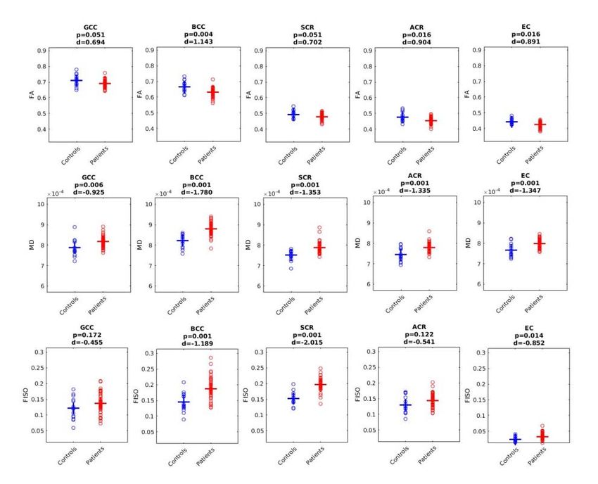
bioRxiv preprint first posted online Jun. 14, 2018; doi: http://dx.doi.org/10.1101/345629. The copyright holder for this preprint
       (which was not peer-reviewed) is the author/funder. All rights reserved. No reuse allowed without permission.

   Brain Injury project (TRACK-TBI) (Yue et al., 2013). A total sample of 40 mTBI patients (age:
   x̅=30.35 years, SD±7.50: education: x̅=15 years, SD±2.68; sex 9F/31M) was included within 24
   hours after injury upon meeting the American Congress of Rehabilitation Medicine (ACRM)
   criteria for mTBI (ACRM, 1993) in which the patient has to exhibit a traumatically induced
   physiological disruption of brain function as manifested by: i) any period of loss of
   consciousness (LOC), ii) any loss of memory for events immediately before or after the accident,
   iii) any alteration of mental state at the time of the accident (feeling dazed, disoriented, and/or
   confused), or iv) focal neurologic deficits that may or may not be permanent. Other inclusion
   criteria for this study were age between 18-55 years, acute brain CT as part of clinical care
   within 24 hours of injury, no significant polytrauma that would interfere with the follow-up and
   outcome assessment, no MRI contraindication, no previous psychiatric or neurological disorder,
   visual acuity and hearing adequate for outcomes testing, fluency in English, and ability to give
   informed consent. Galveston Orientation and Amnesia Test (GOAT) score assessed at the time
   of informed consent was normal (x̅=98.63; SD±2.18).

   Fourteen orthopedic trauma control subjects matched by age (x̅=31.71 years; SD±10.14),
   education (x̅=15.5 years; SD±2.17), and sex (6F/8M) were also recruited from the ED.
   Orthopedic injury causes included falls, pedestrian run overs, and bike accidents. All subjects
   presented with lower extremity fractures except for one that had an upper extremity fracture.
   Control subjects were ruled out for the current study if the emergency room physician required
   CT brain scan for suspicion of head trauma or if by interviewing medical services or subjects,
   participants reported clinical information such as loss of consciousness, amnesia, previous TBI,
   psychiatric or neurological prevalent pathology, and if, according to the abbreviated injury scale
   (AIS), this study would be counterproductive for their sustained systemic injuries.

   All eligible subjects who voluntarily agreed to participate gave written informed consent. All
   study protocols were approved by the University of California, San Francisco Institutional
   Review Board.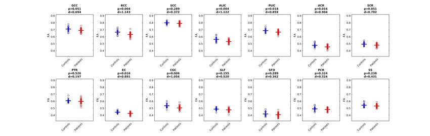
bioRxiv preprint first posted online Jun. 14, 2018; doi: http://dx.doi.org/10.1101/345629. The copyright holder for this preprint
       (which was not peer-reviewed) is the author/funder. All rights reserved. No reuse allowed without permission.

   Neuropsychological Assessment

   Commonly affected neuropsychological domains after mTBI were assessed using self-report and
   performance-based cognitive measures at 2 weeks and 6 month after injury: i) The Rivermead
   Postconcussion Symptoms Questionnaire (RPQ3-13), a self-reported questionnaire consisting of
   16 physical and psychosocial symptoms frequently reported after mTBI; ii) the Rey Auditory
   Verbal Learning Test (RAVLT) to evaluate learning, short, and long-term memory; iii) Trail
   Making Tests A (TMTA) and B (TMTB) to evaluate attention, processing speed, and cognitive
   flexibility to switch tasks (TMTB-A); and iv) the Wechsler Adult Intelligence Scale (WAIS)
   coding and symbol digit subscales for processing speed and visuo-perceptive association
   learning (Lezak et al., 2012).

   Image Acquisition

   All mTBI subjects underwent a standardized MRI protocol acquired on a 3T GE MR750 scanner
   equipped with an eight channel phased array head radiofrequency coil (GE Healthcare,
   Waukesha, WI) at 2 weeks (x̅=13.30 days; SD±2.10) and 6 months (x̅=184 days; SD±8.86) after
   injury. Whole-brain DTI was performed with a multi-slice single-shot spin echo echoplanar
   pulse sequence (echo time [TE] = 81 ms; repetition time [TR] = 9 s) using 64 diffusion-encoding
   directions, isotropically distributed over the surface of a sphere with electrostatic repulsion,
   acquired at b = 1300 s/mm2 and another 64 directions at b = 3000 s/mm2, eight acquisitions at b
   = 0 s/mm2 for each set of 64 directions, slices of 2.7-mm thickness each with no gap between
   slices, a 128 x128 matrix, and a field of view (FOV) of 350 x 350 mm. Sagittal three-
   dimensional (3D) inversion recovery fast spoiled gradient recalled echo T1-weighted images
   (inversion time [TI] = 400 ms; flip angle, 11 degrees), were acquired with 256mm FOV, 200
   contiguous partitions (1.2 mm) at 256x256 matrix. Sagittal 3D gradient echo T2*- weighted
   images (TE = 250 ms; TR = 500 ms; flip angle 10 degrees), were acquired with 256mm FOV,
   and 130 contiguous slices (1.6 mm) at 192x192 matrix. Sagittal 3D T2-weighted fluid-attenuated
   inversion recovery images (FLAIR; TE = 102 ms; TR = 575 ms; TI = 163 ms) were acquired
   with 256mm FOV, 184 contiguous slices (1.2 mm) at 256x256 matrix. The control group scans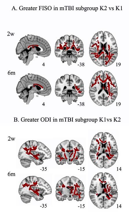
bioRxiv preprint first posted online Jun. 14, 2018; doi: http://dx.doi.org/10.1101/345629. The copyright holder for this preprint
       (which was not peer-reviewed) is the author/funder. All rights reserved. No reuse allowed without permission.

   were acquired with the same parameters of acquisition as the mTBI patients, with data available
   for this study at the 2 week time point after orthopedic injury.

   MRI Image Processing and Analysis

   An overview of the imaging analysis and statistical methods used in this study is presented in
   Figure 1.

   Figure 1. Overview of the imaging analysis and statistical methods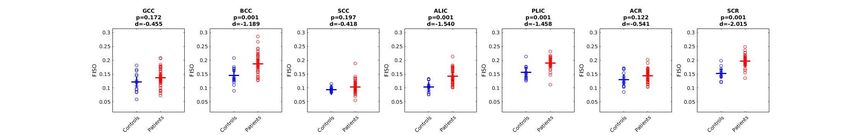
bioRxiv preprint first posted online Jun. 14, 2018; doi: http://dx.doi.org/10.1101/345629. The copyright holder for this preprint
       (which was not peer-reviewed) is the author/funder. All rights reserved. No reuse allowed without permission.

   Radiological Findings

   The structural 3T MRI images were interpreted by a board-certified neuroradiologist (E.L.Y),
   who was blinded to the initial presentation and subject group designation, using the NIH
   Common Data Elements (CDEs) for TBI pathoanatomic classification (Yuh et al., 2013). Table 1
   summarizes the clinical characteristics of the patients and the neuroradiological findings on
   structural 3T MRI. The MRI scans for the control group had no findings specific for TBI.

   Diffusion Tensor Imaging

   The diffusion MRI data were verified to be free of major image artifacts or excessive patient
   movement, defined as more than 2mm of translation and/or rotation. DTI pre-processing and
   analysis were performed using tools from the Oxford Centre for Functional MRI of the Brain
   (FMRIB) Software Library, abbreviated as FSL (v. 5.0.7). First, images were corrected for eddy
   distortions and motion, using an average of the 8 b=0 s/mm2 volumes for each diffusion-
   weighted shell as a reference. The registered images were skull-stripped using the Brain
   Extraction Tool (Smith, 2002). All the resulting brain masks were visually inspected for
   anatomic fidelity. FA, MD, axial diffusivity (AD), and radial diffusivity (RD) maps were
   calculated using the FSL Diffusion Toolbox.

   Multi-compartment Biophysical Modeling of Diffusion MR Imaging

   NODDI              metrics          were           derived          using          the         NODDI             toolbox         v0.9
   (http://www.nitrc.org/projects/noddi_toolbox). We averaged the corresponding 8 b=0 s/mm2
   images for each diffusion weighting. The NODDI code was modified to account for the slightly
   differing minimum echo times (TE) between images acquired at b = 1300 s/mm2 versus b = 3000
   s/mm2 by fitting the NODDI model to the normalized diffusion-weighted images instead of the
   raw images. As per the developers' recommendation, the diffusion-weighted images at each b
   value were normalized by the mean b = 0 s/mm2 images acquired with the same minimal TE
   scan parameter, generating images with TE-independent signal intensity, as we have described
   previously (Owen et al., 2014). NODDI fitting was performed with the NODDI Matlab Toolbox
   using the default settings (http://www.nitrc.org/projects/ noddi_toolbox v0.9). Maps of NDI,
   ODI and FISO were generated. Figure 2 displays examples of DTI and NODDI map parameters.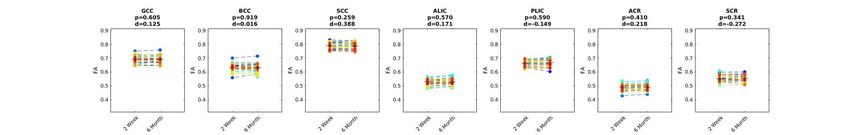
bioRxiv preprint first posted online Jun. 14, 2018; doi: http://dx.doi.org/10.1101/345629. The copyright holder for this preprint
       (which was not peer-reviewed) is the author/funder. All rights reserved. No reuse allowed without permission.

                                           Figure 2. DTI and NODDI map examples.

   Example DTI (FA, MD) and NODDI (NDI, ODI, FISO) parameter maps obtained from a single subject in
   FMRIB58_FA standard-space.

   Statistics

   Tract-Based Spatial Statistics

   After calculation of the FA map, a voxel-wise statistical analysis of the FA data was performed
   using Tract-Based Spatial Statistics (TBSS). FA data were aligned into the common FMRIB58
   FA template, which is in MNI152 (Montreal Neurological Institute) standard space, using the
   non-linear registration algorithm FNIRT (Jenkinson et al., 2002). Next, a mean FA image was
   created from the images for all the subject’s serial scans in this common space and thinned to
   generate a mean FA white matter skeleton that represented the center of all tracts common to the
   entire group of scans and thresholded at > 0.2. The aligned FA volume was then projected onto
   the skeleton by filling the skeleton with FA values from the nearest relevant tract center.

   Twelve main fasciculi were studied using masks obtained from the Jülich histological atlas
   (JHU) mapped onto the standard MNI152 space and resampled to 1mm resolution. Binary mask
   images from the fasciculi of interest were used to mask the individual skeletonized maps
   previously registered to the MNI standard space using the non-linear tools in the TBSS
   procedure. Mean FA, MD, AD, RD, ODI, NDI, and FISO values were obtained from each
   subject’s white matter skeleton as well as each of the skeletonized regions of interest (ROIs).
   Right and left tracts were averaged into one single measurement.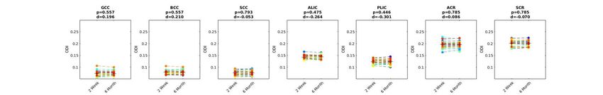
bioRxiv preprint first posted online Jun. 14, 2018; doi: http://dx.doi.org/10.1101/345629. The copyright holder for this preprint
       (which was not peer-reviewed) is the author/funder. All rights reserved. No reuse allowed without permission.

   We compared between subjects using a voxel-wise general linear model (GLM) analysis with
   permutation testing to correct for multiple comparisons (Nichols et al., 2002) using threshold-
   free cluster enhancement (TFCE), family-wise error corrected (FWE) at p≤0.05. An unpaired t-
   test was employed to compare cross-sectionally the group of patients and controls in the voxel-
   wise analysis at 2 weeks. A paired t-test was used to compare differences among DTI & NODDI
   measures within the patient group between 2 weeks and 6 months.

   Machine Learning Analysis

   A percentage of mTBI patients remain functionally impaired after 1 year post-injury (McMahon
   et al., 2014). In an attempt to distinguish these patients in our cohort, we used unsupervised
   machine learning to derive a metric of cognitive and symptom improvement and link it to the
   imaging biomarkers. First, to obtain and define a global improvement measure (GIM) that best
   reflects their outcomes, we first subtracted the 2-week scores from the 6-month scores for each
   of the 9 self-reported and cognitive measures described in Table 2 for each subject, then used a
   Z-score transformation to normalize the values. Since each individual test could be noisy, we
   sought to combine them together into a single composite metric that we defined as the GIM. We
   approached this task through an unsupervised k-means clustering analysis with two clusters and
   thirty replicates in MATLAB 2012b (The MathWorks, Inc., Natick, Massachusetts, United
   States). We then calculated a hyperplane to separate these two clusters equidistantly and each
   subject’s GIM was defined as the signed (positive/negative) distance between the subject’s
   recovery status and this hyperplane. This distance can also be expressed as a weighted average of
   the various symptomatic and cognitive metrics (Fig 3a). Intuitively, this represents a data-driven
   method to combine the various self-reported and performance-based cognitive metrics to provide
   a wide degree of discrimination between patient outcome groups. We used two clusters because
   we were interested in distinguishing the patients with the best improvement from those who did
   not improve. While one cluster represents patients whose testing trend indicates overall
   improvement between two weeks and six months, the other cluster represents patients whose
   overall testing indicates a lack of, or in some cases a regression of, testing performance. Finally,
   we performed the voxel-wise comparison among clusters determined by the GIM measure with
   the DTI and NODDI metrics using randomise, the nonparametric permutation analysis tool in
   FSL, with TFCE correction for multiple comparisons at p
bioRxiv preprint first posted online Jun. 14, 2018; doi: http://dx.doi.org/10.1101/345629. The copyright holder for this preprint
(which was not peer-reviewed) is the author/funder. All rights reserved. No reuse allowed without permission.
Figure 3. Machine Learning Clustering for Global Improvement Measure (GIM)
(A): Two-dimensional schematic illustration of the clustering of change in cognitive and behavioral
scores and the PCA projection used to define global improvement measure (arrows for some subjects
demonstrating this distance). Subjects farther from the line in the upper right direction have better degree
of recovery (black points), and subjects farther from the line in the other direction have less recovery (red
points). (B): Two clusters comprising the actual group of mTBI patients based on their global
improvement measure (GIM) showing Cluster 1 (red; n=24), patients with less recovery, and Cluster 2
(black; n=16), patients with more recovery. The triangles indicate the mean GIM for each cluster.
RESULTS
Neuropsychological Assessment
Table 2 displays the average results for the self-report and performance-based cognitive
measures at 2 weeks and 6 months after injury for the 40 mTBI patients. Overall, patients self-
reported a significant reduction in post-concussive symptoms on the RPQ scores, but a subset of
subjects showed persistent self-reported symptomatology at the 6-month time point. Moreover,bioRxiv preprint first posted online Jun. 14, 2018; doi: http://dx.doi.org/10.1101/345629. The copyright holder for this preprint
(which was not peer-reviewed) is the author/funder. All rights reserved. No reuse allowed without permission.
patients manifested improved performance in processing speed (TMTA), visuo-perceptive
association learning (WAIS coding and symbol), as well as verbal memory (RAVLT) at 6
months vs 2 weeks.
DTI and NODDI Voxel-wise Group Comparisons
Cross-sectional analysis between mild TBI patients and controls at the 2-week time point.
Compared to the trauma controls, the mTBI patients showed decreased FA and increased MD in
the genu and body of the corpus callosum, anterior and posterior limbs of the internal capsule,
anterior corona radiata, anterior thalamic radiation, external capsule and cingulum. FISO was
found to be increased in patients versus controls for the same tracts, but also additionally in the
superior longitudinal fasciculi, posterior corona radiata, and fronto-occipital tracts. To a lesser
extent, NDI also showed decreases mainly in the external capsule, anterior thalamic radiations,
inferior longitudinal fasciculi, fornix and stria terminalis (Fig 4a). Figure 5 shows the mean
values, effect sizes (Cohen’s d), and distributions of the sample for FA, MD, and FISO in the
JHU tracts shown to be most affected by mTBI in the data-driven voxel-wise whole-brain TBSS
analysis of Figure 4. The rest of the JHU tracts for the DTI and NODDI measures are provided
in the Supplemental Material, Section A, Figs S1-S5.
Longitudinal analysis of mTBI patients at 2 weeks vs 6 months
Longitudinal voxel-wise analysis of mTBI patients showed decreases over time of NDI in tracts
including the anterior and posterior corona radiata, posterior thalamic radiation, inferior
longitudinal and inferior fronto occipital fasciculi, posterior and anterior thalamic radiation,
uncinate fasciculi, and external capsules. FISO showed decreases over time in the posterior
corona radiata and inferior longitudinal fasciculi (Fig 4b). NODDI measures were more sensitive
to microstructural damage in posterior tracts than DTI (Fig 6). Mean values and effect sizes, as
well as the distribution of the samples, for all the DTI and NODDI measures are provided for the
remaining JHU tracts in the Supplemental Material, Section B- Figs S6-S10. No other significant
results were found for the rest of the DTI and NODDI parameters.bioRxiv preprint first posted online Jun. 14, 2018; doi: http://dx.doi.org/10.1101/345629. The copyright holder for this preprint
(which was not peer-reviewed) is the author/funder. All rights reserved. No reuse allowed without permission.
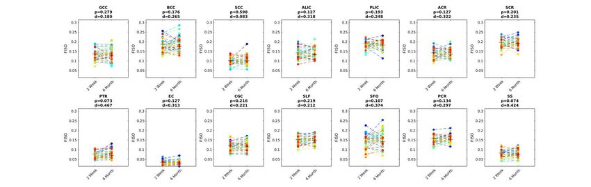
Figure 4. Cross-sectional and Longitudinal Voxel-wise Analysis
(A): Cross-sectional voxel-wise analysis comparison at 2 weeks between patients and controls. FA:
fractional anisotropy; MD: mean diffusivity; ND: neurite dispersion index; FISO: volume fraction of
isotropic water; In yellow: parameter increased in patients related to controls; In blue: parameter
decreased in patients relative to controls. (B): Longitudinal voxel-wise comparison between 2 weeks and
6 months after injury in the patients group. In blue: parameter decreased over time. All results corrected
for multiple comparisons using threshold-free cluster enhancement (TFCE) family-wise error (FWE) at
p≤0.05. The number next to each image is the MNI atlas coordinate defining its plane.bioRxiv preprint first posted online Jun. 14, 2018; doi: http://dx.doi.org/10.1101/345629. The copyright holder for this preprint
(which was not peer-reviewed) is the author/funder. All rights reserved. No reuse allowed without permission.
Figure 5. JHU Tracts Cross-sectional Comparison at 2 weeks Between mTBI and Controls.
Averaged fractional anisotropy (FA), mean diffusivity (MD), and free water fraction (FISO) values of the
left/right JHU tracts for each subject at the 2-week time point. GCC: genu corpus callosum; BCC: body
corpus callosum; SCR: superior corona radiata; ACR: anterior corona radiata; EC: external capsule;
Patient and control comparison FDR corrected p at 0.05. d: Cohen´s d effect size.bioRxiv preprint first posted online Jun. 14, 2018; doi: http://dx.doi.org/10.1101/345629. The copyright holder for this preprint
(which was not peer-reviewed) is the author/funder. All rights reserved. No reuse allowed without permission.
Figure 6. JHU Tracts Longitudinal Comparison at 2 weeks vs 6 month in mTBI .
Averaged fractional anisotropy (FA) and neurite density index (NDI) values of the left/right JHU tracts
for each subject at 2-week vs 6-month time points, showing significant changes over time in posterior
tracts on NDI not captured by the FA parameter. PTR: posterior thalamic radiation; PCR: posterior corona
radiata; SS: sagittal stratum (merged inferior fronto-occipital and inferior longitudinal fasciculi).
Comparison FDR corrected p at 0.05. d: Cohen´s d effect size.
Machine Learning Analysis
Machine learning clusters among mTBI patients and their global improvement measure.
Two clear clusters (subgroups) were obtained dividing the group of mTBI patients based on their
global improvement measure. Cluster K1 included 24 patients who had less improvement by the
GIM metric than Cluster K2, which consisted of the 16 patients with the best global
improvement (Fig 3b). Table 2 shows the change from 2 weeks to 6 months post-injury in thebioRxiv preprint first posted online Jun. 14, 2018; doi: http://dx.doi.org/10.1101/345629. The copyright holder for this preprint
(which was not peer-reviewed) is the author/funder. All rights reserved. No reuse allowed without permission.
self-report and cognitive performance measures that comprise the GIM across all 40 mTBI
patients. While the effect sizes of the change over time in the group means appear small for these
measures, these group averages obscure variation among patients that can be uncovered by the
unsupervised machine learning analysis dividing the group into two clusters based on the GIM.
Is noteworthy to mention that, because of this division, K1 and K2 differed in years of education
(K1: x̅=14.4 years, SD±2.1; K2: x̅=17.7 years; SD±2.5; p=0.002) but not in age.
Self-report and cognitive measures based on machine learning clustering
Table 3 shows the difference between K1 and K2 in each of the nine measures that comprise the
GIM. The patients of K2 were much more symptomatic on the RPQ than those of K1 at 2-weeks
post-injury but recover to symptom levels similar to K1 by 6-months post-injury. The patients of
K2 perform equivalently to those of K1 on the cognitive performance tests at 2-weeks post-
injury, but, at the 6-month time point, are significantly outperforming their counterparts on the
RAVLT and the WAIS, particularly the WAIS Coding subtest. K2 also trended toward better
performance than K1 on the TMTA at both time points.
Voxel-wise group comparison of the DTI and NODDI parameters based on machine learning
clustering
No significant relationship between traditional DTI metrics and patient global recovery GIM was
found. However, NODDI metrics were found to be associated with cluster membership based on
the GIM metric (Fig 7). The voxel-wise group comparison between K1 and K2 revealed
increases in FISO in K2 compared to K1, but the pattern of elevated FISO varied between the 2-
week and 6-month time points. The increased FISO of K2 versus K1 was posterior predominant
at 2 weeks post-injury whereas the increased FISO of K2 versus K1 was anterior predominant at
6 months post-injury. In contradistinction, increased ODI of K1 versus K2 was found, both at 2
weeks and at 6 months in a largely stable pattern encompassing much of the central white matter
tracts, with only the right internal capsule showing resolution of the elevated ODI at 6 months.bioRxiv preprint first posted online Jun. 14, 2018; doi: http://dx.doi.org/10.1101/345629. The copyright holder for this preprint
(which was not peer-reviewed) is the author/funder. All rights reserved. No reuse allowed without permission.
Figure 7. Voxel-wise Comparison of NODDI Metrics between mTBI Patient Subgroups K1 & K2
Voxel-wise group comparison of the DTI and NODDI parameters based on the machine learning cluster
division (A): FISO: volume fraction of isotropic water, increased in patients in cluster 2 (K2). FISO
increased in posterior tracts at 2 weeks while pattern at 6 months showed increases mainly in anterior
tracts (B): ODI: orientation dispersion is increased in patients in cluster 1 (K1). All results corrected for
multiple comparisons using threshold-free cluster enhancement (TFCE) family-wise error (FWE) at
p≤0.05. The number next to each image is the MNI atlas coordinate defining its plane.
DISCUSSION
To the best of our knowledge, there is only one previous NODDI study of TBI, which was a
cross-sectional investigation of sport-related concussion (Churchill et al., 2017). Our study
compares DTI to NODDI longitudinally for the evolution of white matter microstructural injury
and its association with symptomatic and cognitive recovery over time in a sample of civilianbioRxiv preprint first posted online Jun. 14, 2018; doi: http://dx.doi.org/10.1101/345629. The copyright holder for this preprint
(which was not peer-reviewed) is the author/funder. All rights reserved. No reuse allowed without permission.
mTBI patients. The main findings are: i) early decreases in FA and NDI along with early
increases of MD and FISO in the mTBI patient group versus trauma controls; ii) longitudinal
white matter changes within the mTBI group as reflected by decreases in NDI and FISO over
time; and iii) statically reduced ODI in those mTBI patients without symptomatic or cognitive
improvement (K1) and dynamically elevated FISO in those mTBI patients with symptomatic
recovery and progressively improved cognitive function (K2).
TBI involves multiple different time-varying pathophysiological effects, including diffuse axonal
injury, diffuse microvascular injury, and neuroinflammation, that can lead to neurologic
dysfunction (Bigler; Corps, JAMA). Due to this complexity, combining different biophysical
measurements has potential for characterizing the underlying microarchitectural changes in the
brain tissue (Cercignani et al., 2017). Our cross-sectional DTI findings at the 2-week time point
showed decreases in FA and increases in MD in the mTBI group versus trauma controls, mainly
in anterior tracts of the frontal and temporal lobes. These findings are in close agreement with
prior DTI studies at this early stage of mTBI (Yuh et al., 2014; Eirud et al., 2014). In addition to
these established DTI results, NODDI analysis revealed two additional findings at the 2-week
time point. First, NDI was decreased in mTBI patients versus controls, primarily in the external
capsule, thalamic radiations, inferior longitudinal fasciculi, and fornix- stria terminalis. Second,
we also found increases in FISO in the same predominantly anterior distribution as FA and MD.
Overall, these cross-sectional results suggest that the early decrease of FA and increase of MD
after mTBI is due to an increase in free water content, possibly reflecting neuroinflammation.
The sole prior study reporting DTI and NODDI measurements in TBI, in a sample of athletes
with contact exposure, found increases in FA as well as increases in NDI and reduced ODI
values (Churchill et al., 2017). These results may differ from those found in our study due to the
mechanism of injury in young athletes involving repetitive subconcussive hits over long periods
of time, thereby conflating injury and recovery effects, rather than a single episode of mTBI that
can be serious enough to produce anatomic lesions on structural MRI (Table 1).
Longitudinally within the group of mTBI patients, we only observed decreases over time in NDI
values, suggesting progressive axonal degeneration, while there were no significant differences
in the DTI parameters. This indicates that NDI is a more sensitive metric of white matter axonalbioRxiv preprint first posted online Jun. 14, 2018; doi: http://dx.doi.org/10.1101/345629. The copyright holder for this preprint
(which was not peer-reviewed) is the author/funder. All rights reserved. No reuse allowed without permission.
loss than the conventional DTI metrics such as FA or MD. The decreases found in NDI over time
were primarily in the bilateral posterior periventricular and left anterior periventricular white
matter. It has been shown that a disproportionately high number of structural connectome links
between gray matter areas traverse these regions of deep white matter. Indeed, these
periventricular white matter regions have been described as a consistent nexus of network
connectivity in the human brain (Owen et al., 2015; 2016). Additionally, virtual lesioning of
these areas of deep periventricular white matter in tractography simulation experiments are
particularly disruptive to the overall integrity of the whole-brain white matter network,
demonstrating their singular importance to the large-scale structural connectome (Wang et al.,
2017). Abnormal posterior periventricular white matter microstructure has also been described
in sensory processing disorders (Owen 2013; Chang 2015) and were the only consistently
affected white matter regions in a meta-analysis of DTI studies of attention-deficit hyperactivity
disorder (ADHD, Chen et al., 2016). Furthermore, the global integrity of the structural
connectome is linked to attention and executive function (Xiao et al., 2016). This constellation
of recent results may account for the major impairments of concussions and mTBI, which are
sensitivity to sensory stimuli (e.g., to bright lights and loud noises, as also seen in sensory
processing disorder), attention deficits and executive dysfunction. Future studies combining
microstructural characterization of these posterior periventricular white matter tracts with
connectomic mapping may prove especially effective for explaining long-term symptomatic,
cognitive and behavioral outcomes after mTBI.
Data-driven machine learning analysis of a composite global improvement measure based on
nine symptom self-report and cognitive performance measures known to be affected in mTBI
produced two patient clusters. One was a higher performing subgroup (K2) with early self-
reported symptoms that resolved over time and who also improved in the information processing
speed (WAIS Coding) and verbal memory (RAVLT) domains. The other was a lower
functioning subgroup (K1) that displayed relatively few initial symptoms, but still performed less
well than K2 on the cognitive tests, especially at the 6-month time point. Although no significant
DTI differences were seen between these two mTBI subgroups, NODDI showed higher ODI
throughout much of the central white matter in the K1 group at both time points. This greater
fiber orientation dispersion in the low functioning K1 cluster may perhaps represent a premorbidbioRxiv preprint first posted online Jun. 14, 2018; doi: http://dx.doi.org/10.1101/345629. The copyright holder for this preprint
(which was not peer-reviewed) is the author/funder. All rights reserved. No reuse allowed without permission.
characteristic influenced by their lower average educational level than K2, which might also
imply lower cognitive reserve. The less well organized central white matter may help explain
their poorer cognitive performance compared to the higher functioning K2 cluster. The
difference in cognitive and educational levels between the subgroups might possibly also affect
symptom reporting. Specifically, the greater number of symptoms reported by the K2 subgroup
at the early time point may partly represent awareness of actual cognitive decline from baseline,
followed by eventual return to baseline in both symptoms and cognition by 6m, at least at the
subgroup level if not for every patient.
Another finding using NODDI was the higher FISO of K2 versus K1 in predominantly posterior
white matter at 2-weeks post-injury versus predominantly anterior white matter at 6-months post-
injury. Since the overall group of 40 mTBI patients showed elevated anterior white matter free
water, indicating vasogenic edema, at the early time point (Fig. 4a), this means that the K2
subgroup had more extensive early edema than K1 that also included posterior white matter.
However, the anterior white matter edema resolved more slowly in K2 than K1, resulting in the
relative elevation of free water in this distribution at the long-term time point. The greater extent
of early white matter edema corresponds to the greater symptoms reported by the K2 subgroup at
that time, with improving edema by 6 months post-injury matching their improvement in self-
reported and cognitive performance measures. This observed association between white matter
edema and the trajectory of symptomatic and cognitive recovery after mTBI requires more study
to determine if there is a causal relationship.
This study is a pilot longitudinal investigation of metrics from biophysical compartment
modeling of diffusion MRI, exemplified by NODDI, for mTBI with comparison to standard DTI
biomarkers. The orthopedic trauma group provides a rigorous control cohort that shares clinical
and demographic features with the mTBI patients. The results provide support for our original
hypotheses of elevated free water fraction early after injury and of serial decline in white matter
axonal density during the first 6 months post-trauma. Exploratory machine learning analysis of
the relationship between NODDI measures and symptomatic/cognitive recovery show that (i)
dynamically increased free water fraction across semi-acute and chronic time points was
associated with better recovery, suggesting a beneficial role for edema/neuroinflammation; and
(ii) statically reduced fiber orientation dispersion was correlated with better long-term cognition,bioRxiv preprint first posted online Jun. 14, 2018; doi: http://dx.doi.org/10.1101/345629. The copyright holder for this preprint
(which was not peer-reviewed) is the author/funder. All rights reserved. No reuse allowed without permission.
consistent with prior studies showing more highly organized white matter in those with better
intellectual functioning in multiple domains (Fijell, et al. 2011; Yang Y et al., 2015; Roger A
Kievit, et al., 2016). These new hypotheses from the exploratory findings need to be tested in
larger cohorts that have better statistical power for determining imaging-cognition relationships.
In the absence of cognitive control data, we also cannot exclude a learning component for the
improvement across the two cognitive assessments, especially for the better recovery subgroup
that had, on average, a higher educational level than the poorer recovery subgroup. Another
limitation of the study is the small sample size of the controls, which lack a longitudinal
component.
In summary, we found that NODDI parameters are sensitive imaging biomarkers for the subtle
yet complex underlying white matter microstructural pathology after mTBI, such as diffuse
axonal injury and neuroinflammation. Our results show that the early decrease of FA and
increase of MD after mTBI, which are primarily in the anterior white matter, correspond to white
matter regions of elevated FISO, which may reflect inflammatory vasogenic edema. This
elevation of free water is more extensive in the subgroup of patients reporting more post-
concussive symptoms early after trauma. The longer-term changes from 2 weeks to 6 months
after mTBI are marked by declining neurite density in predominantly posterior white matter,
suggesting axonal degeneration from DAI for which NODDI appears more sensitive than any of
the DTI metrics, such as FA. The affected posterior white matter regions are known to be
topologically integral to the structural connectome and are involved in multiple sensory and
cognitive domains, including attention and executive function. The observation of stably
elevated white matter fiber orientation dispersion in the mTBI subgroup with poorer cognitive
performance may represent the sensitivity of ODI to premorbid intellectual functioning. Further
research studies in larger well-phenotyped cohorts are needed to validate these NODDI
biomarkers for mTBI diagnosis, for prediction of symptoms and cognitive performance, and for
treatment monitoring.bioRxiv preprint first posted online Jun. 14, 2018; doi: http://dx.doi.org/10.1101/345629. The copyright holder for this preprint
(which was not peer-reviewed) is the author/funder. All rights reserved. No reuse allowed without permission.
Funding
This research was supported by the following grants of the National Institutes of Health (NIH)
and the United States Department of Defense (DoD): NIH U01 NS086090, NIH R01 NS060776,
NIH RC2 NS069409, DoD W81XWH-14-2-0176.
REFERENCES
1. American Congress of Rehabilitation Medicine (ACRM). Mild Traumatic Brain Injury
Committee. (1993). Definition of mild traumatic brain injury. Journal of Head Trauma
Rehabilitation, 8 (3), 86–87.
2. Basser PJ, Jones DK. Diffusion-tensor MRI: theory, experimental design and data
analysis - a technical review. NMR Biomed. 200;15(7-8):456-67. Review.
3. Bigler ED. Neuroimaging biomarkers in mild traumatic brain injury (mTBI).
Neuropsychol Rev. 2013 Sep;23(3):169-209.
4. Bigler ED. Neuropathology of Mild Traumatic Brain Injury: Correlation to
Neurocognitive and Neurobehavioral Findings. In: Kobeissy FH, editor. Brain
Neurotrauma: Molecular, Neuropsychological, and Rehabilitation Aspects. Boca Raton
(FL): CRC Press/Taylor & Francis; 2015. Chapter 31.
5. Billiet T, Vandenbulcke M, Mädler B, Peeters R, Dhollander T, Zhang H, Deprez S, Van
den Bergh BR, Sunaert S, Emsell L. Age-related microstructural differences quantified
using myelin water imaging and advanced diffusion MRI. Neurobio Aging.
2015;36(6):2107-21.
6. Caverzasi E, Papinutto N, Castellano A, Zhu AH, Scifo P, Riva M, Bello L, Falini A,
Bharatha A, Henry RG. Neurite Orientation Dispersion and Density Imaging Color Maps
to Characterize Brain Diffusion in Neurologic Disorders. J Neuroimaging. 2016
Sep;26(5):494-8.
7. Cercignani M, Bouyagoub S. Brain microstructure by multi-modal MRI: Is the whole
greater than the sum of its parts? Neuroimage. 2017. pii: S1053-8119(17)30886-8.
8. Chang YS, Owen JP, Pojman NJ, Thieu T, Bukshpun P, Wakahiro ML, Berman JI,
Roberts TP, Nagarajan SS, Sherr EH, Mukherjee P. White Matter Changes of NeuritebioRxiv preprint first posted online Jun. 14, 2018; doi: http://dx.doi.org/10.1101/345629. The copyright holder for this preprint
(which was not peer-reviewed) is the author/funder. All rights reserved. No reuse allowed without permission.
Density and Fiber Orientation Dispersion during Human Brain Maturation. PLoS One.
2015;10(6):e0123656.
9. Chen L, Hu X, Ouyang L, He N, Liao Y, Liu Q, Zhou M, Wu M, Huang X, Gong Q. A
systematic review and meta-analysis of tract-based spatial statistics studies regarding
attention-deficit/hyperactivity disorder. Neuroscience and biobehavioral reviews.
2016;68:838-47.
10. Churchill NW, Caverzasi E, Graham SJ, Hutchison MG, Schweizer TA. White matter
microstructure in athletes with a history of concussion: Comparing diffusion tensor
imaging (DTI) and neurite orientation dispersion and density imaging (NODDI). Hum
Brain Mapp. 2017;38(8):4201-4211.
11. Croall ID, Cowie CJ, He J, Peel A, Wood J, Aribisala BS, Mitchell P, Mendelow AD,
Smith FE, Millar D, Kelly T, Blamire AM. White matter correlates of cognitive
dysfunction after mild traumatic brain injury. Neurology. 2014;83(6):494-501.
12. Eierud C, Craddock RC, Fletcher S, Aulakh M, King-Casas B, Kuehl D, LaConte SM.
Neuroimaging after mild traumatic brain injury: review and meta-analysis. Neuroimage
Clin. 2014;4:283-94.
13. Fjell AM, Westlye LT, Amlien IK, Walhovd KB. Reduced white matter integrity is
related to cognitive instability. J Neurosci. 2011; 7;31(49):18060-72.
14. Jelescu OI, Budde MD. Design and validation of diffusion MRI models of white matter.
Frontiers in Physics. 2017; 5:61. Review.
15. Jenkinson M., Bannister P, Brady M, Smith S. Improved optimization for the robust and
accurate linear registration and motion correction of brain images. Neuroimage 2002;17:
825-41.
16. Jones DK, Cercignani M. Twenty-five pitfalls in the analysis of diffusion MRI data.
NMR Biomed. 2010 Aug;23(7):803-20. doi: 10.1002/nbm.1543. Review.
17. Kamagata K, Zalesky A, Hatano T, Ueda R, Di Biase MA, Okuzumi A, Shimoji K, Hori
M, Caeyenberghs K, Pantelis C, Hattori N, Aoki S. Gray Matter Abnormalities in
Idiopathic Parkinson's Disease: Evaluation by Diffusional Kurtosis Imaging and Neurite
Orientation Dispersion and Density Imaging. Hum Brain Mapp. 2017; 38(7): 3704-3722.bioRxiv preprint first posted online Jun. 14, 2018; doi: http://dx.doi.org/10.1101/345629. The copyright holder for this preprint
(which was not peer-reviewed) is the author/funder. All rights reserved. No reuse allowed without permission.
18. Kievit RA, Davis SW, Griffiths J, Correia MM, Cam-Can, Henson RN. A watershed
model of individual differences in fluid intelligence. Neuropsychologia. 2016; 186-
106:417.
19. Levin HS, Diaz-Arrastia RR. Diagnosis, prognosis, and clinical management of mild
traumatic brain injury. Lancet Neurol. 2015;14(5):506-17.
20. Lezak, M D, Howieson, D B, Bigler, E D, & Tranel, D (2012). Neuropsychological
assessment (5th ed.). New York: Oxford University Press.
21. Mah A, Geeraert B, Lebel C. Detailing neuroanatomical development in late childhood
and early adolescence using NODDI. PLoS One. 2017;12(8):e0182340.
22. Manley GT, Maas AI. Traumatic brain injury: an international knowledge-based
approach. JAMA. 2013;310(5):473-4.
23. McMahon PJ, Hricik A, Yue JK, et al. Symptomatology and functional outcome in mild
traumatic brain injury: results from the prospective TRACK-TBI study. Journal of
neurotrauma. 2014;31(1):26-33.
24. Mori S, Zhang J. Principles of diffusion tensor imaging and its applications to basic
neuroscience research. Neuron. 2006 Sep 7;51(5):527-39. Review.
25. Mukherjee P, Berman JI, Chung SW, Hess CP, Henry RG. Diffusion tensor MR imaging
and fiber tractography: theoretic underpinnings. AJNR Am J Neuroradiol.
2008;29(4):632-41.
26. Nichols TE, Holmes AP. Nonparametric permutation tests for functional neuroimaging: a
primer with examples. Hum Brain Mapp.2002; 15: 1-25.
27. Oehr L, Anderson J. Diffusion-Tensor Imaging Findings and Cognitive Function
Following Hospitalized Mixed-Mechanism Mild Traumatic Brain Injury: A Systematic
Review and Meta-Analysis. Arch Phys Med Rehabil. 2017; (11):2308-2319
28. Owen JP, Chang YS, Mukherjee P. Edge density imaging: mapping the anatomic
embedding of the structural connectome within the white matter of the human brain.
Neuroimage. 2015; 1;109:402-17.
29. Owen JP, Marco EJ, Desai S, Fourie E, Harris J, Hill SS, Arnett AB, Mukherjee P.
Abnormal white matter microstructure in children with sensory processing disorders.
Neuroimage Clin. 2013 Jun 23;2:844-53.bioRxiv preprint first posted online Jun. 14, 2018; doi: http://dx.doi.org/10.1101/345629. The copyright holder for this preprint
(which was not peer-reviewed) is the author/funder. All rights reserved. No reuse allowed without permission.
30. Owen JP, Wang MB, Mukherjee P. Periventricular White Matter Is a Nexus for Network
Connectivity in the Human Brain. Brain Connect. 2016;6(7):548-57.
31. Radhakrishnan R, Garakani A, Gross LS, Goin MK, Pine J, Slaby AE, Sumner CR,
Baron DA. Neuropsychiatric aspects of concussion. Lancet Psychiatry. 2016 (12):1166-
1175.
32. Sato K, Kerever A, Kamagata K, Tsuruta K, Irie R, Tagawa K, Okazawa H, Arikawa-
Hirasawa E, Nitta N, Aoki I, Aoki S. Understanding microstructure of the brain by
comparison of neurite orientation dispersion and density imaging (NODDI) with
transparent mouse brain. Acta Radiol Open. 2017;6(4):2058460117703816.
33. Schneider T, Brownlee W, Zhang H, Ciccarelli O, Miller DH, Wheeler-Kingshott G.
Sensitivity of multi-shell NODDI to multiple sclerosis white matter changes: a pilot
study. Funct Neurol. 2017;32(2):97-101.
34. Sepehrband F, Clark KA, Ullmann JF, Kurniawan ND, Leanage G, Reutens DC, Yang,
Z. Brain tissue compartment density estimated using diffusion-weighted MRI yields
tissue parameters consistent with histology. Hum Brain Mapp. 2015;36(9):3687-702.
35. Smith SM. Fast robust automated brain extraction. Hum Brain Mapp. 2002; 17:143-55.
36. Timmers I, Roebroeck A, Bastiani M, Jansma B, Rubio-Gozalbo E, Zhang H. Assessing
Microstructural Substrates of White Matter Abnormalities: A Comparative Study Using
DTI and NODDI. PLoS One. 2016;11(12):e0167884.
37. Wang MB, Owen JP, Mukherjee P, Raj A. Brain network eigenmodes provide a robust
and compact representation of the structural connectome in health and disease. PLoS
Comput Biol. 2017;13(6):e1005550.
38. Xiao M, Ge H, Khundrakpam BS, Xu J, Bezgin G, Leng Y, Zhao L, Tang Y, Ge X, Jeon
S, Xu W, Evans AC, Liu S. Attention Performance Measured by Attention Network Test
Is Correlated with Global and Regional Efficiency of Structural Brain Networks. Front
Behav Neurosci. 2016;10:194.
39. Yang Y, Bender AR, Raz N. Age related differences in reaction time components and
diffusion properties of normal-appearing white matter in healthy adults.
Neuropsychologia. 2015;66:246-58.
40. Yue JK, Vassar MJ, Lingsma HF, Cooper SR, Okonkwo DO, Valadka AB, Gordon WA,
Maas AI, Mukherjee P, Yuh EL, Puccio AM, Schnyer DM, Manley GT; TRACK-TBIbioRxiv preprint first posted online Jun. 14, 2018; doi: http://dx.doi.org/10.1101/345629. The copyright holder for this preprint
(which was not peer-reviewed) is the author/funder. All rights reserved. No reuse allowed without permission.
Investigators. Transforming research and clinical knowledge in traumatic brain injury
pilot: multicenter implementation of the common data elements for traumatic brain
injury. J Neurotrauma. 2013;30(22):1831-44.
41. Yuh EL, Cooper SR, Mukherjee P, Yue JK, Lingsma HF, Gordon WA, Valadka AB,
Okonkwo DO, Schnyer DM, Vassar MJ, Maas AI, Manley GT; TRACK-TBI
INVESTIGATORS. Diffusion tensor imaging for outcome prediction in mild traumatic
brain injury: a TRACK-TBI study. J Neurotrauma. 2014;31(17):1457-77.
42. Yuh EL, Mukherjee P, Lingsma HF, Yue JK, Ferguson AR, Gordon WA, Valadka AB,
Schnyer DM, Okonkwo DO, Maas AI, Manley GT; TRACK-TBI Investigators. Magnetic
resonance imaging improves 3-month outcome prediction in mild traumatic brain injury.
Ann Neurol. 2013;73(2):224-35.
43. Zhang H, Hubbard PL, Parker GJ, Alexander DC. Axon diameter mapping in the
presence of orientation dispersion with diffusion MRI. Neuroimage. 2011;56(3):1301-15.bioRxiv preprint first posted online Jun. 14, 2018; doi: http://dx.doi.org/10.1101/345629. The copyright holder for this preprint
(which was not peer-reviewed) is the author/funder. All rights reserved. No reuse allowed without permission.
Table 1. Demographic and clinical characteristics of mTBI patients
Subject Se Age GCS PTA LOC MRI Radiological findings
1 Male 31 14 Yes Suspected Microhemorrhages (L sup fr gyr, R
pons).
2 Male 29 15 Yes Yes Microhemorrhage (R sup fr gyr
subcortical white matter).
3 Male 39 14 Yes No Microhemorrhage (L ant temp lobe).
Hemorrhagic contusion (L mid orb gyr).
4 Female 42 14 Yes No No evidence of traumatic intracranial
injury.
5 Male 29 15 Yes Yes Hemorrhagic contusion (L fr operc, L
orb).
6 Female 26 15 Yes Yes No evidence of traumatic intracranial
injury.
7 Male 26 15 No No No evidence of traumatic intracranial
injury.
8 Female 44 15 Yes No No evidence of traumatic intracranial
injury.
9 Male 28 15 Yes Yes Microhemorrhages (R fr , ant temp, par).
10 Male 41 15 Yes Yes Microhemorrhages (supratentorial WM,
R putamen).
11 Male 46 15 Yes Yes No evidence of traumatic intracranial
injury.
12 Male 23 10 Yes Yes Microhemorrhages (L fr operc, L
putamen, L mid temp gyr, L par operc).
13 Male 31 15 No No Hemorrhagic contusion (L mid orb gyr,
L rect gyr).
14 Female 31 15 Yes Yes Hemorrhagic contusions (R inf temp gyr,
mild surrounding vasogenic edema, R
sup temp gyr). SDH (R occ).
15 Female 26 14 Yes Yes Microhemorrhages (L fr & temp
subcortical WM).
16 Male 44 15 Yes Yes No evidence of traumatic intracranial
injury.
17 Male 27 15 Yes Yes Hemorrhagic contusions (R sup & mid
temp, R inf temp).
18 Female 30 15 Yes Yes No evidence of traumatic intracranial
injury.
19 Male 35 15 Yes Unknown SDH (L fr temp occ)
20 Male 18 14 Yes No No evidence of traumatic intracranial
injury.
21 Female 25 15 Yes Yes Microhemorrhages (Bil fr, L par).
Hemorrhagic contusions (Bil rect gyr,
mid orb gyr, L sup & inf fr gyr, L ant &
mid temp). SDH (L fr temp). Residual
EDH.
22 Male 28 14 Yes No No evidence of traumatic intracranial
injury.You can also read