Effect of exercise in the recovery process after the inflammation process caused by coronavirus
←
→
Page content transcription
If your browser does not render page correctly, please read the page content below
Review Paper

Effect of exercise in the recovery process after the
inflammation process caused by coronavirus
ALFREDO CÓRDOVA-MARTÍNEZ1         , DANIEL PÉREZ-VALDECANTOS1, ALBERTO CABALLERO-
GARCÍA2, JOSÉ M. SARABIA3,5, ENRIQUE ROCHE4,5,6
1Department    of Biochemistry, Molecular Biology and Physiology, Faculty of Health Sciences, University of Valladolid,
Soria, Spain
2Department of Anatomy and Radiology, Faculty of Health Sciences, University of Valladolid, Soria, Spain
3Sports Research Center, Department of Sport Sciences, Miguel Hernandez University, Elche, Spain
4Institute of Bioengineering and Department of Applied Biology-Nutrition, Miguel Hernández University, Elche, Spain
5Alicante Institute of Health and Biomedical Research (ISABIAL), Alicante, Spain
6CIBER Physiopathology of Obesity and Nutrition (CIBEROBN), Institute of Health Carlos III (ISCIII), Madrid, Spain

                                                          ABSTRACT

Coronavirus (SARS-CoV-2 - COVID-19) disease causes severe acute respiratory syndrome. During infection,
activation of macrophages and pro-inflammatory granulocytes produces cell damage, inducing lung inflammation that
leads to the characteristic symptoms of fever, cough, fibrosis, and high increase in pro-inflammatory cytokine levels. In
general, during the inflammatory process and infection by coronavirus, cytokines are elevated, particularly IL-1, 6 and
12, TNF-α, and TGF-β. In addition, patients with complications and lethal prognosis present increased serum levels of
IF-I and γ compared to healthy individuals or patients with moderate symptoms. On the other hand, it is known that
physical activity favours an adaptation of the immune system function. In this context, we suggest that appropriate
exercise programs could improve recovery of people who have suffered from COVID-19 disease, improving the quality
of life and reinforcing the protection against future infections. The immunomodulatory properties of exercise and
physical activity could act as prevention tools for different chronic diseases in healthy individuals and complement
therapeutic tools in sick patients. Nevertheless, exercise must be adequate both in time and intensity, taking into
account the patient's clinical situation as well as their previous physical activity.
Keywords: Coronavirus; COVID-19; Cytokines; Exercise; Immunomodulators; Inflammation.

    Cite this article as:
    Córdova-Martínez, A., Pérez-Valdecantos, D., Caballero-García, A., Sarabia, J.M., & Roche, E. (2021). Effect of exercise in
        the recovery process after the inflammation process caused by coronavirus. Journal of Human Sport and Exercise, in
        press. doi:https://doi.org/10.14198/jhse.2023.181.08

1
 Corresponding author. Departmento de Bioquímica, Biología Molecular y Fisiología. Facultad de Ciencias de la Salud.
Universidad Valladolid. Campus Universitario "Los Pajaritos". 42004-Soria, Spain. https://orcid.org/0000-0003-0236-2817
   E-mail: jara.gonzalez@ddi.uhu.es
   Submitted for publication February 11, 2021.
   Accepted for publication April 12, 2021.
   Published in press June 01, 2021.
   JOURNAL OF HUMAN SPORT & EXERCISE ISSN 1988-5202.
   © Faculty of Education. University of Alicante.
   doi:10.14198/jhse.2023.181.08
                                                                                   VOLUME -- | ISSUE - | 2021 |           1
Córdova-Martínez, et al. / Inflammation recovery of coronavirus JOURNAL OF HUMAN SPORT & EXERCISE
INTRODUCTION
The World Health Organization (WHO) designated SARS-CoV-2 as the cause of an international outbreak of
respiratory disease known as COVID-19 (Coronavirus disease-2019) (Zhao et al. 2020; Lu et al., 2020).
Around 15-30% of common colds are caused by human coronaviruses, such as HCoV-229E, HCoV-NL63,
HCoV-OC43 and HCoV-HKU1. Coronavirus (CoV) family causes severe acute respiratory syndrome (SARS)
illnesses. In this context, SARS-CoV-2 causes SARS with high transmissibility and pathogenicity (Merad &
Martin, 2020; Rothan & Byrareddy, 2020; Sarzi-Puttini et al., 2020). The virus is transmitted from human to
human through breath droplets and cause respiratory tract pathology. The disease starts as a self-limiting
respiratory tract illness, displaying fever, dry cough, dyspnoea and headache. Then, it progresses to a severe
pneumonia that is potentially life threatening, ending with multiorgan failure and death. The most exposed
population segments with poorer prognosis are older age groups with allergic diseases, asthma and chronic
obstructive pulmonary disease, among other potential risk factors (Al-Shaibani, 2020). During infection,
macrophages and pro-inflammatory granulocytes produce cell damage, inducing lung inflammation that
results in the characteristic symptoms of fever, cough, fibrosis, and high increase of pro-inflammatory
cytokine levels (Chousterman, Swirski, & Weber, 2017).
Cytokines are produced by leukocytes and regulate different functions of cells in the immune system
(Chousterman et al., 2017; Zhang & An, 2007). Cell processes modulated by cytokines include activation,
proliferation, differentiation, maturation and apoptosis as well as particular effector functions on lymphocytes
and accessory cells. Cytokines also participate in the regulation and distribution of circulating leukocytes and
exert modulatory effects on cells in various organs and body systems (Chousterman et al., 2017; Germolec,
Frawley, & Evans, 2010; Zhang & An, 2007). Cytokines are pleiotropic extracellular messengers. Otherwise
said, they can be produced by various cell types and at the same time, exert their action on different targets.
The cytokine family includes growth factors, interferons, interleukins and colony-stimulating factors
(Germolec et al., 2010). In addition, cytokines have overlapping activities to regulate proliferation or
differentiation on the target cells involved. Therefore, cytokines act as messengers between immune cells
and the cells that receive the signal (target cells). These cells can proliferate, secrete additional cytokines,
migrate from the affected area, differentiate into another cell type or die by apoptotic mechanisms
(Chousterman et al., 2017; Germolec et al., 2010; Zhang & An, 2007). Interleukins (IL) are the main group of
cytokines responsible for leukocyte communication. Other cytokines act as colony-stimulating factors for
macrophages and granulocytes, stimulating cell growth. Finally, some cytokines cause tumour cytotoxicity
and inhibition of viral replication, such as tumour necrosis factors (TNF) and interferons (IFN), respectively
(Chousterman et al., 2017; Germolec et al., 2010; Zhang & An, 2007).
During an inflammatory process, such as during an infection by coronavirus, cytokines IL-1, IL-6 and TNF-α
expression are increased. This makes them candidate targets for therapeutic intervention. In this context,
and just like certain drugs do, physical activity and exercise are effective helpful tools for many chronic
diseases at different levels, including prevention, therapy and immunomodulation (Kawanishi, Mizokami,
Niihara, Yada, & Suzuki, 2016; Ozemek, Lavie, & Rognmo, 2019). In addition, the changes caused by
exercise in the immune response may be responsible for better survival after respiratory virus infection
(Lowder, Padgett, & Woods, 2006). Furthermore, exercise has an accumulative effect on the immune system
at both innate and adaptive levels (Walsh et al., 2011). In this context, exercise interventions have not been
clearly defined as part of the strategy to recover from coronavirus infection. In the face of the current situation,
the present mini review aims to address the inflammatory consequences of infection by coronavirus, as well
as to discuss the potential use of exercise as an efficient therapeutic complement against the deleterious
effects caused by COVID-19.
2 | 2021 | ISSUE - | VOLUME -- © 2021 University of AlicanteCórdova-Martínez, et al. / Inflammation recovery of coronavirus JOURNAL OF HUMAN SPORT & EXERCISE
INNATE IMMUNE RESPONSE TO COVID-19
The immune response to coronavirus starts from the first line of defence mechanism which is innate immunity.
The immune signalling pathways are initiated by nucleic acid sensors, a family of proteins that detect the
presence of unusual nucleic acids (DNA or RNA) introduced by a virus. Nucleic acid sensors are located in
different subcellular compartments and include Toll-like receptors (TLRs) in endosomes and retinoic acid-
inducible gene I (RIG-I)-like receptors (RLRs) in the cytosol, among others. When coronavirus RNA enters
the cell, RLRs are activated triggering a transduction pathway that culminates in phosphorylation of several
transcription factors known as interferon regulatory factors. These, together with nuclear factor-κB (NF-κB),
result in the activating the transcription of type I interferons (IFN-I) and additional antiviral genes (Rehwinkel
& Gack, 2020).
Therefore, the result from viral RNA entry is the activation of several immunoregulatory genes and secretion
of a battery of cytokines, including IFN-I, TNF-α, IL-1, IL-6, IL-8 and IL-18. IFN-I can limit CoV infection by
IFN-induced transmembrane family (IFITM) proteins that inhibit SARS-CoV-2 entry. The remaining cytokines
may contribute to cytokine storm formation (Hadjadj et al., 2020). Therefore, IFN-I members are antiviral
agents with immunomodulatory properties, including the anti-inflammatory activity of IFN-α and IFN-β
(McNab, Mayer-Barber, Sher, Wack, & O’Garra, 2015). On the other hand, IFN-γ, the only member of IFN-
II, is considered a proinflammatory cytokine that regulates the expression of TNF receptors, modulates the
activity of TNF-α, and induces NO synthase expression (Billiau, 1996; Green et al., 1994; Ruggiero,
Tavernier, Fiers, & Baglioni, 1986; Schroder, Hertzog, Ravasi, & Hume, 2004). Experimentally, it seems that
IFN-γ increases resistance to viral infection and inhibits viral replication (Lee & Ashkar, 2018). However, the
virus evades IFNs and cytokines by inhibiting IFN-I induction, secretion and signalling from infected bronchial
cells (Hadjadj et al., 2020).
Other mechanisms of virus evasion include shielding viral double-strand RNA by membrane bound
compartments that are formed during viral replication (Knoops et al., 2008). During this process, CoV genome
expression depends on a set mRNAs capped at the 5’-end that promote the synthesis of viral proteins into
the infected cell. Cap methylation is carried out by CoV non-structural proteins (NSPs) 10, 13, 14, and 16
(Bouvet et al., 2010). A component of the virus replication complex is endoribonuclease NSP15 that seems
to evade double-strand RNA recognition by host nucleic acid sensors in macrophages, avoiding RIG-I
activation (Deng & Baker, 2018; Hackbart, Deng, & Baker, 2020).
Other innate immune cells recruited during viral infections are natural killer (NK) cells. NK cells contribute to
the resolution of infection through the expression of receptors that recognize virus-infected cells, inducing
their lysis. Patients with COVID-19 showed reduced numbers of NK cells in the peripheral blood, as well as
impaired maturation and migration of the mature NK cells into the lungs or other peripheral tissues associated
with disease progression (Giamarellos-Bourboulis et al., 2020; Song, Xu, He, & Lu, 2020). NK cells also
express reduced membrane levels of CD16 and killer immunoglobulin-like receptors (KIRs), as well as
reduced intracellular expression of CD107a, killer-specific secretory protein 37 (Ksp37), granzyme B and
granulysin, leading to impaired cytotoxicity (Wilk et al., 2020). During COVID-19 infection, there is a secretion
of immunoglobulin (Ig) G1 and IgG3 that induces CD56(dim)CD16 + NK cell activation through antibody
receptors (Fc receptors) (Amanat & Krammer, 2020). The immune checkpoint NKG2A is increased in NK
cells, inhibiting cell cytotoxicity by binding to the non-classical human immune-compatibility leukocyte antigen
chain E (HLA-E), enabling viral escape (Zheng & Song, 2020).
VOLUME -- | ISSUE - | 2021 | 3Córdova-Martínez, et al. / Inflammation recovery of coronavirus JOURNAL OF HUMAN SPORT & EXERCISE
Finally, other important cells in immunity against COVID-19 are innate lymphoid cells (ILC). ILCs are effector
cells that lack the expression of T cell receptor and B cell receptor. ILCs are divided into two main groups:
the previously mentioned cytotoxic natural killer (NK) cells, and the non-cytotoxic helper ILCs, which include
ILCs1, ILCs2, and ILCs3 that are present in healthy lungs (Vivier et al., 2018). Particularly, ILCs2 improve
lung function following infection through amphiregulin-mediated restoration of the airway epithelium and
oxygen saturation. They produce IL-13 that contributes to the recruitment of macrophages to the lungs as
well as the polarization of alveolar macrophages (Chang et al., 2011). Nevertheless, the role of ILCs in
COVID-19 infection is still under investigation.
ADAPTIVE IMMUNE RESPONSE TO COVID-19
The second line of defence (adaptive immunity) appears when the first line of immune defence mechanism
failed to eradicate COVID-19. This new line includes the participation of T and B lymphocytes.
T lymphocytes responses
T cells play a basic role in viral infections. They can differentiate into effector cells (T helper lymphocytes
CD4+) that help B cell for antibody production. In addition, T cytotoxic (CD8 +) cells can kill viral infected cells
to reduce virus load. Patients with moderate and severe COVID-19 infection showed reduced numbers of
both CD4 T helper and CD8 T cytotoxic lymphocytes (Nie et al. 2020). This reduction in the number of T
lymphocytes is due to increased levels of inflammatory cytokines such as IL-6. In addition, IFN-I and TNF-α
inhibit T-cell recirculation in blood by promoting its retention in lymphoid organs and attachment to the
endothelium. The extensive cell death observed in retained lymphocytes was due to the pro-apoptotic effect
of IL-6 and Fas-FasL interactions. In this context, treating patients with IL-6 receptor antagonist (tocilizumab)
was found to increase the number of circulating lymphocytes (Diao et al., 2020; Guo et al., 2020; Shiow et
al, 2006; Velikova, Kotsev, Georgiev, & Batselova, 2020).
T cells (CD4+) express on its surface COVID-19 immunogenic epitopes, including S, N, M, and ORF3,
together with HLA-I and HLA-II restricted antigenic epitopes. This stimulation leads to memory T cell
formation, which is an important event in vaccination. In this context, the magnitude of CD8+ memory T cells
is more than CD4+ memory T cells, persisting for 6 to 11 years and conferring long term immunity. Infected
patients presented increased serum IFN-γ, TNF-α and IL-2 concomitant with lower levels of anti-inflammatory
IL-5, IL-13, IL-9, IL-10, and IL-22 (Li et al., 2008; Ng et al., 2016). Specific CD4 + T cell responses included
CD154 and CD137 co-expression as antiviral mechanisms together with enhanced expression of CD38,
HLA-DR isotype and the proliferation marker Ki-67 (Braun et al., 2020). The induction of vigorous T cell
immunity is essential for competent virus control, while dysregulated T cell responses may cause
immunopathology and determine severity of COVID-19 disease. This dysregulation includes reduced
frequencies of regulatory T (Treg) cells in severe COVID-19 cases, evolving to acute respiratory distress
syndrome (ARDS) inflammation and facilitating the development of COVID-19 immunopathology in lungs
(Qin et al., 2020).
Another point to consider is the reduction of gd-T (γδ-T) cells, a subset of T cells with protective antiviral
function in severe COVID-19 disease (Braun et al., 2020). Regarding T cell subsets during COVID infection;
there is an increased level of activated T cells characterized by the expression of HLA-DR, CD38, CD69,
CD25, CD44, and Ki-67. CD8 T cells seem to be more activated than CD4 T cells (Thevarajan et al., 2020).
However, the progression of infection leads to an increased level of PD-1 (programmed death-1), an inhibitor
of T cell proliferation by activating apoptotic mechanisms. Increased PD-1 expression together with T-cell
immunoglobulin and mucin-domain containing-3 (TIM-3) expression indicates T cell exhaustion, particularly
4 | 2021 | ISSUE - | VOLUME -- © 2021 University of AlicanteCórdova-Martínez, et al. / Inflammation recovery of coronavirus JOURNAL OF HUMAN SPORT & EXERCISE
CD8+, becoming less cytotoxic, with decreased degranulation of granzymes A and B content, as well as
perforin production. These changes can be observed in COVID-19 patients on ventilator treatment and
severe prognosis (Zhou et al., 2020). On the other hand, patients recovered from COVID-19 displayed higher
frequencies of polyfunctional T cells with T helper CD4+ producing more cytokines such as IFN-γ, TNF-α,
and IL-2, and CD8+ T cytotoxic cells producing more IFN-γ, TNF-α, and CD107a.
In summary, in severe COVID-19 cases, T cells are highly activated ending with exhaustion due to expression
of inhibitory markers such as PD-1 and TIM-3, resulting in decreased function and cytotoxicity. Conversely,
recovered patients display an increase in follicular helper CD4 + T cells (TFH) with decreasing levels of
inhibitory markers along with enhanced levels of effector molecules such as granzymes A and B, and perforin
(Zheng et al., 2020; Channappanavar, Fett, Zhao, Meyerholz & Perlman, 2014).
B lymphocytes responses
The humoral immune response is important for the clearance of the cytopathic effect of viruses and is a main
component of the memory immune responses that prevents viral reinfection. COVID-19 induces a strong B
cell stimulation as observed by the rapid production of virus-specific antibodies such as IgM, IgA and
neutralizing IgG antibodies (Huang et al., 2020). Seven to fourteen days after the onset of symptoms, the
antibody presence increases and persists weeks after recovery (Okba et al., 2020). These antibodies are
directed against external glycoproteins of viral spikes (S), specifically against receptor binding domain (RBD),
which is highly immunogenic, and internal N proteins (To et al., 2020). These antibodies can block the binding
of the virus to ACE2 (angiotensin converting enzyme-2) receptors, neutralizing viral particles (Ju et al., 2020).
High antibody levels may contribute to pulmonary disease and pneumonia due to a type III hypersensitivity
reaction (Mahdi, 2020). The humoral immune response against this virus leads to formation of plasma cells
from B cells during acute and convalescent phases. These cells secrete short half-life antibodies even after
resolution of infection. This results in the formation of memory B cells that can protect from the initial challenge
and offer extended immunity against reinfection. This immunity may disappear 1-2 years after primary
infection.
The first antibody that appears is IgM which represents the primary immune response starting after 5 days
of virus exposure, increasing to reach a maximum after 10-21 days post-infection and decreasing thereafter.
The second antibody formed is IgG which represents the secondary immune response, increasing after 21
days of exposure and formed against RBD and viral N antigens. This antibody decreases after 1-2 years,
being undetectable in 25% of individuals (Liu et al., 2006). During patient recovery, IgG levels began to
decrease 8 weeks after symptom onset (Adams et al., 2020). Therefore, viral load correlates inversely with
levels of IgG antibodies (Algaissi et al., 2020). However, non-neutralizing virus-specific IgG facilitate entry of
virus particles through antibody Fc-receptors of macrophages and monocytes, phenomenon known as
antibody-dependent enhancement (ADE) of infection (Taylor et al., 2015).
INFLAMMATORY RESPONSE TO EXERCISE
There are many levels by which exercise can affect the immune system function, such as intensity of effort,
training programme, length of activity and associated competitive environmental stressors (Shephard, Verde,
Thomas, & Shek, 1991). Exercise produces a stimulation of the immune system that is proportional to
exercise intensity. Exercise increases the function and mobilization of leukocytes, particularly neutrophils and
monocytes (Cordova, Martin, Reyes, & Alvarez-Mon, 2004; Cordova, Monserrat, Villa, Reyes, & Soto, 2006;
Cordova, Sureda, Pons, & Alvarez-Mon, 2015). These effects are more significant when performing high
intensity physical activities that involve eccentric muscle contractions that provoke muscle damage. This is
VOLUME -- | ISSUE - | 2021 | 5Córdova-Martínez, et al. / Inflammation recovery of coronavirus JOURNAL OF HUMAN SPORT & EXERCISE
associated with increased inflammatory markers, such as pro-inflammatory cytokines (TNF-α, IL-1 and IL-6),
C-reactive protein (CRP), oxidative markers and counteracting antioxidant enzymes (Cordova et al., 2004;
Sureda et al., 2007, Córdova et al., 2015).
The acute phase response is associated with stressor elements such as tissue damage and inflammation,
as a result of exercise intensity. IL-6 is a potent intermediate of the acute phase response together with IL-1,
IL-11 and TNF-α (Baumann & Gauldie, 1994; Kammuller, 1995), contributing to the catabolism of muscle
proteins (Pedersen & Hoffman-Goetz, 2000). IL-6 increases ACTH (adrenocorticotropic hormone)
production, subsequently priming the production of glucocorticoids (Baumann & Gauldie, 1994). Initially,
corticoids potentiate the effects of pro-inflammatory cytokines such as IL-6. However, prolonged use
provokes an inhibitory effect, affecting IL-6 production by the macrophages (Kammuller, 1995). At the same
time, the changes of these immunomodulatory and pro-inflammatory cytokines are mediated by other anti-
inflammatory cytokines (IL-1ra, IL-10 and IL-13) and cytokine inhibitor factors such as cortisol and soluble
receptors against TNF-α and IL-2. These immunomodulatory agents increase in response to resistance
exercises (Pedersen & Hoffman-Goetz, 2000).
In addition, the production of reactive oxygen species (ROS) promotes the activation of transcription factors
such as NF-κB (Wang, Zhang, & Li, 2002). On the other hand, cytokines released from the muscle (myokines)
mediate inflammatory and metabolic processes. Therefore, acute exercise primes the connection between
cytokines, immune cells and other intracellular components, creating an inflammatory milieu responsible for
the adaption and post-exercise recovery (Allen, Sun, & Woods, 2015). On the contrary, moderate exercise
improves the recovery from systemic inflammation together with adaptations in endocrine, metabolic and
immune systems. This phenomenon is accompanied by a decrease in acute phase proteins (Suzuki et al.,
2002).
INFLAMMATION ASSOCIATED WITH COVID-19
Altogether, the clinical symptoms in severe cases of COVID-19 reveal an activation of the immune system,
with enhanced expression of pro-inflammatory cytokines such as TNFα and IL-6 and high levels of acute
phase reactants such as ferritin and CRP (Wang et al., 2020). In addition, SARS-CoV infection induces low-
level expression of the antiviral cytokines IFN-α/β (Sarzi-Puttini et al., 2020; Merad & Martin, 2020). In this
context, serum of SARS patients with complications compared to individuals with no complications, displays
high levels of IL-1, IL-6, IFN-γ, IL-12, and TGF-β and very low levels of the anti-inflammatory cytokine IL-10.
Individuals with lethal SARS presented increased gene expression by IFN together with increased IFN-α and
γ levels, compared to controls (healthy) and subjects with moderate disease symptoms (Mehta et al., 2020).
The uncontrolled and acute release of pro-inflammatory messengers could play an important role in severe
COVID-19 infection. In this manner, significantly increased inflammatory cytokines participate in the induction
and effector phases of all immune and inflammatory responses. The major participating cytokines include IL-
1β, IL-6, induced protein 10 (IP10) and monocyte chemoattractant protein 1 (MCP-1) (Lee et al., 2014).
However, the mechanisms involved have not been elucidated. Kinetics of virus clearance is believed to be
the trigger of this process. In this context, IFN-I seems to be the cornerstone, since the SARS process avoids
its recognition by receptors, antagonizing IFN-I responses (Liu, Zhou, & Yang, 2016). In the lung, the
activation of the IFN-I signalling cascade induces the expression of IFN-stimulated genes, attracting
macrophages, neutrophils, dendritic cells and NK-lymphocytes (Channappanavar et al., 2016; Liu, Zhou, &
Yang, 2016). In addition, the severity of the disease in patients with SARS is correlated with elevated levels
of IL-6 (Tanaka, Narazaki, & Kishimoto, 2016; Pathan et al., 2004; Zhang et al., 2004). The increase of IL-6
6 | 2021 | ISSUE - | VOLUME -- © 2021 University of AlicanteCórdova-Martínez, et al. / Inflammation recovery of coronavirus JOURNAL OF HUMAN SPORT & EXERCISE
induces the expression of vascular endothelial growth factor (VEGF), decreasing myocardial contractility and
increasing the permeability of blood vessels (Polidoro, Hagan, de Santis Santiago, & Schmidt, 2020).
ROLE OF EXERCISE ON RECOVERY FROM IMMUNOINFLAMMATION
The components of the innate and adaptive immunity overlap to guarantee a state of immunity against
infection. Physical activity modulates transiently or permanently many elements of the immune system that
play a role in defence reactions against infections such as COVID-19. These changes are mediated by the
nervous and endocrine systems that play a key role in determining exercise induced immune changes
(Nieman, 1997; Surkina et al., 1994).
Prolonged high intensity exercise reduces the number of peripheral blood type 1 T helper cells (Th1) and
their ability to produce pro-inflammatory cytokines such as IFN-γ (Kang, Brown, & Hwang, 2018; Nieman,
1997). Training favours the production of anti-inflammatory cytokines (IL-4 and IL-10) by increasing the
number of peripheral blood type 2 helper cells (Th2) and regulatory T cells. However, due to the cross-
regulatory effect of IL-4 on the production of IFN-γ and the immunosuppressive effect of IL-10, the risk of
upper respiratory symptoms seems to increase (Kang, Brown, & Hwang, 2018).
Altogether, physical activity can lead to modifications in cells of the immune system. High-intensity exercise
may promote a decrease of parameters related to cellular immunity, increasing the risk of infectious diseases.
However, moderate exercise may stimulate the same parameters and hence decrease the risk of infection
(Krüger, Mooren, & Pilat, 2016).
Dysregulation of the adaptive immune response in severe patients with COVID-19 presents a marked
decrease in CD4+, CD8+ and regulatory T cells, accelerating the production of pro-inflammatory cytokines
(Hu et al., 2020; Qin et al., 2020). Regular exercise can allow senescent T cells to be replaced by new T cells
capable of responding to new antigens and increasing the naive T cell repertoire. This fact improves
symptoms and biomarkers associated with immune-senescence and the immunological response to infection
(Shaw, Merien, Braakhuis, & Dulson, 2018).
The immunoregulatory effects of exercise are due to the activation of anti-inflammatory signalling pathways.
In this context, cortisol and adrenaline elevations are instrumental, as are the release of myokines due to
skeletal muscle contraction and increase of immunoregulatory leukocytes (Krüger, Mooren, & Pilat, 2016). In
addition, exercise increases the antioxidant capacity and various compensatory mechanisms in tissues, such
as increased compliance of the vascular system and increased anabolic signalling in the muscle. Physical
activity can modulate cytokine production at the gene level of ligand and receptor proteins (Nieman &
Nehlsen-Cannarella, 1992).
Light to moderate exercise leads to leucocytosis (neutrophils, eosinophils and basophils), lymphocytosis (T
helper, T cytotoxic and B cells), increased number of NK and dendritic cells especially neutrophils and
lymphocytes, during and immediately after exercise. This is due to the activation of the sympathetic nervous
system and hypothalamic–pituitary axis that leads to increased concentration of plasma catecholamines and
cortisol. This hormonal increase leads to de-margination of vascular and pulmonary pools, inducing the
release of leukocytes from bone marrow to circulation. The return to resting levels occurs immediately after
cessation of exercise due to a deactivation of the sympathetic nervous system and hypothalamic–pituitary
axis. Meanwhile, heavy and severe exercise leads to a continuous increase in neutrophil/lymphocyte ratio as
VOLUME -- | ISSUE - | 2021 | 7Córdova-Martínez, et al. / Inflammation recovery of coronavirus JOURNAL OF HUMAN SPORT & EXERCISE
a response to the magnitude of stress induced by exercise (Gleeson et al., 2002; Nieman, 1997; Surkina et
al., 1994).
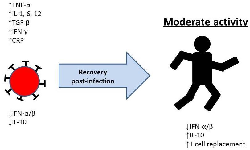
Altogether, adaptation to regular physical activity can affect immune function, especially cellular immunity.
We certainly believe that applying appropriate exercise programs to people who have suffered from COVID-
19 can improve their recovery and quality of life as well as the protection against infection at the long term
(Figure 1).
Figure 1. Cytokines alterations with the infection, effect and/or modulation through physical activity.
CONCLUSION
In conclusion, we think that physical activity and/or physical exercise or recreational sports in general can be
a good alternative for the improvement of the immunological function of patients who have suffered from the
coronavirus infection (Brawner et al., 2021). The anti-inflammatory potential of exercise can also improve the
health status of people and promote an increase in the quality of life. Of course, the practice of exercise must
be adequate in time and intensity, especially taking into account the patient's current situation and previous
sports history.
AUTHOR CONTRIBUTIONS
Conceptualization, A.CM.; Methodology, A.C. and E.R.; Validation, A.CM., A.CG, D PV, JM S and E.R.;
Writing-original draft preparation, A.C.; Writing-Review & Editing, A.C. and E.R.; Visualization, A.CM., A.CG,
D PV, JM S and E.R.; Supervision, A.CM., A.CG, D PV, JM S and E.R.; Funding Acquisition, A.CM, and
A.CG. All authors have read and agreed to the published version of the manuscript.
SUPPORTING AGENCIES
This paper was supported by Caja Rural de Soria.
8 | 2021 | ISSUE - | VOLUME -- © 2021 University of AlicanteCórdova-Martínez, et al. / Inflammation recovery of coronavirus JOURNAL OF HUMAN SPORT & EXERCISE
DISCLOSURE STATEMENT
No potential conflict of interest was reported by the authors.
REFERENCES
Adams, E. R., Ainsworth, M., Anand, R., Andersson, M. I., Auckland, K., Baillie, J. K., … Whitehouse, J.
(2020). Evaluation of antibody testing for SARS-Cov-2 using ELISA and lateral flow immunoassays.
MedRxiv. https://doi.org/10.1101/2020.04.15.20066407
Al-Shaibani, A. (2020). Epidemiology of the domestic and repatriation (Covid-19) Infection in Al Najaf
province, Iraq. Journal of the Faculty of Medicine of Bagdad, 62, 13-19.
https://doi.org/10.32007/jfacmedbagdad.621.21738
Algaissi, A., Alfaleh, M. A., Hala, S., Abujamel, T. S., Alamri, S. S., Almahboud, S. A., … Hashem, A. M.
(2020). SARS-CoV-2 S1 and N-based serological assays reveal rapid seroconversion and induction
of specific antibody response in COVID-19 patients. Scientific Reports, 10, 16561.
https://doi.org/10.1038/s41598-020-73491-5
Allen, J., Sun, Y., & Woods, J. A. (2015). Exercise and the regulation of inflammatory responses.
Progress in Molecular Biology and Translational Science, 135, 337-354.
https://doi.org/10.1016/bs.pmbts.2015.07.003
Amanat, F., & Krammer, F. (2020). SARS-CoV-2 vaccines: Status Report. Immunity, 52, 583-589.
https://doi.org/10.1016/j.immuni.2020.03.007
Baumann, H., & Gauldie J. (1994). The acute phase response. Immunology Today, 15, 74-80.
https://doi.org/10.1016/0167-5699(94)90137-6
Billiau, A. (1996). Interferon-γ: biology and role in pathogenesis. Advances in Immunology, 62, 61-130.
https://doi.org/10.1016/S0065-2776(08)60428-9
Bouvet, M., Debarnot, C., Imbert, I., Selisko, B., Snijder, E. J., Canard, B., & Decroly, E. (2010). In vitro
reconstitution of SARS-coronavirus mRNA cap methylation. PLoS Pathogens, 6, e1000863.
https://doi.org/10.1371/journal.ppat.1000863
Braun, J., Loyal, L., Frentsch, M., Wendisch, D., Georg, P., Kurth, F., … Thiel, A. (2020). Presence of
SARS-CoV-2 reactive T cells in COVID-19 patients and healthy donors. Nature, 587, 270-274.
https://doi.org/10.1038/s41586-020-2598-9
Brawner, C. A, Ehrman, J. K., Bole, S., Kerrigan, D. J., Parikh, S. S., Lewis, B. K., … Keteyian, S. J.
(2019). Inverse relationship of maximal exercise capacity to hospitalization secondary to coronavirus
disease 2019. Mayo Clinic Proceedings, 96, 32-39. https://doi.org/10.1016/j.mayocp.2020.10.003
Chang, Y. J., Kim, H. Y., Albacker, L. A., Baumgarth, N., McKenzie, A. N., Smith, D. E., … Umetsu, D.
T. (2011). Innate lymphoid cells mediateinfluenza-induced airway hyper-reactivity independently of
adaptive immunity. Nature Immunology, 12, 631-638. https://doi.org/10.1038/ni.2045
Channappanavar, R., Fett, C., Zhao, J., Meyerholz, D. K., & Perlman, S. (2014). Virus-specific memory
CD8 T cells provide substantial protection from lethal severe acute respiratory syndrome coronavirus
infection. Journal of Virology, 88, 11034-11044. https://doi.org/10.1128/JVI.01505-14
Channappanavar R., Fehr, A. R., Vijay, R., Mack, M., Zhao, J., Meyerholz, D. K., & Perlman, S. (2016).
Dysregulated type I interferon and inflammatory monocyte-macrophage responses cause lethal
pneumonia in SARS-CoV-infected mice. Cell Host and Microbe, 19, 181-193.
https://doi.org/10.1016/j.chom.2016.01.007
Chousterman, B. G., Swirski, F. K., & Weber, G. F. (2017). Cytokine storm and sepsis disease
pathogenesis. Seminars in Immunopathology, 39, 517-528. https://doi.org/10.1007/s00281-017-
0639-8
VOLUME -- | ISSUE - | 2021 | 9Córdova-Martínez, et al. / Inflammation recovery of coronavirus JOURNAL OF HUMAN SPORT & EXERCISE
Cordova, A., Martin, J. F., Reyes, E., & Alvarez-Mon, M. (2004). Protection against muscle damage in
competitive sports players: the effect of the immunomodulator AM3. Journal of Sports Sciences, 22,
827-833. https://doi.org/10.1080/02640410410001716742
Cordova, A., Monserrat, J., Villa, G., Reyes, E., & Soto, M. A. (2006). Effects of AM3 (Inmunoferon) on
increased serum concentrations of interleukin-6 and tumour necrosis factor receptors I and II in
cyclists. Journal of Sports Sciences, 24, 565-573. https://doi.org/10.1080/02640410500141158
Córdova A., Martorell M., Sureda A., Tur J. A., Pons A. Changes in Circulating Cytokines and Markers
of Muscle Damage in Elite Cyclists during a Multi-stage Competition Clinical Physiology Function
Imaging. 2015;35:351-358. https://doi.org/10.1111/cpf.12170
Cordova, A., Sureda, A., Pons, A., & Alvarez-Mon, M. (2015). Modulation of TNF-alpha, TNF-alpha
receptors and IL-6 after treatment with AM3 in professional cyclists. Journal of Sports Medicine and
Physical Fitness, 55, 345-351.
Deng X., & Baker, S. C. (2018). An "old" protein with a new story: coronavirus endoribonuclease is
important for evading host antiviral defences. Virology, 517, 157-163.
https://doi.org/10.1016/j.virol.2017.12.024
Diao, B., Wang, C., Tan, Y., Chen, X., Liu, Y., Ning, L., … Chen, Y. (2020). Reduction and functional
exhaustion of T Cells in patients with coronavirus disease 2019 (COVID-19). Frontiers in
Immunology, 11, 827. https://doi.org/10.3389/fimmu.2020.00827
Germolec, D. R., Frawley, R. P., & Evans, E. (2010). Markers of inflammation. Methods in Molecular
Biology, 598, 53-73. https://doi.org/10.1007/978-1-60761-401-2_5
Giamarellos-Bourboulis, E. J., Netea, M. G., Rovina, N., Akinosoglou, K., Antoniadou, A., Antonakos, N.,
… Koutsoukou, A. (2020). Complex immune dysregulation in COVID-19 patients with severe
respiratory failure. Cell Host and Microbe, 27, 992-1000.e3.
https://doi.org/10.1016/j.chom.2020.04.009
Gleeson, M., Pyne, D. B., Austin, J. P., Lynn Francis, J., Clancy, R. L., McDonald, W. A., & Fricker, P. A.
(2002). Epstein_Barr virus reactivation and upper-respiratory illness in elite swimmers. Medicine and
Science in Sports and Exercise, 34, 411-417. https://doi.org/10.1097/00005768-200203000-00005
Green, S. J., Scheller, L. F., Marletta, M. A., Seguin, M. C., Klotz, F. W., Slayter, M., … Nacy, C. A.
(1994). Nitric oxide: cytokine-regulation of nitric oxide in host resistance to intracellular pathogens.
Immunology Letters, 43, 87-94. https://doi.org/10.1016/0165-2478(94)00158-8
Guo, C., Li, B., Ma, H., Wang, X., Cai, P., Yu, Q., … Qu, K. (2020). Tocilizumab treatment in severe
COVID-19 patients attenuates the inflammatory storm incited by monocyte centric immune
interactions revealed by single-cell analysis. BioRxiv. https://doi.org/10.1101/2020.04.08.029769
Hackbart, M., Deng, X., & Baker, S. C. (2020). Coronavirus endoribonuclease targets viral polyuridine
sequences to evade activating host sensors. Proceedings of the National Academy of Sciences of
the United States of America, 117, 8094-8103. https://doi.org/10.1073/pnas.1921485117
Hadjadj, J., Yatim, N., Barnabei, L., Corneau, A., Boussier, J., Pere, H., … Terrier, B. (2020). Impaired
type I interferon activity and exacerbated inflammatory responses in severe Covid-19 patients.
Science, 369, 718-724. https://doi.org/10.1126/science.abc6027
Hu, L., Chen, S., Fu, Y., Gao, Z., Long, H., Ren, H.-W., … Deng, Y. (2020). Risk factors associated with
clinical outcomes in 323 coronavirus disease 2019 (COVID-19) hospitalized patients in Wuhan,
China. Clinical Infectious Diseases, 71, 2089-2098. https://doi.org/10.1093/cid/ciaa539
Huang, A.T., Garcia-Carreras, B., Hitchings, M. D. T., Yang, B., Katzelnick, L., Rattigan, S. M., …
Cummings, D. A. T. (2020). A systematic review of antibody mediated immunity to coronaviruses:
antibody kinetics, correlates of protection, and association of antibody responses with severity of
disease. MedRxiv. https://doi.org/10.1101/2020.04.14.20065771
10 | 2021 | ISSUE - | VOLUME -- © 2021 University of AlicanteCórdova-Martínez, et al. / Inflammation recovery of coronavirus JOURNAL OF HUMAN SPORT & EXERCISE
Ju, B., Zhang, Q., Ge, X., Wang, R., Sun, J., Ge, X., … Song, S. (2020). Human neutralizing antibodies
elicited by SARS-CoV-2 infection. Nature, 584, 115-119. https://doi.org/10.1038/s41586-020-2380-z
Kawanishi, N., Mizokami, T., Niihara, H., Yada, K., & Suzuki, K. (2016). Neutrophil depletion attenuates
muscle injury after exhaustive exercise. Medicine and Science in Sports and Exercise, 48, 1917-
1924. https://doi.org/10.1249/MSS.0000000000000980
Kammuller, M. E. (1995). Recombinant human interleukin-6: safety issues of a pleiotropic growth factor.
Toxicology, 105, 91-107. https://doi.org/10.1016/0300-483X(95)03128-3
Kang, S., Brown, H. M., & Hwang, S. (2018). Direct antiviral mechanisms of interferon-gamma. Immune
Network, 18, e33. https://doi.org/10.4110/in.2018.18.e33
Knoops, K., Kikkert, M., Worm, S. H., Zevenhoven-Dobbe, J. C., van der Meer, Y., Koster, A. J., …
Snijder, E.J. (2008). SARS-coronavirus replication is supported by a reticulovesicular network of
modified endoplasmic reticulum. PLoS Biology, 6, e226.
https://doi.org/10.1371/journal.pbio.0060226
Krüger, K., Mooren, F.-C.., & Pilat, C. (2016). The immunomodulatory effects of physical activity. Current
Pharmaceutical Design, 22, 3730-3748. https://doi.org/10.2174/1381612822666160322145107
Lee, A. J., & Ashkar, A. A. (2018). The dual nature of type I and type II interferons. Frontiers in
Immunology, 9, 2061. https://doi.org/10.3389/fimmu.2018.02061
Lee, D. W., Gardner, R., Porter, D. L. Louis, C. U., Ahmed, N., Jensen, M, … Mackall, C. L., (2014).
Current concepts in the diagnosis and management of cytokine release syndrome. Blood, 124, 188-
195. https://doi.org/10.1182/blood-2014-05-552729
Li, C. K.-F., Wu, H., Yan, H., Ma, S., Wang, L., Zhang, M., … Xu, X.-N. (2008). T cell responses to whole
SARS coronavirus in humans. Journal of Immunology, 181, 5490-5500.
https://doi.org/10.4049/jimmunol.181.8.5490
Liu, W., Fontanet, A., Zhang, P. H., Zhan, L., Xin, Z. T., Baril, L., … Cao, W.C. (2006). Two-year
prospective study of the humoral immune response of patients with severe acute respiratory
syndrome. Journal of Infectious Diseases, 193, 792-795. https://doi.org/10.1086/500469
Liu, Q., Zhou, Y. H., & Yang, Z. Q. (2016). The cytokine storm of severe influenza and development of
immunomodulatory therapy. Cellular and Molecular Immunology, 13, 3-10.
https://doi.org/10.1038/cmi.2015.74
Lowder, T., Padgett, D. A., & Woods J. A. (2006). Moderate exercise early after influenza virus infection
reduces the Th1 inflammatory response in lungs of mice. Exercise Immunology Review, 12, 97-111.
Lu, R., Zhao, X., Li, J., Niu, P., Yang, B., Wu, H., … Tan, W. (2020). Genomic characterisation and
epidemiology of 2019 novel coronavirus: implications for virus origins and receptor binding. Lancet,
395, 565-574. https://doi.org/10.1016/S0140-6736(20)30251-8
Mahdi, B.M. (2020). COVID-19 type III hypersensitivity reaction. Medical Hypotheses, 140, 109763.
https://doi.org/10.1016/j.mehy.2020.109763
McNab, F., Mayer-Barber, K., Sher, A., Wack, A., & O'Garra, A. (2015). Type I interferons in infectious
disease. Nature Reviews. Immunology, 15, 87-103. https://doi.org/10.1038/nri3787
Mehta, P., McAuley, D. F., Brown, M., Sanchez, E., Tattersall, R. S., Manson, J. J., & HLH Across
Speciality Collaboration, UK. (2020). COVID-19: consider cytokine storm syndromes and
immunosuppression. Lancet, 395, 1033-1034. https://doi.org/10.1016/S0140-6736(20)30628-0
Merad, M., & Martin, J. C. (2020). Pathological inflammation in patients with COVID-19: a key role for
monocytes and macrophages. Nature Reviews. Immunology, 20, 355-362.
https://doi.org/10.1038/s41577-020-0331-4
Ng, O.-W., Chia, A., Tan, A. T., Jadi, R. S., Leong, H. N., … Tan,Y.-J. (2016). Memory T cell responses
targeting the SARS coronavirus persist up to 11 years post-infection. Vaccine, 34, 2008-2014.
https://doi.org/10.1016/j.vaccine.2016.02.063
VOLUME -- | ISSUE - | 2021 | 11Córdova-Martínez, et al. / Inflammation recovery of coronavirus JOURNAL OF HUMAN SPORT & EXERCISE
Nie, S., Zhao, X., Zhao, K., Zhang, Z. [Zhaohui], Zhang, Z. [Zhentao], & Zhang, Z. [Zhan] (2020).
Metabolic disturbances and inflammatory dysfunction predict severity of coronavirus disease 2019
(COVID-19): a retrospective study. MedRxiv. https://doi.org/10.1101/2020.03.24.20042283
Nieman, D. C. (1997). Immune response to heavy exertion. Journal of Applied Physiology. 82, 1385-
1394. https://doi.org/10.1152/jappl.1997.82.5.1385
Nieman, D. C., & Nehlsen-Cannarella, S. L. (1992). Exercise and infection. In R. R. Watson & M. Eisinger
(Eds.), Exercise and disease (pp. 121-148). Boca Raton, Florida, USA: CRC Press.
https://doi.org/10.1201/9781003068853-8
Okba, N. M. A., Müller, M. A., Li, W., Wang, C., GeurtsvanKessel, C. H., Corman,V. M., … Haagmans,
B. L. (2020). Severe acute respiratory syndrome coronavirus 2-specific antibody responses in
coronavirus disease patients. Emerging Infectious Diseases, 26, 1478-1488.
https://doi.org/10.3201/eid2607.200841
Ozemek, C., Lavie, C. J., & Rognmo, Ø. (2019). Global physical activity levels-Need for intervention.
Progress in Cardiovascular Disease, 62, 102-107. https://doi.org/10.1016/j.pcad.2019.02.004
Pathan, N., Hemingway, C. A., Alizadeh, A. A., Stephens, A. C., Boldrick, J. C., Oragui, E. E., … Levin,
M. (2004). Role of interleukin 6 in myocardial dysfunction of meningococcal septic shock. Lancet,
363, 203-209. https://doi.org/10.1016/S0140-6736(03)15326-3
Pedersen, B. K., & Hoffman-Goetz, L. (2000). Exercise and the immune system: regulation, integration,
and adaptation. Physiological Reviews, 80, 1055-1081.
https://doi.org/10.1152/physrev.2000.80.3.1055
Polidoro, R. B., Hagan, R. S., de Santis Santiago, R., & Schmidt, N. W. (2020). Overview: Systemic
inflammatory response derived from lung injury caused by SARS-CoV-2 infection explains severe
outcomes in COVID-19. Frontiers in Immunology, 11, 1626.
https://doi.org/10.3389/fimmu.2020.01626
Qin, C., Zhou, L., Hu, Z., Zhang, S., Yang, S., Tao, Y., … Tian, D.-S. (2020). Dysregulation of immune
response in patients with COVID-19 in Wuhan, China. Clinical Infectious Diseases, 71, 762-768.
https://doi.org/10.1093/cid/ciaa248
Rehwinkel, J., & Gack, M. U. (2020). RIG-I-like receptors: their regulation and roles in RNA sensing.
Nature Reviews. Immunology, 20, 537-551. https://doi.org/10.1038/s41577-020-0288-3
Rothan, H. A., & Byrareddy, S. N. (2020). The epidemiology and pathogenesis of coronavirus disease
(COVID-19) outbreak. Journal of Autoimmunity, 109, 102433.
https://doi.org/10.1016/j.jaut.2020.102433
Ruggiero, V., Tavernier, J., Fiers, W., & Baglioni, C. (1986). Induction of the synthesis of tumor necrosis
factor receptors by interferon-gamma. Journal of Immunology, 136, 2445-2450.
Sarzi-Puttini, P., Giorgi, V., Sirotti, S., Marotto, D., Ardizzone, S., Rizzardini, G, … Galli, M. (2020).
COVID-19, cytokines and immunosuppression: what can we learn from severe acute respiratory
syndrome? Clinical and Experimental Rheumatology, 38, 337-342.
https://doi.org/10.1016/j.autrev.2020.102574
Schroder, K., Hertzog, P. J., Ravasi, T., & Hume, D.A. (2004). Interferon-gamma: an overview of signals,
mechanisms and functions. Journal of Leukocyte Biology, 75, 163-189.
https://doi.org/10.1189/jlb.0603252
Shaw, D. M., Merien, F., Braakhuis, A., & Dulson, D. (2018). T-cells and their cytokine production: the
anti-inflammatory and immunosuppressive effects of strenuous exercise. Cytokine, 104, 136-142.
https://doi.org/10.1016/j.cyto.2017.10.001
Shephard, R. J., Verde, T. J., Thomas, S. G., & Shek, P. (1991). Physical activity and the immune system.
Canadian Journal of Sport Sciences, 16, 163-185.
12 | 2021 | ISSUE - | VOLUME -- © 2021 University of AlicanteCórdova-Martínez, et al. / Inflammation recovery of coronavirus JOURNAL OF HUMAN SPORT & EXERCISE
Shiow, L. R., Rosen, D. B., Brdicková, N., Xu, Y., An, J., Lanier, L. L., … Matloubian, M. (2006). CD69
acts downstream of interferon-alpha/beta to inhibit S1P1 and lymphocyte egress from lymphoid
organs. Nature, 440, 540-544. https://doi.org/10.1038/nature04606
Song, C.-Y., Xu, J., He, J.-Q., & Lu, Y.-Q. (2020). COVID-19 early warning score: a multi-parameter
screening tool to identify highly suspected patients. MedRxiv.
https://doi.org/10.1101/2020.03.05.20031906
Sureda, A., Ferrer, M. D., Tauler, P., Maestre, I., Aguiló, A., Cordova, A, … Pons, A. (2007). Intense
physical activity enhances neutrophil antioxidant enzyme gene expression. Immunocytochemistry
evidence for catalase secretion. Free Radical Research, 41, 874-883.
https://doi.org/10.1080/10715760701416459
Surkina, I., Danilenko, S., Dudov, N., Gotovtseva, E. P., Koptelov, O. V., Kostina, L. V., … Vorobiev, A.
A. The role of the immune system in processes of adaptation to stress in sportsmen. Clinical Science,
87, 22. https://doi.org/10.1042/cs087s022
Suzuki, K., Nakaji, S., Yamada, M., Totsuka, M., Sato, K., & Sugawara, K. (2002). Systemic inflammatory
response to exhaustive exercise. Cytokine kinetics. Exercise Immunology Review, 8, 6-48.
Tanaka, T., Narazaki, M., & Kishimoto, T. (2016). Immunotherapeutic implications of IL-6 blockade for
cytokine storm. Immunotherapy, 8, 959-970. https://doi.org/10.2217/imt-2016-0020
Taylor, A., Foo, S.-S., Bruzzone, R., Dinh, L. V., King, N. J. C., & Mahalingam, S. (2015). Fc receptors
in antibody-dependent enhancement of viral infections. Immunological Reviews, 268, 340-364.
https://doi.org/10.1111/imr.12367
Thevarajan, I., Nguyen, T. H. O., Koutsakos, M., Druce, J., Caly, L., van de Sandt, C. E.,… Kedzierska,
K. (2020) Breadth of concomitant immune responses prior to patient recovery: a case report of non-
severe COVID-19. Nature Medicine, 26, 453-455. https://doi.org/10.1038/s41591-020-0819-2
To, K. K.-W., Tsang, O. T.-Y., Leung, W.-S., Tam, A. R., Wu, T.-C., Lung, D. C., … Yuen, K.-Y. (2020).
Temporal profiles of viral load in posterior oropharyngeal saliva samples and serum antibody
responses during infection by SARS-CoV-2: an observational cohort study. Lancet. Infectious
Diseases, 20, 565-574. https://doi.org/10.1016/S1473-3099(20)30196-1
Velikova, T. V., Kotsev, S. V., Georgiev, D. S., & Batselova, H. M. (2020). Immunological aspects of
COVID-19: What we do know? World Journal of Biological Chemistry, 11, 14-29.
https://doi.org/10.4331/wjbc.v11.i2.14
Vivier, E., Artis, D., Colonna, M., Diefenbach, A., Di Santo, J.P., Eberl, G., … Spits, H. (2018). Innate
Lymphoid Cells: 10 years on. Cell, 174, 1054-1066. https://doi.org/10.1016/j.cell.2018.07.017
Walsh, N. P., Gleeson, M., Shephard, R. J., Gleeson, M., Woods, J. A., Bishop, N. C., … Simon, P.
(2011). Position statement. Part one: Immune function and exercise. Exercise Immunology Review,
17, 6-63.
Wang, D., Hu, B., Hu, C., Zhu, F., Lui, X., Zhang, J., …Peng, Z. (2020). Clinical characteristics of 138
hospitalized patients with 2019 novel coronavirus-infected pneumonia in Wuhan, China. JAMA, 323,
1061-1069. https://doi.org/10.1001/jama.2020.1585
Wang, T., Zhang, X., & Li, J. J. (2002). The role of NF-κB in the regulation of cell stress responses.
International Immunopharmacology, 2, 1509-1520. https://doi.org/10.1016/S1567-5769(02)00058-9
Wilk, A. J., Rustagi, A., Zhao, N. Q., Roque, J., Martinez-Colon, G. J., McKechnie, J. L., … Blish, C. A.
(2020). A single-cell atlas of the peripheral immune response in patients with severe COVID19.
Nature Medicine, 26, 1070-1076. https://doi.org/10.1038/s41591-020-0944-y
Zhang, J., & An, J. (2007). Cytokines, inflammation and pain. International Anesthesiology Clinics, 45,
27-37. https://doi.org/10.1097/AIA.0b013e318034194e
VOLUME -- | ISSUE - | 2021 | 13Córdova-Martínez, et al. / Inflammation recovery of coronavirus JOURNAL OF HUMAN SPORT & EXERCISE
Zhang, Y., Li, J., Zhan, Y., Wu, L., Yu, X., Zhang, W., … Lou, J. (2004). Analysis of serum cytokines in
patients with severe acute respiratory syndrome. Infection and Immunity, 72, 4410-4415.
https://doi.org/10.1128/IAI.72.8.4410-4415.2004
Zhao, S., Lin, Q., Ran, J., Musa, S. S., Yang, G., Wang, W, … Wang, M. H. (2020). Preliminary estimation
of the basic reproduction number of novel coronavirus (2019-nCoV) in China, from 2019 to 2020: a
data-driven analysis in the early phase of the outbreak. International Journal of Infectious Diseases,
92, 214-217. https://doi.org/10.1016/j.ijid.2020.01.050
Zheng, M., Gao, Y., Wang, G., Song, G., Liu, S., Sun, D., … Tian, Z. (2020). Functional exhaustion of
antiviral lymphocytes in COVID-19 patients. Cellular and Molecular Immunology, 17, 533-535.
https://doi.org/10.1038/s41423-020-0402-2
Zheng, M., & Song, L. (2020). Novel antibody epitopes dominate the antigenicityof spike glycoprotein in
SARS-CoV-2 compared to SARS-CoV. Cellular and Molecular Immunology, 17, 536-538.
https://doi.org/10.1038/s41423-020-0385-z
Zhou, Y., Fu, B., Zheng, X., Wang, D., Zhao, C., Qi, Y., … Wei, H. (2020). Pathogenic T cells and
inflammatory monocytes incite inflammatory storm in severe COVID-19 patients. National Science
Review, 7, 998-1002. https://doi.org/10.1093/nsr/nwaa041
This work is licensed under a Attribution-NonCommercial-NoDerivatives 4.0 International (CC BY-NC-ND 4.0).
14 | 2021 | ISSUE - | VOLUME -- © 2021 University of AlicanteYou can also read