In-Vivo Toxicity of Bioreactor-Grown Biomass And Exopolysaccharides From Malaysian Tiger Milk Mushroom Mycelium For Potential Future Health ...
←
→
Page content transcription
If your browser does not render page correctly, please read the page content below
In-Vivo Toxicity of Bioreactor-Grown Biomass And Exopolysaccharides From Malaysian Tiger Milk Mushroom Mycelium For Potential Future Health Applications Siti Rokhiyah Ahmad Usuldin Universiti Malaya Imad Wan-Mohtar Universiti Malaya Zul Ilham Universiti Malaya Adi Ainurzaman Jamaludin Universiti Malaya Nur Raihan Abdullah International Islamic University Malaysia Neil Rowan ( nrowan@ait.ie ) Athlone Institute of Technology Research Article Keywords: mycelial biomass, exopolysaccharide, Zebra sh Embryo Toxicity, therapeutic DOI: https://doi.org/10.21203/rs.3.rs-778200/v1 License: This work is licensed under a Creative Commons Attribution 4.0 International License. Read Full License
In-vivo Toxicity of Bioreactor-Grown Biomass and
Exopolysaccharides from Malaysian Tiger Milk Mushroom
Mycelium for Potential Future Health Applications
Siti Rokhiyah Ahmad Usuldin1,2, Wan Abd Al Qadr Imad Wan-Mohtar2,3,6, +, Zul Ilham3,4, Adi
Ainurzaman Jamaludin4, Nur Raihan Abdullah5, and Neil Rowan 6,7,+, *

1
  Agro-Biotechnology Institute, Malaysia (ABI), National Institutes of Biotechnology Malaysia (NIMB), c/o HQ
MARDI, 43400, Serdang,Selangor, Malaysia
2
  Functional Omics and Bioprocess Development Laboratory, Institute of Biological Sciences, Faculty of Science,
Universiti Malaya, 50603 Kuala Lumpur, Malaysia
3
  Bioresources and Bioprocessing Research Group, Institute of Biological Sciences, Faculty of Sciences, Universiti
Malaya, 50603, Kuala Lumpur, Malaysia
4
  Environmental Science and Management Program, Institute of Biological Sciences, Faculty of Science, Universiti
Malaya, 50603, Kuala Lumpur, Malaysia
5
  Department of Biotechnology, Kulliyyah of Science, International Islamic University Malaysia, 25200 Kuantan,
Pahang, Malaysia
6
  Bioscience Research Institute, Athlone Institute of Technology, Ireland
7
  Empower Eco Innovation Hub, Boora, Co. Offaly, Ireland

*nrowan@ait.ie
+
 these authors contributed equally to this work

ABSTRACT
Natural mycelial biomass (MB) and exopolysaccharide (EPS) produced from bioreactor-grown
Malaysian Tiger Milk Mushroom Lignosus rhinocerus are considered high-end components due to their
high commercial potential value in drug discovery. Both MB (0.16-10 mg/mL) and EPS (0.16-10 mg/mL)
extracts were tested for Zebrafish Embryo Toxicity (ZFET) and early development effects on Zebrafish
Embryos (ZE) during 24-120 h post-fertilization (HPF) in order to assess toxicity given potential
application as a new therapy for treating asthma. Finding revealed that MB was deemed to be safe with
an LC50 of 0.77 mg/mL; EPS was shown to be non-toxic (LC50 of 0.41 mg/mL). Neither MB nor EPS
delayed hatching nor teratogenic defects in treated ZE at a dose of 2.5 mg/mL. There were no significant
changes in the ZE heart rate after MB or EPS treatments with MB (0.16-10 mg/mL: 130 beats/min) and
EPS (0.16-10 mg/mL: 140 beats/min), as compared to normal ZE (120-180 beats/min). In addition,
mixing of both natural compounds MB and EPS did not affect toxicity using ZFET testing; thus,
intimating their safe future use as therapeutic interventions.
Introduction Mushrooms are filamentous fungi that have been used worldwide since prehistoric times all across the world 1,2, and medically since at least 3000 BCE 3,4. Mushrooms have become increasingly important as producing organisms in research and industry; food, medicines, cosmetics, detergents and biofuels are examples of high-value products produced by fungi 5,6. In addition, there has been increased interest in exploiting mushroom-derived extracts as potential solutions for pressing health applications 7,8, including a focused interest in biorefining these products such as through Green New Deal innovations from food waste streams that attests to enhanced focus on growing ‘circularity’ and bioeconomy 9,10. Mushrooms are classified in the kingdom of Fungi and have many active constituents including, but possibly not limited to, polysaccharides, polysaccharide peptides, proteins, terpenoids, and nucleotides 11. The most studied and used medicinally active ingredient in mushrooms is -glucan. Previous research has revealed that β-glucans have broad metabolic and gastro-intestinal effects, including modulating the gut microbiome, altering lipid and glucose metabolism, reducing cholesterol; thus, leading to use of -glucan as potential therapies for treating metabolic syndrome, obesity and diet regulation, gastrointestinal conditions, and to reduce the risk of cardiovascular and diabetes 12. It is appreciated that bioactive extracts derived from mushrooms can modulate the immune response affecting hematopoietic stem cells, lymphocytes, macrophages, T cells, dendritic cells and natural killer cells. Moreover, the pronounced immune-modulatory effects of β-glucans 13,14 has also lead to their investigation as adjuvant agents for treating cancers (solid and haematological malignancies), for immune-mediated conditions (such as allergic rhinitis, respiratory infections), and to enhance wound healing 12,15. Murphy et al. 14 have reviewed over 200 patents that highlighted the therapeutic potential of β-glucans; this is evidenced by the fact that two glucans were licensed as immune-adjuvant therapy for treating cancer in Japan. However, significant challenges exist to further clinical testing and translation of β-glucans, which includes important differences that appear to exist in the effects of apparently similar β-glucan preparations that may be due to differences in sources and extraction procedures 12. The present authors have recently identified active ingredients of Lignosus rhinocerus using 2D COSY, TOCSY, HMQC and HMBC analyses and reported on anti-oxidative potential of (1,3)--D-glucan (in the form of exopolysaccharide crude extract) as an important constituent 16. However, it is appreciated that there are many other compounds extracted from medicinal mushroom have yet to be named, such as often referred to by gel chromatography fraction 11; thus, highlighting the need to conduct and report on safety. The Tiger Milk mushroom, Lignosus rhinocerus, belongs to the Basidiomycota section of the Polyporaceae family and is classified as a filamentous fungus 17,18. L. rhinocerus was grown in submerged-liquid fermentation (SLF) using a laboratory-scale stirred-tank bioreactor in order to achieve bulk cultivation and commensurate production of polysaccharides 16. When compared to solid-state fermentation (SSF), SLF has several advantages, including limited space requirements, ease of scale-up, reliable and reproducible processing, ease to monitoring, and versatility 19. Artificially-cultivated L. rhinocerus is also an excellent replacement in the development of therapeutic items; for example, exopolysaccharides (EPS) isolated from mushroom mycelial biomass (MB) have pharmacological properties, such as immunomodulatory, anti-inflammatory, antibacterial, antiviral, and antioxidant properties 20. Chen et al.21 discovered that L. rhinocerotis mycelium grown in SLF does not cause
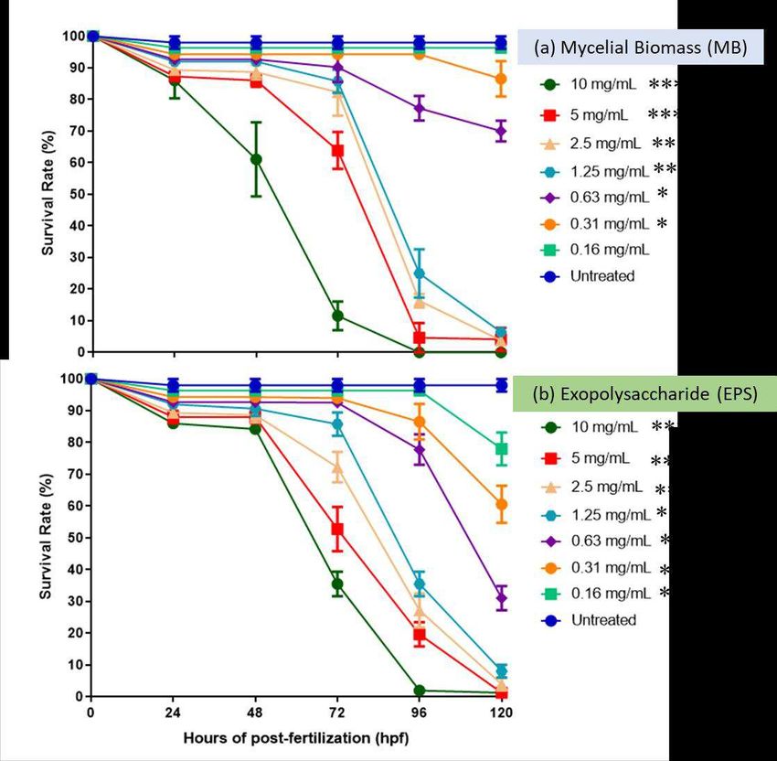
mutagenicity or genotoxicity. FDA standards, on the other hand, demand substantial proof of no hazard
for commercial usage 21.

Asthma affects 300 million individuals worldwide and is caused by a complex combination of inherited
and environmental variables 18. Allergic asthma is a long-term condition characterized by wheezing,
shortness of breath, chest tightness, and coughing. In Malaysia, indigenous peoples have long used L.
rhinocerus to treat asthma. The majority of today's asthma medications are made up of steroids and other
anti-inflammatory drugs 18. Recent studies have reported on the efficacy of EPS from medicinal
mushrooms to ameliorate pro- and anti-inflammatory responses using ex vivo and in vivo infection models
with therapeutic potential 12,14,22. Aqueous extracts of L. rhinocerotis were reported to help reduce asthma-
related variables in asthma model 23. In addition, a previous toxicity study indicated that feeding 1000
mg/kg of L. rhinocerus extract to rats had no detrimental consequences, hence considered safe 24. As a
result, a more effective asthma treatment is required and L. rhinocerus has the potential to be a useful
adjuvant or alternative to currently available asthma medications.

A recent publication on the evaluation of the toxicity of biomass-EPS comparable medicinal mushroom
mycelial extracts revealed that the zebrafish embryo toxicity (ZFET) assay can be deployed as safety
screening approach before pre-clinical testing according to national and international standards 25. In
comparison to human cell lines, the ZFET model research has been shown to be quick, resilient, efficient,
and cost-effective for early development investigations; in addition, it also represents relevant genetic
structure and equivalent key organs and tissues26,27. Thus, this study will help determine mushroom
extract's toxicity before it is developed and potentially commercialized as new therapeutic intervention.
To date, there has been no toxicity studies using the ZFET model describing use of MB and EPS of L.
rhinocerus generated in bioreactor.

As a result, this constitutes the second study to determine the levels of toxicity of extracted MB and EPS
using a zebrafish-embryo-toxicity (ZFET) model. The toxicity of MB and EPS produced from L.
rhinocerus was tested using the Zebrafish embryo toxicity model to ensure product safety throughout the
pre-commercialization phase. It is notable that the present rare L. rhinocerus strain ABI was successfully
isolated and identified from a tropical forest near Lata Iskandar, Pahang, Malaysia 16; however, there has
been limited information published on its’ therapeutic potential.

Results

Zebrafish Embryo Survival Rate after MB and EPS Exposure
The survival rate of zebrafish embryos following MB and EPS exposure was studied between 0 and 120
h at MB and EPS extract concentrations of 0.16-10 mg/mL. For five days, the survival rate of embryos
(prior to hatching) and larvae (post hatching) treated with MB and EPS extract were measured. The
survival rate of embryos (prior to hatching) and larvae (post hatching) treated with MB and EPS extract
was assessed for a maximum of five days (120 h) as the zebrafish embryos hatch typically 48 to 72 h-
post-fertilization (HPF). The survival rate of untreated embryos, between 0 and 120 HPF, was shown to
be 100% (Figure 1a). At 48 HPF, the survival percentage for embryos treated with MB fell to 85% at
concentrations >5mg/mL and 60% at 10 mg/mL. At 72 HPF, the survival rate declined to 80% at
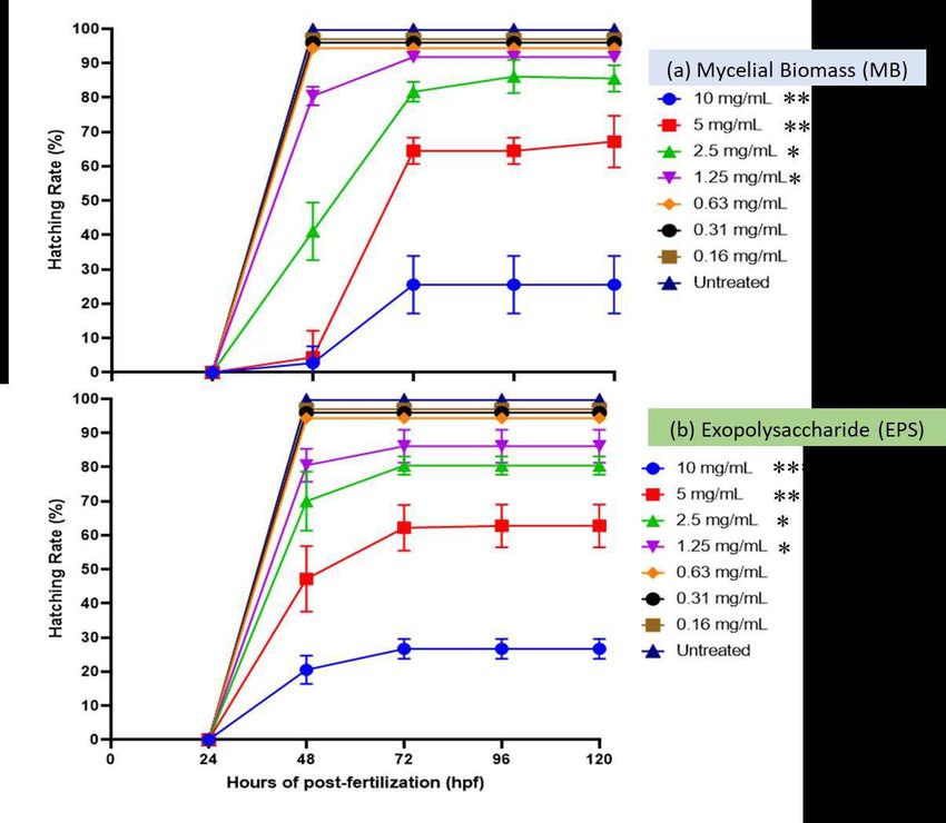
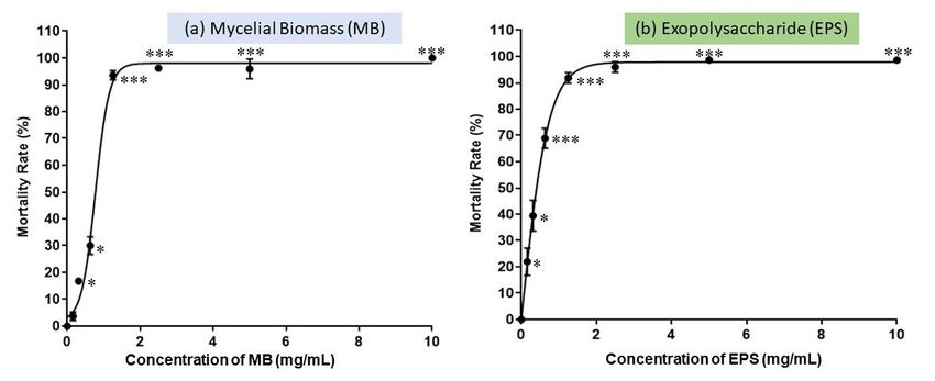
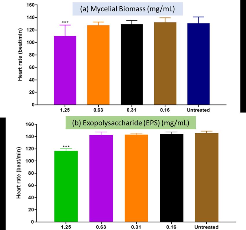
concentrations less than 2.5 mg/mL, 65% at concentrations greater than 5 mg/mL, and 10% at
concentrations greater than 10 mg/mL. At concentrations >1.25 mg/mL, the survival rate was 20% at 96
HPF, and after 120 HPF; it was observed that no embroys survived at concentrations >1.25 mg/mL
(Figure 1a). The survival rate of embryos (prior to hatching) and larvae (post hatching) treated with EPS
(0.110 mg/mL) throughout a five-day period is shown in Figure 1b. Between 0 and 120 h of HPF,
untreated embryos (control) exhibited a 100% survival rate. After 72 h of HPF exposure, the survival rate
declined to 90% at a concentration of 0.63 mg/mL, 85% at a dose of 1.25 mg/mL, and 50% at a dosage
of 5 mg/mL. At 96 HPF, the survival rate declined to 75% at concentrations less than 0.63 mg/mL and
30% at concentrations greater than 1.25 mg/mL. At 120 HPF, survival rates at concentrations 0.63 mg/mL
declined to 30%, while survival rates at concentrations >1.25 mg/mL were 0%, with no embryos
surviving. Overall, the results suggest that MB and EPS extracts delay hatching at doses less than 1.25
mg/mL.

The Zebrafish Embryos Mortality after MB and EPS Exposure
Overall, MB and EPS extracts had dose- and time-dependent fatal effects. Figure 2 shows a high survival
rate, (90%) of zebrafish embryos at concentrations of MB and EPS extracts below 1.25 mg/mL. Both MB
and EPS extracts had a low survival rate at high concentrations (over 1.25 mg/mL), and none survived
after 96 HPF. As a result, the fatal concentration at 50% (LC50 value) of zebrafish embryos exposed to
MB was 0.77 mg/mL, while the LC50 value of the EPS extract was 0.41 mg/mL.

The Zebrafish Embryos Hatching after MB and EPS Exposure
Based on the embryo observation, different mushroom extract concentrations will impact upon the
embryo's hatchability. The percentage of hatchability fell with increasing concentrations of mushroom
extracts. Figure 3a illustrates the hatching rate of zebrafish embryos treated with MB (0.1610 mg/mL)
and EPS (0.1610 mg/mL) at 0–120 HPF. No significant changes in the hatching rate were found when
MB was treated with MB extract at a concentration of 0.63 mg/mL. However, at 48 HPF, the rate declined
to 80% at concentrations >1.25 mg/mL. At 72 HPF, the hatching rate was lowered to 65% at 5 mg/mL.
Further reduction was seen (25% hatching rate) when treated with MB at a dose of 10 mg/mL, intimating
that a high mortality rate occurred after 72 HPF. The hatching rate of EPS did not alter significantly after
treatment with MB extract at a dosage of 0.63 mg/mL. Less than 85% of the embryos hatched was
observed after a 48-h treatment with EPS at a concentration of >1.25 mg/mL. However, due to a
significant mortality rate at 72 HPF, zebrafish larvae treated with EPS at doses of 10 mg/mL had the
lowest hatching rate (30%).

The Zebrafish Embryos Heart Rate after MB and EPS Exposure
During the development of many model species, including zebrafish, the heart is the major functioning
organ 28. The heart rates of zebrafish larvae at 96 HPF (4 days) for both the MB (Fig. 4a) and EPS (Fig.
4b) treatments were 130 and 140 beats/min, respectively, according to Figure 4. According to a
previous study, the normal heart rate of zebrafish embryos is 120–180 beats per minute, which is much
closer to that of humans 29. Both extracts exhibited no significant difference in the heart rate of zebrafish
larvae at 96 HPF at lower concentrations (relative to higher doses in Figure 3) ranging between 0.161.25
mg/mL for MB and 0.161.25 mg/mL for EPS. The heart rate of zebrafish larvae at these concentrations
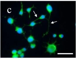
was not determined because both MB and EPS extracts at 2.5, 5 and 10 mg/mL demonstrated very little to no survival at 96 HPF. The Morphology of Larvae and Zebrafish Embryos after MB and EPS Exposure Potential morphological abnormalities in embryos and larvae were detected and measured from 0 HPF to 120 HPF. At 120 h following exposure to MB and EPS at doses of 0.63 mg/mL and 1.25 mg/mL respectively, no obvious teratogenic effect on embryos and larvae (Figure 5). These findings infer that MB and EPS have no teratogenic effects on zebrafish embryo development prior to- and post hatching. The development of zebrafish embryos and larvae were unaffected by exposure to 0.63 mg/mL MB (Figure 6) and 1.25 mg/mL EPS (Figure 7); however, numerous defects were observed when the concentration of MB and EPS was increased to 10 mg/mL MB (Figure 8) and 10 mg/mL EPS (Figure 9). Coagulated embryos observed from 24 HPF (segmentation) to 48 HPF (pharyngula), along with loss of yolk sac resulting in un-hatched were most common abnormalities reported using MB treatments. Moreover, EPS-treated zebrafish hatched at 72 HPF, where tail deformity and damaged blood cells were observed after 120 HPF; defects included missing fins, guts, and melanophores. Discussion This work investigates and reports on acute toxicity of zebrafish embryos post exposure to MB and EPS derived from a rare Malaysian-origin Tiger Milk mushroom L. rhinocerus grown in a bioreactor. Bioactive compounds, such as -glucans, were found in these L. rhinocerus MB-EPS extracts; use of - glucans have gained appeal in recent years for a number of emerging applications including biopolymers 30 and biomedicines 31. It is notable that mushroom-derived -glucans have many new therapeutic properties, including (a) potential new or complementary immunotherapies against Coronavirus disease (SARS-CoV-2) 14, (b) potential new therapeutic agent for mitigating diseases associated with gastrointestinal mucosal damage, such as peptic ulcers and inflammatory bowel disease 32, (c) potential as anticancer drugs for lung and breast cancer 33, and (d) potentially asthmatic treatment 23,34. However, there is a marked gap in knowledge surrounding the toxicity (if any), of these mushroom-derived bioactive compounds; particularly use of Zebrafish trials that would aid product development and implementation. Zebrafish embryos have been extensively studied and documented as a reliable and popular model for developmental biology, toxicity, and, more recently, drug discovery 35. Zebrafish are easy to breed and keep. Zebrafish embryos develop quickly; where they are fully developed five days after conception. Light microscopy can easily examine morphological structures and internal organs, such as the brain, eyes, heart, liver, and kidney due to the embryo's transparency. Dyes can be used to measure organ- specific and overall developmental toxicity visually or quantitatively. Due to its small size, a single Zebrafish embryo can be maintained in low fluid volumes for the first six days of development including use of microtiter plates. The permeability of zebrafish embryos is prominent; for example, small chemicals added to fish water permeate into the embryos easily simplifying drug administration and assay processing 36. Chemical screening can be completed after a few days due to the embryo's rapid development. The zebrafish is a unique vertebrate model for high-throughput chemical screening, which is beneficial for preclinical drug discovery and toxicity assessment 37,38.
The ZFET approach was used to expose fertilized zebrafish embryos to quantities of L. rhinocerus extract,
MB (0.16-10 mg/mL), and EPS (0.16-10 mg/mL) that were shown to be non-toxic. Overall, MB at
concentrations of 2.5 mg/mL and EPS at concentrations of 2.5 mg/mL did not delay embryo hatching and
had a >80% survival rate at 24 to 120 HPF. In addition, there were no significant differences in the embryo
heart rate between the MB and EPS concentrations of 1.25 mg/mL and the typical ones. Only use of MB
and EPS doses of >0.63 mg/mL and >1.25 mg/mL respectively were teratogenic effects observed with
zebrafish embryo defects evident. The test revealed that MB has a larger LC50 value of 0.77 mg/mL than
EPS, which has a lower LC50 value of 0.41 mg/mL. Despite the fact that both MB and EPS extracts were
obtained from L. rhinocerus mycelium, they may differ in terms of compound composition produced
from the fruiting body and mycelial extraction procedures 39,40. L. rhinocerus mycelium and culture broth
demonstrated similar or increased bioactivities including antioxidant capacities, compared to use of
fruiting bodies 39. Moreover, EPS exhibited lower LC50 value than MB due to its different mycelial
extraction methodology. This is possibly related to the fact that MB is directly obtained from dried fungal
mycelium, whereas EPS is derived post series of physico-chemical extractions using active fungal
mycelia 16,41. The embryo's ability to burst through the chorion (Fig. 8) and hatch after 5 days, may be
limited by morphological defects such as tail deformity. A coagulated embryo and the absence of a
heartbeat are both considered deadly.

Certain medicinal mushrooms have been tested on the effects of toxicity on zebrafish embryos in
comparison to Lignosus species. Recent research on Ganoderma lucidum exposure found that MB had
no effect on ZE hatching at concentrations ranging from 250 to 5000 g/mL and EPS at 3000 g/mL. It is
notable that neither MB nor EPS were found to be teratogenic at concentrations of
used the Zebrafish 3.0 toxicity model to evaluate and assess what was to be non-toxic mycelial biomass
(0.77 mg/mL) and EPS (0.41 mg/mL) in L. rhinocerus bioreactor samples. This Zebrafish model was
used to offer evidence for the safe use of Malaysian bioactive mycelial biomass and polysaccharides L.
rhinocerus that offers potential as new therapeutic intervention.

The findings from this research highlight the increasing trend towards the intensive exploiting and
sustaining bio-based resources, such as developing food and marine ecosystems including bioeconomy
10
   , where these bio-inspired materials may be refined and scaled up for commercial use through advances
in biotechnology as described here. Notably, this emerging area will be future-proofed through
accelerating digitalization, where metadata outputs will potentially inform food for good, therapeutics,
cosmetics, personal care products, smart packaging along with offering putative interventions to help
mitigate against Covid-19 disease 9,47,51.

Methods

Tiger Milk Mushroom
Wild-Malaysian Tiger milk mushroom, Lignosus rhinocerus strain ABI was isolated from Lata Iskandar,
Pahang, Malaysia at tropical rainforest (23 to 28°C), coordinated of 4.1949° N, 101.1923° E 16. The
sclerotium was cultured on potato dextrose agar (PDA) plate (Sigma-Aldrich, Dorset, UK) and incubated
under the dark condition at room temperature. The strain was stored and maintained on PDA slants at
4°C 48.

Culture Conditions
The fungal inoculum was prepared according to Wan Mohtar et al.49 blueprint fungal production plan,
which includes two seed culture stages. The mycelium was cultivated for ten days under dark conditions
at an initial pH of 5, 150 rpm, and 30 C with slight adjustments for the first seed culture. Four mycelial
agar squares (1 cm x 1 cm each) were cut from a ten-day-old plate culture and inoculated into a 250 mL
Erlenmeyer flask using sterile scalpels (100 mL of medium). The first seed culture was then homogenized
for 10 seconds with a sterile Waring hand mixer in order to produce more hyphal tips with uniform
mycelium diameters. The homogenized mycelial culture was transferred to a 500 mL shake flask (200
mL medium) as the inoculum for the second seed culture and incubated for eleven days under dark
circumstances on an orbital shaker at initial pH5, 150 rpm, and 30°C. Unless otherwise stated, the liquid
culture medium of seed cultures contained glucose (3 percent (w/ v), yeast extract (0.1 percent (w/ v),
peptone (0.2 percent (w/ v), potassium dihydrogen phosphate (KH2PO4) (0.046 percent (w/ v),
dipotassium hydrogen phosphate (K2HPO4) (0.1 percent (w/ v), and magnesium sulphate heptahydrate
(MgSO4.7H).

High-Scale Bioreactor fermentation
A stirred-tank (STR) bioreactor was used with total volume of 5-L (3.5-L working volume) (Sartorius
Stedim, Biostat B-plus, Germany). Blueprint of Usuldin et al.16 was followed; 10% (v/v) of the seed
culture was used to inoculate the STR using parameters as follows: temperature (30 °C); pH 5.0; dissolved
oxygen (DO) (20 % - 40 %); air flow rate 3 L/min; agitation speed (200 rpm). The mycelium was cultured
in the bioreactor for 11 days and the resulting mycelial pellets were isolated. The media formulation for
bioreactor use was the same as for shake flask, unless otherwise stated.
Mycelial biomass and Exopolysaccharide Production
The harvested mycelial biomass (MB) from the bioreactor was filtered three times with distilled water
using a vacuum Buchner funnel filter. The filtered MB was dried at 35 °C in a food dehydrator until it
reached a consistent weight 41. The filtrate was precipitated by adding 95% (v/ v) ethanol at a ratio of 1:4
to the filtrate and leaving it overnight at 4 °C to obtain EPS. After that, the sample was centrifuged for 15
minutes at 10,000 rpm. The supernatant was discarded, and the pellet was dried at 35°C in a food
dehydrator until it reached a consistent weight.

Sample preparation for toxicity test
Dried MB and EPS were prepared at room temperature for toxicity testing. 10 mg/mL of stock solution
was prepared by dissolving each of dried MB and dried EPS in embryo media (Danio-SprintM media).
Later, it was diluted in two-fold and further diluted in a 96-well microplate (200 µL/well) using serial
dilution to obtain seven different concentrations in the range between 0.16 to 10 mg/mL. For a standard
control, zebrafish embryos in embryo media solution were used as untreated sample (0 mg/mL).

Upkeep and Breeding of Zebrafish System
For setting up the breeding system, a couple of adult zebrafish were placed in a breeding tank the day
before the breeding occurred. The following day, embryos were taken, cleansed and incubated in embryo
medium (Danio-SprintM media) for two hours. Only healthy fertilized embryos were selected for the
zebrafish embryo toxicity testing, meanwhile the dead and coagulated embryos were discarded 25.

The Zebrafish Embryo Toxicity (ZFET) Test
Firstly, at 0 HPF, zebrafish embryos (were exposed to samples (200 µL) in 96-well microplates
(embryo/well) at seven different concentrations ranging from 0.16-10 mg/mL. The experiments were
designed with exposure group, both treated and untreated, contained a total of 12 embryos each. The
successfully treated embryos were cultured at ambient temperature (25 to 28°C) for 5 days. The
cumulative mortality and development abnormalities of embryos and larvae of zebrafish were observed
and examined for every 24 HPF from 0 – 120 HPF. Observation data for the survival rate, hatching rate,
heart rate, morphological malformations and teratogenic defects were captured and recorded using an
inverted microscope with a digital camera. The heartbeats were counted using a stopwatch (three
embryos/ minute). Lethal endpoints were defined based on coagulation and the nonappearance of a
heartbeat. Developmental defects such as pericardial oedema, yolk sac oedema, non-hatched, twisted
body, and twisted tail were observed and recorded. The LC50 values were considered based on the
principle of toxicity in which >1 mg/mL are considered relatively harmless, 0.1-1 mg/mL are practically
non-toxic, 0.01-0.1 mg/mL are considered slightly toxic, 0.001-0.01 mg/mL are considered moderately
toxic, 0.0001-0.001 mg/mL are considered highly toxic, and 0.0001 mg/mL are considered super toxic.

Ethics declaration
The breeding of Zebrafish (Danio rerio F. Hamilton, 1822) brood stocks and the in vivo methodology was
approved by Institutional Animal Care and Use Committee (IACUC) of Universiti Putra Malaysia
(UPM), Malaysia and a licensed Danio Assay Laboratories Sdn. Bhd. (1075617-T), Director, Animal
Biochemistry & Biotechnology Laboratory (ABBTech), Department of Biochemistry, Faculty of
Biotechnology & Biomolecular Sciences, UPM, Selangor. danioassay@gmail.com. The research was
carried out in accordance with the Organization for Economic Cooperation and Development (OECD) 50
No. 236: Fish Embryo Acute Toxicity (FET) Test (OECD, 2013), under compliance of IACUC UPM
with triplicate on all samples, and ARRIVE guidelines.

Statistical Evaluation
All of the graphs and figures were made with GraphPad Prism version 8.0. (GraphPad Soft-ware, Inc.).
The lethal concentration at 50% (LC50) of treated samples toward zebrafish embryos was evaluated using
the same methods. The heart rates of three different animals were presented as a mean standard error of
mean (S.E.M). A one-way analysis of variance (ANOVA) was used to find significant differences,
followed by a post hoc test using Dunnett's Multiple Comparison. To determine if there was a significant
difference between the means of the treated group and embryos in embryo media, p 0.001***, p 0.01**,
p 0.05*, and were deployed.

References
1      Waktola, G. & Temesgen, T. Application of mushroom as food and medicine. Advances in
       Biotechnology and Microbiology 113, 1-4, doi:https://doi.org/10.19080/AIBM.2018.11.555817 (2018).
2      Vunduk, J. et al. Polysaccharides of Pleurotus flabellatus strain Mynuk produced by submerged
       fermentation as a promising novel tool against adhesion and biofilm formation of foodborne pathogens.
       Lwt 112, 108221, doi:https://doi.org/10.1016/j.lwt.2019.05.119 (2019).
3      Sullivan, R., Smith, J. E. & Rowan, N. J. Medicinal mushrooms and cancer therapy: translating a
       traditional practice into Western medicine. Perspectives in biology and medicine 49, 159-170,
       doi:https://doi.org/10.1353/pbm.2006.0034 (2006).
4      Smith, J. E., Rowan, N. J. & Sullivan, R. Medicinal mushrooms: a rapidly developing area of
       biotechnology for cancer therapy and other bioactivities. Biotechnology Letters 24, 1839-1845 (2002).
5      Cerimi, K., Akkaya, K. C., Pohl, C., Schmidt, B. & Neubauer, P. Fungi as source for new bio-based
       materials: a patent review. Fungal biology and biotechnology 6, 1-10,
       doi:https://doi.org/10.1186/s40694-019-0080-y (2019).
6      Meyer, V. et al. Current challenges of research on filamentous fungi in relation to human welfare and a
       sustainable bio-economy: a white paper. Fungal biology and biotechnology 3, 1-17,
       doi:https://doi.org/10.1186/s40694-016-0024-8 (2016).
7      Wan-Mohtar, W. et al. Fruiting-body-base flour from an oyster mushroom-a waste source of
       antioxidative flour for developing potential functional cookies and steamed-bun. AIMS Agriculture and
       Food (2018).
8      Wan‐Mohtar, W. A. A. Q. I. et al. Fruiting‐body‐base flour from an Oyster mushroom waste in the
       development of antioxidative chicken patty. Journal of food science 85, 3124-3133,
       doi:https://doi.org/10.1111/1750-3841.15402 (2020).
9      Rowan, N. J. & Galanakis, C. M. Unlocking challenges and opportunities presented by COVID-19
       pandemic for cross-cutting disruption in agri-food and green deal innovations: Quo Vadis? Science of the
       Total Environment, 141362, doi:https://doi.org/10.1016/j.scitotenv.2020.141362 (2020).
10     Rowan, N. J. & Casey, O. Empower Eco Multi-Actor HUB: a triple helix “academia-industry-authority”
       approach to creating and sharing potentially disruptive tools for addressing novel and emerging new
       Green Deal opportunities under a United Nations’ Sustainable Development Goals framework. Current
       Opinion in Environmental Science & Health, 100254 (2021).
11     Guggenheim, A. G., Wright, K. M. & Zwickey, H. L. Immune modulation from five major mushrooms:
       application to integrative oncology. Integrative Medicine: A Clinician's Journal 13, 32 (2014).
12 Murphy, E. J., Rezoagli, E., Major, I., Rowan, N. J. & Laffey, J. G. β-glucan metabolic and
immunomodulatory properties and potential for clinical application. Journal of Fungi 6, 356,
doi:https://doi.org/10.3390/jof6040356. (2020).
13 Pogue, R., Murphy, E. J., Fehrenbach, G. W., Rezoagli, E. & Rowan, N. J. Exploiting
immunomodulatory properties of beta-glucans derived from natural products for improving health and
sustainability in aquaculture-farmed organisms: concise review of existing knowledge, innovation and
future opportunities. Current Opinion in Environmental Science & Health, 100248 (2021).
14 Murphy, E. J. et al. β-Glucan extracts from the same edible shiitake mushroom Lentinus edodes produce
differential in-vitro immunomodulatory and pulmonary cytoprotective effects—Implications for
coronavirus disease (COVID-19) immunotherapies. Science of the Total Environment 732, 139330
(2020).
15 Pushparajah, V. et al. Characterisation of a New Fungal Immunomodulatory Protein from Tiger Milk
mushroom, Lignosus rhinocerotis. Scientific Reports 6, 30010, doi:https://doi.org/10.1038/srep30010
(2016).
16 Usuldin, S. R. A. et al. In-depth spectral characterization of antioxidative (1, 3)-β-D-glucan from the
mycelium of an identified tiger milk mushroom Lignosus rhinocerus strain ABI in a stirred-tank
bioreactor. Biocatalysis and Agricultural Biotechnology 23, 101455 (2020).
17 Abdullah, N., Haimi, M. Z. D., Lau, B. F. & Annuar, M. S. M. Domestication of a wild medicinal
sclerotial mushroom, Lignosus rhinocerotis (Cooke) Ryvarden. Industrial crops and products 47, 256-
261, doi: https://doi.org/10.1016/j.indcrop.2013.03.012 (2013).
18 Johnathan, M., Gan, S. H., Ezumi, M. F. W., Faezahtul, A. H. & Nurul, A. A. Phytochemical profiles and
inhibitory effects of Tiger Milk mushroom (Lignosus rhinocerus) extract on ovalbumin-induced airway
inflammation in a rodent model of asthma. BMC Complementary and Alternative Medicine 16, 167,
doi:https://doi.org/10.1186/s12906-016-1141-x (2016).
19 Supramani, S. A. R. I. Z. A. M. S. M. K. A. W.-M. W. A. A. Q. I. Optimisation of biomass,
exopolysaccharide and intracellular polysaccharide production from the mycelium of an identified
Ganoderma lucidum strain QRS 5120 using response surface methodology. AIMS Microbiology 5, 19-38,
doi:http://dx.doi.org/10.3934/microbiol.2019.1.19 (2019).
20 Phan, C.-W., David, P. & Sabaratnam, V. Edible and medicinal mushrooms: emerging brain food for the
mitigation of neurodegenerative diseases. Journal of medicinal food 20, 1-10,
doi:https://doi.org/10.1089/jmf.2016.3740 (2017).
21 Chen, T.-I., Zhuang, H.-W., Chiao, Y.-C. & Chen, C.-C. Mutagenicity and genotoxicity effects of
Lignosus rhinocerotis mushroom mycelium. Journal of ethnopharmacology 149, 70-74,
doi:https://doi.org/10.1016/j.jep.2013.06.001 (2013).
22 Masterson, C. H. et al. Purified β‐glucans from the Shiitake mushroom ameliorates antibiotic‐resistant
Klebsiella pneumoniae‐induced pulmonary sepsis. Letters in Applied Microbiology 71, 405-412,
doi:https://doi.org/10.1111/lam.13358. (2020).
23 Johnathan, M. et al. Lignosus rhinocerotis Cooke Ryvarden ameliorates airway inflammation, mucus
hypersecretion and airway hyperresponsiveness in a murine model of asthma. PloS one 16, e0249091,
doi:https://doi.org/10.1371/journal.pone.0247639 (2021).
24 Lee, S. S., Tan, N. H., Fung, S. Y., Pailoor, J. & Sim, S. M. Evaluation of the sub-acute toxicity of the
sclerotium of Lignosus rhinocerus (Cooke), the Tiger Milk mushroom. Journal of ethnopharmacology
138, 192-200, doi: https://doi.org/10.1016/j.jep.2011.09.004 (2011).
25 Taufek, N. M. et al. Performance of mycelial biomass and exopolysaccharide from Malaysian
Ganoderma lucidum for the fungivore red hybrid Tilapia (Oreochromis sp.) in Zebrafish embryo.
Aquaculture Reports 17, doi:https://doi.org/10.1016/j.aqrep.2020.100322 (2020).
26 Paatero, I. et al. Analyses in zebrafish embryos reveal that nanotoxicity profiles are dependent on
surface-functionalization controlled penetrance of biological membranes. Scientific Reports 7, 8423,
doi:https://doi.org/10.1038/s41598-017-09312-z (2017).
27 Saleem, S. & Kannan, R. R. Zebrafish: an emerging real-time model system to study Alzheimer’s disease
and neurospecific drug discovery. Cell Death Discovery 4, 45, doi:https://doi.org/10.1038/s41420-018-
0109-7 (2018).
28 Bakkers, J. Zebrafish as a model to study cardiac development and human cardiac disease.
Cardiovascular research 91, 279-288 (2011).29 Baker, K., Warren, K. S., Yellen, G. & Fishman, M. C. Defective “pacemaker” current (Ih) in a zebrafish
mutant with a slow heart rate. Proceedings of the National Academy of Sciences 94, 4554-4559 (1997).
30 Choudhary, S. in New and Future Developments in Microbial Biotechnology and Bioengineering 171-
181 (2020).
31 van Steenwijk, H. P., Bast, A. & de Boer, A. Immunomodulating Effects of Fungal Beta-Glucans: From
Traditional Use to Medicine. Nutrients 13, doi:https://doi.org/10.3390/nu13041333 (2021).
32 Veeraperumal, S. et al. Restitution of epithelial cells during intestinal mucosal wound healing: The effect
of a polysaccharide from the sclerotium of Lignosus rhinocerotis (Cooke) Ryvarden. J Ethnopharmacol
274, 114024, doi:https://doi.org/10.1016/j.jep.2021.114024 (2021).
33 Vetvicka, V., Teplyakova, T. V., Shintyapina, A. B. & Korolenko, T. A. Effects of Medicinal Fungi-
Derived β-Glucan on Tumor Progression. Journal of Fungi 7, 250,
doi:https://doi.org/10.3390/jof7040250 (2021).
34 Tan, E. S. S., Leo, T. K. & Tan, C. K. Effect of tiger milk mushroom (Lignosus rhinocerus)
supplementation on respiratory health, immunity and antioxidant status: an open-label prospective study.
Scientific Reports 11, 11781, doi:https://doi.org/10.1038/s41598-021-91256-6 (2021).
35 Zhang, C., Willett, C. & Fremgen, T. Zebrafish: an animal model for toxicological studies. Current
Protocols in Toxicology 17, 1.7. 1-1.7. 18, doi:https://doi.org/10.1002/0471140856.tx0107s17 (2003).
36 Su, T. et al. The feasibility of the zebrafish embryo as a promising alternative for acute toxicity test using
various fish species: A critical review. Science of The Total Environment, 147705,
doi:https://doi.org/10.1016/j.scitotenv.2021.147705 (2021).
37 Abramenko, N. et al. Acute Toxicity of Cu-MOF Nanoparticles (nanoHKUST-1) towards Embryos and
Adult Zebrafish. International Journal of Molecular Sciences 22, 5568,
doi:https://doi.org/10.3390/ijms22115568 (2021).
38 Dubińska-Magiera, M., Migocka-Patrzałek, M., Lewandowski, D., Daczewska, M. & Jagla, K. Zebrafish
as a Model for the Study of Lipid-Lowering Drug-Induced Myopathies. International Journal of
Molecular Sciences 22, 5654, doi:https://doi.org/10.3390/ijms22115654 (2021).
39 Lau, B. F. et al. The Potential of Mycelium and Culture Broth of Lignosus rhinocerotis as Substitutes for
the Naturally Occurring Sclerotium with Regard to Antioxidant Capacity, Cytotoxic Effect, and Low-
Molecular-Weight Chemical Constituents. PloS one 9, e102509,
doi:http://dx.doi.org/10.1371/journal.pone.0102509 (2014).
40 Lau, B. F., Abdullah, N. & Aminudin, N. Chemical composition of the tiger's milk mushroom, Lignosus
rhinocerotis (Cooke) Ryvarden, from different developmental stages. J Agric Food Chem 61, 4890-4897,
doi:https://doi.org/10.1021/jf4002507 (2013).
41 Abdullah, N. R. et al. Pellet diameter of Ganoderma lucidum in a repeated-batch fermentation for the trio
total production of biomass-exopolysaccharide-endopolysaccharide and its anti-oral cancer beta-glucan
response. AIMS Microbiol 6, 379-400, doi:https://doi.org/10.3934/microbiol.2020023 (2020).
42 Wan-Mohtar, W. A. A. Q. I. et al. Investigations on the use of exopolysaccharide derived from mycelial
extract of Ganoderma lucidum as functional feed ingredient for aquaculture-farmed red hybrid Tilapia
(Oreochromis sp.). Future Foods 3, doi:https://doi.org/10.1016/j.fufo.2021.100018 (2021).
43 Wan-Mohtar, W. A. A. Q. I., Ilham, Z., Jamaludin, A. A. & Rowan, N. Use of Zebrafish Embryo Assay
to Evaluate Toxicity and Safety of Bioreactor-Grown Exopolysaccharides and Endopolysaccharides from
European Ganoderma applanatum Mycelium for Future Aquaculture Applications. International journal
of molecular sciences 22, 1675 (2021).
44 Lau, B. F., Abdullah, N., Aminudin, N., Lee, H. B. & Tan, P. J. Ethnomedicinal uses, pharmacological
activities, and cultivation of Lignosus spp. (tigers milk mushrooms) in Malaysia - A review. J
Ethnopharmacol 169, 441-458, doi:http://dx.doi.org/10.1016/j.jep.2015.04.042 (2015).
45 Phan, C. W., David, P., Naidu, M., Wong, K. H. & Sabaratnam, V. Neurite outgrowth stimulatory effects
of culinary-medicinal mushrooms and their toxicity assessment using differentiating Neuro-2a and
embryonic fibroblast BALB/3T3. BMC Complement Altern Med 13, 261,
doi:https://doi.org/10.1186/1472-6882-13-261 (2013).
46 Jhou, B. Y., Liu, H. H., Yeh, S. H. & Chen, C. C. Oral reproductive and developmental toxicity of
Lignosus rhinocerotis mycelium in rat. J Ethnopharmacol 208, 66-71,
doi:https://doi.org/10.1016/j.jep.2017.06.029 (2017).47 Galanakis, C. M., Aldawoud, T. M. S., Rizou, M., Rowan, N. J. & Ibrahim, S. A. Food Ingredients and
Active Compounds against the Coronavirus Disease (COVID-19) Pandemic: A Comprehensive Review.
Foods 9, 1701, doi:https://doi.org/10.3390/foods9111701 (2020).
48 Wan-Mohtar, W. A. et al. Antimicrobial properties and cytotoxicity of sulfated (1,3)-beta-D-glucan from
the mycelium of the mushroom Ganoderma lucidum. J Microbiol Biotechnol 26, 999-1010,
doi:10.4014/jmb.1510.10018 (2016).
49 Wan-Mohtar, W. A. A. Q. I., Abd Malek, R., Harvey, L. M. & McNeil, B. Exopolysaccharide production
by Ganoderma lucidum immobilised on polyurethane foam in a repeated-batch fermentation.
Biocatalysis and Agricultural Biotechnology 8, 24-31, doi:https://doi.org/10.1016/j.bcab.2016.08.002
(2016).
50 OECD. Test No. 236: Fish Embryo Acute Toxicity (FET) Test. (2013).
51 Rowan, N.J. Pogue, R. Editorial overview: Green new deal era — Current challenges and emerging
opportunities for developing sustaining and disruptive innovation. Current Opinion Environ Sci Health,
22 doi: https://doi.org/10.1016/j.coesh.2021.100294 (2021).
Acknowledgements (not compulsory)
We want to thank Universiti Malaya under the GPF084A-2020 and IIRG003A-2020IISS awarded to
Dr Wan-Mohtar and Agro-Biotechnology Institute (ABI), Malaysia under the Bio-Analytical Industry
Development Programme (BIDP) awarded to Ms Siti Usuldin. The work was also funded by H2020
MSCA RISE project [number 872217/19] awarded to Prof Neil Rowan.
Author contributions statement
Conceptualization, A.U.; methodology, W.M.; software, W.M.; validation, A.J; formal analysis,
N.R.; investigation, Z.I.; resources, N.R.; data curation, N.R.; writing—original draft preparation,
A.U.; writing—review and editing, A.J.; visualization, R.A.; supervision, Z.I. ; project administration,
W.M. ; funding acquisition and writing – review and editing, N.R. All authors reviewed the
manuscript.
A.U: Ahmad Usuldin; R.A: Raihan Abdullah; Z.I: Zul Ilham; A.J: Ainurzaman Jamaludin; W.M.:
Wan-Mohtar; N.R: Neil Rowan
Additional information
The corresponding author and co-author(s) declare no competing interests.Figure 1. The performance of Tiger milk mushroom, Lignosus rhinocerus strain ABI (a) MB and
(b) EPS extract at concentrations of 0.16-10 mg/mL on the survival rate of zebrafish embryos at 0-
120 h. Symbols: *** p < 0.001, ** p < 0.01 and * p ˂ 0.05. No embryos survived for both samples
at concentration tested >5.0 mg/mL after 96 h-post-fertilization (HPF).Figure 2. Effect of Tiger milk mushroom, Lignosus rhinocerus strain ABI (a) MB extract at concentrations of 0.16-10 mg/mL and (b) EPS at concentrations of 0.01-10 mg/mL on zebrafish embryos mortality rate after 120 HPF. Symbols: *** p < 0.001, ** p < 0.01 and * p ˂ 0.05. The LC50 value for MB extract was 0.77 mg/mL while LC50 value for EPS extract was 0.41 mg/mL.
Figure 3. Hatching rate of zebrafish embryos at 0 to 120 HPF with Tiger milk mushroom Lignosus
rhinocerus strain ABI, with MB and EPS extract at concentrations of 0.16-10 mg/mL. Symbols: ***
    p < 0.001, ** p < 0.01 and * p ˂ 0.05. (a) For MB, low hatching rate (80%).
Figure 4. Effect of Tiger milk mushroom Lignosus rhinocerus strain ABI, with MB and EPS extract
at concentrations of 0.16-10 mg/mL on heart rate of zebrafish embryos at 96 HPF. *** p < 0.05
significantly different from the untreated group (zebrafish embryos in medium only). *P2.5 mg/mL due to embryo death. Meanwhile, (b) for EPS, no data at
concentrations >2.5 mg/mL recorded due to embryo death.Figure 5. Effect of MB-EPS extracts (0.16-10 mg/mL) of Tiger Milk mushroom Lignosus
rhinocerus strain ABI showing normal zebrafish embryogenesis at different HPF development.
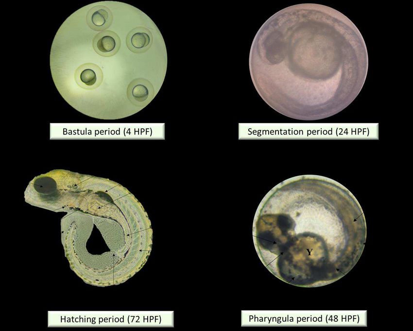
There were 4 periods depicted as according to Taufek et al., [16]: Blastula (4 HPF), b)
Segmentation (24 HPF), c) Pharyngula (48 HPF), and d) Hatching (72 HPF). A – eye anlage; An –
anus; Bc – blood cells; C – chorda; Ch – chorion; F – fin; G – gut; M – melanophores; O – ear
bud; P – pericardium; S – somites; Y – yolk sac. Scale bar =0.5 mm. The inverted microscope
procedure was used to produce the images.Figure 6. Illustrations of zebrafish embryo and larvae development after treated with Tiger Milk mushroom Lignosus rhinocerus strain at EPS concentration of 0.63 mg/mL. Descriptions were captured using inverted microscope at 100X (0 and 24 HPF) and 40X magnification (48-20 HPF). Figure 7. Illustrations of zebrafish embryo and larvae development after treated with Tiger Milk mushroom Lignosus rhinocerus strain ABI at EPS concentration of 1.25 mg/mL. Descriptions were captured using inverted microscope at 100X (0 and 24 HPF) and 40X magnification (48-20 HPF).
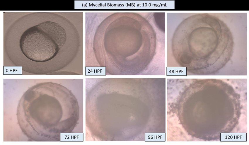
Figure 8. Illustrations of zebrafish embryo and larvae development after treated with Tiger Milk
mushroom Lignosus rhinocerus strain ABI at high EPS concentration of 10.0 mg/mL. Descriptions
 were captured using inverted microscope at 100X (0 and 24 HPF) and 40X magnification (48-20
                                               HPF).
Figure 9. Illustrations of zebrafish embryo and larvae development after treated with Tiger Milk Lignosus rhinocerus strain ABI at high EPS concentration of 10.0 mg/mL. Descriptions were captured using inverted microscope at 100X (0 and 24 HPF) and 40X magnification (48-20 HPF).
Non-toxic          concentrations
                                                        (mg/mL)
Fungal source     Toxicity model        Image                                               References
                                                        Mycelial
                                                                    Exopolysaccharide
                                                        Biomass
                                                                    (EPS)
                                                        (MB)

                  In vivo – Zebrafish
L. rhinocerus     embryos        and                    0.77        0.41                    Current study
                  larvae

                  In vitro - Cervical                                                       44
L. rhinocerotis                        NA               25          -
                  cancer cells (Ca
                  Ski, HPV-16)
                  In            vitro-
L. rhinocerotis   Differentiating
                  mouse                                 1.75–                               45
                                                                    -
                  neuroblastoma                         5.93
                  (N2a) cells

                  In    vitro-MTT                                                           39
L. rhinocerotis                   NA                    0.2         -
                  assay

                  In       vivo    -
L. rhinocerotis
                  Developmental                                                             46
                                     NA                 3.4         -
                  toxicity       in
                  pregnant Sprague-
                  Dawley (SD) rats

      Table 1. Similarity with literature for non-toxicity evaluation of mycelium biomass (MB) and
     exopolysaccharide (EPS) from the rare Tiger milk mushroom Lignosus sp. [NA: Not Available]
You can also read