Performance and Limitation of Machine Learning Algorithms for Diabetic Retinopathy Screening: Meta-analysis - Journal of Medical Internet Research
←
→
Page content transcription
If your browser does not render page correctly, please read the page content below
JOURNAL OF MEDICAL INTERNET RESEARCH                                                                                                               Wu et al

     Review

     Performance and Limitation of Machine Learning Algorithms for
     Diabetic Retinopathy Screening: Meta-analysis

     Jo-Hsuan Wu1, MD; T Y Alvin Liu2, MD; Wan-Ting Hsu3, MSc; Jennifer Hui-Chun Ho4, PhD, MD; Chien-Chang
     Lee5,6,7, SCD, MD
     1
      Shiley Eye Institute and Viterbi Family Department of Ophthalmology, University of California San Diego, La Jolla, CA, United States
     2
      Retina Division, Wilmer Eye Institute, The Johns Hopkins Medicine, Baltimore, MD, United States
     3
      Harvard TH Chan School of Public Health, Boston, MA, United States
     4
      National Yang-Ming University, Taipei City, Taiwan
     5
      Health Data Science Research Group, National Taiwan University Hospital, Taipei, Taiwan
     6
      The Centre for Intelligent Healthcare, National Taiwan University Hospital, Taipei, Taiwan
     7
      Department of Emergency Medicine, National Taiwan University Hospital, Taipei, Taiwan

     Corresponding Author:
     Chien-Chang Lee, SCD, MD
     Department of Emergency Medicine, National Taiwan University Hospital
     No 7, Chung-Shan South Road
     Taipei, 100
     Taiwan
     Phone: 886 2 23123456 ext 63485
     Fax: 886 2 23223150
     Email: hit3transparency@gmail.com

     Abstract
     Background: Diabetic retinopathy (DR), whose standard diagnosis is performed by human experts, has high prevalence and
     requires a more efficient screening method. Although machine learning (ML)–based automated DR diagnosis has gained attention
     due to recent approval of IDx-DR, performance of this tool has not been examined systematically, and the best ML technique for
     use in a real-world setting has not been discussed.
     Objective: The aim of this study was to systematically examine the overall diagnostic accuracy of ML in diagnosing DR of
     different categories based on color fundus photographs and to determine the state-of-the-art ML approach.
     Methods: Published studies in PubMed and EMBASE were searched from inception to June 2020. Studies were screened for
     relevant outcomes, publication types, and data sufficiency, and a total of 60 out of 2128 (2.82%) studies were retrieved after study
     selection. Extraction of data was performed by 2 authors according to PRISMA (Preferred Reporting Items for Systematic Reviews
     and Meta-Analyses), and the quality assessment was performed according to the Quality Assessment of Diagnostic Accuracy
     Studies 2 (QUADAS-2). Meta-analysis of diagnostic accuracy was pooled using a bivariate random effects model. The main
     outcomes included diagnostic accuracy, sensitivity, and specificity of ML in diagnosing DR based on color fundus photographs,
     as well as the performances of different major types of ML algorithms.
     Results: The primary meta-analysis included 60 color fundus photograph studies (445,175 interpretations). Overall, ML
     demonstrated high accuracy in diagnosing DR of various categories, with a pooled area under the receiver operating characteristic
     (AUROC) ranging from 0.97 (95% CI 0.96-0.99) to 0.99 (95% CI 0.98-1.00). The performance of ML in detecting more-than-mild
     DR was robust (sensitivity 0.95; AUROC 0.97), and by subgroup analyses, we observed that robust performance of ML was not
     limited to benchmark data sets (sensitivity 0.92; AUROC 0.96) but could be generalized to images collected in clinical practice
     (sensitivity 0.97; AUROC 0.97). Neural network was the most widely used method, and the subgroup analysis revealed a pooled
     AUROC of 0.98 (95% CI 0.96-0.99) for studies that used neural networks to diagnose more-than-mild DR.
     Conclusions: This meta-analysis demonstrated high diagnostic accuracy of ML algorithms in detecting DR on color fundus
     photographs, suggesting that state-of-the-art, ML-based DR screening algorithms are likely ready for clinical applications.
     However, a significant portion of the earlier published studies had methodology flaws, such as the lack of external validation and
     presence of spectrum bias. The results of these studies should be interpreted with caution.

     https://www.jmir.org/2021/7/e23863                                                                    J Med Internet Res 2021 | vol. 23 | iss. 7 | e23863 | p. 1
                                                                                                                         (page number not for citation purposes)
XSL• FO
RenderX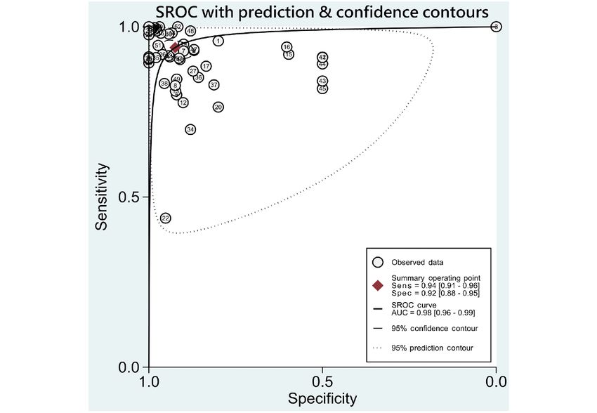
JOURNAL OF MEDICAL INTERNET RESEARCH                                                                                                       Wu et al

     (J Med Internet Res 2021;23(7):e23863) doi: 10.2196/23863

     KEYWORDS
     machine learning; diabetic retinopathy; diabetes; deep learning; neural network; diagnostic accuracy

                                                                          Although ML has garnered significant attention with the recent
     Introduction                                                         US Food and Drug Administration (FDA) approval of the first
     Diabetic retinopathy (DR) is the leading cause of vision             ML-based, fully automatic DR screening machine in April 2018
     impairment and blindness among working-aged people in the            [15], skepticism within the medical community remains
     world [1]. Approximately one-third of people with diabetes           regarding the robustness of ML techniques in real-world clinical
     mellitus have signs of DR, among whom one-third have                 applications. Given that ophthalmology is among the medical
     vision-threatening DR (VTDR). A meta-analysis estimated              disciplines that have reaped the most benefits from recent AI
     global prevalence of any DR and proliferative diabetic               advancements and that DR screening is one of the most
     retinopathy (PDR) among patients with diabetes to be 35.4%           promising ML applications in ophthalmology, we have set out
     and 7.5%, respectively [2]. The number of patients with DR is        to systematically survey, through meta-analysis, the current
     approximately 93 million and is expected to rise to 191 million      status of ML as applied in DR screening based on color fundus
     by 2030, as type 2 diabetes has attained the status of a global      photographs. Specifically, we have examined the range of
     pandemic, spreading from affluent industrialized nations to the      performances reported by different studies and have determined
     developing world [3].                                                which ML technique is superior for this clinical purpose.

     Vision impairment due to DR can be significantly reduced if          Methods
     diagnosed in early stages and treated appropriately [4]. However,
     fewer than 60% of patients with diabetes undergo regular eye         Search Methods for Identifying Studies
     examinations at intervals recommended by guidelines due to           This meta-analysis was performed in accordance with the
     the high cost and low accessibility of ophthalmologic services       PRISMA (Preferred Reporting Items for Systematic Reviews
     [3]. The number of people with diabetes that need regular eye        and Meta-Analyses) guidelines [16]. A literature search for
     examinations has quadrupled in the past three decades.               relevant studies published through June 2020 was performed
     Therefore, the development of an automatic, low-cost, accurate       with 2 publicly available databases, PubMed and EMBASE.
     eye screening tool has become an important public health issue       There were 3 stages to the literature search. No language or
     [5]. The gold standard for DR screening is based on clinical         population filters were applied, while nonhuman experiments,
     examinations by human clinicians or the analysis of color fundus     case reports, guidelines, conference papers, letters, editorials,
     photographs via telemedicine [6]. However, both approaches           and review articles were excluded. Filter for publication year
     are time-consuming, labor-intensive, and prone to inconsistency      was applied only in the second and third stages of the literature
     due to inherent human subjectivity [7]. Automated systems that       search in order to avoid overlapping of search results. Duplicated
     are capable of interpreting color fundus photographs with high       references in different stages of the literature search were
     sensitivity and specificity are critical for widespread              manually excluded. The major search key combination terms
     implementation of DR screening, and the rise of artificial           were “diabetic retinopathy” OR “diabetic macular edema” OR
     intelligence (AI), specifically machine learning (ML), has made      “macular edema” OR “retinopathy” OR “neovascularized
     such automated approaches a possibility.                             retinopathy” OR “proliferative retinopathy” OR “referable
     ML uses existing data to train a computer to recognize a specific    diabetic retinopathy” OR “diabetic macular oedema” OR
     pattern or predict a specific outcome in a new data set [6].         “proliferative diabetic retinopathy” OR “retinal disorders” OR
     Exploration of automated image analysis can be dated back to         “diabetic eye disease” OR “vision loss” OR “retinal diseases”
     1980, when classical ML methods, such as support vector              OR “macular disease” OR “macular degeneration” OR “macular
     machines and random forests, were used to detect predefined          disorders” crossed with “artificial intelligence” OR “deep
     features [8]. These early ML techniques for detecting DR             learning” OR “transfer learning” OR “machine learning” OR
     employed mathematical image transformation techniques and            “deep learning system”. The detailed search strategy is provided
     image engineering guided by expert-designed rules [9]. The           in Multimedia Appendix 1.
     accuracy of this type of analysis did not reach the standard of
                                                                          Eligibility Criteria for Considering Studies for This
     clinical application. In recent years, the advent of deep learning
                                                                          Review
     (DL), a subtype of ML, has transformed the field of automated
     image analysis [10]. Briefly, DL methods are representation          We included studies that evaluated ML algorithms on the
     learning methods that use multilayered neural networks, the          accuracy of automated image analysis for screening or diagnosis
     performance of which can be enhanced by reiteratively changing       of DR. We included studies that detected pathological findings
     the internal parameters [11,12]. Unlike other ML approaches,         of DR, diagnosed DR status, and staged DR severity.
     DL does not require image engineering. It develops its own           Study Selection
     representations needed for pattern recognition after being fed
     raw data and has shown superior accuracy as compared with            The study selection and data extraction were independently
     other classical ML algorithms [13,14].                               performed by 2 authors (JHW and CCL). After duplicates were
                                                                          removed, titles and abstracts were screened for exclusion of

     https://www.jmir.org/2021/7/e23863                                                            J Med Internet Res 2021 | vol. 23 | iss. 7 | e23863 | p. 2
                                                                                                                 (page number not for citation purposes)
XSL• FO
RenderX
JOURNAL OF MEDICAL INTERNET RESEARCH                                                                                                        Wu et al

     studies with potentially nonrelevant outcome or publication           logit-transformed sensitivity and specificity simultaneously to
     types and studies applying information other than images in           account for the inherent negative correlation between sensitivity
     analytical work. When there were multiples studies derived            and specificity that might have arisen due to different thresholds
     from the same cohort with overlapping study periods, earlier          in different studies. Heterogeneity was tested using the Cochran
     studies were considered duplicates and only the study with the        Q statistic (P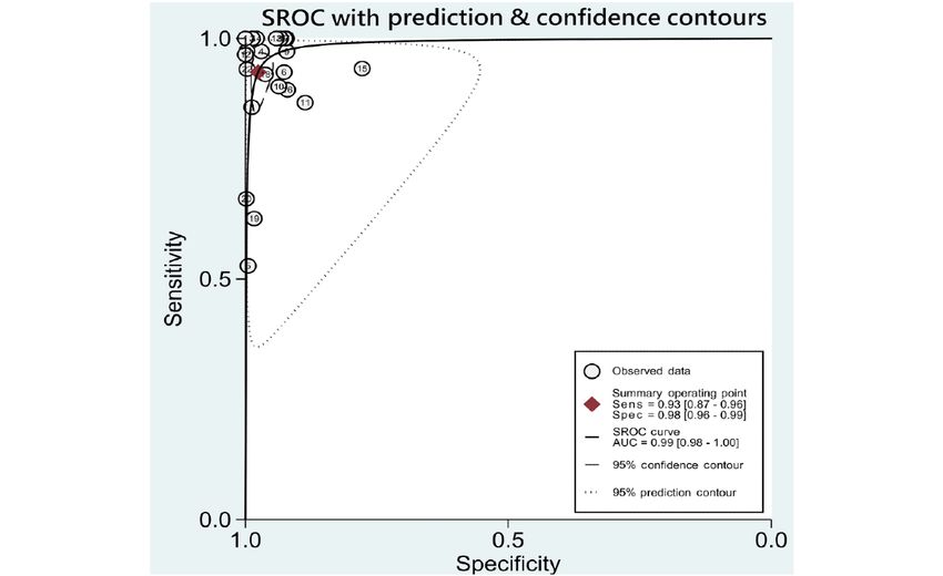
JOURNAL OF MEDICAL INTERNET RESEARCH                                                                                                        Wu et al

     Figure 1. Flowchart of study selection.

                                                                           criteria and processes, and were at a low risk for bias. Of the
     Study Characteristics                                                 60 studies, 3 (5%) reported limited information on the
     Multimedia Appendix 2 Table S1 summarizes the study-level             establishment of reference standard and were at a high risk for
     characteristics of studies assessing the diagnostic accuracy of       bias, and 4 (7%) reported insufficient blinding to a reference
     ML algorithms for different categories of DR. Of the 60 studies,      standard during interpretation of the index test results and were
     35 studies (58%) evaluated any DR, 23 (38%) mtmDR, 12                 at a high risk for bias. For study applicability, 1 study (2%) in
     (20%) VTDR, and 12 (20%) PDR. Publicly available benchmark            the index test section and 4 (7%) studies in the patient selection
     databases, such as Messidor, Structured Analysis of the Retina        section were recorded to be at a high risk of concern, due to
     (STARE), Digital Retinal Images for Vessel Extraction                 insufficient information reported.
     (DRIVE), DIARETDB, e-Ophtha, and EyePACs were used for
     testing of the ML algorithms in 40 of the 60 (67%) studies. The       Synthesis of Results
     characteristics of these publicly available retinal image databases   A summary of data of included studies is presented in
     are summarized in Multimedia Appendix 3. The distribution of          Multimedia Appendix 6. Pooled sensitivities, specificities,
     categories of ML algorithms used was as follows: SVM (6/60,           likelihood ratios, AUROCs, and I2 statistics for the 5 DR
     10%), RF (2/60, 3%), NN (37/60, 62%), and others (17/60,              categories, including any DR, mtmDR, VTDR, PDR, and
     28%). The general principles of these ML algorithms are               VTDR+PDR, are presented in Table 1. As some studies might
     described in Multimedia Appendix 4.                                   have used more than 2 data sets for validation, performance of
     Quality Assessment                                                    ML derived from each data set was viewed as individual data,
                                                                           and we used “data” as the unit for calculation (eg, 35 included
     Quality assessments using the QUADAS-2 criteria are                   studies performed evaluation of ML on identifying any DR,
     summarized in Multimedia Appendix 5. Most studies (56/60,             resulting in a total of 53 data for synthesis and analysis). The
     93%) presented a clear source of patient recruitment or selection

     https://www.jmir.org/2021/7/e23863                                                             J Med Internet Res 2021 | vol. 23 | iss. 7 | e23863 | p. 4
                                                                                                                  (page number not for citation purposes)
XSL• FO
RenderX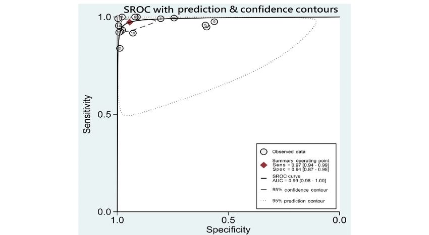
JOURNAL OF MEDICAL INTERNET RESEARCH Wu et al
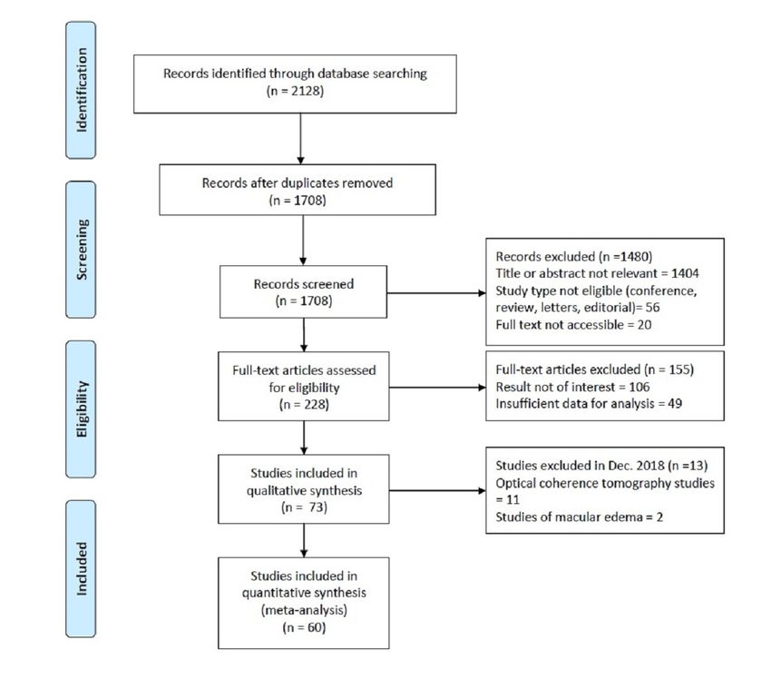
hierarchical summary ROC plots for the 4 main DR categories, (95% CI 0.87-0.93) for mtmDR to 0.98 (95% CI 0.96-0.99) for
including DR, mtmDR, VTDR, and PDR, are also presented PDR. The Fagan plots for different DR categories are presented
(Figures 2-5). ML showed a high overall accuracy in detecting in Multimedia Appendix 7. For images that were classified as
the 5 categories of DR, with a pooled AUROC ranging from positive by the ML algorithms, the posttest probability for DR,
0.97 (95% CI 0.96-0.99) for mtmDR and VTDR+PDR to 0.99 mtmDR, VTDR, and PDR was 87%, 71%, 66%, and 77%,
(95% CI 0.98-1.00) for VTDR and PDR. The pooled sensitivity respectively. For images that were classified as negative by the
for all 5 categories was high, ranging from 0.93 (95% CI ML algorithms, the posttest probability for DR, mtmDR, VTDR,
0.87-0.96) for PDR to 0.97 (95% CI 0.94-0.99) for VTDR. The and PDR was 4%, 1%, 0%, and 1%, respectively.
pooled specificity, however, showed more variation: from 0.90
Table 1. Pooled analysis for diagnostic accuracy of diabetic retinopathy by machine learning on color fundus photographs.
Goal of detec- Dataa, n Senb,c Sped,c LR+e,c LR–f,c AUROCg,c I2 statisticc Publication bias
tion (P value)
h 53 0.94 (0.91- 0.92 (0.88- 12.4 (8.0- 0.07 (0.05- 0.98 (0.96-0.99) 32 (22-42) .01
Any DR
0.96) 0.95) 19.3) 0.09)
mtmDRi 40 0.95 (0.93- 0.90 (0.87- 9.7 (7.4-12.7) 0.05 (0.04- 0.97 (0.96-0.99) 29 (18-40) .11
0.97) 0.93) 0.08)
VTDRj 15 0.97 (0.94- 0.94 (0.87- 17.3 (7.5- 0.03 (0.01- 0.99 (0.98-1.00) 32 (9-56) .33
0.99) 0.98) 39.9) 0.06)
PDRk 22 0.93 (0.87- 0.98 (0.96- 38.5 (21.7- 0.07 (0.04- 0.99 (0.98-1.00) 29 (11-46) .11
0.96) 0.99) 68.4) 0.13)
VTDR and 37 0.96 (0.93- 0.97 (0.94- 24.3 (14.5- 0.07 (0.05- 0.97 (0.96-0.99) N/A .06
PDR 0.98) 0.98) 38.5) 0.10)
a
Machine learning data derived from each data set was viewed as individual data, and we used “data” as the unit for calculation.
b
Sen: sensitivity.
c
Values in this column are as follows: mean (95% confidence interval).
d
Spe: specificity.
e
LR+: positive likelihood ratio.
f
LR–: negative likelihood ratio.
g
AUROC: area under the receiver operating characteristic.
h
DR: diabetic retinopathy.
i
mtmDR: more-than-mild diabetic retinopathy.
j
VTDR: vision-threatening diabetic retinopathy.
k
PDR: proliferative diabetic retinopathy.
https://www.jmir.org/2021/7/e23863 J Med Internet Res 2021 | vol. 23 | iss. 7 | e23863 | p. 5
(page number not for citation purposes)
XSL• FO
RenderXJOURNAL OF MEDICAL INTERNET RESEARCH Wu et al
Figure 2. SROC curves for diagnosis of any diabetic retinopathy on color fundus photographs. AUC: area under the curve; Sens: sensitivity; Spec:
specificity; SROC: summary receiver operating characteristics.
Figure 3. SROC curves for diagnosis of more-than-mild diabetic retinopathy on color fundus photographs. Sens: sensitivity; Spec: specificity; SROC:
summary receiver operating characteristics; AUC: area under the curve.
https://www.jmir.org/2021/7/e23863 J Med Internet Res 2021 | vol. 23 | iss. 7 | e23863 | p. 6
(page number not for citation purposes)
XSL• FO
RenderXJOURNAL OF MEDICAL INTERNET RESEARCH Wu et al
Figure 4. SROC curves for diagnosis of vision-threatening diabetic retinopathy on color fundus photographs. AUC: area under the curve; Sens:
sensitivity; Spec: specificity; SROC: summary receiver operating characteristics.
Figure 5. SROC curves for diagnosis of proliferative diabetic retinopathy on color fundus photographs. AUC: area under the curve; Sens: sensitivity;
Spec: specificity; SROC: summary receiver operating characteristics.
with a total of 40 data obtained from different testing data sets.
Subgroup Analyses and Sensitivity Analysis Of the 23 studies, the 22 studies that applied NN algorithms
We performed subgroup analyses for mtmDR studies to explore demonstrated high pooled performance (summary AUROC
the possible heterogeneity in test accuracy (Table 2). The main 0.98; 95% CI 0.96-0.99), sensitivity (sensitivity 0.95; 95% CI
causes of heterogeneity included in the analysis were algorithm 0.93-0.97), and specificity (specificity 0.91; 95% CI 0.88-0.93).
type, mean age of subject populations, and validation set The only study that used a different kind of ML algorithm
selection. For this subgroup analysis, 23 studies were included, (instance learning) reported significantly inferior sensitivity
https://www.jmir.org/2021/7/e23863 J Med Internet Res 2021 | vol. 23 | iss. 7 | e23863 | p. 7
(page number not for citation purposes)
XSL• FO
RenderXJOURNAL OF MEDICAL INTERNET RESEARCH Wu et al
(0.84) under preset specificity (0.50). Of the 60 studies, 19 sensitivities of algorithms validated by clinical data sets
(83%) tested the algorithm’s performance on data sets with (sensitivity 0.97; 95% CI 0.95-0.98) and independent data sets
subject populations with a mean age greater than 50 years. (sensitivity 0.96; 95% CI 0.93-0.97) were not inferior. The
Pooled sensitivity of data from these studies was high results of pooled AUROCs validated by these 3 types of data
(sensitivity 0.95; 95% CI 0.93-0.97), and the pooled specificity sets were similar, implying that robust performance of ML
was moderate (specificity 0.89; 95% CI 0.85-0.92). Compared algorithms can be generalized to images collected in clinical
with algorithms that used benchmark data sets for validation practice.
(pooled sensitivity 0.92; 95% CI 0.87-0.95), the pooled
Table 2. Subgroup analysis for diagnostic accuracy of mtmDR retinopathy on color fundus photographs.
Features of sub- Dataa, n Senb,c Sped,c LR+e,c LR–f,c AUROCg,c I2 statisticc Publication bias
group (P value)
Overall mtm- 40 0.95 (0.93, 0.90 (0.87, 9.7 (7.4, 12.7) 0.05 (0.04, 0.97 (0.96, 0.99) 29 (18, 40) .11
DRh 0.97) 0.93) 0.08)
Mean age> 50 32 0.95 (0.93, 0.89 (0.85, 8.8 (6.4, 12.0) 0.05 (0.03, 0.97 (0.95, 0.98) 33 (20, 46) .22
years 0.97) 0.92) 0.08)
NNi algorithms 38 0.95 (0.93, 0.91 (0.88, 10.1 (7.7, 0.05 (0.03, 0.98 (0.96, 0.99) 30 (19, 41) .14
0.97) 0.93) 13.2) 0.07)
Benchmark test 15 0.92 (0.87, 0.90 (0.82, 9.0 (4.8, 16.6) 0.09 (0.05, 0.96 (0.94, 0.98) 25 (10, 39) .22
sets 0.95) 0.94) 0.16)
Clinical 25 0.97 (0.95, 0.90 (0.88, 10.0 (7.9, 0.04 (0.02, 0.97 (0.96, 0.98) 30 (15, 45) .06
Test sets 0.98) 0.92) 12.6) 0.06)
External valida- 31 0.96 (0.93, 0.90 (0.87, 9.7 (7.2, 13.0) 0.05 (0.03, 0.98 (0.96, 0.99) 29 (16, 42) .08
tion 0.97) 0.93) 0.07)
a
Machine learning data derived from each data set was viewed as individual data, and we used “data” as the unit for calculation.
b
Sen: sensitivity.
c
Values in this column are as follows: mean (95% confidence interval).
d
Spe: specificity.
e
LR+: positive likelihood ratio.
f
LR–: negative likelihood ratio.
g
AUROC: area under the receiver operating characteristic.
h
mtmDR: more-than-mild diabetic retinopathy.
i
NN: neural network.
[75], 34% [76], and 33% [77] achieved by board-certified
Publication Bias ophthalmologists performing indirect ophthalmoscopy and to
The test for publication bias was generally not significant in reported sensitivities of 92% [78] and 89% [79] achieved by
different categories of DR (Deeks test P=.01; Multimedia ophthalmologists interpreting digital fundus photographs. Our
Appendix 8), except for any DR. Trim-and-fill analysis showed analysis suggests that the performance of ML algorithms in
the diagnostic OR remained insignificant (OR 0.50, 95% CI detecting DR based on color fundus photographs is likely to be
0.25-1.01) after hypothetical unpublished data were included on par with human clinicians and supports a previous study that
for analysis (Multimedia Appendix 9). compared humans head to head with ML. Rajalakshmi et al
[37] compared the performance of an AI DR screening software
Discussion (EyeArt) on smartphone-based fundus photographs of 296
patients to the performance of human graders who evaluated
Principal Results and Comparison With Prior Work the same data set. The EyeArt achieved a high sensitivity of
This systematic review synthesizes the available evidence and 95.8% for any retinopathy and 99.1% for VTDR, both of which
compares the diagnostic accuracy of ML algorithms for the were on par with human graders. Our pooled data suggest that
detection of DR based on color fundus photographs. The primary ML techniques are more sensitive than specific in DR detection.
meta-analysis included 60 studies with 445,175 interpretations. It is unclear whether this is a reflection of the limitations of ML
Out of the 60 studies, 35 (58%) were validated by external techniques for this clinical purpose or whether it is by design.
testing data sets that were completely independent of the training It is possible that model developers of these studies chose
data sets. Overall, ML demonstrated a robust performance in optimal statistical thresholds that favored sensitivity over
detecting different DR categories, with a pooled sensitivity of specificity. Regardless, the lower specificity should not pose a
0.93 to 0.97 and a pooled specificity of 0.90 to 0.98. The pooled major issue, as false negatives are much more problematic than
sensitivity compares favorably to reported sensitivities of 73% are false positives in the context of disease screening.
https://www.jmir.org/2021/7/e23863 J Med Internet Res 2021 | vol. 23 | iss. 7 | e23863 | p. 8
(page number not for citation purposes)
XSL• FO
RenderXJOURNAL OF MEDICAL INTERNET RESEARCH Wu et al
Furthermore, the major causes of false positives in retinal image Use and Predominance of NN Algorithms
interpretation, including inadequate image quality and artifacts NN algorithms, especially deep convolutional NN algorithms,
[55], are modifiable with future improvement in image quality were generally recognized as the best ML technique for
control. automated medical image analysis. NN algorithms were also
To further facilitate direct clinical interpretations, we used Fagan the most-used technique in diagnosing DR of all categories in
nomograms to determine whether a patient with a positive or our study, being used in 37 of the 60 (62%) studies. As for the
negative finding by ML actually has that particular finding as 23 studies evaluating mtmDR (Table 2), NN algorithms were
per the gold standard. For any DR, in a population with a DR used in 22 studies and contributed to the high pooled AUROC
prevalence of 36%, a positive likelihood ratio of 12.4 translates of 0.98 (95% CI 0.96-0.99). In addition, we ranked the
into a posttest probability of 87%. In other words, approximately performance of the included studies by sensitivity, specificity,
9 out of 10 patients with a positive ML diagnosis of DR can be and quality. The top-5 performing, high-quality (based on
expected to have DR as per the gold standard. The diagnostic QUADUS-2 and study design) studies are listed in Multimedia
value for ML to rule out DR performed as well as its rule-in Appendix 10, and 4 out of the 5 studies used NN algorithms.
value. In the same population, a negative ML diagnosis This result confirms that NN is the cutting-edge ML technique
translates into a 4% posttest probability of any DR (negative for medical image classification, at least in the context of DR
likelihood ratio 0.07) and only a 1% posttest probability of detection.
mtmDR (negative likelihood ratio 0.05). These numbers again
Limitations
suggest that ML is extremely sensitive in detecting overall DR
and mtmDR based on color fundus photographs and that the Our study was based on a rigorous literature search, and a
rate of false negatives are low. validated appraisal tool was used to determine the risk of bias
of included studies. Several limitations should be considered,
We performed an in-depth analysis of studies that involved the however. First, of the 60 studies included for final analysis,
detection of mtmDR, as Abramoff et al’s [15] pivotal trial only 35 applied true external validation. For those studies
involved the detection of mtmDR and led to the FDA’s approval without external validation, the generalizability of their ML
of the first fully automated ML system. Among the 16 mtmDR algorithms was not adequately evaluated, and thus their reported
studies conducted by other research teams that were also performance should be interpreted with caution. Second, without
externally validated, 14 showed performance superior to the sufficient details, we were unable to conduct subgroup analysis
preset end points (sensitivity >85%; specificity > 82.5%) used on populations with available key factors of DR that could
in Abramoff et al’s trial. Although only 5 out of these studies influence the clinical practicability of the diagnostic tool. Bias
were prospectively evaluated in a real-world setting as the could have been introduced by poor reporting of patient
Abramoff algorithm was, this suggests that Abramoff et al’s characteristics of the included studies. Finally, except for
trial was no accident and that ML algorithms in general are Abramoff et al’s trial [15] and 5 other prospectively conducted
likely capable of producing clinical grade detections of mtmDR studies [48,52,55,60,66], all other studies on ML-based DR
based on color fundus photographs. In addition, no statistically diagnosis were validated by retrospective data. Due to spectrum
significant difference in pooled AUROC between studies bias, an overestimation of ML’s performance in a real-world
validated by benchmark databases and studies validated by setting is possible and should be considered.
clinical databases was identified within this group.
Conclusions
To the best of our knowledge, previous meta-reviews on DR
ML algorithms for diagnosing DR based on color fundus
screening have focused on the performance of DL algorithms
photographs have shown high diagnostic accuracy for different
alone [80,81]. DL is only a subtype of ML, and other ML
categories of DR. Specifically, the performances of ML
techniques, such as SVM and RF, can be used to detect DR as
algorithms in detecting mtmDR, the widely accepted threshold
well. Therefore, our meta-analysis was more comprehensive
for clinically relevant DR, compare favorably to those of clinical
than these previous studies, as it included all ML studies,
examinations by ophthalmologists and to those of expert grading
including DL studies, published through 2020. In addition, the
of digital fundus photographs. To the best of our knowledge,
review by Nielsen et al [80] did not conduct pooled analysis on
this is the first meta-analysis in the published literature that
the results of past studies, while our study did. The meta-analysis
quantitatively assessed the performance of ML algorithms for
by Islam et al [81] focused mainly on detection of referable DR,
a specific medical image classification task. As evidence-based
while our study was broader and more fine grained, as it
medicine expands from therapy to diagnosis, the information
evaluated the ability of ML to detect different categories of DR,
from this systematic review may provide important evidence
including any DR, mtmDR (referrable DR), VTDR, and PDR.
in the determination of the proper and efficacious use of ML
These analyses are clinically meaningful, as different categories
algorithms in the diagnosis or screening of DR and may serve
of DR require different management strategies. For example,
as a framework for similar analyses of other medical conditions
while patients with moderate nonproliferative DR (a subset
conducted in the future. However, our meta-analysis also
within referrable DR) should be further evaluated by
showed that a significant portion of the published studies had
ophthalmologists at some point, patients with VTDR require
methodological flaws, such as the lack of external validation
immediate referrals to retinal specialists.
and presence of spectrum bias. Therefore, more rigorous
prospective studies would be helpful in establishing the true
efficacy of these algorithms in real-life clinical care.
https://www.jmir.org/2021/7/e23863 J Med Internet Res 2021 | vol. 23 | iss. 7 | e23863 | p. 9
(page number not for citation purposes)
XSL• FO
RenderXJOURNAL OF MEDICAL INTERNET RESEARCH Wu et al
Acknowledgments
This study was funded by the Taiwan National Ministry of Science and Technology (grants MOST 104-2314-B-002 -039 -MY3
and MOST 106-2811-B-002-048). The sponsor or funding organization had no role in the design or conduct of this research.
Conflicts of Interest
None declared.
Multimedia Appendix 1
Search detail for PubMed and EMBASE.
[DOCX File , 33 KB-Multimedia Appendix 1]
Multimedia Appendix 2
Summary of characteristics of the included studies.
[DOCX File , 74 KB-Multimedia Appendix 2]
Multimedia Appendix 3
Characteristics of public databases for benchmarking diabetic retinopathy detection from color fundus photographs.
[DOCX File , 40 KB-Multimedia Appendix 3]
Multimedia Appendix 4
Principles of major machine learning algorithms and methods applied in automated image analysis.
[DOCX File , 39 KB-Multimedia Appendix 4]
Multimedia Appendix 5
Summary of QUADAS-2 assessment for included studies.
[PDF File (Adobe PDF File), 145 KB-Multimedia Appendix 5]
Multimedia Appendix 6
Summary of data of included studies.
[DOCX File , 81 KB-Multimedia Appendix 6]
Multimedia Appendix 7
Fagan plot for diagnosis of different categories of diabetic retinopathy on color fundus photographs.
[PDF File (Adobe PDF File), 728 KB-Multimedia Appendix 7]
Multimedia Appendix 8
Deeks funnel plot for 4 main types of diabetic retinopathy lesions on color fundus photograph.
[PDF File (Adobe PDF File), 374 KB-Multimedia Appendix 8]
Multimedia Appendix 9
A filled funnel plot that filled the potential missing studies (square) due to publication bias.
[DOCX File , 264 KB-Multimedia Appendix 9]
Multimedia Appendix 10
Characteristics and best results of 5 high-quality studies with top performances.
[DOCX File , 46 KB-Multimedia Appendix 10]
References
1. Fong DS, Aiello L, Gardner TW, King GL, Blankenship G, Cavallerano JD, American Diabetes Association. Diabetic
retinopathy. Diabetes Care 2003 Jan;26(1):226-229. [doi: 10.2337/diacare.26.1.226] [Medline: 12502685]
https://www.jmir.org/2021/7/e23863 J Med Internet Res 2021 | vol. 23 | iss. 7 | e23863 | p. 10
(page number not for citation purposes)
XSL• FO
RenderXJOURNAL OF MEDICAL INTERNET RESEARCH Wu et al
2. Yau JWY, Rogers SL, Kawasaki R, Lamoureux EL, Kowalski JW, Bek T, et al. Global prevalence and major risk factors
of diabetic retinopathy. Diabetes Care 2012 Mar;35(3):556-564 [FREE Full text] [doi: 10.2337/dc11-1909] [Medline:
22301125]
3. Zheng Y, He M, Congdon N. The worldwide epidemic of diabetic retinopathy. Indian J Ophthalmol 2012;60(5):428-431.
[doi: 10.4103/0301-4738.100542] [Medline: 22944754]
4. Vashist P, Singh S, Gupta N, Saxena R. Role of early screening for diabetic retinopathy in patients with diabetes mellitus:
an overview. Indian J Community Med 2011 Oct;36(4):247-252 [FREE Full text] [doi: 10.4103/0970-0218.91324] [Medline:
22279252]
5. Zheng Y, Ley SH, Hu FB. Global aetiology and epidemiology of type 2 diabetes mellitus and its complications. Nat Rev
Endocrinol 2018 Feb;14(2):88-98. [doi: 10.1038/nrendo.2017.151] [Medline: 29219149]
6. Viswanath K, McGavin DDM. Diabetic retinopathy: clinical findings and management. Community Eye Health
2003;16(46):21-24 [FREE Full text] [Medline: 17491851]
7. Heitmar R, Kalitzeos AA, Patel SR, Prabhu-Das D, Cubbidge RP. Comparison of subjective and objective methods to
determine the retinal arterio-venous ratio using fundus photography. J Optom 2015;8(4):252-257 [FREE Full text] [doi:
10.1016/j.optom.2014.07.002] [Medline: 26386537]
8. Hosny A, Parmar C, Quackenbush J, Schwartz LH, Aerts HJWL. Artificial intelligence in radiology. Nat Rev Cancer 2018
Dec;18(8):500-510 [FREE Full text] [doi: 10.1038/s41568-018-0016-5] [Medline: 29777175]
9. Wernick MN, Yang Y, Brankov JG, Yourganov G, Strother SC. Machine learning in medical imaging. IEEE Signal Process
Mag 2010 Jul;27(4):25-38 [FREE Full text] [doi: 10.1109/MSP.2010.936730] [Medline: 25382956]
10. Wong TY, Bressler NM. Artificial intelligence with deep learning technology looks into diabetic retinopathy screening.
JAMA 2016 Dec 13;316(22):2366-2367 [FREE Full text] [doi: 10.1001/jama.2016.17563] [Medline: 27898977]
11. LeCun Y, Bengio Y, Hinton G. Deep learning. Nature 2015 May 28;521(7553):436-444. [doi: 10.1038/nature14539]
[Medline: 26017442]
12. Jiang F, Jiang Y, Zhi H, Dong Y, Li H, Ma S, et al. Artificial intelligence in healthcare: past, present and future. Stroke
Vasc Neurol 2017 Dec;2(4):230-243 [FREE Full text] [doi: 10.1136/svn-2017-000101] [Medline: 29507784]
13. Gulshan V, Peng L, Coram M, Stumpe MC, Wu D, Narayanaswamy A, et al. Development and validation of a deep learning
algorithm for detection of diabetic retinopathy in retinal fundus photographs. JAMA 2016 Dec 13;316(22):2402-2410. [doi:
10.1001/jama.2016.17216] [Medline: 27898976]
14. Esteva A, Robicquet A, Ramsundar B, Kuleshov V, DePristo M, Chou K, et al. A guide to deep learning in healthcare. Nat
Med 2019 Jan;25(1):24-29. [doi: 10.1038/s41591-018-0316-z] [Medline: 30617335]
15. Abràmoff MD, Lavin PT, Birch M, Shah N, Folk JC. Pivotal trial of an autonomous AI-based diagnostic system for detection
of diabetic retinopathy in primary care offices. NPJ Digit Med 2018;1:39 [FREE Full text] [doi: 10.1038/s41746-018-0040-6]
[Medline: 31304320]
16. Moher D, Shamseer L, Clarke M, Ghersi D, Liberati A, Petticrew M, et al. Preferred reporting items for systematic review
and meta-analysis protocols (PRISMA-P) 2015 statement. Syst Rev 2015 Jan;4:1 [FREE Full text] [doi:
10.1186/2046-4053-4-1] [Medline: 25554246]
17. Abbas Q, Fondon I, Sarmiento A, Jiménez S, Alemany P. Automatic recognition of severity level for diagnosis of diabetic
retinopathy using deep visual features. Med Biol Eng Comput 2017 Nov;55(11):1959-1974. [doi: 10.1007/s11517-017-1638-6]
[Medline: 28353133]
18. Abràmoff MD, Lou Y, Erginay A, Clarida W, Amelon R, Folk JC, et al. Improved automated detection of diabetic retinopathy
on a publicly available dataset through integration of deep learning. Invest Ophthalmol Vis Sci 2016 Oct 01;57(13):5200-5206.
[doi: 10.1167/iovs.16-19964] [Medline: 27701631]
19. Adal KM, Sidibé D, Ali S, Chaum E, Karnowski TP, Mériaudeau F. Automated detection of microaneurysms using
scale-adapted blob analysis and semi-supervised learning. Comput Methods Programs Biomed 2014 Apr;114(1):1-10. [doi:
10.1016/j.cmpb.2013.12.009] [Medline: 24529636]
20. Agurto C, Barriga ES, Murray V, Nemeth S, Crammer R, Bauman W, et al. Automatic detection of diabetic retinopathy
and age-related macular degeneration in digital fundus images. Invest Ophthalmol Vis Sci 2011 Jul 29;52(8):5862-5871.
[doi: 10.1167/iovs.10-7075] [Medline: 21666234]
21. Annie GVG, Kaja MS. Diabetic retinopathy screening system: A validation analysis with multiple fundus image databases.
Biomedical Research (India) 2017;28(4):A.
22. Chaum E, Karnowski TP, Govindasamy VP, Abdelrahman M, Tobin KW. Automated diagnosis of retinopathy by
content-based image retrieval. Retina 2008;28(10):1463-1477. [doi: 10.1097/IAE.0b013e31818356dd] [Medline: 18997609]
23. Ganesan K, Martis RJ, Acharya UR, Chua CK, Min LC, Ng EYK, et al. Computer-aided diabetic retinopathy detection
using trace transforms on digital fundus images. Med Biol Eng Comput 2014 Aug;52(8):663-672. [doi:
10.1007/s11517-014-1167-5] [Medline: 24958614]
24. Gardner GG, Keating D, Williamson TH, Elliott AT. Automatic detection of diabetic retinopathy using an artificial neural
network: a screening tool. Br J Ophthalmol 1996 Nov;80(11):940-944 [FREE Full text] [doi: 10.1136/bjo.80.11.940]
[Medline: 8976718]
https://www.jmir.org/2021/7/e23863 J Med Internet Res 2021 | vol. 23 | iss. 7 | e23863 | p. 11
(page number not for citation purposes)
XSL• FO
RenderXJOURNAL OF MEDICAL INTERNET RESEARCH Wu et al
25. Gargeya R, Leng T. Automated identification of diabetic retinopathy using deep learning. Ophthalmology 2017
Dec;124(7):962-969. [doi: 10.1016/j.ophtha.2017.02.008] [Medline: 28359545]
26. Gupta G, Kulasekaran S, Ram K, Joshi N, Sivaprakasam M, Gandhi R. Local characterization of neovascularization and
identification of proliferative diabetic retinopathy in retinal fundus images. Comput Med Imaging Graph 2017 Jan;55:124-132.
[doi: 10.1016/j.compmedimag.2016.08.005] [Medline: 27634547]
27. Jelinek HF, Cree MJ, Leandro JJG, Soares JVB, Cesar RM, Luckie A. Automated segmentation of retinal blood vessels
and identification of proliferative diabetic retinopathy. J Opt Soc Am A Opt Image Sci Vis 2007 May;24(5):1448-1456.
[doi: 10.1364/josaa.24.001448] [Medline: 17429492]
28. Malathi K, Nedunchelian R. A recursive support vector machine (RSVM) algorithm to detect and classify diabetic retinopathy
in fundus retina images. biomedicalresearch 2018:57-64. [doi: 10.4066/biomedicalresearch.29-16-2328]
29. Fadafen MK, Mehrshad N, Razavi SM. Detection of diabetic retinopathy using computational model of human visual
system. biomedicalresearch 2018;29(9):1956-1960. [doi: 10.4066/biomedicalresearch.29-18-551]
30. Li Z, Keel S, Liu C, He Y, Meng W, Scheetz J, et al. An automated grading system for detection of vision-threatening
referable diabetic retinopathy on the basis of color fundus photographs. Diabetes Care 2018 Dec;41(12):2509-2516. [doi:
10.2337/dc18-0147] [Medline: 30275284]
31. Bala MP, Vijayachitra S. Extraction of retinal blood vessels and diagnosis of proliferative diabetic retinopathy using extreme
learning machine. J Med Imaging Hlth Inform 2015 Apr 01;5(2):248-256. [doi: 10.1166/jmihi.2015.1380]
32. Orlando JI, Prokofyeva E, Del Fresno M, Blaschko MB. An ensemble deep learning based approach for red lesion detection
in fundus images. Comput Methods Programs Biomed 2018 Jan;153:115-127. [doi: 10.1016/j.cmpb.2017.10.017] [Medline:
29157445]
33. Orlando JI, van Keer K, Barbosa Breda J, Manterola HL, Blaschko MB, Clausse A. Proliferative diabetic retinopathy
characterization based on fractal features: Evaluation on a publicly available dataset. Med Phys 2017 Dec;44(12):6425-6434.
[doi: 10.1002/mp.12627] [Medline: 29044550]
34. Pires R, Carvalho T, Spurling G, Goldenstein S, Wainer J, Luckie A, et al. Automated multi-lesion detection for referable
diabetic retinopathy in indigenous health care. PLoS One 2015;10(6):e0127664 [FREE Full text] [doi:
10.1371/journal.pone.0127664] [Medline: 26035836]
35. Quellec G, Charrière K, Boudi Y, Cochener B, Lamard M. Deep image mining for diabetic retinopathy screening. Med
Image Anal 2017 Jul;39:178-193 [FREE Full text] [doi: 10.1016/j.media.2017.04.012] [Medline: 28511066]
36. Quellec G, Lamard M, Abràmoff MD, Decencière E, Lay B, Erginay A, et al. A multiple-instance learning framework for
diabetic retinopathy screening. Med Image Anal 2012 Aug;16(6):1228-1240. [doi: 10.1016/j.media.2012.06.003] [Medline:
22850462]
37. Rajalakshmi R, Subashini R, Anjana RM, Mohan V. Automated diabetic retinopathy detection in smartphone-based fundus
photography using artificial intelligence. Eye (Lond) 2018 Jun;32(6):1138-1144 [FREE Full text] [doi:
10.1038/s41433-018-0064-9] [Medline: 29520050]
38. Raju M, Pagidimarri V, Barreto R, Kadam A, Kasivajjala V, Aswath A. Development of a deep learning algorithm for
automatic diagnosis of diabetic retinopathy. Stud Health Technol Inform 2017;245:559-563. [Medline: 29295157]
39. Ramachandran N, Hong SC, Sime MJ, Wilson GA. Diabetic retinopathy screening using deep neural network. Clin Exp
Ophthalmol 2018 May;46(4):412-416. [doi: 10.1111/ceo.13056] [Medline: 28881490]
40. Sangeethaa SN, Uma Maheswari P. An intelligent model for blood vessel segmentation in diagnosing DR using CNN. J
Med Syst 2018 Aug 15;42(10):175. [doi: 10.1007/s10916-018-1030-6] [Medline: 30109508]
41. B. Sumathy, S. Poornachandra. Automated DR and prediction of various related diseases of retinal fundus images.
biomedicalresearch 2018:325-332. [doi: 10.4066/biomedicalresearch.29-17-480]
42. Ting DSW, Cheung CY, Lim G, Tan GSW, Quang ND, Gan A, et al. Development and Validation of a Deep Learning
System for Diabetic Retinopathy and Related Eye Diseases Using Retinal Images From Multiethnic Populations With
Diabetes. JAMA 2017 Dec 12;318(22):2211-2223 [FREE Full text] [doi: 10.1001/jama.2017.18152] [Medline: 29234807]
43. Usman Akram M, Khalid S, Tariq A, Younus Javed M. Detection of neovascularization in retinal images using multivariate
m-Mediods based classifier. Comput Med Imaging Graph 2013;37(5-6):346-357 [FREE Full text] [doi:
10.1016/j.compmedimag.2013.06.008] [Medline: 23916066]
44. Welikala RA, Fraz MM, Dehmeshki J, Hoppe A, Tah V, Mann S, et al. Genetic algorithm based feature selection combined
with dual classification for the automated detection of proliferative diabetic retinopathy. Comput Med Imaging Graph 2015
Jul;43:64-77. [doi: 10.1016/j.compmedimag.2015.03.003] [Medline: 25841182]
45. Yu S, Xiao D, Kanagasingam Y. Machine learning based automatic neovascularization detection on optic disc region. IEEE
J. Biomed. Health Inform 2018 May;22(3):886-894. [doi: 10.1109/jbhi.2017.2710201]
46. Zhang Y, An M. An active learning classifier for further reducing diabetic retinopathy screening system cost. Comput Math
Methods Med 2016;2016:4345936 [FREE Full text] [doi: 10.1155/2016/4345936] [Medline: 27660645]
47. Badgujar R, Deore P. Hybrid nature inspired SMO-GBM classifier for exudate classification on fundus retinal images.
IRBM 2019 Mar;40(2):69-77 [FREE Full text] [doi: 10.1016/j.irbm.2019.02.003] [Medline: 32595044]
https://www.jmir.org/2021/7/e23863 J Med Internet Res 2021 | vol. 23 | iss. 7 | e23863 | p. 12
(page number not for citation purposes)
XSL• FO
RenderXJOURNAL OF MEDICAL INTERNET RESEARCH Wu et al
48. Bellemo V, Lim ZW, Lim G, Nguyen QD, Xie Y, Yip MYT, et al. Artificial intelligence using deep learning to screen for
referable and vision-threatening diabetic retinopathy in Africa: a clinical validation study. The Lancet Digital Health 2019
May;1(1):e35-e44. [doi: 10.1016/s2589-7500(19)30004-4]
49. Bhaskaranand M, Ramachandra C, Bhat S, Cuadros J, Nittala MG, Sadda SR, et al. The value of automated diabetic
retinopathy screening with the EyeArt system: a study of more than 100,000 consecutive encounters from people with
diabetes. Diabetes Technol Ther 2019 Nov;21(11):635-643 [FREE Full text] [doi: 10.1089/dia.2019.0164] [Medline:
31335200]
50. Chowdhury AR, Chatterjee T, Banerjee S. A Random Forest classifier-based approach in the detection of abnormalities in
the retina. Med Biol Eng Comput 2019 Jan;57(1):193-203. [doi: 10.1007/s11517-018-1878-0] [Medline: 30076537]
51. Colomer A, Igual J, Naranjo V. Detection of early signs of diabetic retinopathy based on textural and morphological
information in fundus images. Sensors (Basel) 2020 Feb 13;20(4):1005 [FREE Full text] [doi: 10.3390/s20041005] [Medline:
32069912]
52. Gulshan V, Rajan RP, Widner K, Wu D, Wubbels P, Rhodes T, et al. Performance of a deep-learning algorithm vs manual
grading for detecting diabetic retinopathy in India. JAMA Ophthalmol 2019 Jun 13:987-993 [FREE Full text] [doi:
10.1001/jamaophthalmol.2019.2004] [Medline: 31194246]
53. He J, Cao T, Xu F, Wang S, Tao H, Wu T, et al. Artificial intelligence-based screening for diabetic retinopathy at community
hospital. Eye (Lond) 2020 Mar;34(3):572-576 [FREE Full text] [doi: 10.1038/s41433-019-0562-4] [Medline: 31455902]
54. Hemanth DJ, Anitha J, Son LH, Mittal M. Diabetic retinopathy diagnosis from retinal images using modified Hopfield
neural network. J Med Syst 2018 Oct 31;42(12):247. [doi: 10.1007/s10916-018-1111-6] [Medline: 30382410]
55. Kanagasingam Y, Xiao D, Vignarajan J, Preetham A, Tay-Kearney M, Mehrotra A. Evaluation of artificial intelligence-based
grading of diabetic retinopathy in primary care. JAMA Netw Open 2018 Sep 07;1(5):e182665 [FREE Full text] [doi:
10.1001/jamanetworkopen.2018.2665] [Medline: 30646178]
56. Khojasteh P, Passos Júnior LA, Carvalho T, Rezende E, Aliahmad B, Papa JP, et al. Exudate detection in fundus images
using deeply-learnable features. Comput Biol Med 2019 Jan;104:62-69 [FREE Full text] [doi:
10.1016/j.compbiomed.2018.10.031] [Medline: 30439600]
57. Li F, Liu Z, Chen H, Jiang M, Zhang X, Wu Z. Automatic detection of diabetic retinopathy in retinal fundus photographs
based on deep learning algorithm. Transl Vis Sci Technol 2019 Nov;8(6):4 [FREE Full text] [doi: 10.1167/tvst.8.6.4]
[Medline: 31737428]
58. Long S, Huang X, Chen Z, Pardhan S, Zheng D. automatic detection of hard exudates in color retinal images using dynamic
threshold and SVM classification: algorithm development and evaluation. Biomed Res Int 2019;2019:3926930 [FREE Full
text] [doi: 10.1155/2019/3926930] [Medline: 30809539]
59. Natarajan S, Jain A, Krishnan R, Rogye A, Sivaprasad S. Diagnostic accuracy of community-based diabetic retinopathy
screening with an offline artificial intelligence system on a smartphone. JAMA Ophthalmol 2019 Aug 08:1182-1188 [FREE
Full text] [doi: 10.1001/jamaophthalmol.2019.2923] [Medline: 31393538]
60. Nazir T, Irtaza A, Shabbir Z, Javed A, Akram U, Mahmood MT. Diabetic retinopathy detection through novel tetragonal
local octa patterns and extreme learning machines. Artif Intell Med 2019 Aug;99:101695 [FREE Full text] [doi:
10.1016/j.artmed.2019.07.003] [Medline: 31606114]
61. Pires R, Avila S, Wainer J, Valle E, Abramoff MD, Rocha A. A data-driven approach to referable diabetic retinopathy
detection. Artif Intell Med 2019 May;96:93-106 [FREE Full text] [doi: 10.1016/j.artmed.2019.03.009] [Medline: 31164214]
62. Raumviboonsuk P, Krause J, Chotcomwongse P, Sayres R, Raman R, Widner K, et al. Deep learning versus human graders
for classifying diabetic retinopathy severity in a nationwide screening program. NPJ Digit Med 2019;2:25 [FREE Full text]
[doi: 10.1038/s41746-019-0099-8] [Medline: 31304372]
63. Riaz H, Park J, Choi H, Kim H, Kim J. Deep and densely connected networks for classification of diabetic retinopathy.
Diagnostics 2020 Jan 02;10(1):24. [doi: 10.3390/diagnostics10010024]
64. Shah P, Mishra DK, Shanmugam MP, Doshi B, Jayaraj H, Ramanjulu R. Validation of Deep Convolutional Neural
Network-based algorithm for detection of diabetic retinopathy - Artificial intelligence versus clinician for screening. Indian
J Ophthalmol 2020 Feb;68(2):398-405 [FREE Full text] [doi: 10.4103/ijo.IJO_966_19] [Medline: 31957737]
65. Son J, Shin JY, Kim HD, Jung K, Park KH, Park SJ. Development and validation of deep learning models for screening
multiple abnormal findings in retinal fundus images. Ophthalmology 2020 Jan;127(1):85-94 [FREE Full text] [doi:
10.1016/j.ophtha.2019.05.029] [Medline: 31281057]
66. Sosale B, Aravind SR, Murthy H, Narayana S, Sharma U, Gowda SGV, et al. Simple, Mobile-based Artificial Intelligence
Algorithm in the detection of Diabetic Retinopathy (SMART) study. BMJ Open Diabetes Res Care 2020 Jan;8(1):e000892
[FREE Full text] [doi: 10.1136/bmjdrc-2019-000892] [Medline: 32049632]
67. Stevenson CH, Hong SC, Ogbuehi KC. Development of an artificial intelligence system to classify pathology and clinical
features on retinal fundus images. Clin Exp Ophthalmol 2019 May;47(4):484-489. [doi: 10.1111/ceo.13433] [Medline:
30370587]
68. Ullah H, Saba T, Islam N, Abbas N, Rehman A, Mehmood Z, et al. An ensemble classification of exudates in color fundus
images using an evolutionary algorithm based optimal features selection. Microsc Res Tech 2019 Jan 24;82(4):361-372.
[doi: 10.1002/jemt.23178]
https://www.jmir.org/2021/7/e23863 J Med Internet Res 2021 | vol. 23 | iss. 7 | e23863 | p. 13
(page number not for citation purposes)
XSL• FO
RenderXJOURNAL OF MEDICAL INTERNET RESEARCH Wu et al
69. Verbraak FD, Abramoff MD, Bausch GCF, Klaver C, Nijpels G, Schlingemann RO, et al. Diagnostic accuracy of a device
for the automated detection of diabetic retinopathy in a primary care setting. Diabetes Care 2019 Apr;42(4):651-656. [doi:
10.2337/dc18-0148] [Medline: 30765436]
70. Voets M, Møllersen K, Bongo LA. Reproduction study using public data of: Development and validation of a deep learning
algorithm for detection of diabetic retinopathy in retinal fundus photographs. PLoS One 2019;14(6):e0217541 [FREE Full
text] [doi: 10.1371/journal.pone.0217541] [Medline: 31170223]
71. Wang H, Yuan G, Zhao X, Peng L, Wang Z, He Y, et al. Hard exudate detection based on deep model learned information
and multi-feature joint representation for diabetic retinopathy screening. Comput Methods Programs Biomed 2020
Jul;191:105398 [FREE Full text] [doi: 10.1016/j.cmpb.2020.105398] [Medline: 32092614]
72. Xie L, Yang S, Squirrell D, Vaghefi E. Towards implementation of AI in New Zealand national diabetic screening program:
Cloud-based, robust, and bespoke. PLoS ONE 2020 Apr 10;15(4):e0225015. [doi: 10.1371/journal.pone.0225015]
73. Zago GT, Andreão RV, Dorizzi B, Teatini Salles EO. Diabetic retinopathy detection using red lesion localization and
convolutional neural networks. Comput Biol Med 2020 Jan;116:103537 [FREE Full text] [doi:
10.1016/j.compbiomed.2019.103537] [Medline: 31747632]
74. Yang W, Zheng B, Wu M, Zhu S, Fei F, Weng M, et al. An evaluation system of fundus photograph-based intelligent
diagnostic technology for diabetic retinopathy and applicability for research. Diabetes Ther 2019 Oct;10(5):1811-1822
[FREE Full text] [doi: 10.1007/s13300-019-0652-0] [Medline: 31290125]
75. Lawrence MG. The accuracy of digital-video retinal imaging to screen for diabetic retinopathy: an analysis of two digital-video
retinal imaging systems using standard stereoscopic seven-field photography and dilated clinical examination as reference
standards. Trans Am Ophthalmol Soc 2004;102:321-340 [FREE Full text] [Medline: 15747766]
76. Lin DY, Blumenkranz MS, Brothers RJ, Grosvenor DM. The sensitivity and specificity of single-field nonmydriatic
monochromatic digital fundus photography with remote image interpretation for diabetic retinopathy screening: a comparison
with ophthalmoscopy and standardized mydriatic color photography. Am J Ophthalmol 2002 Aug;134(2):204-213. [doi:
10.1016/s0002-9394(02)01522-2] [Medline: 12140027]
77. Pugh JA, Jacobson JM, Van Heuven WA, Watters JA, Tuley MR, Lairson DR, et al. Screening for diabetic retinopathy.
The wide-angle retinal camera. Diabetes Care 1993 Jun;16(6):889-895. [doi: 10.2337/diacare.16.6.889] [Medline: 8100761]
78. Lopez-Bastida J, Cabrera-Lopez F, Serrano-Aguilar P. Sensitivity and specificity of digital retinal imaging for screening
diabetic retinopathy. Diabet Med 2007 Apr;24(4):403-407. [doi: 10.1111/j.1464-5491.2007.02074.x] [Medline: 17298591]
79. Saari JM, Summanen P, Kivelä T, Saari KM. Sensitivity and specificity of digital retinal images in grading diabetic
retinopathy. Acta Ophthalmol Scand 2004 Apr;82(2):126-130 [FREE Full text] [doi: 10.1111/j.1600-0420.2004.00240.x]
[Medline: 15043527]
80. Nielsen KB, Lautrup ML, Andersen JKH, Savarimuthu TR, Grauslund J. Deep learning-based algorithms in screening of
diabetic retinopathy: a systematic review of diagnostic performance. Ophthalmol Retina 2019 Apr;3(4):294-304. [doi:
10.1016/j.oret.2018.10.014] [Medline: 31014679]
81. Islam MM, Yang H, Poly TN, Jian W, Jack Li Y. Deep learning algorithms for detection of diabetic retinopathy in retinal
fundus photographs: A systematic review and meta-analysis. Comput Methods Programs Biomed 2020 Jul;191:105320
[FREE Full text] [doi: 10.1016/j.cmpb.2020.105320] [Medline: 32088490]
Abbreviations
AI: artificial intelligence
AUROC: area under the receiver operating characteristics
DL: deep learning
DR: diabetic retinopathy
DRIVE: Digital Retinal Images for Vessel Extraction
FDA: US Food and Drug Administration
ML: machine learning
mtmDR: more-than-mild diabetic retinopathy
NN: neural network
OR: odds ratio
PDR: proliferative diabetic retinopathy
PRISMA: Preferred Reporting Items for Systematic Reviews and Meta-Analyses
QUADAS-2: Quality Assessment of Diagnostic Accuracy Studies 2
RF: random forest
ROC: receiver operating characteristics
STARE: Structured Analysis of the Retina
SVM: support vector machine
VTDR: vision-threatening diabetic retinopathy
https://www.jmir.org/2021/7/e23863 J Med Internet Res 2021 | vol. 23 | iss. 7 | e23863 | p. 14
(page number not for citation purposes)
XSL• FO
RenderXJOURNAL OF MEDICAL INTERNET RESEARCH Wu et al
Edited by R Kukafka; submitted 26.08.20; peer-reviewed by G Lim, K Koshechkin; comments to author 06.11.20; revised version
received 19.11.20; accepted 30.04.21; published 05.07.21
Please cite as:
Wu JH, Liu TYA, Hsu WT, Ho JHC, Lee CC
Performance and Limitation of Machine Learning Algorithms for Diabetic Retinopathy Screening: Meta-analysis
J Med Internet Res 2021;23(7):e23863
URL: https://www.jmir.org/2021/7/e23863
doi: 10.2196/23863
PMID: 34407500
©Jo-Hsuan Wu, T Y Alvin Liu, Wan-Ting Hsu, Jennifer Hui-Chun Ho, Chien-Chang Lee. Originally published in the Journal
of Medical Internet Research (https://www.jmir.org), 05.07.2021. This is an open-access article distributed under the terms of
the Creative Commons Attribution License (https://creativecommons.org/licenses/by/4.0/), which permits unrestricted use,
distribution, and reproduction in any medium, provided the original work, first published in the Journal of Medical Internet
Research, is properly cited. The complete bibliographic information, a link to the original publication on https://www.jmir.org/,
as well as this copyright and license information must be included.
https://www.jmir.org/2021/7/e23863 J Med Internet Res 2021 | vol. 23 | iss. 7 | e23863 | p. 15
(page number not for citation purposes)
XSL• FO
RenderXYou can also read