Inflammatory Bowel Disease - Open Access eBooks
←
→
Page content transcription
If your browser does not render page correctly, please read the page content below
Inflammatory Bowel Disease
Chapter 5

   Dilemmas and Pitfalls in Diagnostic
Evaluation of Inflammatory Bowel Disease:
A Review on the Available Armamentarium
               for Diagnosis
Gunjan S Desai MS, MRCS, FMAS2*; Ujwala Sitaram Chavan MD1; RajvilasNarkhede MS, DNB [GI
Surgery] 2; Prasad Pande MS, FMAS2; Nikunj Khatri MD, DNB3; Chandralekha Tampi MD1
1
    Department of pathology, Lilavati hospital and research centre, Mumbai, Maharashtra, India. 400050.
2
    Department of Gastrointestinal surgery, Lilavati hospital and research centre, Mumbai, Maharashtra,
India. 400050.
3
    Department of radiology, Lilavati hospital and research centre, Mumbai, Maharashtra, India. 400050.
*
    Correspondence to: Gunjan S Desai, Department of Gastrointestinal surgery, Lilavati hospital and research
centre, Mumbai, Maharashtra, India. 400050.
Email: dsshlsh@yahoo.com

1. Introduction

       Inflammatory Bowel Disease (IBD) can be broadly divided into Ulcerative Colitis (UC)
and Crohn’s Disease (CD), with 10-15% patients showing overlapping features of both, termed
as Indeterminate Colitis (IC). In practice, however, the diagnosis is never straightforward, and
a plethora of conditions of varying severity, including both benign and malignant diseases,
have remarkably similar presentation and features as any of the IBD spectrum.

       In the present chapter, we highlight the common mimics of IBD that make the diagnosis
and management of this entity difficult. The differences are highlighted across clinical pre-
sentation and laboratory parameters, imaging features, endoscopic findings, and pathological
findings.

2. Common Differentials

      The most common conditions mimicking IBD are listed in (Table 1). While some of
them can usually be differentiated from IBD based on clinical features, that is not always
the case, and as a result, patients require all the above mentioned investigations to arrive at a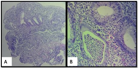
Inflammatory Bowel Disease

           diagnosis [1].
           Table 1: Conditions mimicking IBD
                                                     Conditions mimicking IBD
                                                          Infective colitides
                                                      Pseudomembranous colitis
Desai GS

                                                     Ischemic enteritis and colitis
                                                    Radiation enteritis and colitis
                                                      Irritable bowel syndrome
                                                          Colorectal cancer
                                                        Intestinal tuberculosis
                                         Chronic nonspecific ulcers of small intestine {CNSU}
                                     Cryptogenic multifocal ulcerous stenosing enteritis {CMUSE}
                                                     Eosinophilic gastroenteritis
                                                       Intestinal Behcet disease

           3. Clinical Presentation

                  Since the origin of medicine, clinical findings have been given the utmost importance
           for diagnosing a disease. The same key applies to differential diagnosis of IBD. A multitude of
           clinical hints are available to a good clinician to rule out differentials. Clinico-epidemiological
           profile of the patients, demographics, dietary habits and general medical condition of the patient
           all provide hints to diagnosis and are discussed next.

                  UC is more common in whites and in jewish population. Nonsmokers are at risk and so
           are patients with positive family history. It commonly presents with bleeding per rectum (90%-
           95%) [2], mucus per rectum, diarrhea, abdominal pain (65%) and tenesmus. The presentation
           varies with the severity and extent of disease, ranging from long duration of loose stools with
           mucus and weight loss to toxic megacolon with severe sepsis in acute severe disease [2].
           Abdominal or pelvic pain of unkown origin is also one of the rare presentations of UC. Many
           times, infective etiology may prompt investigations and diagnosis of asymptomatic UC and
           in these scenarios, it is important to suspect this co-existence of disease to avoid missing the
           diagnosis.

                  CD, on the other hand, has abdominal pain as the dominant symptom (80-85%), and
           weight loss (60%) and chronic diarrhea with mucus and blood (40-50%) are encountered less
                                                                                                                                www.openaccessebooks.com

           frequently [3]. More importantly, CD presents with small bowel predominant disease and hence
           mucus per rectum and tenesmus are less common than UC. Also, CD can present with perianal
           predominant disease as atypical or recurrent/refractory perianal abscess, complex fistulas or
           fissures/ulcers. CD can also affect upper gastrointestinal tract and presents with dyspepsia,
           nausea, vomiting or severe epigastric pain. Up to 25% patients of IBD can present with extra-
           intestinal manifestations such as oral ulcers, arthralgia, skin rashes or ocular symptoms before
           diagnosis of IBD [4-6].
                                                                    2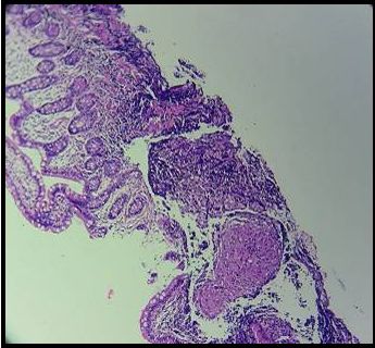
Inflammatory Bowel Disease

       In India and in other lower socioeconomic areas, intestinal tuberculosis {TB} is the most
common differential diagnosis of CD and less commonly UC. The disease most commonly
affects the age group 20-50 years of age and the presentation can be with an ileocecal mass,
appendicitis, small bowel obstruction or nonspecific abdominal pain, colic, nausea, vomiting,
anorexia, loss of weight and fever with an evening rise, high grade with chills and rigors [7].
These patients often have concomitant pulmonary/extrapulmonary TB or a past history of
treated or incompletely treated TB or a positive case in their family or close community. These
rarely present with colorectal involvement and hence, rarely confused with UC [8].

      History of recent travel to endemic areas for infections such as Entameba histolytica,
consumption of contaminated food/water, history of contact and presence of more affected
members in family or close community provide a hint towards infective colitis. These patients
present with abdominal pain, diarrhea, fever, generalized fatigue and malaise and may or
may not have blood in stools [9]. Hemorrhagic colitis is a fatal form of Enterohemorrhagic
Escherichia coli {EHEC} infection and is an important differential for acute severe toxic colitis
due to ulcerative colitis as well as ischemic colitis. Immunocompromised patients (human
immunodeficiency virus infection or prolonged steroid intake or chemotherapy/malignancy
patients or patients post-transplant on immunosuppression) are at risk for infective colitis,
most commonly due to cytomegalovirus and some opportunistic parasites such as isospora and
cyclospora [11].

        Patients with prolonged hypotensive states, patients with history of peripheral arterial
insufficiency/cerebrovascular or coronary insufficiency, obesity, diabetes and hypertension
with risk factors for thromboembolic arteriopathy, patients with hypercoaguable states,
pregnancy, patients on long term steroids or oral contraceptives are all at risk for arteriovenous
thromboembolic or vasospastic events which can lead to ischemic enteritis {Embolic>Non-
occlusive vasospastic > thrombotic > venous thrombotic} or ischemic colitis {Vasospastic >
thromboembolic} [11]. These patients presenting with symptoms such as postprandial pain
and fullness, who are afraid to eat owing to this pain, have weight loss and malabsorption in
chronic ischemia which can progress to intestinal obstruction due to stricture formation. On
the other hand, acute ischemic event can present with persistent and severe abdominal pain,
abdominal distension and gastrointestinal bleeding which can be confused with acute severe
colitis.

       Behcet’s disease [12] is a chronic, relapsing and remitting systemic vasculitis which can
be neurologic/ocular/intestinal or vascular types or may be a combination of all these. It can
present in a wide variety of symptoms owing to this multisystem involvement. Neurological
symptoms such as stroke/papillitis/papilledema, ocular symptoms such as scleritis/episcleritis/
anterior or posterior uveitis/retinitis, vascular manifestations such as endocarditis/myocarditis/
pulmonary arteritis/arthritis and triad of recurrent oro-oculo-genital aphthoid ulcers can be
                                                3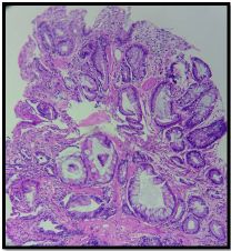
Inflammatory Bowel Disease
the presenting features in these patients that help in differentiating intestinal Behcet’s disease
from CD as the abdominal symptoms are otherwise identical to CD. The most common site
involved here is ileocecal valve and most common differential is CD amongst other causes of
ileocecal ulcers.

      CNSU [13] and CMUSE [14] are relatively newer disease entities that affect the
proximal ileum more than terminal ileum and are differential diagnosis for CD. CNSU affects
young females who most commonly present with long standing symptoms of hypochromic,
microcytic anemia and/or growth retardation. They rarely have any other symptoms such as
diarrhea, hematochezia or fever. On the other hand, CMUSE is more common in males and
presents with diarrhea, weight loss and edema. It also has a chronic, relapsing course and
can present with recurrent intestinal obstruction due to unexplained small intestinal strictures.
There are no signs of infection or inflammation in these patients.

       Drug history is also very important as in patients with symptoms of enterocolitis, a history
of antibiotics/chemotherapy drugs suggest a possibility of pseudomembranous colitis. On other
hand, history of allergic diseases, especially food allergy with similar presentation points to
a differential diagnosis of eosinophilic gastroenteritis. In eosinophilic enteritis, periumbilical
pain, nausea, vomiting and diarrhea occur with mucosal involvement, obstruction occurs when
the muscularis is involved, whereas subserosal and serosal involvement can lead to ascites or
pleural effusion {Klein classification for the extent of involvement} [15].

       Two other important differentials for ulcerative colitis are solitary rectal ulcer syndrome
(SRUS) and endometriotic involvement of rectosigmoid junction. SRUS is a misnomer and
these patients have history of functional constipation with pelvic floor dyssynergy that results
in single or multiple anterior rectal ulcers with/out inflammation and occasional rectal polyps.
The disease is limited to the last 10-12 cm of rectum. Endometriosis of rectosigmoid junction is
a rare condition presenting with recurrent pelvic pain or intestinal obstruction. The condition is
peculiar because the endometriotic deposits here are not under hormonal influence and hence,
the symptoms are not cyclical.

       Irritable Bowel Syndrome (IBS) is a functional bowel disorder (FBD) characterized
by chronic recurrent abdominal pain associated with defecation, accompanied by abdominal
distension or bloating and changes in bowel habits (constipation, diarrhea, or a combination
of both). The diagnostic criteria are: Recurrent abdominal pain, on average, at least 1 day per
week in the last 3 months, associated with 2 or more of 1) Relation to defecation, 2) Associated
with a change in frequency of stool or 3) Associated with a change in form (appearance) of
stool. The criteria should be fulfilled for the last 3 months with symptom onset at least 6
months before diagnosis [17].

      The diagnosis of IBS should be made based on the following 4 key features: clinical
                                                 4
Inflammatory Bowel Disease
history; physical examination; minimal laboratory tests; and, when clinically indicated, a
colonoscopy or other appropriate tests. Abdominal pain, usually in the lower abdomen, is
an essential feature. Abnormal stool frequency (>3 bowel movements/day and 25% constipation, 25% constipation,
>25% diarrhoea and IBS unclassified (IBS-U): Patients who meet diagnostic criteria for IBS
but whose bowel habits cannot be accurately categorized into 1 of the 3 groups above [16].
Female gender, ageInflammatory Bowel Disease
which are highly sensitive for IBD. Inflammatory markers, including faecal calprotectin, may
not be useful in patients with constipation symptoms. Thyroid function tests, serologic tests for
celiac disease, and stool analysis, should be individualized [17].
Stool culture, stool routine and microscopy are important to rule out infective colitis
which may be individually responsible for the symptoms or may co-exist with IBD. Stool
assay for toxin A/B are to be performed in cases with suspicion of Clostridium difficile colitis
[23]. Blood culture, serology for anti-salmonella antibodies are helpful for enteric fever. Long
standing anemia and multiple positive fecal occult blood tests in a young female patient points
towards CNSU. Peripheral eosinophilia is present in patients with eosinophilic gastroenteritis.
Elevated lactate, metabolic acidosis, and leucocytosis point towards bowel ischemia and
gangrene. These patients need to be worked up for thrombophilic states such as factor V leiden
mutation, prothrombin gene mutation, antiphospholipid antibody syndrome, etc.
5. Endoscopy Findings and Role in Differential Diagnosis
After a proper clinical history, physical examination and appropriate laboratory
investigations, the most important part of investigation is endoscopy ± biopsy and imaging.
The sequence of these investigations depend on the clinical situation, however, most patients
undergo a non-invasive investigation first followed by endoscopy.
5.1. Aims of Endoscopic Assessment
• Diagnosis of inflammatory bowel disease
• Distinguish between ulcerative colitis and crohn’s disease
• Monitor the grade the extent and severity of disease
• Assess the response to treatment
• Assess the exacerbations and document remissions following treatment
• Identify complications and surveillance for malignancy
• Therapeutic role of endoscopy
5.2. Endoscopic methods of assessment
• Colonoscopy with mandatory ileal intubation
• Flexible Sigmoidoscopy
• Esophagogastroduodenoscopy {OGDscopy}
6Inflammatory Bowel Disease
• Capsule Endoscopy
• Balloon enteroscopy – single/double balloon for small bowel evaluation
• Spiral enteroscopy for small bowel evaluation
• Endoscopic Ultrasonography
Here, we will keep our discussion to diagnostic features of IBD and the differential
diagnosis. Ulcerative colitis has continuous, circumferential mucosal and submucosal
inflammation that extends from rectum proximally. The endoscopic findings are hyperemic
and erythematous colorectal mucosa with a sharp demarcation at the proximal limit between
the normal and the involved mucosa [24]. Uneven, irregular fine or rough granular mucosa is
seen which bleeds on touch. Also, the normal mucosal vascular pattern is lost due to mucosal
and submucosal edema. Ulcers in UC are superficial and are surrounded by inflamed mucosa.
Stricture in UC and polyps should be evaluated to rule out adenomas.
In atypical cases, up to 40% patients with UC can have skip lesions whereas around
10% cases can have rectal sparing. Some patients may have colorectal disease with isolated
appendiceal orifice inflammation. Patients can have backwash ileitis characterized by mild
inflammation in terminal ileum due to incompetent ileocecal valve. However, the inflammation
is not as severe as in CD. These cases are difficult to characterize as either CD or UC based
only on endoscopic findings.
CD is characterized endoscopically by skip lesions, longitudinal, serpigenous ulcers with
transmural inflammation most commonly in terminal ileum. The ulcers are usually located at
the site of mesenteric attachments to bowel. Aphthous ulcers are also seen in CD. Coalescing
ulcers result in a cobblestone appearance. In CD, ileal involvement is patchy, deep and more
than cecal inflammation with discrete ulcers and chronic disease can result in strictures [3,
25]. OGDscopy is important in CD and can show solitary or multiple longitudinal ulcers/
erosions of esophagus and/or aphthous ulcers in gastric antrum, gastric body and/or part 2 of
duodenum.
Differential diagnosis for ulcers in terminal ileum can be done with help of some of the
endoscopic features specific to each disease. Intestinal tuberculosis most commonly affects
the ileocecal junction and presents with small and shallow to large and deep but transversely
oriented ulcers in terminal ileum. The ulcers more commonly has circumferential >serpigenous
arrangement. Scars, strictures or pseudpolyps can be seen in TB too. Patulous ileocecal valve
is more common in TB than CD. However, apart from the finding of transverse orientation of
ulcer, none of the other endoscopic finding is characteristic of TB [26,27].
CNSU and CMUSE also have endoscopic findings of ileal ulcers. CNSU presents with
7Inflammatory Bowel Disease
sharply demarcated flat proximal ileal ulcers in circumferential or oblique alignment which
when heal, produce spiral stenosis that looks in endoscopy like a coiled spring. CMUSE has
multifocal, superficial ulcers in small bowel, most commonly in terminal ileum [13,14]. These
ulcers never progress to cobblestoning, adhesions, fistula or fissure formation and remain
confined to mucosa and submucosa. Gastroduodenal and/or colonic superficial ulcers are also
common in CNSU.
Intestinal Behcet’s disease is characterized by presence of aphthous ulcer which is a
single and usually less than 5 round or oval, white ulcer with red peripheral rim of 2-15 mm
size and usually is deep ulcer in ileocecal area. The erythematous rim is nodular and elevated
with normal surrounding mucosa and base covered in exudates. This description of ulcer is
known as volcano-type ulcer [28]. The ulcer usually heals without scarring. The characteristic
ulcer pattern, absence of anorectal and distal colonic lesions and well defined discrete border
of the ulcer help identifying Behcet’s disease over CD.
Pseudomembranous colitis is characterized endoscopically by well defined, yellowish
white plaques ranging from 1 mm – 2 cm in diameter which cover the distal colon and rectum
most commonly. Occasionally, the disease can extend into proximal colon. Bowel preparation
can remove the pseudomembranes, and hence, if this disease is suspected, the colonoscopy is
recommended without bowel preparation [23].
Infective colitis has non-specific endoscopy findings such as mucosal erosions, edema,
punctate hemorrhages and mucoid exudates. Enteric fever shows longitudinal ulcers in terminal
ileum. Typhoid affects the right colon very rarely and in those cases can be confused with
CD. Similarly, Yersinia colitis and amebic colitis affect the right colon in 70% cases with
pathognomonic flask shaped ulcers, whereas it affects rectosigmoid in 30% cases. Yersinia
has a characteristic octopus sucker shaped ulcer with mucosal elevation in ileocecal area [29].
Yersinia can also present with mesenteric lymphadenopathy.
On the other hand, campylobacter colitis and shigella colitis affects rectosigmoid area
and can be differentiated from UC by absence of severe desquamation and presence of uneven
discrete lesions in rectosigmoid mucosa. Also, shigella colitis presents initially with watery
diarrhea which gradually decreases and changes to stool with blood, mucus and tenesmus.
Solitary rectal ulcer syndrome also presents a single ulcer in the rectum or can have multiple
ulcers or polyps [29].
Chronic ischemic enteritis or colitis will present with a stricture in the characteristic
watershed locations such as jejunum, splenic flexure or rectosigmoid junction. The stricture can
be concentric, short or long and single or multiple. In acute cases, mucosa appears edematous,
ulcerated and/or haemorrhagic. Severe cases can have transmural necrosis and/or gangrene.
Thus, endoscopic findings help in differential diagnosis of a lot of diseases that have clinical
8Inflammatory Bowel Disease
presentation confusing with IBD.
6. Pathological Dilemmas in Diagnosis of IBD
Biopsy diagnosis is one of the crucial steps in diagnosis, further management, assessment
of response and for prognostication of IBD. Many types of colitis mimic the histological features
of IBD and therefore, it is important to distinguishing between them. The morphological
features include endoscopic features and histopathological findings of multiple labelled biopsy
specimens from different parts of colorectum [30,31].
Various lesions mimicking IBD pathologically are as shown in table 2
Table 2: IBD mimics in pathology
Indeterminate colitis
Reparative change in IBD mimicking dysplasia
Dysplasia associated lesion or mass (DALM) versus adenoma
Morphologic variants of ulcerative colitis with crohn’s-like features.
• Backwash ileitis
• Upper GI tract involvement
• Superficial fissuring ulcers and aphthous ulcers
• Transmural inflammation
• Granulomas
• Iatrogenic procedure/ manoeuvres mimicking IBD
Diversion colitis and defunctioned rectum
Ileal reservoirs or pouchitis
Drugs
Other forms of colitis mimicking IBD
• Microscopic / lymphocytic colitis
• Diverticular disease associated colitis
• ischemic enterocolitis and Bechet’s syndrome
• Radiation colitis
Infections
Polyposis disorder mimicking IBD
Solitary rectal ulcer syndrome
Inflammatory cap Polyposis
Juvenile Polyposis
Others
• Malignant lymphoma
• Eosinophilic infiltrate of the gut
• Chronic Granulomatous Disease (CGD)
• Graft vs host disease
• Mass lesion
It is important
•
to distinguish between ulcerative colitis from crohn’s disease, because their
Common variable immunodeficiency
9Inflammatory Bowel Disease
management differs and both have different clinical behaviour. The pathological distinguishing
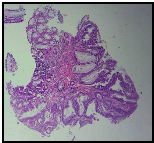
features between Ulcerative colitis and Crohn’s disease are as shown in table 3. Figure 1
shows the pathological characteristics of ulcerative colitis.
Table 3: Pathological differences between ulcerative colitis and crohn’s disease [1,2,3,4,5]
Features Ulcerative colitis Crohn’s disease
Macroscopic
Continuous and diffuse; left Segmental with skip areas.
Disease involvement sided and involves mucosa and Predominantly right sided and
submucosa involves transmurally
Rectal involvement Always present( adult) Can be involved
Perianal disease Rare Present 75%
Occasional 15% (backwash ileitis)
Ileal involvement Common
not more than 10 cms
Serositis Absent (except in fulminant colitis) Present
Fat wrapping Usually absent Frequently present
Fistulas and sinuses Absent Present
Microscopic
Discrete mucosal ulcers Absent (except in fulminant colitis) Present
Mucosal edema Usually absent Present
Rare, in fulminant colitis
Fissures Present , located deep
superficially located
Granulomas Absent, except crypt related. Common
Mucosal regeneration and
crypt atrophy, abnormal crypt Frequent Minimal
architecture
Mucosal inflammation and
Diffuse Focal
architectural involvement
Cytoplasmic mucin Diminished Maintained
Lymphoid aggregates Rare Frequent
Paneth cell metaplasia Sometimes present Absent
Pyloric gland metaplasia Rare Common in crohn’s enteritis.
6.1 Indeterminate colitis
The diagnosis is restricted to the cases showing overlapping pathological features, in
which it is difficult to differentiate between Ulcerative colitis and Crohn’s disease even after
the surgically resected specimen is examined. Its prevalence is less than 15% of IBD cases.
Most common pathological presentation is a case showing all features of Ulcerative colitis
with superficial fissures initially, most of which, on follow up behave like ulcerative colitis
with fewer having the biological behaviour of Crohn’s disease [30,31].
10Inflammatory Bowel Disease
Figure 1: Ulcerative colitis. [A] The microscopic diagnosis is based on diffuse dense inflammation, architectural chang-
es that are marked loss and mild distortion of crypts.(H & E X 100). [B] Chronic active phase showing crypt abscess
and cryptitis (H & E 400X)
6.2 Reparative change in IBD mimicking dysplasia [31,32]
The important features differentiating the two entities are as follows (Table 4)
Table 4: Differentiating features between dysplasia and reparative change
Characteristic Dysplasia Reparative change
Loss of nuclear polarity Present (in high grade) Absent
Glandular complexity
(cribriform change and luminal Present (in high grade ) Absent
bridging)
Nuclear enlargement Mild to severe Mild
Nuclear pleomorphism,
hyperchromasia, irregular Mild to severe Absent / mild
nuclear contours
Nuclear stratification and
Mild to severe Absent / mild
crowding
Inflammatory infiltrate Mild to moderate Moderate
High N:C ratio Moderate to severe Absent / mild
Cytoplasmic eosinophilia Absent Mild
6.3 DALM verses adenoma
It is important to distinguish these two entities as DALM (Table 5 and Figure 2) is
associated with a higher risk for malignancy even when the dysplasia is of low grade. It is
therefore, an indication for colectomy [31].
11Inflammatory Bowel Disease
Table 5: Differentiating features between adenoma and DALM
Features Adenoma DALM
Age Usually more than 40yrs Usually less than 40 yrs
Endoscopic appearance
Present Present or absent
Pedunculated polyp
Stalk base region and biopsy
Negative for dysplasia Dysplasia seen.
of adjacent colonic mucosa
If the lesion is in an area unaffected by ulcerative colitis then adenoma is favoured.
Figure 2:DALM: Rectal polyp, showing mildly dysplastic epithelial lining with adjacent epithelium showing changes
of Ulcerative Colitis ( H& E 100X)
6.4 Morphologic variants of Ulcerative colitis with Crohn’s-like features
6.4.1 Backwash ileitis
It important to recognise this entity as, this can be misdiagnosed as Crohn’s disease.
It is seen in approximately 15% of cases with pancolitis and is generally associated with
incompetent ileocaecal valve causing regurgitation of colonic content. The inflammation is
mild, patchy and limited to mucosa. It is not associated with stricture or thickening and other
features of Crohn’s disease such as granulomas, deep fissures and submucosal inflammation
are absent [31,33].
6.4.2 Upper GI tract involvement
Very rarely in UC, duodenal involvement is present and is represented by diffuse
inflammation, thickening of duodenum and fistula [34]. Gastric involvement in UC shows
superficial plasmacytosis, basal mixed inflammation and focal gastritis [35]. These patterns
of involvement, in the absence of granulomas can be categorised as UC. However as these
changes are fairly nonspecific, a diagnosis of UC has to be established, elsewhere in the colon
[34].
12Inflammatory Bowel Disease
6.4.3 Aphthous ulcers
It is a shallow irregular ulcer or mucosal erosion, located over a lymphoid follicle. It is
characteristic of CD. However, in one study aphthous like ulcer were present in 17 % of cases
with UC. It can also be seen in other conditions like infectious colitis, diversion colitis and
diverticular disease [31,33,36].
6.4.4 Transmural inflammation
Transmural mononuclear inflammation can be seen in cases of UC, with fissuring ulcers
extending to deep submucosa and superficial muscularis propria. It can also be seen in toxic
megacolon with prominent myocyte necrosis and serosal inflammation.
6.4.5 Granulomas [2]
Two types of granulomas are seen in UC [31].
• Crypt related - Damaged crypt causing extravasation of mucin causing histiocytic
collection in surrounding mucosa.
• Deep seated foreign body granulomas usually associated with fulminant colitis.
Such granulomas need to be differentiated from epithelioid granulomas seen in case of
Crohn’s disease.
6.4.6 Iatrogenic procedure/ manoeuvres mimicking IBD
6.4.6.1 Diversion colitis and defunctioned rectum [3,9,10]
When part of rectum or colon is surgically placed out of the fecal stream for any reason,
it acquires histological changes of defunctioning. It is probably related to loss of exposure to
essential fatty acids and physiological response to stasis. It occurs in patients with defunctioned
large intestine for disorders like colorectal cancer, diverticular disease, Hirschsprung’s
disease.
The defunctioned rectum shows acute and chronic inflammation, architectural distortion,
transmural inflammation, fissures, lymphoid hyperplasia and poorly formed granulomas,
mimicking Crohn’s disease. In such cases the clinical history is critical, so also if any histological
examination of the rectum, colon prior to fecal stream diversion is available [32,37,38].
6.4.6.2 Pouchitis [3,4,9,10]
In some operations requiring total proctocolectomy, one of the late complications of
pouch construction for patients with UC or familial adenomatous polyposis is pouchitis.
13Inflammatory Bowel Disease
It is primary chronic relapsing inflammation of the pouch and can be acute or chronic. In
some patients, unresponsive pouchitis may develop as a complication and this mimics CD.
Microscopically it shows acute on chronic inflammation, atrophy of villi and elongation of
crypts.
Rare cases show transmural inflammation and granulomas formation. In these case,
if the colectomy was done for UC, the original case should be re-examined to exclude the
possibility of a true CD, which can be seen in 2 to 7% of patients [32,33,37,38].
6.4.6.3 Drugs
Various drugs causes active inflammation of the large bowel which includes non-steroidal
anti-inflammatory drug (NSAIDS) causing mucosal damage, occasional granulomas and IBD
like changes. It can be differentiated by increased intraepithelial lymphocytes and epithelial
cell apoptosis and history of drug intake, with regression of symptoms and microscopic features
on cessation of the drug [38,39].
Antineoplastic drugs such as 5-flurouracil cause acute colitis. Epithelial necrosis is an
important feature in the acute phase. Crypt regeneration and distortion are seen in the resolving
phase, mimicking ulcerative colitis.
6.4.7 Other forms of colitis mimicking IBD
6.4.7.1 Microscopic / lymphocytic/collagenous colitis [32,33,37,38]
Microscopic colitis is defined only by its microscopic abnormality, with normal endoscopy
findings and clinical complaints of watery diarrhoea. It includes two entities: lymphocytic
colitis and collagenous colitis.
The characteristic features of lymphocytic colitis are as follows
• Increase in intraepithelial lymphocytes ≥ 25/ 100 epithelial cells. {Normal ≤ 6 / 100
epithelial cells}.
• Mucin depletion and decreased cell height which implies surface epithelial damage.
• Relatively preserved crypt architecture.
• The lamina propria shows increased in lymphocytes, plasma cells, eosinophils and mast
cells {usually clustered at crypt bases}.
14Inflammatory Bowel Disease
The characteristic features of collagenous colitis are as follows
• Subepithelialy located thickened collageneous bands > 10 micrometre. {normal< 7
micrometer}.
• The collageneus layer may extend into the lamina propria with irregular lower border.
Normally the lower edge of basement membrane should be sharp and distinct.
• The presence of chronic inflammation can suggest an IBD. However, the absence of
mucosal architectural distortion and atrophy differentiate it.
2 to 5 % of cases show aberrant histology including ulcers, crypt abnormalities, paneth
cell metaplasia, inflammatory membranes and occasional cryptitis. Histology of both these
entities do not correlate with the symptoms, response to treatment or outcome and no patient
with this aberrant histology has yet developed IBD [32].
6.4.7.2 Ischemic enterocolitis and Bechet’s syndrome
In chronic ischemic bowel disease, stricture development may mimic CD grossly.
However superficial epithelial damage, lamina propria showing hemosiderin laden macrophages,
fibrosis and relative absence of chronic inflammation differentiate it from IBD.
Behcet’s disease is a multisystem disorder, which can affect the intestine. Ulceration is
an important feature. It can be diffuse or limited to the ileocaecal region. Colitis in Bechet’s
syndrome shows mucosal cobblestoning,apthous ulcers and sometimes granulomas can also be
seen. Presence of characterstic perivascular inflammation, necrotizing vasculitis and absence
of transmural inflammation and lymphocytic aggregates differentiate it from CD [32,38].
6.4.7.3 Radiation colitis
This can cause architectural distortion, crypt atrophy, mucin depletion and chronic
inflammation mimicking IBD. Other features of obliterative arteritis with hyalinization
of vessel wall, vascular ectasia and submucosal and intramural fibrosis with the history of
radiation exposure help in differentiating it from IBD [37].
6.4.8 Infections [32,38,39]
The differentiating features between IBD and infective colitis are as shown in Table 6.
15Inflammatory Bowel Disease
Table 6: Differentiating features between Infective colitis, UC and CD
Feature Infective colitis UC CD
Diffuse change Sometimes present Present Sometimes present
Focal change Usually present Absent Usually present
Architectural
Focal Diffuse Focal
abnormality
Neutrophils in lamina
Usually present Absent Absent
propria
Granulomas Absent Absent Present
6.4.8.1 Granulomatous inflammation simulating crohn’s disease is seen in the following
conditions:
• Tuberculosis (TB) is the most common cause of granulomas associated. The mucosal
architectural changes with crypt distortion, crypt loss, cryptitis and crypt abscess is a close
mimic of IBD. It is the prominence of granulomas that subtly indicate a tubercular etiology.
Florid coalescent granulomatous inflammation with extensive caseous necrosis is the most
definitive feature favouring TB. In 50% of intestinal tuberculosis, acid fast bacilli can be
identified on special stains, in which case the diagnosis of TB is definitive.
• Yersiniosis – Relative lack of transmural inflammation and granulomas exhibiting
central necrosis favours yersiniosis.
• Other conditions with identifiable organism in granulomatous inflammation include
schistosomiasis, deep mycoses and larval infestation.
Figure 3: Mimic of Crohn’s disease: ileal biopsy showing single large granuloma with adjacent ileum showing relatively
preserved architecture. (H & E 100X)
6.4.8.2 Viral infections [32,38]
In immunosuppressed patients, infection by Cytomegalovirus(CMV), Herpes simplex
virus (HSV) and cryptosporidiosis mimic active UC. CMV causes necrotizing gut injury
16Inflammatory Bowel Disease
secondary to vasculitis. HSV infects distal rectum causing ulceration and microscopy shows
ulceration with acute inflammation in lamina propria with cryptitis and crypt abscess. In the
rectum, HSV inclusions are not seen. Characteristic inclusion bodies, multinucleated giant
cells, demonstration of organism and their typical cytopathic changes help in differentiating
from IBD.
Amoebic colitis can also causes crypt distortion [36]. Clostridium difficile causes early
patchy changes, with epithelial cell degeneration, and later on regenerative crypt architectural
changes, can be seen.
6.4.9 Others
6.4.9.1 Solitary rectal ulcer syndrome (SRUS) (Figure 4)
It is an endoscopically and pathologically defined entity. It occurs in the anterior wall
of rectum, in relatively young patients with female preponderance. Histological features can
mimic IBD because of marked crypt distortion with crypt atrophy seen in IBD.
In SRUS, crypt hyperplasia and elongation is seen. The inflammatory cells are scant.
Fibromuscular replacement of the lamina propria is seen. These features help in differentiating
it from IBD [36,37].
Figure 4: Mimic of IBD, (SRUS): Rectal biopsy showing architectural distortion with fibromuscular replacement of
lamina propria (H &E 100X).
6.4.9.2 Malignant lymphoma
In high grade small and large cell lymphoma of B and T cell type, deep destructive
ulceration is seen, which can mimic Crohn’s disease specially in cases with few neoplastic cells
and numerous eosinophils. In such cases immunohistochemistory can help to differentiate the
two [38].
Rarely UC can coexist with a malignant lymphoma. Large bowel primary malignant
lymphoma is one of the rare yet known complications of chronic ulcerative colitis.
17Inflammatory Bowel Disease
6.4.9.3 Eosinophilic colitis
Eosinophilia of the intestine can be seen in several conditions eg- parasitic infestation
such as strongyloides stercoralis or eosinophilic colitis in which marked eosinophilic infiltrate
of the mucosa is seen without architectural distortion or eosinophilic gastroenteritis and
peripheral blood eosiniphilia [37,38].
In the quiescent phase of UC, eosinophils can be the prominent inflammatory infiltrate.
Crypt architectural abnormality would help to differentiate it from eosinophilic colitis.
6.4.9.4 Chronic Granulomatous Disease (CGD)
This is autosomal recessive disorder. When complicated by colitis it can mimic CD.
Presence of histiocytes containing lipid vacuoles and pigment helps in the differentiation. In
CD leucocyte bactericidal activity is normal [38].
6.4.9.5 Graft versus host disease
Acute graft vs host disease causes crypt epithelial apoptosis, crypt distortion and atrophy.
Chronic inflammation may be mild or absent. In chronic graft vs host disease crypt architectural
abnormality with or without fibrosis is seen. Here history would help in differentiation [39].
6.4.9.6 Mass lesion
In subserosal or intramural mass (eg- carcinoma, diverticular disease, endometriosis)
the overlying mucosa due to pathological distortion may show features resembling IBD [39].
6.4.9.7 Common variable immunodeficiency
In absence of or with mild chronic inflammation with crypt atrophy and distortion with
or without fibrosis is seen in common variable immunodeficiency. The mild or absent chronic
inflammation differentiates this condition from IBD [39].
7. Radiological Imaging modalities for IBD
7.1 X-ray
• Not very useful for primary diagnosis of IBD.
• Useful for imaging in complicated IBD
• Perforation: visible extraluminal air/free air under diaphragm
• Toxic megacolon: Diameter of transverse colon > 6cm
18Inflammatory Bowel Disease
• Extra-intestinal manifestations
• Spondyloarthropathy evaluation
• Immunosuppression induced pneumonia
• To rule out tuberculosis before starting infliximab
7.2 Fluoroscopic imaging {Barium meal follow through, barium enteroclysis for small bowel
and barium enema for large bowel} [40]
• Findings on fluoroscopy of small bowel
• Ulcerative colitis: Patulous, incompetent IC valve with nodular ileitis
• Crohn’s disease: Stenotic ileocecal junction with luminal narrowing and ileal ulcerations.
Fat wrapping is radiologically seen as bowel loop angulation and kinking. Cobblestone
appearance is seen due to transverse and longitudinal ulcerations and strictures are seen in
chronic disease.
• Limitations
• Invasive procedure and not definitive.
• Patient intolerance to oral contrast and rapid distension in enteroclysis.
• No extraluminal information is obtained. Inadequate bowel distension in small
bowel follow through.
• Limited availability especially in peripheral centers.
• Limited experience of radiologists
• Findings on Barium enema [Large bowel]
• Barium enema is performed when clinical suspicion of ulcerative colitis is high and
helps in differentiation between ulcerative colitis and crohn’s disease.
• Ulcerative colitis: Continuous involvement, rectum is involved early, aphthous ulceration
and lead pipe pattern due to loss of haustrations and widening of presacral space > 20 mm
• Crohn’s disease: Skip pattern, rectum may be spared or involved late, cobblestone
pattern and strictures
Table 7 highlights the differences between barium enema and colonoscopy for diagnosis
of inflammatory bowel disease
19Inflammatory Bowel Disease
Table 7: Differences in utility of barium enema and colonoscopy for diagnosis of IBD
Barium enema Colonoscopy
Noninvasive Invasive
Indirect and subjective evaluation of mucosa Direct objective mucosal evaluation,
Doubtful reproducibility Reproducible
Screening modality Diagnostic modality
Strictures are not a limitation and proximal loops
Strictures may limit the study
can be visualized using dilute barium suspension
Biopsy cannot be done Biopsy can be done simultaneously
Less complications Perforation rate 1 in 5000
Less costly Expensive
7.3 Ultrasound abdomen and pelvis
It does not play a major role in diagnosis of inflammatory bowel disease. Wall thickening
of bowel and loss of gut signature may be seen in crohn’s disease.
7.4 Computed tomography [CT] enterography and CT enteroclysis [41]
It is now preferred over fluoroscopy studies as it has a rapid scan time with multiplanar
reformatting options and also provides extraluminal information. CT enteroclysis similar to
barium enteroclysis requires a nasojejunal tube placement for contrast instillation. Enterography
is hence preferred except for cases of partial obstruction wherein, enteroclysis is the procedure
of choice.
Neutral contrast is now favored over iodinated contrast because it is tasteless so more
palatable, mucosal enhancement by intravenous contrast can be adequately visualized with
neutral contrast whereas it is obscured with iodinated contrast. Stricture evaluation is better
with iodinated contrast. Oral contrast is followed by intravenous low osmolar contrast such as
iohexol to look for bowel wall enhancement and extraluminal findings.
In cases of suspected bowel obstruction or perforation, positive oral contrast is preferred
as transit across the perforation site is identified better with positive contrast. Also, after the
diagnosis of rectal IBD on colonoscopy, the extraluminal findings can be better evaluated
with CT with positive rectal contrast. Pattern of disease [skip versus continuous], submucosal
extent, ulceration, lymphadenopathy, and presence of fistula can be evaluated
Findings on CT
• In acute cases, colonic/small bowel wall thickening and edema is seen
• Mucosal hyperenhancement, mesenteric inflammation and increased vascularity
are seen in the form of fat stranding and ‘comb sign’ at the mesenteric edge of the bowel
20Inflammatory Bowel Disease
• Fistula/perforation/abscess are better evaluated
• Fibrostenotic strictures can be seen and evaluated
• Chronic cases show mesenteric fat proliferation known as creeping fat sign and
submucosal fat infiltration
• Gross bowel distension with bowel wall thinning and/or pneumatosis are seen in
toxic megacolon
CT enterography allows more interobserver agreement, is reproducible, is widely
available and can be used in children without prolonged sedation. Also, the scan time is
short, radiologists are more familiar with the procedure and scan readings, cost is less when
compared to magnetic resonance [MR] enterography and the scan can be done in patients with
claustrophobia or patients with MR sensitive implants/devices. Limitation is contrast induced
complications such as nephropathy or allergy and radiation exposure in follow up of cases.
7.5 MR enterography [42]
MR enterography is helpful in follow up of the cases to look for disease activity. Also,
frequent follow up scans are feasible as there is no radiation in MR and non-contrast T1, T2,
diffusion and cine images are good enough to provide a lot of information. Important MR
sequences are
• T2 steady state free precession [SSFP] sequence helpful for bowel wall edema
• Post contrast T1 imaging is used for mucosal hypervascularity and comb sign
• Dynamic cine T2 imaging is useful for distinguishing between peristalsis and
stricture
• T2 fat saturated sequences [Short tau inversion recovery {STIR}/ Spectral and
inversion recovery {SPAIR}] are also useful for better evaluation of bowel wall edema, abscess
and fistula.
• Diffusion weighted imaging will show restricted diffusion in areas of active
inflammation and abscess. This is seen in T2 sequences without contrast.
Findings on MR [42]
• Active disease – Mucosal edema on T2 and mucosal hyperenhancement and
transmural enhancement on post contrast T1 images.
• Inflammatory strictures are T2 hyperintense whereas fibrotic strictures are T2
hypointense.
• MR pelvis is the gold standard for imaging of perianal fistula due to better
evaluation of tracts as well as the relationship of the fistula with the sphincter complex.
21Inflammatory Bowel Disease
• MR is also a means to evaluate mucosal healing in response to treatment.
• MR cholangiopancreaticography[MRCP] helps in evaluation of primary sclerosing
cholangitis.
Table 8 summarizes the role of radiology in diagnosis of IBD.
Table 8: Role of different imaging modalities on diagnosis of IBD
o CT enterography is first line
o Capsule endoscopy if no evidence of obstruction and/or
Small bowel disease enteroscopy if required
o CT enteroclysis useful in cases of suspicious partial small
bowel obstruction
• Colonoscopy for diagnosis
Large bowel disease • CT to look for pattern of involvement and for evaluation of
extra-luminal disease
o X-ray to rule out perforation/ toxic megacolon
o If megacolon, CT done without positive oral or rectal contrast
o If no megacolon, CT done with positive oral and/or rectal
Acute abdomen
positive contrast to look for fistula or abscess.
o This is continued till intravenous urography phase if ureteric
stricture or fistula is suspected in cases with hydronephrosis
For follow up of cases on treatment MR enterography
For sacroiliac and hip joint MR of the involved area
Biliary system MRCP
Perianal disease MR pelvis
7.6 Differential diagnosis based on imaging findings
7.6.1 Crohn’s disease
Stratified enhancement of the intestinal wall with wall thickness > 3 mm is the most
characteristic finding in CD. This is also known as mural stratification sign which is because
of hyperenhancing mucosa and muscularis on CECT and hypoenhancingsubmucosa. This
becomes reverse with a hyperintensesubmcosa on T2 MR imaging. MR also shows mesenteric
edema and inflammation is the form of T2 hyperintense mesentery and CECT/MR can also
show nodes, sinuses, fistulas or sinuses. Sacculations of intestinal wall due to compensatory
dilatation of antimesenteric wall due to fibrosis and shortening at the mesenteric side of the
bowel wall are a feature of chronic CD along with the presence of fat wrapping/ creeping fat.
Imaging will also reveal extra-intestinal manifestations such as primary sclerosing cholangitis,
gallstones, pancreatitis, arthropathy and portal vein thrombosis amongst others.
7.6.2 Ischemic enteritis/ colitis
Splenic flexure, rectosigmoid junction and jejunum are the most common sites involved in
ischemic enterocolitis. Thumb printing is the most common sign described for ischemic colitis.
22Inflammatory Bowel Disease
Target sign, bowel wall thickening and pericolonic infiltration and stranding is also common
on CT. Stricture suggests chronic disease and location of the stricture in an appropriate clinical
setting suggests ischemia as the cause. Penumatosis, portal gas or free air suggests ischemic
gangrene/ perforation and are surgical emergencies. Imaging can also hint towards possible
cause for ischemia with the help of CT angiography which has now replaced conventional
angiography for diagnosis. Arterial thrombus and embolus can be visualised as meniscus sign
or planar defect on angiography whereas prolongation of arterial phase can be seen in patients
with vasospastic states. Phlebosclerosis is suggested by presence of calcification in the venous
system. Presence of target sign, absence of long segment stricture and absence of mucosal
hyperenchancement suggest the presence of ischemic bowel disease.
7.6.3 Intestinal tuberculosis
Being most common type of abdominal tuberculosis (90%) [43], Ileocecal tuberculosis
often shows abdominal lymphadenopathy with mesenteric lymphadenopathy being the most
common of them [44]. Generalised lymphadenopathy involving peripancreatic, periportal and
periaortic lymphadenopathy is seen in generalised disease. Early disease may show enhancing
mucosa associated wall edema and thickening which later form strictures. Associated
symmetrical wall thickening of IC valve and adjacent cecal wall is often seen in early intestinal
tuberculosis which often confuses with radiological findings of Crohn’s disease, malignancy
or lymphoma however in advanced stage associated mesenteric inflammation along with
clumping of bowel can be seen in as high as 45% patients [45]. Multifocal bowel involvement
can often mimic skip lesions of Crohn’s disease. Isolated TB involving colon is relatively less
common which can mimic ulcerative colitis. Important differences between CD and TB are
shown in Table 9
Table 9: Differences in imaging characteristics of CD and intestinal TB
CD TB
Long segment small bowel involvement at more than 4 Short segment ileocecal> small bowel involvement is
sites is characteristic with eccentric strictures seen at less than 4 sites with concentric strictures
Comb sign and fat wrapping is common Comb sign and fat wrapping is uncommon
Lymphadenopathy is uncommon and reactive Lymphadenopathy is common with necrotic nodes
Omentum is rarely involved and ascites is not common Omentum is frequently involved and ascites is common
Cobblestoning pattern, deep ulcers, enterocutaneous Cobblestoning pattern, deep ulcers, enterocutaneous
fistulas are common fistulas are uncommon
7.6.4 Intestinal Behcet’s disease
The most common site of involvement is ileocecal region. Double contrast barium
examination is better than single contrast and shows the characteristic deep collar button
shaped lesions with ring like protrusion. It very rarely presents as ileocecal mass when, only
the pathological findings can differentiate it from the other diagnostic mimics. On contrast
23Inflammatory Bowel Disease
enchanced CT, the bowel wall appears thickened with/out a mass and a central ulceration may
be present in the mass. The involved segment enhances brightly on IV contrast administration.
The absence of significant lymphadenopathy and peri-enteric stranding helps in differentiation
from malignancy and other inflammatory bowel diseases [46].
Other sites of involvement are rare. There is no specific pattern of involvement in other
organs such as esophagus, stomach or proximal small bowel but, deep penetrating ulcers in an
appropriate clinical setting should raise a suspicion of Intestinal Behcet’s disease. The disease
can also present in its complicated form with perforation, hemorrhage, peritonitis or fistula
[47].
7.6.5 Pseudomembranous colitis
Pseudomembrane may be visible on double contrast barium study. Findings on CT that
support the diagnosis include the accordion sign that presents due to trapping of oral contrast in
between the thickened edematous folds, shaggy mucosal outline and/or bowel wall thickening.
Other findings such as grossly dilated colon in cases of toxic megacolon, thumb printing due
to ischemic colitis and pericolonic stranding can be present as in other inflammatory bowel
diseases [48].
7.6.6 CNSU, CMUSE and infective colitis
These diseases are diagnosed based on history, endoscopy and biopsy and/or cultures
and radiology does not have a role in establishing a diagnosis.
8. Conclusion
From the above discussion, it is clear that correlation between clinical features, laboratory
parameters, radiologic and endoscopic findings, and histopathological examination, is the key
to overcome the diagnostic dilemmas and hence, all these features should be used to arrive at
a diagnosis in all cases suspected to have IBD.
9. References
1. Magro F, Gionchetti P, Eliakim R, Ardizzone S, Armuzzi A, Barreiro-de Acosta M, et al. Third European Evidence-
based Consensus on Diagnosis and Management of Ulcerative Colitis. Part 1: Definitions, Diagnosis, Extra-intestinal
Manifestations, Pregnancy, Cancer Surveillance, Surgery, and Ileo-anal Pouch Disorders. J Crohn’s Colitis. Oxford
University Press; 2017 Jun 1; 11(6): 649–70.
2. Tontini GE, Vecchi M, Pastorelli L, Neurath MF, Neumann H. Differential diagnosis in inflammatory bowel disease
colitis: state of the art and future perspectives. World J Gastroenterol. Baishideng Publishing Group Inc; 2015 Jan 7;
21(1): 21–46.
3. Gomollón F, Dignass A, Annese V, Tilg H, Van Assche G, Lindsay JO, et al. 3rd European Evidence-based Consensus
on the Diagnosis and Management of Crohn’s Disease 2016: Part 1: Diagnosis and Medical Management. J Crohn’s
Colitis. Oxford University Press; 2017 Jan 1; 11(1): 3–25.
24Inflammatory Bowel Disease
4. Jose FA, Heyman MB. Extraintestinal manifestations of inflammatory bowel disease. J Pediatr Gastroenterol Nutr.
NIH Public Access; 2008 Feb; 46(2): 124–33.
5. Levine JS, Burakoff R. Extraintestinal manifestations of inflammatory bowel disease. Gastroenterol Hepatol (N Y).
Millenium Medical Publishing; 2011 Apr;7(4): 235–41.
6. Vavricka SR, Rogler G, Gantenbein C, Spoerri M, Prinz Vavricka M, Navarini AA, et al. Chronological Order of
Appearance of Extraintestinal Manifestations Relative to the Time of IBD Diagnosis in the Swiss Inflammatory Bowel
Disease Cohort. Inflamm Bowel Dis. 2015 Aug; 21(8): 1794–800.
7. PATEL N, AMARAPURKAR D, AGAL S, BAIJAL R, KULSHRESTHA P, PRAMANIK S, et al. Gastrointestinal
luminal tuberculosis: Establishing the diagnosis. J Gastroenterol Hepatol. 2004 Nov; 19(11): 1240–6.
8. Sharma MP, Bhatia V. Abdominal tuberculosis. Indian J Med Res. 2004; 120(4): 305–15.
9. Singh R, Balekuduru A, Simon EG, Alexander M, Pulimood A. The differentiation of amebic colitis from inflammatory
bowel disease on endoscopic mucosal biopsies. Indian J Pathol Microbiol. Medknow Publications and Media Pvt. Ltd.;
2015; 58(4): 427–32.
10. Huh CW, Youn YH, Jung DH, Kim DW, Kho BG, Kim J-H, et al. A Case of Cytomegalovirus Colitis with Endoscopic
Finding Resembling Crohn’s Disease. Korean J Gastroenterol. 2012; 59(4): 303.
11. Gandhi SK, Hanson MM, Vernava AM, Kaminski DL, Longo WE. Ischemic colitis. Dis Colon Rectum. 1996 Jan;
39(1): 88–100.
12. Ebert EC. Gastrointestinal Manifestations of Behçet’s Disease. Dig Dis Sci. Springer US; 2009 Feb 2; 54(2):
201–7.
13. Matsumoto T, Iida M, Matsui T, Yao T. Chronic nonspecific multiple ulcers of the small intestine: a proposal of the
entity from Japanese gastroenterologists to Western enteroscopists. Gastrointest Endosc. 2007 Sep; 66(3 Suppl): S99-
107.
14. Perlemuter G, Guillevin L, Legman P, Weiss L, Couturier D, Chaussade S. Cryptogenetic multifocal ulcerous
stenosing enteritis: an atypical type of vasculitis or a disease mimicking vasculitis. Gut. 2001 Mar; 48(3): 333–8.
15. Klein NC, Hargrove RL, Sleisenger MH, Jeffries GH. Eosinophilic gastroenteritis. Medicine (Baltimore). 1970 Jul;
49(4): 299–319.
16. Lacy BE, Mearin F, Chang L, Chey WD, Lembo AJ, Simren M, et al. Bowel disorders. Gastroenterology. Elsevier,
Inc; 2016;150(6): 1393–1407e5.
17. Drossman DA, McKee DC, Sandler RS, Mitchell CM, Cramer EM, Lowman BC, et al. Psychosocial factors
in the irritable bowel syndrome. A multivariate study of patients and nonpatients with irritable bowel syndrome.
Gastroenterology. 1988 Sep; 95(3): 701–8.
18. Lovell RM, Ford AC. Global Prevalence of and Risk Factors for Irritable Bowel Syndrome: A Meta-analysis. Clin
Gastroenterol Hepatol. 2012 Jul; 10(7): 712–721.e4.
19. Ford AC, Talley NJ, Veldhuyzen van Zanten SJO, Vakil NB, Simel DL, Moayyedi P. Will the History and Physical
Examination Help Establish That Irritable Bowel Syndrome Is Causing This Patient’s Lower Gastrointestinal Tract
Symptoms? JAMA. 2008 Oct 15; 300(15): 1793.
20. Saibeni S, Folli C, de Franchis R, Borsi G, Vecchi M. Diagnostic role and clinical correlates of anti-Saccharomyces
cerevisiae antibodies (ASCA) and anti-neutrophil cytoplasmic antibodies (p-ANCA) in Italian patients with inflammatory
bowel diseases. Dig Liver Dis. 2003 Dec; 35(12): 862–8.
21. Khan R, Abid S, Jafri W, Abbas Z, Hameed K, Ahmad Z. Diagnostic dilemma of abdominal tuberculosis in non-HIV
25Inflammatory Bowel Disease
patients: an ongoing challenge for physicians. World J Gastroenterol. 2006 Oct 21; 12(39): 6371–5.
22. Alvares JF, Devarbhavi H, Makhija P, Rao S, Kottoor R. Clinical, Colonoscopic, and Histological Profile of Colonic
Tuberculosis in a Tertiary Hospital. Endoscopy. 2005 Apr; 37(4): 351–6.
23. Hookman P, Barkin JS. Clostridium difficile associated infection, diarrhea and colitis. World J Gastroenterol. 2009
Apr 7; 15(13): 1554–80.
24. Hommes DW, van Deventer SJH. Endoscopy in inflammatory bowel diseases. Gastroenterology. 2004 May; 126(6):
1561–73.
25. Annese V, Daperno M, Rutter MD, Amiot A, Bossuyt P, East J, et al. European evidence based consensus for
endoscopy in inflammatory bowel disease. J Crohn’s Colitis. 2013 Dec; 7(12): 982–1018.
26. PULIMOOD AB, PETER S, RAMAKRISHNA B, CHACKO A, JEYAMANI R, JEYASEELAN L, et al. Segmental
colonoscopic biopsies in the differentiation of ileocolic tuberculosis from Crohn’s disease. J Gastroenterol Hepatol.
2005 May; 20(5): 688–96.
27. Naga MI, Okasha HH, Ismail Z, El-Fatatry M, Hassan S, Monir BE. Endoscopic diagnosis of colonic tuberculosis.
Gastrointest Endosc. 2001 Jun; 53(7): 789–93.
28. Lee S, Kim B, Kim T, Kim W. Differential diagnosis of intestinal Behçet’s disease and Crohn’s disease by colonoscopic
findings. Endoscopy. 2009 Jan 21; 41(1): 9–16.
29. Shen B, Khan K, Ikenberry SO, Anderson MA, Banerjee S, Baron T, et al. The role of endoscopy in the management
of patients with diarrhea. Gastrointest Endosc. 2010 May; 71(6): 887–92.
30. Juan Rosai. Rosai and Ackerman’s Surgical Pathology. 10e ed. Elsevier; 2012. 731-802 p.
31. Chandrasoma P. Chapter 11 Idiopathic Inflammatory Bowel Disease. In: Gastrointestinal Pathology. Hong
Kong :Appleton and Lange; 1999. p. 283–312.
32. Stacey E. Mills. Chapter 33 Nonneoplastic intestinal diseases. In: Sternberg’s Diagnostic Surgical Pathology. 6e ed.
China: Wolters Kluwer Health; 2015. p. 1447–504.
33. Yantiss RK, Odze RD. Diagnostic difficulties in inflammatory bowel disease pathology. Histopathology. Blackwell
Science Ltd; 2006 Jan 1; 48(2): 116–32.
34. Chandrasoma P. Chapter 6 The Duodenum. In: Gastrointestinal Pathology. Hong Kong :Appleton and Lange; 1999.
p. 145–166.
35. Lin J, McKenna BJ, Appelman HD. Morphologic Findings in Upper Gastrointestinal Biopsies of Patients With
Ulcerative Colitis. Am J Surg Pathol. 2010 Oct; 34(11): 1.
36. Odze R D Goldblum JR. Chapter 14 Inflammatory Disorder of the Small Intestine. In: Surgical Pathology of the GI
Tract, LIVER, BILIARY TRACT and Pancreas. Philadelphia: Saunders Elsevier; 2009. p. 355–94.
37. Chandrasoma P. Chapter 10 Nonneoplastic diseases of colon. In: Gastrointestinal Pathology. Hong Kong :Appleton
and Lange; 1999. p. 249–82.
38. Shepherd NA. Pathological mimics of chronic inflammatory bowel disease. J Clin Pathol. BMJ Publishing Group;
1991 Sep; 44(9): 726–33.
39. Feakins RM, British Society of Gastroenterology. Inflammatory bowel disease biopsies: updated British Society of
Gastroenterology reporting guidelines. J Clin Pathol. 2013 Dec; 66(12): 1005–26.
40. Maglinte DD, Kelvin FM, O’Connor K, Lappas JC, Chernish SM. Current status of small bowel radiography.
Abdom Imaging. 21(3): 247–57.
26You can also read