DEGENERATIVE CERVICAL MYELOPATHY: A REVIEW OF CURRENT CONCEPTS - SciELO
←
→
Page content transcription
If your browser does not render page correctly, please read the page content below
Review Article/Artigo               de   Revisão/Artículo             de   Revisión

                    DEGENERATIVE CERVICAL MYELOPATHY: A REVIEW OF
                    CURRENT CONCEPTS
                    MIELOPATIA CERVICAL DEGENERATIVA: REVISÃO DOS CONCEITOS ATUAIS

                    MIELOPATÍA CERVICAL DEGENERATIVA: REVISIÓN DE LOS CONCEPTOS ACTUALES

                    Eduardo Moreira Pinto,1 Artur Teixeira,1 Ricardo Frada,1 Pedro Atilano,1 Filipa Oliveira,1 António Miranda1
                    1. Centro Hospitalar de Entre Douro e Vouga, Orthopaedic and Traumatology Surgery Spine Division, Santa Maria da Feira, Portugal.

ABSTRACT
    Herbert von Luschka, a German anatomist, was the first to describe the developmental changes in the anatomical struc-
tures of the cervical spine. Degenerative cervical myelopathy (DCM) represents a collection of pathological entities that cause
compression of the cervical spinal cord, resulting in a clinical syndrome typified by spasticity, hyperreflexia, pathologic reflexes,
finger/hand clumsiness, gait disturbance and sphincter dysfunction. In the cervical spine, certain patients are more likely to
have myelopathy due to a congenitally narrowed cervical spine canal. Degenerative changes are more common at C5 and C6
or C6 and C7 due to the increased motion at these levels. Additional contributors to canal narrowing are infolding of the liga-
mentum flavum, olisthesis, osteophytes, and facet hypertrophy. Myelopathy will develop in approximately 100% of patients with
canal stenosis greater than 60% (less than 6 mm sagittal disc cord space). Classically it has an insidious onset, progressing
in a stepwise manner with functional decline. Without treatment, patients may progress toward significant paralysis and loss of
function. Treatment requires surgery with either anterior or posterior decompression of the area of narrowing, and probable fu-
sion. Factors of a poor prognosis include symptoms lasting for more than 18 months, increased range of motion in the cervical
spine, and female gender. In this study, we give an overview of the state-of-the-art in DCM, with a focus on the pathophysiology,
clinical presentation, differential diagnosis, imaging evaluation, natural history, treatment options and complications. Level of
evidence III; Review article.

Keywords: Spinal Cord Compression; Spondylosis; Spine.

RESUMO
    Herbert von Luschka, anatomista alemão, foi o primeiro a descrever as mudanças no desenvolvimento das estruturas anatômi-
cas da coluna cervical. A mielopatia cervical degenerativa (MCD) representa um conjunto de entidades patológicas que causam
compressão da medula espinhal cervical, resultando em uma síndrome clínica caracterizada por espasticidade, hiperreflexia,
reflexos patológicos, perda de destreza manual, distúrbios de marcha e disfunção de esfíncteres. Certos pacientes têm maior
probabilidade de desenvolver mielopatia na coluna cervical em decorrência de estenose congênita do canal cervical. As alterações
degenerativas são mais comuns em C5 e C6 ou C6 e C7 devido ao aumento da mobilidade nesses níveis. Outros fatores que
contribuem para a estenose do canal medular são hipertrofia do ligamento amarelo, listese, osteofitose e hipertrofia de facetas.
A mielopatia cervical ocorre em aproximadamente 100% dos pacientes com estenose do canal superior a 60%, isto é, espaço
da medula discal sagital menor que 6 mm. Em geral, tem início insidioso, progredindo gradualmente com declínio funcional.
Sem tratamento, os pacientes podem progredir para paralisia significativa e perda de função. O tratamento requer cirurgia de
descompressão anterior ou posterior da área estenosada e provável fusão. Os fatores de mau prognóstico prevalecem no sexo
feminino e incluem sintomatologia com duração superior a 18 meses e aumento da amplitude de movimento da coluna cervical.
Neste estudo, apresentamos uma visão geral do estado da arte em MCD, com ênfase de fisiopatologia, apresentação clínica,
diagnóstico diferencial, avaliação por imagem, história natural, opções de tratamento e complicações. Nível de evidência III;
Artigo de revisão.

Descritores: Compressão da Medula Espinhal; Espondilose; Coluna Vertebral.

RESUMEN
     Herbert von Luschka, anatomista alemán, fue el primero en describir los cambios en el desarrollo de las estructuras anatómicas
de la columna cervical. La mielopatía cervical degenerativa (MCD) representa un conjunto de entidades patológicas que causan
compresión de la médula espinal cervical, resultando en un síndrome clínico caracterizado por espasticidad, hiperreflexia, reflejos
patológicos, pérdida de destreza manual, disturbios de marcha y disfunción de esfínteres. Ciertos pacientes tienen mayor proba-
bilidad de desarrollar mielopatía en la columna cervical como consecuencia de estenosis congénita del canal cervical. Las altera-
ciones degenerativas son más comunes en C5 y C6 o C6 y C7, debido al aumento de la movilidad en esos niveles. Otros factores
que contribuyen para la estenosis del canal medular son la hipertrofia del ligamento amarillo, listesis, osteofitosis e hipertrofia de
facetas. La mielopatía cervical ocurre en aproximadamente 100% de los pacientes con estenosis del canal superior a 60%, esto es,
espacio de la médula discal sagital menor que 6mm). En general, tiene inicio insidioso, progresando gradualmente con disminución

Study conducted at Centro Hospitalar de Entre Douro e Vouga, Santa Maria da Feira, Portugal.
Correspondence: Eduardo Moreira Pinto. Rua do Rustelhal, 523, Santa Maria da Feira, Portugal. 4520-819. eduardoampinto@gmail.com

                                                                                                                             http://dx.doi.org/10.1590/S1808-185120201904233163

                                                                                                                                                        Coluna/Columna. 2020;19(4):302-7
Received on 01/19/2020 accepted on 04/02/2020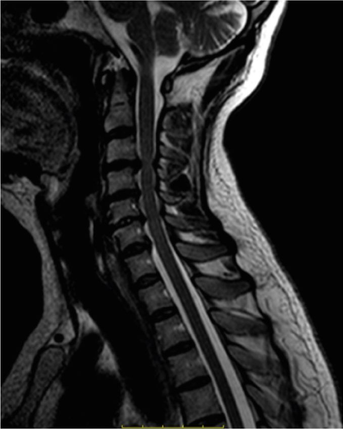
DEGENERATIVE CERVICAL MYELOPATHY: A REVIEW OF CURRENT CONCEPTS
303
funcional. Sin tratamiento, los pacientes pueden progresar para parálisis significativa y pérdida de función. El tratamiento requiere
cirugía de descompresión anterior o posterior del área estenosada y probable fusión. Los factores de mal pronóstico prevalecen
en el sexo femenino e incluyen sintomatología con duración superior a 18 meses y aumento de la amplitud de movimiento de
la columna cervical. En este estudio, presentamos una visión general del estado del arte en MCD, con énfasis de fisiopatología,
presentación clínica, diagnóstico diferencial, evaluación por imagen, historia natural, opciones de tratamiento y complicaciones.
Nivel de evidencia III; Artículo de revisión.
Descriptores: Compresión de la Médula Espinal; Espondiloses; Columna Vertebral.
INTRODUCTION posterior longitudinal ligament (PLL).6,7 (Figure 1) Dynamic factors
relate to the exacerbation of spinal cord compression seen with physi-
The overall frequency of neck pain is estimated to be 34%, and
ological movement, and in the context of degenerative subluxation,
previous studies have shown that the frequency of complaints lasting
pathological motion of the cervical spine. Asians are at increased
1 month or longer was higher in women than in men. The prevalence
risk of cervical myelopathy (1.9% to 4.3% of individuals older than 30
of chronic pain and longer pain duration increases with age.1-5 years) due to increased prevalence of ossification of the PLL.
Myelopathy is a disorder that results from severe compression When considering injury to the spinal cord itself, there is histopath-
of the spinal cord. It is caused by a variety of conditions, including ological evidence to suggest that there is a reduction in blood supply
congenital stenosis, degenerative changes, rheumatoid arthritis, leading to considerable ischemia within the spinal cord, as well as
and trauma. Cervical spondylotic myelopathy is the most common physical compression.8 Pathological features of DCM include gray and
cause of cervical spinal cord dysfunction in individuals older than 55 white matter degeneration, anterior horn cell loss, cystic cavitation, and
years.3 This debilitating condition is called cervical spondylotic my- Wallerian degeneration of the posterior columns adjacent to the site
elopathy (CSM), which reflects the fact that the myelopathy is often of compression. In rat models, Karadimas et al.,9,10 demonstrated de-
associated with and caused by the normal osteoarthritic changes creased capillary density in the compressed spinal cord compared to
of the axial spine.3,6 controls, indicating disruption of the blood-spinal cord barrier (BSCB)
in the context of progressive stenosis due to DCM. In addition to the
Pathophysiology
above, with breakdown of the normal BSCB, a secondary cascade of
The pathophysiology of degenerative cervical myelopathy (DCM) neuroinflammation consisting of microglia activation and macrophage
is multifactorial and can be divided into static and dynamic factors. recruitment occurs at the site of mechanical compression within the
Static factors result from congenital stenosis or acquired stenosis sec- spinal cord.10 Recent literature has investigated the inflammatory path-
ondary to spondylosis and disk degeneration. (Figure 1) Intervertebral ways involved in DCM, in particular, the increased local expression of
discs lose elasticity and hydration due to the loss of proteoglycan the CX3CR1 gene and its association with increased microglia and
matrix with age, which leads to a biomechanical incompetence that macrophage accumulation at the compression sites.11
predisposes the disc to collapse.6 This may or may not result in her-
niation of the annulus, causing symptoms. The resulting cervical cord Clinical Evaluation
stenosis may be aggravated by ossification and hypertrophy of the The onset of symptoms is usually after the age of 40, often
posterior facet joints, uncinate processes, ligamentum flavum, and between 50 and 70 years, but it can also occur in the elderly. It is
C2 Dura
Posterior longotudinal
ligament (PLL)
Ligamentum flavum
Dura
CSF
Hypertrophy of lig. flavum
Increased ant-post C3
vertebral body length Spinal cord
Osteophyte
Spinal cord compression
with cavitation
Loss of vertebral body C4
height Dissociation of PLL from
vertebral
Loss of intervertebral
disc height with migration
of disc material into canal
C5
Hourglass reshaping Ossification of lig. flavum
Hypertrophy of PLL
C6
Hypermobility and listhesis Ossification of PLL
C7
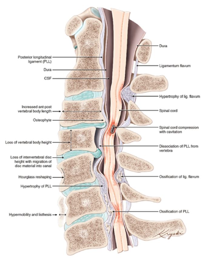
Figure 1. Pathology of DCM. (Left) Anatomy of an initially healthy spine (C2 level), with examples of the potential pathological changes that can occur and
cause DCM (shown at lower spinal levels; C3-7). (Figure reproduced with permission from Diana Kryski and Michael G Fehlings).
Coluna/Columna. 2020;19(4):302-7304
more prevalent in men, at a ratio of 3 to 2.12 Typically, the onset is MRI, flexion-extension radiography, and computed tomography (CT).
insidious, although symptoms can develop acutely or subacutely, Narrowing of the disc space, bone spurs, osteophytes, joint sub-
often after minor head and neck trauma. A prospective study by luxation, facet joint arthrosis, spondylolisthesis, and ossification of
Rhee et al.,13 found that 79% of CSM patients had at least one posterior longitudinal ligament may be visualized on standard films.
myelopathic sign on objective physical examination, compared to Patients with a congenitally narrow canal (< 13 mm) or with
57% of healthy, normal patients. a cord compression greater than 30% are at a higher risk for de-
The lateral corticospinal tracts (voluntary skeletal muscle control) veloping clinical features from static mechanical compression.21
and the spinocerebellar tracts (proprioception) are frequently involved White et al., showed that cervical myelopathy should be strongly
in DCM. Together, these deficits are responsible for the wide-based suspected when the dynamic canal space during extremes of
spastic gait with clumsy upper extremity function that is classic to cer- flexion or extension is < 11 mm21. In two studies, published in
vical myelopathy.14 Additional commonly involved spinal cord regions 1986 and 1987,22,23 American orthopedic surgeon Joseph Torg
are the spinothalamic tracts, which are responsible for contralateral and American radiologist Helene Pavlov introduced the ratio
pain and temperature sensation, the posterior columns, which are method for reducing inter- and intraobserver error caused by
responsible for ipsilateral position and vibration sense, and the dorsal variance in magnification and landmarking. This error can be
nerve root, which is responsible for dermatomal sensation.15,16 calculated by dividing the AP diameter of the spinal canal by
Examination of patients with CSM typically reveals lower motor neu- the AP diameter of the vertebral body. A Pavlov’s ratio < 0.8 is
ron signs at the level of the cervical lesions (weakness and hyporeflexia) suggestive of stenosis.
and upper motor neuron signs below the lesions (spasticity, hyperreflexia Weight-bearing films, including flexion-extension radiographs,
of the deep tendons, clonus, Hoffman sign, Babinski sign and absence are the definitive study for the assessment of cervical lordosis and
of fasciculation or fibrillation). Other tests such as the Spurling sign, the evidence of instability that may predispose to a repetitive trauma
Lhermitte sign, crossed radial reflex, inverted radial reflex, finger escape mechanism underlying CSM.24 CT scans are the best method to
sign and grip test should be included in the initial evaluation. identify bony and calcific changes, including calcified disks, ossifi-
Symptoms in CSM are characteristically persistent rather than cation of the posterior longitudinal ligament, and facet hypertrophy.
transient or fluctuating. About 75% of patients report clumsy hands CT may also demonstrate ankylosis of the uncovertebral and/or
and over 80% report unsteady gait and/or upper extremity sensory facet joints, which are important for the restoration of lordosis.
changes such as shock-like paresthesias with neck flexion (Lhermitte Finally, noncontrast CT scan suggests the course of the vertebral
sign).17 Pain is a less frequently noted complaint and its absence arteries, potentially revealing anomalies such as tortuous vertebral
may often lead to a delay in diagnosis. It has been strongly suggest- artery or ponticulus posticus, which must be considered before
ed that subtle gait disturbance is the most common presentation, corpectomy and placement of C1 screws, respectively. In the pres-
followed by loss of fine motor control of the hands with associated ence of an abnormally tortuous vertebral artery, using anatomic
numbness. Bladder and/or bowel incontinence and quadriparesis landmarks to guide decompression may not prevent iatrogenic
affect 15 to 50% of patients, and are usually associated with late injury to the vertebral artery.25,26
stage myelopathy. Its development is due to long tract involvement, MRI demonstrates disk bulges, disk-osteophyte complexes,
resulting in reduced sphincter control.6 facet and ligamentous hypertrophy, as well as possible listhe-
In their study of 120 patients with cervical myelopathy, Clarke and sis, all of which contribute to spinal canal stenosis and spinal
Robinson described the natural history of this condition as following cord compression. Increased T2 signal is sometimes seen in the
one of several patterns: 5% had rapid onset of symptoms followed by spinal cord, suggesting spinal cord injury, including myelomala-
long periods of remission; 20% had a slow gradual decline in func- cia, from either cord compression or repetitive trauma. Patients
tion; and 75% had a stepwise decline in function.18 Over the years, presenting with multisegmental T2 changes on MRI often have
additional studies have documented a highly variable natural history. longstanding CSM and poor neurological outcome even with
A systematic review conducted in 2013 by Karadimas et al.17 decompression surgery.27
sought to more specifically quantify the rate of clinic deterioration Since conventional MRI is not able to reveal the microstructural
in patients presenting with DCM treated nonoperatively. Of the 6 characteristics of the spinal cord, including the fiber tract of the
studies that considered Japanese Orthopaedic Association (JOA) or white matter, alternative approaches, such as diffusion tensor im-
modified JOA (mJOA) scores, 20% to 62% of patients experienced aging (DTI), have been developed, which may enable detailed in
clinical deterioration, defined by at least a 1-point reduction in mJOA/ vivo analysis of the diffusion of water molecules. DTI is based on
JOA compared to baseline, at 3 to 6 years of follow-up. anisotropic diffusion parameters, and takes conventional MRI one
Many disease severity classifications have been used, such step further by measuring differences in the amount and orienta-
as the European Myelopathy Score, Nurick’s Functional Scale, the tion of water diffusion, particularly in the white matter.28,29 It is a
Ranawat Classification of Neurological Deficit and the modified promising novel imaging modality offering increased diagnostic
Japanese Orthopaedic Association scoring system (mJOA). A study sensitivity as compared to standard MRI, and enabling earlier de-
by Kopjar et al. validated the mJOA, and marked the first step in the tection of CSM before T2 changes appear on conventional MRI.
use of this scale as the standard for assessing severity in patients Once the diagnosis of CSM is established, repeated imaging is
with DCM. Fehlings et al. defined mild myelopathy as a score greater not needed unless there has been significant clinical worsening,
than or equal to 15, moderate myelopathy as a score from 12 to 14, or to aid in surgery planning.
and severe disease as a score of less than 12.19 The spectrum of neurophysiological assessment consists of
Multiple sclerosis, amyotrophic lateral sclerosis, osteomyelitis, electromyography (EMG), electroneurography (ENG), and evoked
borreliosis (Lyme disease), polyradiculitis (Landry-Guillain-Barré), potentials. Electromyography (EMG) and Somatosensory Evoked
vascular and neoplastic causes and psychogenic disorders should Potentials (SSEPs) are diagnostic investigations that are infrequently
be considered in the differential diagnosis of cervical spondylosis with used to exclude differential diagnoses such as multiple sclerosis,
involvement of neural structures leading to cervical spinal myelopathy. amyotrophic lateral sclerosis, and peripheral neuropathy.30
Imaging Treatment
Evidence of radiological degenerative changes to the cervical Studies comparing conservative versus surgical treatment in
spine in the aging population are common. By the fourth decade mild CSM are conflicting. Some studies showed no difference
of life, 30% of asymptomatic subjects show degenerative changes between the two groups, while others showed a deterioration in
in the intervertebral discs, while by the seventh decade, up to 90% patients treated conservatively.31
have developed degenerative alterations.20 A Cochrane review of randomized controlled trials to investigate
Imaging techniques that assist in the diagnosis of CSM include the role of surgery in mild CSM found only one small controlled trial.
Coluna/Columna. 2020;19(4):302-7DEGENERATIVE CERVICAL MYELOPATHY: A REVIEW OF CURRENT CONCEPTS
305
This trial showed no significant differences between surgical and restoration of cervical lordosis. It is considered the gold standard tre-
conservative management in 68 patients after 3 years of follow-up.32 atment for single-level and 2-level disease and is indicated for anterior
The AOSpine CSM North America Study33 remains one of the pathology, particularly in the setting of cervical kyphosis. It provides
largest prospective studies conducted to evaluate the impact of relatively easy access to the vertebral column, and the surgical ou-
surgical management on clinical outcomes. At 1-year follow-up, tcome is satisfactory in most cases.39 However, ACDF is relatively
across the entire spectrum of injury severity, statistically and clinically contraindicated in patients with stenosis primarily from a posterior
significant improvements were noted for functional outcome (mJOA origin, located behind the vertebral body rather than in the disk space,
and Nurick grade) and disability outcome (Neck Disability Index in patients with ossification of the posterior longitudinal ligament, and
[NDI]), as well as generic health-related quality of life (SF-36). The is less commonly performed in patients with >3 levels of disease.
results of that study seem to indicate that surgery is most effective Cervical disk arthroplasty is a relatively new surgical treatment
in restoring function in those with moderate or severe myelopathy at option. A recent multicenter randomized controlled trial comparing
presentation, but also that cervical decompression prevents further cervical disk arthroplasty with anterior cervical decompression sho-
deterioration and improves neurological outcomes, functional status wed similar improvement in neurologic status at 2-year follow-up.40
and quality of life (QOL). In one of the largest studies to evaluate this question, Fehlings et
A subsequent prospective study (AOSpine International CSM)34 al.40 performed a retrospective cohort study comparing 169 subjects
confirmed some of the findings reported in the North American treated anteriorly (ACDF or anterior cervical corpectomy and fusion)
Study, providing external validity to its conclusions. with 95 subjects treated posteriorly (laminectomy and fusion (86%)
In another prospective study on this topic, Sampath et al.35 asses- or with laminoplasty). They found no differences in mJOA recovery
sed rating of neurological symptoms, activities of daily living, pain, and or other outcomes, including NDI, SF-36, Nurick and complications.
ability to work, amongst two cohorts of DCM patients: one cohort trea- To collect evidence on this topic, Lawrence et al.41 performed a
ted with surgical intervention and the other with nonsurgical treatment systematic review that summarized the findings of 8 retrospective
(pharmacological therapy with either narcotic or nonsteroidal drugs, cohort studies comparing anterior and posterior surgery for multi-
steroids, bed rest, home exercise, cervical traction, neck bracing, or level CSM. In summary, regarding the change in mJOA score from
spinal injections). Compared to baseline, nonsurgical patients had preoperatively to postoperatively, neither approach was consistently
an increased number of neurological symptoms, worsened activities associated with superior outcomes.
of daily living, and a more severe pain rating at 11-month follow-up. In a patient with multilevel cervical spondylosis in whom an an-
Several retrospective studies have demonstrated no benefit of rigid terior approach is decided upon, the choice of optimal operative
immobilization in the absence of surgery.36 intervention remains controversial. Shamji et al.42 demonstrated similar
In summary, a large body of literature has shown that surgical trajectories of neurological recovery for patients managed by these va-
management of DCM has the potential to improve outcomes, and rious options, although sagittal correction and neck pain scores were
to maintain these results after a long period of follow-up. best among the multiple discectomy cohort and worst among the
Although it remains unclear whether patients with mild CSM corpectomy cohort. A similar incidence of perioperative complications
benefit from surgery when compared to conservative management, was noted among the different procedures, namely pseudoarthrosis,
patients with moderate to severe CSM or progressive neurologic dysphagia, and infection. The strong recommendations arising from
deficits definitely do. The decision to proceed with surgery must this systematic review were that when minimal retrovertebral disease
consider the patient’s age, baseline function, severity of symp- is present, multiple discectomy should be selected over hybrid or
toms, and the historical rate of progression. Nonsurgical treatment corpectomy procedures. On the other hand, in patients with significant
is a viable option in patients with radiographic evidence of cervical retrovertebral disease, hybrid procedures should be selected.
stenosis without clinical signs or symptoms of myelopathy. Non-
-surgical management for those with mild symptoms should include Posterior approaches
anti-inflammatories, physical therapy, ultrasound modalities, and Posterior procedures, such as laminoplasty and laminectomy,
occasionally, corticosteroid injections. If there is a deterioration in can achieve spinal cord decompression by expanding the canal in
the patient’s clinical condition, these short-term alternatives will not a posterior direction.43
provide definitive treatment. The objective of both laminectomy with fusion and laminoplasty
is to decompress the spinal cord. The primary indication for these
Surgical Treatment procedures is multilevel cord compression in the absence of kyphosis.
Prolonged compression of the spinal cord can result in irreversi- Although multilevel laminectomy was historically the most popu-
ble damage, and the results of operative treatment are better when lar posterior operation for the treatment of DCM, the relatively high
there is early intervention. Surgical intervention can be considered in rates (15% - 20%) of postlaminectomy kyphosis reported have led
two anatomical areas: the upper (C0-C2) and lower (C3-C7) cervical to a gradual rejection of this approach and to the nearly uniform
spine, and by three general approaches: anterior, posterior, and adoption of either laminectomy with fusion or laminoplasty when
combined anterior and posterior. considering a posterior operation.44 Laminectomy with instrumen-
The four surgical procedures commonly performed to treat CSM tation was also associated with loss of lordosis and increase in C2
are anterior cervical discectomy and fusion (ACDF), anterior cervical SVA, these variables being negatively correlated with health-related
corpectomy and fusion (ACCF), laminectomy, and laminoplasty. The outcomes. On the contrary, the anterior approach has been shown to
appropriate choice depends on the presence of preoperative neck be more associated with a superior capacity of kyphosis correction
pain, the level and degree of spondylotic change, sagittal align- and the maintenance of postoperative lordosis than laminoplasty
ment and stability of the spine, individual characteristics including and laminectomy with instrumentation.15
previous surgery, and surgical expertise. Regardless of the chosen Laminoplasty can be an advantageous option in lordotic spines
procedure, the goals of operative treatment are to decompress the in younger patients where fusion is undesirable. The two main la-
spinal cord and its circulation, preserve the alignment and stability minoplasty techniques are the ‘French Door’ and the ‘Open Door’
of the spine, and prevent further neural injury. techniques. One study reported that laminoplasty is not an effective
treatment for axial neck pain, and that axial symptoms may worsen
Anterior approaches post-procedure. It is also contraindicated in patients with instability
Initially described by Robinson and Smith in 1955 and Cloward resulting from trauma or rheumatoid arthritis.45
in 1958, ACDF has become one of the most common surgical pro-
cedures for both CSM and cervical radiculopathy.37,38 The goals of Combined approaches
ACDF are direct decompression of the cervical spinal cord from its In complex cases, especially when there is compression from
ventral pathology, direct and indirect foraminal decompression, and both anterior and posterior structures, both approaches may be
Coluna/Columna. 2020;19(4):302-7306
used concurrently. Indications include patients with fixed kyphosis, for single-level fusions and up to 50% for multilevel fusions.58 Rates
multilevel stenosis, instability, and poor bone quality. Adding pos- after posterior laminectomy with fusion range from 1% to 38%.59
terior stabilization may address the issue of suboptimal stability, C5 palsy is an established neurological complication and gene-
particularly in cases where there is extensive resection.46,47 rally manifests as delayed-onset painful deltoid and biceps weakness,
Shamji et al.48 demonstrated that patients with preoperative kypho- either unilateral or bilateral.60 No statistically significant differences in
sis exhibit less neurological recovery than those with preoperative the rates of C5 palsy between anterior and posterior approaches have
lordosis, independently of whether alignment was corrected at the been reported. A retrospective review of 1001 cases by Bydon et al.61
time of surgery. The kyphosis group also deteriorated more frequen- found that in anterior surgeries, older age most strongly predicted
tly, particularly when only the posterior approach was used, which development of C5 palsy while in posterior approaches, the strongest
supports the importance of restoring the cervical sagittal alignment. predictor was C4–C5 foraminotomy. The authors also reported a hi-
gher incidence of C5 palsy in corpectomies versus ACDF.
Surgical complications Postlaminectomy kyphosis has a significant harmful complica-
Fehlings et al.49 described an overall perioperative complication tion in adult patients undergoing a laminectomy without fusion for
rate of 15.6% and an overall delayed complication rate of 4.4%. CSM.44 The incidence of postlaminectomy kyphosis in the setting
Perioperative worsening of myelopathy occurred in 1.3% and the of CSM in the adult population is approximately 20%.45 Cumula-
mortality rate was 0.33%. Most of the reported complications were tive data suggest that kyphosis following cervical laminectomy
treatable and had no long-term impacts. The occurrence of perio- without fusion develops more rapidly the less spondylotic and
perative complications has been associated with increased age, the younger the patient is.
combined procedures, increased surgical time, and greater ope-
rative blood loss. CONCLUSION
The anterior approach to the cervical spine is one of the most
commonly performed spinal procedures. Fountas et al.50 stated that Cervical spondylotic myelopathy is a debilitating condition com-
the overall morbidity rate in their ACDF series was 19.3%, while monly affecting the elderly that occurs as a result of degenerative
the mortality rate was 0.1%. Similarly, Veeravagu et al.51 estimated changes, leading to cord compression.
that the overall mortality at 2 years was 0.1% in single level ACDF The clinical diagnosis of myelopathy requires a detailed history
procedures, and 0.18% among their multilevel cases. Bilbao et al. and physical examination to define the syndrome. Because of the
reported a complication rate of 25% in patients undergoing cervical wide-ranging pathologies contributing to DCM development, the
spondylotic corpectomies.52 constellation of findings in any given patient may vary considerably.
Complications of anterior cervical surgery include dysphagia, re- Neuroimaging is indicated in most instances of new onset myelopa-
current laryngeal nerve palsy, vertebral artery injury, Horner syndrome, thy. Static and dynamic factors should be considered when estab-
postoperative hematoma, esophageal injury, unintended dural tear, lishing the causative features in the development of this condition.
superficial wound infection, and hardware complications.52,53 Some There is scant evidence for nonoperative treatment of cervi-
studies have shown that recurrent laryngeal nerve (RLN) palsy is un- cal myelopathy, and further studies are needed to establish its
derreported.54,55 Dysphonia and/or hoarseness are the most common role between treatment modalities. At present, acknowledging the
clinical expression of unilateral vocal paralysis. Incidental durotomy methodological limitations of existing studies, the body of evidence
is rare, with a reported incidence of 0.2–0.5%.50 Although rare, one supports the efficacy of surgical treatment for symptomatic patients
of the most serious complications that has been reported in anterior with moderate to severe DCM. Although these procedures are as-
cervical corpectomy is inadvertent laceration of the vertebral artery, sociated with severe potential risks, most perioperative and delayed
with a reported incidence during cervical surgery of 0.3–0.5%.53 postoperative complications from surgery are treatable and do not
Cloward reported one of the first graft migrations.56 Since then, have any long-term impacts.
mechanical failure and/or screw migration have been described.
Initial misplacement of the implant is the most common reason for
mechanical fatigue and eventual failure of the implant.57 All authors declare no potential conflict of interest related to
Nonunion can produce persistent neck pain and radicular symp-
this article.
toms. Rates of pseudarthrosis following ACDF range from 1% to 20%
CONTRIBUTION OF THE AUTHORS: Each author made significant individual contributions to this manuscript. EMP: writing, statistical analysis,
intellectual concept, preparation of the entire research project and approval of the final version of the work; AT: analysis of the data for the work, critical
review of its intellectual content and approval of the final version of the work; RF: analysis of the data for the work, critical review of its intellectual content
and approval of the final version of the work; PA: analysis of the data for the work, critical review of its intellectual content and approval of the final version
of the work; FO: analysis of the data for the work, critical review of its intellectual content and approval of the final version of the work; AM: analysis of the
data for the work, critical review of its intellectual content and approval of the final version of the work.
REFERENCES
1. Luschka H. Die Halbgelenke des menschlichen Körpers. Berlin: Reimers; 1858. tion of the posterior longitudinal ligament: A minimum 10-year cohort study. J Neurosurg.
2. Tetreault L, Goldstein CL, Arnold P, Harrop J, Hilibrand A, Nouri A, et al. Degenera- 2004;100(3 Suppl Spine):245-8.
tive cervical myelopathy: a spectrum of related disorders affecting the aging spine. 8. Baptiste DC, Fehlings MG. Pathophysiology of cervical myelopathy. Spine J. 2006;6(6
Neurosurgery. 2015;77(suppl 4):S51-67. Suppl):190S-7S.
3. Nouri A, Tetreault L, Singh A, Karadimas SK, Fehlings MG. Degenerative cervi- 9. Karadimas SK, Moon E, Fehlings M. The sodium channel/glutamate blocker riluzole is com-
cal myelopathy: epidemiology, genetics, and pathogenesis. Spine (Phila Pa 1976). plementary to decompression in a preclinical experimental model of cervical spondylotic
2015;40(12):E675-93. myelopathy: implications for translational clinical application. Spine J. 2012;12(9):S88-9.
4. Nagoshi N, Tsuji O, Okada E, Fujita N, Yagi M, Tsuji T, et al. Clinical indicators of surgi- 10. Karadimas SK, Laliberte AM, Tetreault L, Chung YS, Arnold P, Foltz WD, et al. Riluzo-
cal outcomes after cervical single open-door laminoplasty assessed by the Japanese le blocks perioperative ischemia-reperfusion injury and enhances postdecompression
Orthopaedic Association Cervical Myelopathy Evaluation Questionnaire. Spinal Cord. outcomes in cervical spondylotic myelopathy. Sci Transl Med. 2015;7(316):316ra194.
2019;57(8):644-51. 11. Yu WR, Karadimas S, Fehlings M. Human and animal model evidence supporting a role for
5. Nakajima H, Uchida K, Taguchi T, Yamashita T, Tominaga T, Tanaka M, et al. Multi- Cx3cr1 in mediating the inflammatory response in cervical spondylotic myelopathy. Abstract
center cross-sectional study of the clinical features and types of treatment of spinal Presented at: The 2012 Society for Neuroscience meeting in October, New Orleans; 2012.
cord-related pain syndrome. J Orthop Sci. 2019;24(5):798-804. 12. Northover JR, Wild JB, Braybrooke J, Blano J. The epidemiology of cervical spondylotic
6. Clark C, Frymoyer JW. The adult spine: Principles and practice, (2nd ed). Philadelphia, USA: myelopathy. Skeletal Radiol. 2012;41(12):1543-6.
Lippincott-Raven publications; 1997. 13. Rhee JM, Heflin JA, Hamasaki T, Freedman B. Prevalence of physical signs in cervical myelo-
7. Matsunaga S, Sakou T, Taketomi E, Komiya S. Clinical course of patients with ossifica- pathy: a prospective, controlled study. Spine (Phila Pa 1976). 2009;34(9):890–5.
Coluna/Columna. 2020;19(4):302-7DEGENERATIVE CERVICAL MYELOPATHY: A REVIEW OF CURRENT CONCEPTS
307
14. Baron EM, Young WF. Cervical spondylotic myelopathy: a brief review of its pathophysiolo- 40. Fehlings MG, Barry S, Kopjar B, Yoon ST, Arnold P, Massicotte EM, et al. Anterior versus
gy, clinical course, and diagnosis. Neurosurgery. 2007;60(1 Suppl 1):S35–41. posterior surgical approaches to treat cervical spondylotic myelopathy: outcomes of the
15. Hitchon PW, Woodroffe RW, Noeller JA, Helland L, Hramakova N, Nourski KV. An- prospective multicenter AOSpine North America CSM study in 264 patients. Spine (Phila Pa
terior and Posterior Approaches for Cervical Myelopathy: Clinical and Radiographic 1976). 2013;38(26):2247-52.
Outcomes. Spine (Phila Pa 1976). 2019;44(9):615-23. 41. Lawrence BD, Jacobs WB, Norvell DC, Hermsmeyer JT, Chapman JR, Brodke DS. Anterior
16. Grelat M, Gimenez C, Madkouri R. Cervical Cord Compression by Exostosis. J Orthop versus posterior approach for treatment of cervical spondylotic myelopathy: a systematic
Sports Phys Ther. 2019;49(2):112. review. Spine (Phila Pa 1976). 2013;38(22 Suppl 1):S173-82.
17. Karadimas SK, Erwin WM, Ely CG, Dettori JR, Fehlings MG. Pathophysiology and natural history 42. Shamji MF, Massicotte EM, Traynelis VC, Norvell DC, Hermsmeyer JT, Fehlings MG. Compa-
of cervical spondylotic myelopathy. Spine (Phila Pa 1976). 2013;38 (22 Suppl 1):S21-36. rison of anterior surgical options for the treatment of multilevel cervical spondylotic myelopa-
18. Clarke E, Robinson P. Cervical myelopathy: a complication of cervical spondylosis. Brain. thy: a systematic review. Spine (Phila Pa 1976). 2013;38(22 Suppl 1):S195- 209.
1956;79(3):483-510. 43. Zhou X, Cai P, Li Y, Wang H, Xia S, Wang X. Posterior or single-stage combined ante-
19. Fehlings MG, Wilson JR, Kopjar B, Yoon ST, Arnold PM, Massicotte EM, et al. Efficacy rior and posterior approach decompression for treating complex cervical spondylotic
and safety of surgical decompression in patients with cervical spondylotic myelopathy: myelopathy coincident multilevel anterior and posterior compression. Clinical Spine
results of the AOSpine North America prospective multi-center study. J Bone Joint Surg. Surgery. 2017;30(10):E1343–51.
2013;95(18):1651–8. 44. Kaptain GJ, Simmons NE, Replogle RE, Pobereskin L. Incidence and outcome of kyphotic de-
20. Dvorak J, Sutter M, Herdmann, J. Cervical myelopathy: clinical and neurophysiological eva- formity following laminectomy for cervical spondylotic myelopathy. J Neurosurg. 2000;93(2
luation. Eur Spine J. 2003;12(Suppl 2):S181–7. Suppl):199-204.
21. White AA, Panjabi MM. Biomechanical considerations in the surgical management of cervi- 45. Ohnari H, Sasai K, Akagi S, Iida H, Takanori S, Kato I. Investigation of axial symptoms after
cal spondylotic myelopathy. Spine (Phila Pa 1976). 1988;13(7):856-60. cervical laminoplasty, using questionnaire survey. Spine J. 2006;6(3):221-7.
22. Torg JS, Pavlov H, Genuario SE, Sennett B, Wisneski RJ, Robie RH, et al. Neurapraxia of the 46. Williams KE, Paul R, Dewan Y. Functional outcome of corpectomy in cervical spondylotic
cervical spinal cord with transient quadriplegia. J Bone Joint Surg Am. 1987;68(9):1354-70. myelopathy. Indian J Orthop. 2009;43(2):205–9.
23. Pavlov H, Torg JS, Robie B, Jahre C. Cervical spinal stenosis: determination with vertebral 47. Kim PK, Alexander JT. Indications for circumferential surgery for cervical spondylotic my-
body ratio method. Radiology. 1987;164(3):771-5. elopathy. Spine J. 2006;6(6 Suppl):299S–307S.
24. Vedantam A, Rajshekhar V. Does the type of T2-weighted hyperintensity influence surgical outco- 48. Shamji MF, Mohanty C, Massicotte EM, Fehlings MG. The Association of Cervi-
me in patients with cervical spondylotic myelopathy? A review. Eur Spine J. 2013;22(1):96–106. cal Spine Alignment with Neurologic Recovery in a prospective cohort of patients
25. Young JP, Young PH, Ackermann MJ, Anderson PA, Riew KD. The ponticulus posticus: with surgical myelopathy: analysis of a series of 124 cases. World Neurosurg.
implications for screw insertion into the first cervical lateral mass. J Bone Joint Surg Am. 2016;86:112-9.
2005;87(11):2495–8.
49. Fehlings MG, Smith JS, Kopjar B, Arnold PM, Yoon ST, Vaccaro AR, et al. Perioperative
26. Curylo LJ, Mason HC, Bohlman HH, Yoo JU. Tortuous course of the vertebral artery and
and delayed complications associated with the surgical treatment of cervical spondylotic
anterior cervical decompression: a cadaveric and clinical case study. Spine (Phila Pa 1976).
myelopathy based on 302 patients from the AOSpine North America Cervical Spondylotic
2000;25(22):2860–4.
Myelopathy Study. J Neurosurg Spine. 2012;16(5):425–32.
27. Rota JJF, Meschian S, Rota AF, Urbano V, Baron M. Cervical spondylotic myelopathy due to
50. Fountas KN, Kapsalaki EZ, Nikolakakos LG, Smisson HF, Johnston KW, Grigorian AA, et al.
chronic compression: The role of signal intensity changes in magnetic resonance images. J
Anterior cervical discectomy and fusion associated complications. Spine (Phila Pa 1976).
Neurosurg Spine. 2007;6(1):17–22.
2007;32(21):2310-7.
28. Shabani S, Kaushal M, Budde MD, Wang MC, Kurpad SN. Diffusion tensor imaging
51. Veeravagu A, Cole T, Jiang B, Ratliff JK. Revision rates and complication incidence in single-
in cervical spondylotic myelopathy: a review. J Neurosurg Spine. 2020:1-8.
and multilevel anterior cervical discectomy and fusion procedures: an administrative data-
29. Stino MA, LoRusso SJ. Myelopathies due to structural cervical and thoracic disease.
Continuum (Minneap Minn). 2018;24(2, Spinal Cord Disorders):567-83. base study. Spine J. 2014;14(7):1125-31.
30. Campbell WW. Focal neuropathies. In: Campbell WW (ed). Essentials of Electrodiagnostic 52. Bilbao G, Duart M, Aurrecoechea JJ, Pomposo I, Igartua A, Catalán G, et al. Surgical results
Medicine. Baltimore, USA: Williams &Willkins publications; 1999. 255-78. and complications in a series of 71 consecutive cervical spondylotic corpectomies. Acta
31. Matz PG, Anderson PA, Holly LT, Groffy MW, Heary RF, Kaiser MG, et al. The natural history Neurochir (Wien). 2010;152(7):1155-63.
of cervical spondylotic myelopathy. J Neurosurg Spine. 2009;11(2):104–11. 53. Burke JP, Gerszten PC, Welch WC. Iatrogenic vertebral artery injury during anterior cervical
32. Nikolaidis I, Fouyas IP, Sandercock PA, Stathan PF. Surgery for cervical radiculopathy or spine surgery. Spine J. 2005;5(5):508–14; discussion 14.
myelopathy. Cochrane Database Syst Rev. 2010;20(1):CD001466. 54. Ebraheim NA, Lu J, Yang H, Heck BE, Yeasting RA. Vulnerability of the sympathetic
33. Fehlings MG, Wilson JR, Kopjar B, Yoon ST, Arnold PM, Massicotte EM, et al. Efficacy trunk during the anterior approach to the lower cervical spine. Spine (Phila Pa 1976).
and safety of surgical decompression in patients with cervical spondylotic myelopathy: 2000;25(13):1603–6.
results of the AOSpine North America prospective multi-center study. J Bone Joint Surg 55. Netterville JL, Koriwchak MJ, Winkle M, Courey MS, Ossoff RH. Vocal fold paralysis following
Am. 2013;95(18):1651-8. the anterior approach to the cervical spine. Ann Otol Rhinol Laryngol. 1996;105(2):85-91.
34. Fehlings MG, Ibrahim A, Tetreault L, Albanese V, Alvarado M, Arnold P, et al. A global 56. Cloward RB. Complications of anterior cervical disc operation and their treatment. Sur-
perspective on the outcomes of surgical decompression in patients with cervical gery. 1971;69(2):175-82.
spondylotic myelopathy: results from the prospective multicenter AOSpine internatio- 57. Yen CP, Hwang TY, Wang CJ, Howng SL. Fracture of anterior cervical plate implant - report
nal study on 479 patients. Spine (Phila Pa 1976). 2015;40(17):1322-8. of two cases. Acta Neurochir (Wien). 2005;147(6):665-7; discussion 667.
35. Sampath P, Bendebba M, Davis JD, Ducker TB. Outcome of patients treated for cervical 58. Wang JC, McDonough PW, Endow KK, Delamarter RB. Increased fusion rates with cervi-
myelopathy: A prospective, multicenter study with independent clinical review. Spine (Phila cal plating for two-level anterior cervical discectomy and fusion. Spine (Phila Pa 1976).
Pa 1976). 2000;25(6):670-6. 2000;25(1):41–5.
36. Gok B, Sciubba DM, McLoughlin GS, McGirt M, Ayhan S, Wolinsky JA, et al. Surgical treat- 59. Yoon ST, Hashimoto RE, Raich A, Shaffrey CI, Rhee JM, Riew KD. Outcomes after lami-
ment of cervical spondylotic myelopathy with anterior compression: a review of 67 cases. J noplasty compared with laminectomy and fusion in patients with cervical myelopathy: a
Neurosurg Spine. 2008;9(2):152–7. systematic review. Spine (Phila Pa 1976). 2013;38(22 Suppl 1):S183–94.
37. Cloward RB. The anterior approach for removal of ruptured cervical disks. J Neurosurg. 60. Radcliff KE, Limthongkul W, Kepler CK, Sidhu GDS, Anderson DG, Rihn JA, et al. Cervical
1958;15(6):602–17. laminectomy width and spinal cord drift are risk factors for postoperative C5 palsy. J Spinal
38. Cloward RB. History of the anterior cervical fusion technique. J Neurosurg. 1985;63(5):817-9. Disord Tech. 2014;27(2):86–92.
39. Sasso RC, Anderson PA, Riew KD, Heller JG. Results of cervical arthroplasty compared with 61. Bydon M, Macki M, Kaloostian P, Sciubba DM, Wolinsky JP, Gokaslan ZL, et al. Incidence
anterior discectomy and fusion: four-year clinical outcomes in a prospective, randomized and prognostic factors of c5 palsy: a clinical study of 1001 cases and review of the literature.
controlled trial. J Bone Joint Surg Am. 2011;93(18):1684–92. Neurosurgery. 2014;74(6):595–604; discussion 5.
Coluna/Columna. 2020;19(4):302-7You can also read