SARS-COV-2 ENGAGES INFLAMMASOME AND PYROPTOSIS IN HUMAN PRIMARY MONOCYTES - NATURE
←
→
Page content transcription
If your browser does not render page correctly, please read the page content below
Ferreira et al. Cell Death Discovery (2021)7:43
                                    https://doi.org/10.1038/s41420-021-00428-w                                                                                                           Cell Death Discovery

                                     ARTICLE                                                                                                                                                    Open Access

                                    SARS-CoV-2 engages inflammasome and
                                    pyroptosis in human primary monocytes
                                    André C. Ferreira1,2,3, Vinicius Cardoso Soares1,4, Isaclaudia G. de Azevedo-Quintanilha1, Suelen da Silva Gomes Dias1,
                                    Natalia Fintelman-Rodrigues1,3, Carolina Q. Sacramento1,3, Mayara Mattos1,3, Caroline S. de Freitas1,3,
                                    Jairo R. Temerozo5,6, Lívia Teixeira1, Eugenio Damaceno Hottz 1,7, Ester A. Barreto 1, Camila R. R. Pão1,
                                    Lohanna Palhinha1, Milene Miranda8, Dumith Chequer Bou-Habib5,6, Fernando A. Bozza9,10, Patrícia T. Bozza1 and
                                    Thiago Moreno L. Souza1,3

                                      Abstract
                                      Infection by the severe acute respiratory syndrome coronavirus 2 (SARS-CoV-2) has been associated with leukopenia
                                      and uncontrolled inflammatory response in critically ill patients. A better comprehension of SARS-CoV-2-induced
                                      monocyte death is essential for the identification of therapies capable to control the hyper-inflammation and reduce
                                      viral replication in patients with 2019 coronavirus disease (COVID-19). Here, we show that SARS-CoV-2 engages
                                      inflammasome and triggers pyroptosis in human monocytes, experimentally infected, and from patients under
                                      intensive care. Pyroptosis associated with caspase-1 activation, IL-1ß production, gasdermin D cleavage, and enhanced
                                      pro-inflammatory cytokine levels in human primary monocytes. At least in part, our results originally describe
                                      mechanisms by which monocytes, a central cellular component recruited from peripheral blood to respiratory tract,
                  1234567890():,;
1234567890():,;
                  1234567890():,;
1234567890():,;

                                      succumb to control severe COVID-19.

                                    Introduction                                                                                        inflammatory mediators TNF-α and IL-6 in the respira-
                                      Severe acute respiratory coronavirus 2 (SARS-CoV-2),                                              tory tract in peripheral blood4. Plasmatic levels of IL-6
                                    the etiological agent of the 2019 coronavirus disease                                               have been associated with mortality, intensive care
                                    (COVID-19), emerged in China, causing a major public                                                admission and hospitalization, representing a poor prog-
                                    health burden in decades. Although patients with severe                                             nostic factor for COVID-195. The uncontrolled inflam-
                                    COVID-19 may present an asymptomatic/mild disease,                                                  mation triggered by SARS-CoV-2 occurs 7 to 10 days after
                                    others experience acute respiratory distress syndrome                                               onset of illness and associates with decreased viral
                                    (ARDS) characterized by elevated serum levels of proin-                                             loads6,7.
                                    flammatory mediators—the cytokine storm1–3. Patients                                                   In severe COVID-19, the cytokine storm associates with
                                    with severe COVID-19, monocytes/macrophages may be                                                  high levels of tissue insult, judged by increased levels
                                    the main source of uncontrolled levels of the pro-                                                  lactate dehydrogenase (LDH) and D-dimer in the
                                                                                                                                        plasma6–8. Moreover, high LDH levels and leukopenia in
                                                                                                                                        severe COVID-19 points out that white cells loses the
                                    Correspondence: André C. Ferreira (tmoreno@cdts.fiocruz.br) or Thiago                                integrity of plasma membrane8–11. Among these cells,
                                    Moreno L. Souza (andre.bio2009@gmail.com)
                                    1                                                                                                   monocytes should orchestrate the equilibrium between
                                     Laboratório de Imunofarmacologia, Instituto Oswaldo Cruz (IOC), Fundação
                                    Oswaldo Cruz (Fiocruz), Rio de Janeiro, RJ, Brazil                                                  innate and adaptative immune responses, which may be
                                    2
                                     Laboratório de Pesquisa Pré-clínica—Universidade Iguaçu - UNIG, Nova                               presumably affected during cytokine storm. The leuko-
                                    Iguaçu, RJ, Brazil
                                                                                                                                        penia of patients with severe COVID-19 seem to precede
                                    Full list of author information is available at the end of the article
                                    These authors contributed equally: André C. Ferreira, Vinicius Cardoso Soares                       the cytokine storm11,12. Moreover, in other virus-induced
                                    Edited by Ivano Amelio

                                    © The Author(s) 2021, corrected publication 2021
                                                        Open Access This article is licensed under a Creative Commons Attribution 4.0 International License, which permits use, sharing, adaptation, distribution and reproduction
                                                        in any medium or format, as long as you give appropriate credit to the original author(s) and the source, provide a link to the Creative Commons license, and indicate if
                                    changes were made. The images or other third party material in this article are included in the article’s Creative Commons license, unless indicated otherwise in a credit line to the material. If
                                    material is not included in the article’s Creative Commons license and your intended use is not permitted by statutory regulation or exceeds the permitted use, you will need to obtain
                                    permission directly from the copyright holder. To view a copy of this license, visit http://creativecommons.org/licenses/by/4.0/.

                                    Official journal of the Cell Death Differentiation Association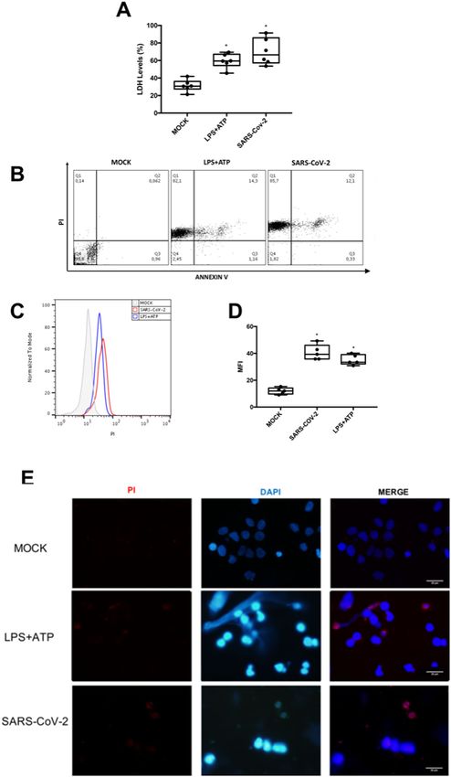
Ferreira et al. Cell Death Discovery (2021)7:43                                                               Page 2 of 12

cytokine storm episodes in the respiratory tract, such as       lytic death (Fig. 2A). Next, we measured caspase-1 acti-
induced by influenza A virus, monocytes and macro-               vation, a downstream event to inflammasome engage-
phages are severely affected13–16.                              ment20. SARS-CoV-2 induced pro-caspase-1 cleavage,
   There are various mechanisms involved in lytic cell          similarly to LPS + ATP, by western blotting and flow
death, which are differently engaged from development to        cytometry analysis (Fig. 2B, C). Apoptotic caspases-3/-7
responses to infection17. For certain diseases, such as         were not upregulated in SARS-CoV-2-infected monocytes
COVID-19, in which the immunopathogenesis mechan-               (Fig. 2F, G).
isms associate with poor clinical outcomes, controlling           Caspase-1 activation is known to trigger IL-1β pro-
the way cells collapse to infection is vital for the host17.    duction and GSDMD cleavage20–24. Indeed, SARS-CoV-2
Lytic cell death may be triggered by pyroptosis or              infection increased IL-1β production, which was pre-
necroptosis, in monocytes/macrophages this phenom-              vented by selective inhibition of capase-1 (AC-YVAD-
enon exacerbates inflammation, because of release of             CMK) or by treatment with pan-caspase inhibitor
intracellular damage-associated molecular patterns18,19.        (Z-VAD-FMK) (Fig. 2H). Impairment of NLPR3, by gly-
Pyroptosis is a caspase-1-dependent event that leads to         buride, also prevented SARS-CoV-2-induced IL-1β pro-
gasdermin D (GSDMD) pore formation20. Necroptosis               duction (Fig. 2H). RIPK inhibition by necrostatin could
depends on intracellular signaling of receptor-interacting      not prevent significantly IL-1β production (Fig. 2H).
protein kinases (RIPK) and mixed lineage kinase domain-           Based on the pharmacological characterization, SARS-
like phosphorylation to form membrane pores20. Thus, we         CoV-2 depends on cellular components associated with
hypothesized that the monocyte cell death induced by            pyroposis to promote lytic monocyte death. We thus
SARS-COV-2 exacerbates the production of inflamma-               measured the cleaved GSDMD as a proxy of the pore
tory cytokines, as well as impairs the immune balance in        forming structure. In fact, SARS-CoV-2, similarly to LPS
the hosts. In fact, we found that SARS-CoV-2 engages            + ATP, enhanced cleaved GSDMD levels (Fig. 2I).
inflammasomes and pyroptosis in human monocytes, by              Importantly, inhibition of IL-1R engagement reduced
experimental or natural infection. These events were            SARS-CoV-2-mediated caspase-1 activation and cell lysis
associated with caspase-1 activation, IL-1ß production,         (Fig. S1A, B), suggesting that inflammasome-dependent
GSDMD cleavage and dysregulation of cytokine release.           IL-1β secretion amplify caspase-1 activation and pyr-
Finally, we show that inhibition of early steps of SARS-        optosis in SARS-CoV-2 infection.
CoV-2 life cycle by atazanavir (ATV) could block pyr-
optosis in SARS-CoV-2-infected human primary                    Inflammasome activation amplify pro-inflammatory
monocytes.                                                      cytokines secretion in infected monocytes
                                                                  We subsequently quantified the levels of IL-6 and TNF-
Results                                                         α, in SARS-CoV-2-infected monocytes upon pharmaco-
Lytic cell death in SARS-CoV-2-infected human primary           logical treatment to block inflammasome and pyroptosis.
monocytes                                                       Inhibition of caspase-1, NLP3, and IL-1 receptor pre-
  For initial assessments of SARS-CoV-2-induced                 vented SARS-CoV-2-induced enhancement of IL-6 and
monocyte death, we quantified the LDH activity in the            TNF-α levels (Fig. 3A, B). In addition, inhibition of IL-1
culture supernatant and annexinV/propidium iodide (PI)          receptor engagement also reduced SARS-CoV-2-
labeling through flow cytometry and fluorescence                  dependent production of IL-1 β (Fig. S1C). Moreover,
microscopy. SARS-CoV-2 increased LDH levels similarly           IL-8 production, which is independent of inflammasome
to the positive control, LPS + ATP (Fig. 1A). SARS-CoV-2        engagement and pyroptosis, was only modulated by pan-
was as potent as LPS + ATP to increase PI+ cells by 300-        caspase inhibition in SARS-CoV-2-infeced monocytes
fold and PI+/annexinV+ cells by 200-fold (Fig. 1B). Flow        (Fig. S2).
cytometry and fluorescence microscopy also reconfirm
plasma membrane disruption triggered by SARS-CoV-2              ATV prevented SARS-CoV-2-induced inflammasome-
in a similar magnitude to the positive control (Fig. 1B–E).     engagement and pyroptosis
These data suggest that SARS-CoV-2 infection can induce           Since lytic cell death in monocytes may depend on
lytic cell death in human primary monocytes.                    SARS-CoV-2 replication, we tested whether orally avail-
                                                                able generic antivirals could prevent these events. Flow
Inflammasome engagement and pyroptotic events in                 cytometry analysis of SARS-CoV-2-infected monocytes
SARS-CoV-2-infected monocytes                                   treated with ATV demonstrated a significant reduction in
  Severe COVID-19 was associated with inflammasome               caspase-1 activity (Fig. 4A, B). Other orally available
activation21. Indeed, we observed that glyburide, which         repurposed anti-SARS-CoV-2 drugs, such as lopinavir
prevents NLR family pyrin domain containing 3 (NLRP3)           (LPV) and ribavirin (RBV), did not affect SARS-CoV-2-
activation22, abolished SARS-CoV-2-dependent monocyte           induced caspase-1 activation (Fig. 4C). Moreover,

Official journal of the Cell Death Differentiation Association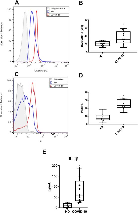
Ferreira et al. Cell Death Discovery (2021)7:43                                                                                     Page 3 of 12

  Fig. 1 SARS-CoV-2 induces lytic monocyte cell death. Human monocytes were infected with SARS-CoV-2 (MOI 0.1) for 24 h. As a positive control,
  monocytes were stimulated with LPS (500 ng/mL) for 23 h and, after this time, incubated with ATP (2 mM) for 1 h. A Cell viability was assessed
  through the measurement of LDH levels in the culture supernatant. B–D Monocytes were stained with propidium iodide (PI) and Annexin V.
  E Monocytes were stained with PI and DAPI, scale bar 20 μm. Images and graph data are representative of six independent experiments. Data are
  presented as the mean ± SEM *P < 0.05 versus control group (MOCK-infected).

Official journal of the Cell Death Differentiation Association
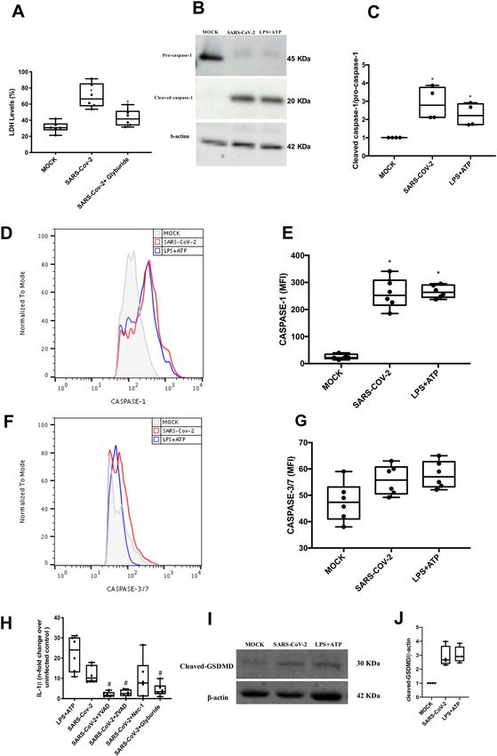
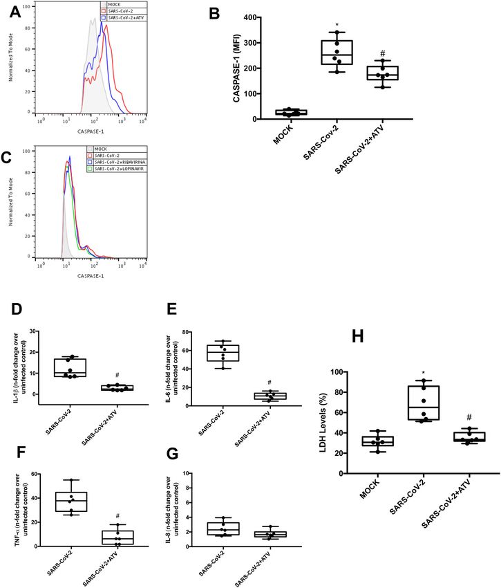
Ferreira et al. Cell Death Discovery (2021)7:43                                                                                            Page 4 of 12

  Fig. 2 Lytic cell death in SARS-CoV-2-infected monocytes associates with inflammasome engagement and pyroptosis. Human monocytes
  were treated with pharmacological inhibitors to impair the function of the following proteins: NLPR3 (Gliburyde; 100 µM), caspase-1 (AC-YVAD-CMK;
  1 µM), or pan-caspase (Z-VAD-FMK; 10 µM) or RIPK (Nec-1; 25 µM). Monocytes were treated since 1 h prior to infection with SARS-CoV-2 (MOI 0.1), for
  24 h. As a control, monocytes were stimulated with LPS (500 ng/mL) for 23 h and, after this time, stimulated with ATP (2 mM) for 1 h. A Cell viability
  was assessed through the measurement of LDH levels in the supernatant of monocytes. B, C Monocytes were lysed and used for determination of
  pro-caspase-1 and cleaved caspase-1 levels by western blotting. D, E Monocytes were stained with FAM-YVAD-FLICA to determine the caspase-1
  activity by flow cytometry. F, G Monocytes were stained with FAM-FLICA to determine caspase-3/7 activity by flow cytometry. H Cell culture
  supernatants were collected for the measurement of the levels of IL-1β. I, J Monocytes were lysed and cleaved GSDMD levels were determined
  western blotting. Western blotting images, histogram and graph data are representative of six independent experiments. Data are presented as the
  mean ± SEM #P < 0.05 versus infected and untreated group; *P < 0.05 versus control group (MOCK-infected).

Official journal of the Cell Death Differentiation Association
Ferreira et al. Cell Death Discovery (2021)7:43                                                                                           Page 5 of 12

  Fig. 3 Caspase-1- and IL-1 receptor-dependent amplification pro-inflammatory cytokines secretion in SARS-CoV-2-infected monocytes.
  Human monocytes were pharmacologically treated to impair the function of caspase-1 (AC-YVAD-CMK; 1 µM), pan-caspase (ZVAD-FMK; 10 µM), RIPK
  (Nec-1; 25 µM), IL-1ß receptor (IL-1RA; 1 µM), or NLPR3 (glyburide 100 µM) since 1 h prior to infection at MOI of 0.1 with SARS-CoV-2. Monocytes were
  stimulated with LPS (500 ng/mL) for 23 h and after this time were stimulated with ATP (2 mM) for 1 h as a positive control. Cell culture supernatants
  were collected for the measurement of the levels of A IL-6 and B TNF-α. Graphs data are representative of six independent experiments. Data are
  presented as the mean ± SEM #P < 0.05 versus infected and untreated group.

treatments with ATV did not alter the activity of caspase-                    sections stand on the shoulders of the clinical relevance of
1, -3 and -7 in monocytes exposed to LPS + ATP (Fig. S3),                     monocytes in patients with severe COVID-19.
indicating a specific antiviral activity of this drug. Con-
sistently, treatment with ATV also reduced the levels of                      Discussion
IL-1β, IL-6, and TNF-α in SARS-CoV-2-infected mono-                             COVID-19 has caused in over 100,000 deaths/month
cytes, when compared to the untreated cells (Fig. 4D–F).                      worldwide21 and represent the major public health crisis of
ATV did not interfere with the production of IL-8, which                      the beginning of 21st century, leading to an unpredictable
is independent of virus-induced inflammasome engage-                           impact in global economics25,26. SARS-CoV-2 infection
ment (Fig. 4G). Consistently, lower levels of LDH were                        triggers an uncontrolled inflammatory response and
detected in the supernatants of SARS-CoV-2-infected                           marked leukopenia with consequent lung/respiratory dys-
monocytes treated with ATV, when compared to infected                         function27,28. Similarly, to other respiratory viruses29–31,
and untreated cells (Fig. 4H). Since ATV inhibits the early                   SARS-CoV-2 induces a cytokine storm, characterized by an
proteolytic processing of viral antigens, an early event                      uncontrolled inflammatory response mediated by mono-
during SARS-CoV-2 replication, this drug represented the                      cytes/macrophages, when they should orchestrate the
most upstream process pharmacologically inhibited in                          antiviral immune response32. In this work, we demonstrate
this investigation to prevent lytic monocyte death.                           that SARS-CoV-2 engages inflammasome, with subsequent
                                                                              caspase-1 activation, increase IL-1ß levels and GSDMD
Caspase 1- activation, lytic monocyte death, and Il-1β                        pore formation in human primary monocytes, pointing
associate with severe COVID-19                                                toward a pyroptotic cell death. This deleterious immune
  To clinically validate our findings, we evaluated if                         dysregulation loop triggered by SARS-CoV-2 may be
monocytes obtained from critically ill patients with                          impaired by ATV, glyburide and the blockage of the IL-1
COVID-19 would also display signals of pyroptosis-like                        receptor.
caspase-1-dependent cell death. We observed that                                Our results demonstrate that SARS-CoV-2 leads to an
monocytes from COVID-19 patients had increased                                intense cell death in human primary monocytes, observed
caspase-1 activation (Fig. 5A, B) and significantly higher                     by the increase in LDH release in infected cultures, as well
lytic death, by PI+ staining, when compared to monocytes                      as by the higher number of PI+ cells when compared to
from healthy donors (HD) (Fig. 5C, D). Corroborating                          uninfected controls, both in experimental infection and in
with our in vitro data, we also detected higher levels of IL-                 patients with severe COVID-19. Pyroptosis is an inflam-
1β in the plasma of critically ill patients, compared to HD                   matory and lytic programmed cell death triggered after
(Fig. 5E). Therefore, the in vitro results from the previous                  inflammasome engagement18–20, it has been demonstrated

Official journal of the Cell Death Differentiation Association
Ferreira et al. Cell Death Discovery (2021)7:43 Page 6 of 12 Fig. 4 Atazanavir prevented capase-1-dependent lytic monocyte death. Human monocytes were infected with SARS-CoV-2 and treated with atazanavir (ATV; 10 µM), ribavirina (10 µM), or Lopinavir (10 µM) for 24 h. A, B Monocytes were stained with FAM-YVAD-FLICA to determine caspase-1 activity. C Monocytes were stained with FAM-FLICA to determine caspase-3/7 activity. D–G Culture supernatants were collected and the levels of IL- 1β, IL-6, TNF-α, and IL-8 were determined by ELISA. H Assessment of cell viability through the measurement of LDH levels in the supernatant of monocytes. Histogram and graphs data are representative of six independent experiments. Data are presented as the mean ± SEM * P < 0.05 versus control group (MOCK-infected); #P < 0.05 versus only infected group. Official journal of the Cell Death Differentiation Association
Ferreira et al. Cell Death Discovery (2021)7:43                                                                                           Page 7 of 12

  Fig. 5 Caspase 1 activation, lytic monocyte death, and increased Il-1β lvels associate with severe COVID-19. Monocytes were isolated from
  blood samples of critically ill patients with COVID-19 and healthy donors. These cells were stained with FAM-YVAD-FLICA (A, B) or propidium iodide
  (PI) (C, D). Plasma samples were analyzed for IL-1β levels (E). Western blotting, histogram and graphs data are representative of nine critically ill
  patients and eight healthy donors. Data are presented as the mean ± SEM *P < 0.05 versus healthy donors (HD).

Official journal of the Cell Death Differentiation Association
Ferreira et al. Cell Death Discovery (2021)7:43                                                               Page 8 of 12

that NLRP3 inflammasome activation in peripheral blood           monocytes/macrophages are key cells in the deleterious
mononuclear cells (PBMCs) associated with severe                pro-inflammatory events that characterize the most ser-
COVID-1921. We confirmed that SARS-CoV-2 infection               ious cases of COVID-1947–49.
triggers NLRP3 inflammasome engagement in monocytes,               In this work, we originally describe that infection by
because glyburide, a drug for treatment of type 2 diabetes      SARS-CoV-2 can engage inflammasome activation and
which modulates ATP-dependent K+ channel22, could               pyroptotic cell death, which may be related to the intense
prevent virus-induced cell lysis. Next, we further demon-       leukopenia and exacerbated inflammation seen in severe
strated that downstream events could be prevented by            cases of the COVID-19. Since there is no specific therapy
early inhibition of virus replication, at protein translation   for this disease, our results point out that ATV has a
levels, by ATV. Conversely, inhibition of viral RNA             promising therapeutic potential against SARS-CoV-2-
synthesis, by RBV did not affect cell death. Based on           induced cell death.
analogy with SARS-CoV from 2002, late viral proteins E,
Orf3, and Orf8, which are synthetized after RNA synthesis,      Material and methods
have been implicated in inflammasome activation33. Thus,         Reagents
it likely that other nonstructural proteins (nsp) from            ATV and RBV were received as donations from Insti-
SARS-CoV-2 can affect intracellular K+ imbalance to             tuto de Tecnologia de Fármacos (Farmanguinhos, Fio-
trigger NLRP3. Among the possible SARS-CoV-2 proteins,          cruz). The antiviral LPV/ritonavir (4:1 proportion) was
nsp6 may affect K+ channels34.                                  pruchased from AbbVie (Ludwingshafen, Germany).
   Moreover, activation of caspase-1, increased production      ELISA assays were purchased from R&D Bioscience.
of IL-1ß and GSDMD pore was documented here in                  Lipopolysacchadides—LPS,       adenosine     triphosphate
SARS-CoV-2-infected monocytes, points towards pyr-              (ATP), the specific inhibitor of caspase-1 (AC-YVAD-
optotic cell death These in vitro results are in accordance     CMK), pan-caspase inhibitor (ZVAD-FMK), RIPK1
with the literature and with our finding described here          (Necrostatin-1—Nec-1), IL-1 receptor (IL-1RA) and gly-
that indicate the formation of inflammasomes in patients         buride were all purchased from Sigma-Aldrich (St. Louis,
with severe COVID-1935. We also showed that the release         MO, USA). All small molecule inhibitors were dissolved
of IL-1ß could be promoted by the activation of inflam-          in 100 % dimethylsulfoxide (DMSO) and subsequently
masomes during the SARS-CoV-2 infection, because                diluted at least 104-fold in culture or reaction medium
blockage of IL-1ß receptors reduced caspase-1 activation        before each assay. The final DMSO concentrations
and cell death. These results corroborate with studies          showed no cytotoxicity. The materials for cell culture
showing that the increase in IL-1ß production is asso-          were purchased from Thermo Scientific Life Sciences
ciated with severe COVID-1936–43. Under our experi-             (Grand Island, NY), unless otherwise mentioned.
mental conditions, the increase in IL-1ß precedes the
unbalanced IL-6 release. These information should not be        Cells and virus
neglected when considering biopharmaceuticals to tackle           African green monkey kidney (Vero, subtype E6) cells
cytokine storm in severe COVID-19.                              were cultured in DMEM high glucose supplemented with
   To establish the clinical relevance of SARS-CoV-2-           10% fetal bovine serum (FBS; HyClone, Logan, Utah) and
induced pyroptosis in monocytes44,45, we analyzed per-          100 U/mL penicillin, and 100 μg/mL streptomycin (P/S).
ipheral monocytes isolated from patients with severe            Vero cells were incubated at 37 °C in 5% CO2 atmosphere.
COVID-19. We found that the cells from the patients               Human primary monocytes were obtained through
displayed higher caspase-1 activation, when compared            plastic adherence of PBMCs, which were obtained from
with monocytes isolated from HD. Recent clinical data           buffy coat preparations of HDs by density gradient cen-
reveal high levels of LDH and consistent leukopenia in          trifugation (Ficoll–Paque, GE Healthcare). In brief,
critically ill COVID-19 patients6,7,35,42–46. Our data also     PBMCs (2.0 × 10⁶ cells) were plated onto 48-well plates
demonstrate intense monocyte death in COVID-19                  (NalgeNunc) in RPMI-1640 without serum for 2–4 h;
patients, as detected through flow cytometry analyzes.           then, non-adherent cells were removed by washing and
Altogether, these data suggest that the severity of COVID-      the remaining monocytes were maintained in DMEM
19 may be associated with inflammasome activation in             with 5% human serum (HS; Millipore) and penicillin/
monocytes that results in large amounts of IL-1ß and            streptomycin.
generates an excessive inflammatory response, further              SARS-CoV-2 was isolated and expanded on Vero E6
characterized by high levels of IL-6 and TNF-α. Con-            cells from a nasopharyngeal swab of a confirmed case
sistently, treatment inhibition of IL-1 receptor has been       from Rio de Janeiro, Brazil. Experiments were performed
associated with clinical and inflammatory improvements           after one passage in cell culture, when Vero E6 cells with
in critically ill COVID-19 patients42. These results are in     DMEM plus 2% FBS in 150 cm2 flasks were incubated at
line with clinical case reports that demonstrate that           37 °C in 5% CO2 atmosphere. Cytopathic effect was

Official journal of the Cell Death Differentiation Association
Ferreira et al. Cell Death Discovery (2021)7:43                                                                 Page 9 of 12

observed daily and peaked 4–5 days after infection. All         Infection was performed for 2 h at 37 °C and then fresh
procedures related to virus culture were handled at bio-        medium with 2% FBS was added. After 24 h, the cells were
safety level 3 (BSL3) multiuser facility, according to WHO      washed with Binding buffer and stained with PI (0.5 µg/mL)
guidelines. Virus titers were determined as the tissue          for 5 min. Next, the cells were fixed with 3.7% formaldehyde
culture infectious dose at 50% (TCID50/mL). Virus stocks        for 30 min at room temperature. The nuclei were stained
were kept in −80 °C ultralow freezers. The virus strain         with DAPI (1 µg/mL) for 5 min and the coverslips were
was sequenced to confirm the virus identity and its              mounted using an antifade mounting medium (VECTA-
complete genome is publicly deposited (GenBank acces-           SHIELD). Fluorescence was analyzed by fluorescence
sion No. MT710714).                                             microscopy with an ×100 objective lens (Olympus, Tokyo,
                                                                Japan).
Yield-reduction assay
  Human primary monocytes were infected with multi-             Measurements inflammatory mediators and cell death
plicity of infection (MOI) of 0.01 at density of 2–8 × 10⁵      marker
cells/well in 48-well culture plates, depending on the total      The levels of IL-1ß, TNF-α, IL-6, IL-8, and LDH were
cell number from each donor. After 1 h at 37 °C, cells were     quantified in the culture supernatants from infected and
washed, and various concentrations of compounds were            uninfected monocytes using ELISA kits, according to the
added in DMEM with 2% FBS. After 48 h, the supernatants         manufacturer’s intructions (R&D System).
were harvested and virus replication was quantified by real        Extracellular LDH was quantified using Doles® kit
time RT-PCR and infectious titers by TCID50/mL.                 according to manufacturer’s instructions. In brief, cell
                                                                culture supernatants were centrifuged at 5000 rpm for
Virus titration                                                 1 min, to remove cellular debris, and then 25 μL were
  Monolayers of Vero cells (2 × 10⁴ cell/well) in 96-well       placed into 96-well plates and incubated with 5 μL of
culture plates were infected with log-based dilutions of        ferric alum and 100 μL of LDH substrate for 3 min at
the supernatants containing SARS-CoV-2 for 1 h at 37 °C.        37 °C. Nicotinamide adenine dinucleotide (oxidized form)
The cells were washed and fresh medium with 2% FBS              was added followed by the addition of a stabilizing solu-
was added. After 3–5 days, the cytopathic effects were          tion. After 10 min, the reaction was read in a spectro-
scored in at least ten replicates per dilution by indepen-      photometer at 492 nm.
dent readers, who were blind with respect to source of the
supernatant. Reed and Muench scoring method was                 Western blot assay
employed to determine TCID50/mL.                                  Cellular extracts of 1 × 10⁶ cells were homogenized in
                                                                the RIPA lysis buffer in the presence of proteinase inhi-
Flow cytometer analysis                                         bitor cocktail (Roche), and the protein levels were mea-
  For flow cytometry analysis, monocytes were diluted in         sured by bicinchoninic acid assay protein assay kit. A total
labeling buffer (106 cells/mL). Then, 100 µL of cell sam-       of 20 μg of protein was loaded onto a 10% sodium dodecyl
ples were marked with 5 µL of AnnexinV and PI for               sulfate polyacrylamide gel for separation by electrophor-
15 min for cell death evaluation. Around 10,000 gated           esis and the protein bands were then transferred to a
events were acquired using FACSCalibur and the analysis         polyvinylidene difluoride membranes (ImmobilonP-SQ,
was performed using the CellQuest software. Monocytes           Millipore). The membranes were blocked with 5% albu-
were gated through cell size (foward light scatter) and         min diluted in Tris-buffered saline containing 0.05% of
granularity (side light scatter) analysis.                      Tween 20 for 2 h at room temperature and incubated with
  Human monocytes were stained for caspase‐1 activity           the specific primary antibodies (Cell Signaling Technol-
with FAM‐YVAD‐FMK (fluorescent‐labeled inhibitor of              ogy), to detect pro-caspase-1 and cleaved-caspase-1, after
caspase‐1 [FLICA] and FAM-FLICA Caspase-3/7 activity            overnight incubation at 4 °C. After washing, membranes
or HLA-DR APC.H7 or IgG APC.H7. Caspase-1 and                   were incubated with secondary antibodies (IRDye®
caspase-3/7 activity was determined via flow cytometry           800CW Goat-anti-Mouse and IRDye® 680LT Goat anti-
(FACSCalibur) by detecting FLICA fluorescence and                Rabbit IgG Antibody, LI-COR, Lincoln) for 30 min at RT.
expression of HLA-DR as mean fluorescence intensity              The protein bands were visualized by digital fluorescence
(MFI) value for each sample. Acquisition of data was set        (Odyssey® CLx Imaging System), and protein density was
to count a total of 10,000 events, and the FLOWJO soft-         analyzed by the ImageJ software. All the data were nor-
ware package was used to analyze the data.                      malized by β-actin expression quantification.

Microscopic analysis                                            Human subjects
   Human primary monocytes were plated on glass cover-            We prospectively enrolled severe COVID19 RT-PCR-
slips at density of 2–8 × 10⁵ cells/well in 48-well plates.     confirmed cases, as well as SARS-CoV-2-negative healthy

Official journal of the Cell Death Differentiation Association
Ferreira et al. Cell Death Discovery (2021)7:43 Page 10 of 12
controls. Blood samples were obtained from 12 patients Table 1 Characteristics of COVID-19 patients and control
with severe COVID-19 within 72 h from intensive care subjects.
unit admission in two reference centers (Instituto Esta-
Characteristicsa Control (n = 8) Covid-19 (n = 9)
dual do Cérebro Paulo Niemeyer and Hospital Copa Star,
Rio de Janeiro, Brazil). Severe COVID-19 was defined as Age, years 52.6 (47–60) 61 (33–79)
critically ill patients presenting viral pneumonia on
Sex, male 8 (100%) 6 (66.7%)
computed tomography scan and in mechanical ventila-
tion. All patients had SARSCoV-2 confirmed diagnostic Respiratory support
through RT-PCR of nasal swab or tracheal aspirates. Oxygen supplementation 0 (0%) 3 (33.3%)
Peripheral vein blood was also collected from eight SARS- Mechanical ventilation 0 (0%) 6 (66.7%)
CoV-2-negative healthy control participants as tested by
SAPS 3 59.25 (31–75)
RT-PCR on the day of blood sampling. The characteristics
of severe (n = 12), and control (n = 8) participants are PaO2/FiO2 ratio – 210.75 (87–509.5)
presented in Table 1. Severe COVID-19 patients usually Vasopressor – 3 (33.3%)
present older age and higher prevalence of comorbidities Time from symptom onset to – 10 (6–18)
as obesity, cardiovascular diseases and diabetes as in blood sample, days
previously reported patient cohorts. In the present study,
Status on Jun 30th
the SARS-CoV-2-negative control group was designed to
include subjects of older age and chronic non- Dead – 5 (55.55%)
communicable diseases, so it is matched with critically Discharged – 1 (11.1%)
ill COVID-19 patients (Table 1). Patients with ARDS were Hospitalized – 3 (33.3%)
managed with neuromuscular blockade and a protective
Comorbidities
ventilation strategy that included low tidal volume (6 mL/
kg of predicted body weight) and limited driving pressure Obesity 1 (11.1%) 1 (11.1%)
(less than 16 cm H2O) as well as optimal PEEP calculated Hypertension 3 (33.3%) 3 (33.3%)
based on the best lung compliance and PaO2/FiO2 ratio. In Diabetes 0 (0%) 0 (0%)
those with severe ARDS and PaO2/FiO2 ratio below 150
Cancer 0 (0%) 0 (0%)
despite optimal ventilatory settings, prone position was
initiated. Our management protocol included antith- Chronic heart diseaseb 0 (0%) 0 (0%)
rombotic prophylaxis with enoxaparin 40–60 mg per day. Presenting symptoms
Patients did not receive routine steroids, antivirals, or Cough 0 (0%) 3 (33,3%)
other anti-inflammatory or anti-platelet drugs. The SARS-
Fever 0 (0%) 3 (33,3%)
CoV-2-negative control participants were not under anti-
inflammatory or anti-platelet drugs for at least 2 weeks. All Dyspnea 0 (0%) 4 (44,4%)
clinical information were prospectively collected using a Headache 0 (0%) 0 (0%)
standardized form—ISARIC/WHO Clinical Characteriza- Anosmia 0 (0%) 0 (0%)
tion Protocol for Severe Emerging Infections (CCPBR).
Laboratory findings on admission
Ethics statement Lymphocyte count, cells/mm3 – 1355.8 (552–2564)
Experimental procedures involving human cells from Platelet count, ×1000/mm 3
– 169.6 (92–278)
HDs were performed with samples obtained after written Leukocytes – 8036 (8.03–18,670)
informed consent and were approved by the Institutional
C reactive protein, mg/L – 14.78 (0.1–30.8)
Review Board (IRB) of the Oswaldo Cruz Foundation/Fio-
a
cruz (Rio de Janeiro, RJ, Brazil) under the number 397-07. Numerical variables are represented as the median and the max range, and
qualitative variables are represented as the number and the percentage.
The National Review Board approved the study protocol b
Coronary artery disease or congestive heart failure
(CONEP 30650420.4.1001.0008), and informed consent was
obtained from all participants or patients’ representatives. were generated based on R2 values ≥ 0.9. Student’s t test was
used to access statistically significant P values < 0.05.
Statistical analysis
The assays were performed in blinded way. They were Acknowledgements
performed by one professional, codified and read by We thank the Hemotherapy Service from Hospital Clementino Fraga Filho
another fellow. All experiments were carried out at least six (Federal University of Rio de Janeiro, Brazil) for providing buffy-coats. This work
was supported by grants from Fundação de Amparo a Pesquisa do Estado do Rio
independent times, including a minimum of two technical de Janeiro (FAPERJ), Inova Fiocruz, Conselho Nacional de Desenvolvimento
replicates in each assay. The equations to fit the best curve Científico e Tecnológico (CNPq) and Coordenação de Aperfeiçoamento de
Official journal of the Cell Death Differentiation AssociationFerreira et al. Cell Death Discovery (2021)7:43 Page 11 of 12
Pessoal de Nível Superior (CAPES) and Mercosur Fund for Structural Convergence 9. Chen, Z., John & Wherry, E. T cell responses in patients with COVID-19. Nat.
(FOCEM, Mercosur, grant number 03/11) granted for Thiago Moreno L. Souza, Rev. Immunol. 20, 529–536 (2020).
Patrícia T. Bozza, Fernando A. Bozza, and Dumith Chequer Bou-Habib. 10. Xu, Z. et al. Pathological findings of COVID-19 associated with acute respira-
tory distress syndrome. Lancet Respir. Med. 8, 420–422 (2020).
Funding 11. Han, Y. et al. Lactate dehydrogenase, an independent risk factor of severe
Funding was also provided by CNPq, CAPES, and FAPERJ through the National COVID19 patients: a retrospective and observational study. Aging 12,
Institutes of Science and Technology Program (INCT) to Carlos Morel (INCT-IDPN) 11.245–11.258 (2020).
and to Wilson Savino (INCT-NIM). Thanks are due to Oswaldo Cruz Foundation/ 12. Terpos, E. et al. Hematological findings and complications of COVID-19. Am. J.
Fiocruz under the auspicious of Inova program. The funding sponsors had no Hematol. 95, 834–847 (2020).
role in the design of the study; in the collection, analyses, or interpretation of 13. Woo, P. C. Y. et al. Cytokine profiles induced by the novel swine-origin
data; in the writing of the paper, and in the decision to publish the results. influenza A/H1N1 virus: Implications for treatment strategies. J. Infect. Dis. 201,
346–53 (2010).
14. Li, Y. X. et al. Characteristicsof peripheral blood leukocyte differential counts in
patients with COVID-19. Zhonghuanei Ke Za Zhi 59, E003 (2020).
Author details
1 15. Ding, X. et al. Dynamic profile and clinical implications of hematological
Laboratório de Imunofarmacologia, Instituto Oswaldo Cruz (IOC), Fundação
parameters in hospitalized patients with coronavirus disease 2019. Clin. Chem.
Oswaldo Cruz (Fiocruz), Rio de Janeiro, RJ, Brazil. 2Laboratório de Pesquisa Pré-
Lab. Med. 58, 1365–1371 (2020).
clínica—Universidade Iguaçu - UNIG, Nova Iguaçu, RJ, Brazil. 3National Institute
16. Singh, A., Sood, N., Narang, V. & Goyal, A. Morphology of COVID-19-affected
for Science and Technology on Innovation in Diseases of Neglected
cells in peripheral blood film. BMJ Case Rep. 13, e236117 (2020).
Populations (INCT/IDPN), Center for Technological Development in Health
17. Danthi, P. Viruses and the diversity of cell death. Annu. Rev. Virol. 3, 533–553
(CDTS), Fiocruz, Rio de Janeiro, RJ, Brazil. 4Program of Immunology and
(2016).
Inflammation, Federal University of Rio de Janeiro, UFRJ, Rio de Janeiro, RJ,
18. Jorgensen, I. & Miao, E. A. Pyroptotic cell death defends against intracellular
Brazil. 5Laboratório de Pesquisas sobre o Timo, IOC, Fiocruz, Rio de Janeiro, RJ,
pathogens. Immunol. Rev. 265, 130–42 (2015).
Brazil. 6National Institute for Science and Technology on
19. Man, S. M., Karki, R. & Kanneganti, T. D. Molecular mechanisms and functions of
Neuroimmunomodulation, Oswaldo Cruz Institute, Fiocruz, Rio de Janeiro, RJ,
pyroptosis, inflammatory caspases and inflammasomes in infectious diseases.
Brazil. 7Laboratório de Imunotrombose, Departamento de Bioquímica,
Immunol. Rev. 277, 61–75 (2017).
Universidade Federal de Juiz de Fora, Juiz de Fora, MG, Brazil. 8Laboratório de
20. Frank, D. & Vince, J. Pyroptosis versus necroptosis: similarities, differences, and
Vírus Respiratório e do Sarampo, IOC, Fiocruz, Rio de Janeiro, RJ, Brazil.
9 crosstalk. Cell Death Differ. 26, 99–114 (2019).
Instituto Nacional de Infectologia Evandro Chagas, Fiocruz, Rio de Janeiro, RJ,
21. Rodrigues, T. S. et al. Inflammasomes are activated in response to SARS-CoV-2
Brazil. 10Instituto D’or de Pesquisa e Ensino, Rio de Janeiro, RJ, Brazil
infection and are associated with COVID-19 severity in patients. J. Exp. Med.
218, 3 e20201707 (2020).
Conflict of interest 22. Mangan, M. S. J. et al. Targeting the NLRP3 inflammasome in inflammatory
The authors declare no competing interests. diseases. Nat. Rev. Drug Discov. 17, 588–606 (2018).
23. Zalinger, Z. B., Elliott, R. & Weiss, S. R. Role of the inflammasome-related
cytokines Il-1 and Il-18 during infection with murine coronavirus. J. Neurovirol.
Publisher’s note 23, 845–854 (2017).
Springer Nature remains neutral with regard to jurisdictional claims in 24. Franchi, L., Eigenbrod, T., Muñoz-Planillo, R. & Nuñez, G. The inflammasome: a
published maps and institutional affiliations. caspase-1-activation platform that regulates immune responses and disease
pathogenesis. Nat. Immunol. 10, 241–7 (2009).
Supplementary information The online version contains supplementary 25. OPAS. COVID-19 (doença causada pelo novo coronavírus). Folha Inf. Print in
material available at https://doi.org/10.1038/s41420-021-00428-w. https://www.paho.org/pt/covid19 (2020).
26. Dong, E., Du, H. & Gardner, L. An interactive web-based dashboard to track
Received: 7 December 2020 Revised: 15 January 2021 Accepted: 3 February COVID-19 in real time. Lancet Infect. Dis. 20, 533–534 (2020).
2021 27. Wissel, B. D. et al. An interactive online dashboard for tracking COVID-19 in U.S.
counties, cities, and states in real time. J. Am. Med. Inf. Assoc. 27, 1121–1125
(2020).
28. Lucas, C. et al. Longitudinal analyses reveal immunological misfiring in severe
References COVID-19. Nature 584, 463–469 (2020).
1. Conti, P. et al. Induction of pro-inflammatory cytokines (IL-1 and IL-6) and lung 29. Tay, M. Z., Poh, C. M., Rénia, L., MacAry, P. A. & Ng, L. F. P. The trinity of COVID-
inflammation by COVID-19: anti-inflammatory strategies. J. Biol. Regul. Home- 19: immunity, inflammation and intervention. Nat. Rev. Immunol. 20, 363–374
ost. Agents 34, 327–331 (2020). (2020).
2. Long, B., Brady, W. J., Koyfman, A. & Gottlieb, M. Cardiovascular complications 30. Teijaro, J. R. The role of cytokine responses during influenza virus pathogenesis
in COVID-19. Am. J. Emerg. Med. 38, 1504–1507 (2020). and potential therapeutic options. Curr. Top. Microbiol. Immunol. 386, 3–22
3. Li, J. & Fan, J. G. Characteristics and mechanism of liver injury in 2019 cor- (2014).
onavirus disease. J. Clin. Transl. Hepatol. 28, 13–17 (2020). 31. Szretter, K. J. et al. Role of host cytokine responses in the pathogenesis of avian
4. Giamarellos-Bourboulis, E. J. et al. Complex immune dysregulation in COVID- H5N1 influenza viruses in mice. J. Virol. 81, 2736–44 (2007).
19 patients with severe respiratory failure. Cell Host Microbe 27, 992–1000 32. Peschke, T., Bender, A., Nain, M. & Gemsa, D. Role of macrophage cytokines in
(2020). influenza A virus infections. Immunobiology 89, 340–55 (1993).
5. Du, R. H. et al. Predictors of mortality for patients with COVID-19 pneumonia 33. Yap, J. K. Y., Moriyama, M. & Iwasaki, A. Inflammasomes and pyroptosis as
caused by SARS-CoV- 2: a prospective cohort study. Eur. Respir. J. 55, 2000524 therapeutic targets for COVID-19. J. Immunol. 3, ji2000513 (2020).
(2020). 34. Gordon, D. E. et al. A SARS-CoV-2 protein interaction map reveals targets for
6. Zhou, F. et al. Clinical course and risk factors for mortality of adult inpatients drug repurposing. Nature 583, 459–468 (2020).
with COVID-19 in Wuhan, China: a retrospective cohort study. Lancet 395, 35. Jamilloux, Y. et al. Should we stimulate or suppress immune responses in
1054–1062 (2020). COVID-19? Cytokine and anti-cytokine interventions. Autoimmun. Rev. 9,
7. Wölfel, R. et al. Virological assessment of hospitalized patients with COVID- 102567 (2020).
2019. Nature 581, 465–469 (2020). 36. Zheng, Z. et al. Risk factors of critical & mortal COVID-19 cases: a systematic
8. Wang, J., Jiang, M., Chen, X. & Montaner, L. J. Cytokine storm and leukocyte literature review and meta-analysis. J. Infect. 81, e16–e25 (2020).
changes in mild versus severe SARS-CoV-2 infection: review of 3939 COVID-19 37. Abbasifard, M. & Khorramdelazad, H. The bio-mission of interleukin-6 in the
patients in China and emerging pathogenesis and therapy concepts. J. Leukoc. pathogenesis of COVID-19: a brief look at potential therapeutic tactics. Life Sci.
Biol. 108, 17–41 (2020). 257, 118097 (2020).
Official journal of the Cell Death Differentiation AssociationFerreira et al. Cell Death Discovery (2021)7:43 Page 12 of 12
38. Yao, Z., Zheng, Z., Wu, K. & Zheng, J. Immune environment modulation in 44. Qin, C. et al. Dysregulation of immune response in patients with COVID-19 in
pneumonia patients caused by coronavirus: SARS-CoV, MERS-CoV and SARS- Wuhan, China. Clin. Infect. Dis. 71, 762–768 (2020).
CoV-2. Aging 12, 7639–7651 (2020). 45. McGonagle, D., Sharif, K., O’Regan, A. & Bridgewood, C. The role of cytokines
39. Zhang, Y. et al. Analysis of serum cytokines in patients with severe acute including interleukin-6 in COVID-19 induced pneumonia and macrophage
respiratory syndrome. Infect. Immun. 72, 4410–5 (2004). activation syndrome-like disease. Autoimmun. Rev. 19, 102537 (2020).
40. Ong, E. Z. et al. A Dynamic immune response shapes COVID-19 progression. 46. Merad, M. & Martin, J. C. Pathological inflammation in patients with COVID-19:
Cell Host Microbe 27, 879–882.e2 (2020). a key role for monocytes and macrophages. Nat. Rev. Immunol. 20, 448 (2020).
41. Laing, A. G., et al. A dynamic COVID-19 immune signature includes 47. Park, M. D. Macrophages: a Trojan horse in COVID-19? Nat. Rev. Immunol. 20,
associations with poor prognosis. Nat. Med. https://doi.org/10.1038/ 351 (2020).
s41591-020-1038-6 (2020). 48. Fintelman-Rodrigues, N. et al. Atazanavir inhibits SARS-CoV-2 replication and
42. Yan, L. et al. An interpretable mortality prediction model for COVID-19 pro-inflammatory cytokine production. Antimicrob. Agents Chemother. 64,
patients. Nat. Mach. Intell. 2, 283–288 (2020). e00825–20 (2020).
43. Cauchois, R. et al. Early IL-1 receptor blockade in severe inflammatory 49. Hantoushzadeh, S. & Norooznezhad, A. H. Possible cause of inflammatory
respiratory failure complicating COVID-19. Proc. Natl Acad. Sci. USA 117, storm and septic shock in patients diagnosed with (COVID-19). Arch. Med. Res.
18951–18953 (2020). 51, 347–348 (2020).
Official journal of the Cell Death Differentiation AssociationYou can also read


