TESTING STRATEGY UPDATE AUGUST 2020: POOLING, SALIVA TESTING, RT-LAMP, RAPID ANTIGEN TESTING, SELF-COLLECTED NOSE, THROAT AND NASOPHARYNGEAL SWABS ...
←
→
Page content transcription
If your browser does not render page correctly, please read the page content below
TESTING STRATEGY UPDATE AUGUST 2020:
POOLING, SALIVA TESTING, RT-LAMP, RAPID
ANTIGEN TESTING, SELF-COLLECTED NOSE,
THROAT AND NASOPHARYNGEAL SWABS AND
MULTIPLEX
RAG 19/08/2020
1. Context
In the context of the rapidly evolving COVID-19 epidemic, the coming season with circulation of several
respiratory viruses, the evolution of the type of tests available and the possible limitations in testing and
sampling capacity, an update of the testing strategy is necessary, in order to use the available resources
in the best way, potentially with inclusion of new techniques.
Therefore we revised the existing evidence and other countries’ strategies on pooling, saliva testing,
RT-LAMP, rapid antigen test and self-swabbing of nasal, throat and nasopharyngeal samples in order
to make recommendations about the conditions in which these techniques can be used.
Additionally, a recommendation for multiplex testing for the coming season of respiratory viruses, is
included.
2. Pooling of samples
2.1. BACKGROUND POOLING
Pooling of samples implies mixing of samples (before or after RNA extraction) and performing the first
diagnostic test on this mixture of samples.
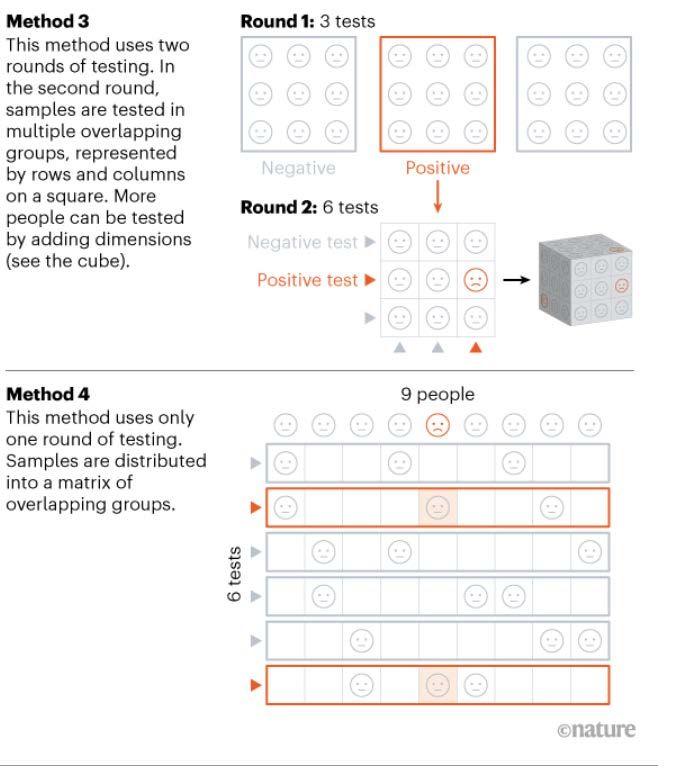
Several pooling strategies can be used, a recent news report published in Nature (1) gives an overview
of 4 pooling strategies that are used or evaluated in the COVID-19 pandemic (a schematic overview can
be found in Annex 1).
• Method 1: involves two rounds of testing, where each sample of a positive pool is retested
individually, most straighforward, used in Wuhan with up to 5 samples per pool, most efficient if
prevalence is ≤1% according to researchers.
• Method 2: involves three or more rounds of testing, where pools with positive samples are
further divided into smaller pools and then are tested individually or re-divided in smaller pools.
Not time effcicient, needs several rounds before obtaining final results for positive samples.
• Method 3: involves two rounds of testing (and if needed an extra round for individual testing), in
the second round samples are tested in multiple overlapping groups. Difficult pooling scheme,
will be trialed in Rwanda.
• Method 4: involves one round of testing, where samples are distributed into a matrix of
overlapping groups, fast but very complex pooling scheme (but apps being developed), being
trialed in India and Israel (with promising results according to the article).
The benefits of pooling are:
• Can save reagents, costs and time.
1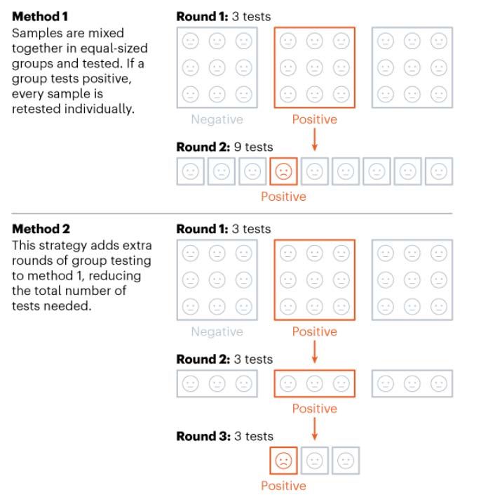
•   Allows for high throughput.

The possible drawbacks are:
   • Efficiency decreases at higher prevalences/test positivity ratios.
   • Sensitivity decreases due to dilution of viral RNA (theoretically increase of 1 Ct for 2-fold
       dilution). The bigger the pool, the greater the decrease in sensitivity.
   • Can imply longer time till test result (especially for samples in a positive pool that needs retesting
       in case of method 1-3). However, because throughput is extensively increased, this is likely to
       be counterbalanced in the context of large scale screening in low prevalence settings.
   • Pooling schemes can be complex (method 3 and 4).
   • Sample mix up and contamination can more easily happen.

CDC has issued an interim guidance on the use of pooling procedures for diagnostic, screening and
surveillance testing, recommending pooling only to be used in areas or situations where the number of
positive test results is expected to be low (for example in areas with a low prevalence of SARS-CoV-2
infections) (2).

ECDC has issued a technical report on Methodology for estimating point prevalence of SARS-CoV-2
infection by pooled RT-PCR testing. They present a pooling methodology for point prevalence studies,
including an PooledTesting R package to help identify optimal study design. The tool requires user
defined operational limits (maximum pool size, maximum number of pools to test, maximum number of
individual samples) and a hypothetical prevalence of the disease in the target population (3).

Some implementations of pooling worldwide:
   • China has used pooling up to 5 samples (method 1) to test the population of Wuhan (1).
   • Nebraska (US) has used pooling of 5 samples (method 1), but discontinued when prevalence
       rose above 10% (4).
   • Israel has used pooling of 8 samples (method 1) for the routine testing of nasopharyngeal swab
       samples from screened asymptomatic healthcare personnel, employees of essential industries,
       and residents and employees of nursing homes (5) and is evaluating method 4.
   • In Germany, pools ranging in size from four to 30 samples were used to test nursing home
       residents and staff (6).

2.2. NON-EXHAUSTIVE OVERVIEW OF STUDIES ON POOLING
Evaluations of method 1:
•   Yelin et al. evaluated method 1 with pooling after RNA extraction and found a sensitivity of 96% (when using a
    cut of Ct of 40) for a 16-sample pool with 1 positive sample (observing an increase of 1.24 in Ct for each dilution
    by a factor of 2). They also evaluated pooling before RNA extraction, and found that successful results were
    obtained for 8-sample pools with 1 positive sample, with an average increase in Ct of 2.9. (7)
•   Lohse et al. evaluated method 1 (unclear if extraction before or after pooling) and found that over a range of
    pool sizes (4-30 samples), Ct values in pools were up to 5 higher. They found that, even with Ct values of
    individual samples up to 34, positive pools could still be confidently identified. (8)
•   Sahajpal et al. evaluated method 1 with pooling before RNA extraction. 940 samples (of which 6 positive) were
    tested in 94 pools of 10 samples resulting in 148 reactions to test 940 samples. The authors state that Ct values
    in the pool- and individually tested samples were comparable. (9)
•   Ben-Ami et al. evaluated method 1 with pooling before RNA extraction. They found that test results were not
    significantly affected when using pools of 8 samples. They also evaluated method 3 and found that this strategy
    is more efficient when prevalence is higher than 2%. Additionally, they created a tool
    (https://github.com/matanseidel/pooling_optimization) to help choose the approach and pool size based on the
    prevalence. They implemented this strategy in Israël to test 26,576 samples from asymptomatic individuals. (5)
•   Abdelhamid et al. used method 1 with pooling before RNA extraction. They tested 25 pools of 5 samples with
    1 positive and found that all 25 pools were positive with Ct values within 0 and 5.03 Ct of the original individual
    specimens. They calculated that, with a prevalence below 10%, pooling can increase testing capability by at

                                                                                                                     2
least 69%.They calculated optimal pool size using a web-based application https://www.chrisbilder.com/shiny
(4)
• Gupta et al. evaluated method 1 with pooling after RNA extraction. They found that in pools of 8 RNA samples
the method can easily detect even up to a single positive sample with Ct value as high as 38. The study
suggests the results of pool testing are not affected by number of positive samples in a pool. (10)
• Torres et al. evaluated method 1 (unclear if extraction before or after pooling) in 40 samples (10 positive in
pools of 5 and 10 (both containing 1 positive)). They found that positive samples were detected in minipools
of both sizes, as long as Ct values of the individual positive samples were < 32 for the E gene and 1%,
simple pooling schemes and smaller pools (eg, 6:1 “minipools” for prevalence of 5%) were more comparable
in efficiency to larger and/or more complex pooling schemes. Below 1% prevalence, larger minipools could be
several-fold more efficient (in terms of results per test used) than 5:1 minipools. When prevalence was 0.1%,
larger pools and particularly 3-stage pools were substantially more efficient. Below 1% prevalence, adding the
intermediate pool stage generally resulted in much higher testing efficiency. They provide a free, publicly-
available web calculator is provided to help inform laboratory decisions on SARS-CoV-2 pooling algorithms
http://www.bios.unc.edu.vdicp.health.fgov.be:8080/~mhudgens/SARS-CoV-2.pooling.home.html (13)
• In a not yet peer reviewed paper, Pikovski et al. present a model to estimate the ideal pool size for method 1
and estimated it to be 4 samples in 1 pool. (14)
2.3. CONCLUSION POOLING
• Literature on pooling is promising.
• Pooling is more efficient at low prevalences/test positivity ratios (although one study suggests
pooling is efficient up to a prevalence of 20%). With increasing prevalence/test positivity ratio,
pooling will increasingly become less efficient and may lead to delayed test results (for positive
pools, due to retesting of all individual samples).
• Pooling could be used in a context of shortages of capacities (reagents, human resources in
lab). However, since such shortages are more likely to occur in a context of high virus circulation
(and high seroprevalence), it will probably not be cost-effective.
• Optimal pooling size depends on:
o Prevalence/test positivity ratio.
o Pooling method used.
o Ct values of positive samples.
• Optimal pooling strategy depends on:
o Prevalence/test positivity ratio.
o Acceptable complexity of pooling scheme.
• Sensitivity decreases with pooling, the extent (which is small according to literature) to which
depends on the protocol used. A decrease of sensitivity will mainly impact samples with high Ct
values (theoretically a 2-fold dilution of RNA increases the Ct with 1). Some studies suggest to
use an alternative cut off when pooling.
• Particular caution is required with regards to pre and post analytical errors. The risk of cross-
contamination indeed greater when using pooling approaches.
3• Pooling can be done before and after RNA extraction. The impact on sensitivity of both
techniques is not clear from literature. Pooling before RNA extraction will additionally save
extraction reagents and decrease the risk of contamination.
• Several models and applications have been developed to determine the best pool size or
pooling strategy.
• Limited information is available on experiences with/capacity of pooling for COVID-19 in Belgian
labs.
2.4. RECOMMENDATION POOLING
• Currently, it is not recommended to use pooling as a diagnostic tool for symptomatic individuals
or contacts, since pooling reduces sensitivity to a certain extent, increases the risk of
errors/contamination and is probably not efficient in reducing the use of capacities in case of
shortages (when seroprevalence is probably high).
• Pooling could be used for screening of large asymptomatic populations expected to have a low
prevalence. When screening in low risk asymptomatic populations (e.g. schools), a certain loss
in sensitivity is acceptable, as those most contagious/superspreaders (ie lowest CT values)
should still be detected. The impact of the reduced sensitivity will depend on the setting, being
possibly more problematic in high risk settings (e.g. WZC/MRS) than in low-risk settings (e.g.
schools).
• It is recommended that laboratories that implement pooling use a validated protocol (extension
of the RT-PCR cycles may be considered. For development of a pooling protocol, the
applications and methods developed and referred to in this document can be useful to determine
the best pooling method and/or the number of samples in a pool. Deconstruction of the pools
should also be well described to reduce risk of cross-contamination. The experience of
veterinary departments and transfusion centers in developing pooling protocols should be used,
as they have a lot of experience in pooling for mass screening.
• It is recommended to pool before RNA extraction to reduce the risk of contamination and limit
the use of reagents.
• An overview of Belgian laboratories experienced with/capable of pooling for COVID-19 is
needed.
• RIZIV/INAMI reimbursement of pooling is currently administratively not possible.
• In the context of platform bis, pooling for screening should be integrated in its ‘own’ flow.
See point 4. for a Note on pooling of saliva samples.
3. RT-PCR on saliva
3.1. BACKGROUND RT-PCR ON SALIVA
Since salivary glands are a potential target of infection due to high level of ACE2 expression on the
epithelial cells of the oral mucosa, saliva testing instead of using nasopharyngeal swabs (NPS) for RT-
PCR has been suggested as a way to:
• Overcome a possible shortage of swabbing material.
• Facilitate the sampling procedure.
• Decrease discomfort of sampling, likely leading to higher acceptability.
• Decrease exposure and requirement of health care workers (possibility of self-sampling).
The Netherlands will, according to several news websites, start testing children younger than 6 years
old with saliva tests. (15)
In France, the Haute Autorité de Santé, recently concluded that there is still uncertainty around the
performance of saliva tests as an alternative for nasopharyngeal swabs. They agreed for a ‘forfait
4d’innovation’ which enables to have the necessary budget for a wide scale evaluation of these tests.
(16)
Some laboratories report concerns about contamination through received saliva samples (not well
closed, saliva out of the container).
3.2. NON-EXHAUSTIVE OVERVIEW OF STUDIES ON RT-PCR ON SALIVA
Systematic reviews and meta-analyses:
• In a not yet peer reviewed Belgian rapid systematic review, Peeters et al. concluded that the relative sensitivity
of SARS-CoV-2 testing on saliva versus NPS was 0.97 (no significant difference; 95% CI=0.92-1.02). In the
second outcome (incorporating more data), they estimated that a pooled proportion 86% (95% CI=77-93%) of
NPS positive cases was also positive on saliva. They conclude that saliva could potentially be considered as
an alternative sampling method. But that studies were often small and involved inclusion of subjects with
insufficient information on clinical covariates. Most studies included patients who were symptomatic (78%,
911/1167). Therefore, additional and larger studies should be performed to verify the relative performance of
saliva in the context of screening of asymptomatic populations and contact-tracing. (17)
• A not yet peer reviewed meta-analysis by Czumbel et al. found 91% (95%CI = 80%-99%) sensitivity for saliva
tests and 98% (95%CI 89%-100%) sensitivity for NPS in previously confirmed COVID-19 infected patients,
with moderate heterogeneity among studies. (18)
Other studies:
• A not yet peer reviewed Belgian study collected two saliva samples (saliva swab and saliva container) and
NPS from more than 2000 people from triage centers. They found that for people with a low viral load (<
20 000 copies/ml nasopharyngeal transport medium), the correspondence between NPS and saliva samples
is < 5%. For people with a medium to high viral load, the correspondence between NPS and saliva samples
is high (up to 97%), and this for collection in a container (swabs performed less good) and irrespective of the
symptomatic status. They concluded that saliva testing is not suitable for individual diagnosis of COVID-19
virus in symptomatic patients and high-risk contacts. But is likely to have value to identify asymptomatic
individuals with medium to high viral load in the context of systematic screening campaigns. (19)
• Belgian data (unpublished) evaluated different methods for collecting saliva (Salivette, stimulated drooling
en unstimulated drooling) and unstimulated drooling provides the best Ct results of RNAseP.
• Landry et al. tested NPS and saliva (collected in a container, not drink or eat 30 minutes prior to saliva
collection) of 124 symptomatic individuals. Thirty-five were RT-PCR positive, 33 by NPS (sensitivity = 94.3
% (95 % CI 81.4%-99.0%)) and 30 by pure saliva (sensitivity = 85.7 % (95 % CI 70.6%-93.7%)), for an overall
agreement of 117/124 (94.4 %). The Ct’s were significantly lower for NPS than for saliva. (20)
• Williams et al. tested the saliva (collected in a container, keep saliva in mouth for 1 to 2 min prior to
collection) of 39 NPS-positive patients at a screening clinic. Thirty-three (84.6 %) of patients with positive
NPS had SARS CoV-2 RNA detected in saliva. In 1/50 (2%) of saliva samples from patients with negative
NPS were also positive. The Ct’s were significantly lower for NPS than for saliva. (21)
• Rao et al. tested the deep throat saliva (collected in a container, early morning saliva) and NPS among
217 individuals who tested positive for SARS-CoV-2 (NPS) in day 8-10 of their quarantine. At the moment of
testing they were asymptomatic (no information about their symptom history available). In total, 160
persons still tested positive by either test. In 76/160 patients virus was detected in saliva but not in swab and
in 11/160 virus was detected in swab but not in saliva. Among the 73 individuals with concordant results, the
Ct-values were significantly lower in saliva. (22)
• Iwasaki et al. tested the saliva (collected in a container) and NPS among 10 COVID-19 patients and 66
patients suspected for COVID-19 and found a concordance rate of 97.4% (95%CI: 90.8-99.7). Viral load was
equivalent at earlier time points but lower in saliva than in NPS samples at convalescent phase. (23)
• Azzi et al. tested saliva (drooling technique, collected in a container) 25 SARS-CoV-2 infected patients
with severe or very severe disease (NPS-positive). All tested positive in the saliva test. (24)
• Guo et al. investigated the effect of throat washing on the detection of SARS-CoV-2 in 11 laboratory-
confirmed COVID-19 participants (6 hospitalized, 5 discharged). They collected 24 paired throat washing
and NPS (unclear in which sequence) and found that in 18/24 the results were concordant, the other 6 tested
positive for throat washing, but negative for NPS. (25)
• Pasomsub et al. tested saliva samples (void of coughing, collected in a container), throat swabs and
nasopharyngeal swabs of 200 symptomatic individuals under investigation (median onset of symptoms
was 3 (2–7) days before). They estimated sensitivity and specificity was 84.2% (95% CI 60.4%–96.6%) and
98.9% (95% CI 96.1%–99.9%) when using NPs and throat swabs as the gold standard. (26)
5• Lai et al. tested 150 saliva samples (early morning deep throat saliva, DTS), 309 NPS or throat swabs and
104 sputum samples of 50 hospitalized (2 asymptomatic). They found DTS had the lowest overall RT-PCR
positive rate (68.7% vs. 89.4% [sputum] and 80.9% [pooled NPS and throat swabs]), and the lowest viral RNA
concentration (mean log copy/mL 3.54 vs. 5.03 [sputum] and 4.63 [pooled NPS and throat swabs]). (27)
• In a not yet peer reviewed study Becker et al. simultaneously tested NPS and saliva specimens, collected
with Orasure OM-505 Microbiome and OGD-610 DNA collection kits, of 88 persons from a community
testing environment (non-hospitalized) . With a Bayesian latent class model, they estimated the NPS
sensitivity to be 98.9% (95% CI: 67.6%- 99.7%) and saliva sensitivity 69.2% (95% CI: 38.6%- 97.6%). In an
additional study, they included 24 patients with prior positive COVID-19 results and found that overall, the
sensitivity using the NPS samples was higher than for the saliva samples. (28)
• In a not yet peer reviewed study, Wyllie et al. tested saliva (collected in a container, avoid food, water and
brushing of teeth until the saliva sample was collected) and NPS from 44 hospitalized COVID-19
patients (multiple saliva samples) and 98 asymptomatic health care workers (NPS also self-collected for
the latter group). They found that the sensitivity of saliva is comparable, if not superior to NPS in early
hospitalization and is more consistent during extended hospitalization and recovery. Moreover, SARS-CoV-2
was detected in the saliva of two asymptomatic healthcare workers with negative NPS. (29)
• In a not-yet peer reviewed study, Ott et al. investigated the stability of SARS-CoV-2 RNA and infectious
virus detection from saliva without supplementation. RNA stability was tested over 2-25 days and at
temperatures representing at-home storage and elevated temperatures which might be experienced when
cold chain transport may be unavailable. They concluded that RNA in saliva from infected individuals is stable
at 4°C, room temperature (~19°C), and 30°C for prolonged periods and found limited evidence for viral
replication in stored saliva samples. They conclude that expensive saliva collection options involving RNA
stabilization and virus inactivation buffers are not always needed, permitting the use of cheaper collection
options. (30)
• The University of Gent has collected 1400 saliva samples of mainly asymptomatic individuals in nursing homes
(residents and personnel). Saliva was collected in a container after people were asked to mix 2ml water with
saliva in their mouth and gargle (the latter only for personnel). They found a sensitivity 80% (which is in line with the results of the meta-
analyses), however some studies suggest a sensitivity below 70% (especially at low viral load
and/or in asymptomatic individuals, albeit not all).
• Recent studies showed higher sensitivity if early morning saliva is collected (before breakfast).
• Most studies evaluating saliva testing studied symptomatic and hospitalized persons, few
results of studies on asymptomatic persons are available. No studies included children.
• In some cases, saliva yields a positive result, whereas the nasopharyngeal swab is negative,
which could be due to differences in the kinetics of viral load in saliva as compared to
nasopharyngeal swabs. The clinical/public health relevance of this is unknown.
6• The biggest study so far (2000 people) concluded that correspondence between
nasopharyngeal swabs and saliva (collected in a container) is up to 97% for viral loads above
20.000 copies/ml.
• Saliva testing with collection in a container works better than with saliva swabs.
• Limited data suggest that there is no need for expensive collection options involving RNA
stabilization and virus inactivation buffers.
• Saliva could be an excellent sample in specific settings. However, in elderly people saliva is
more difficult to collect.
• People will very likely accept more to be tested repeatedly with saliva samples than with
nasopharyngeal swabs.
3.4. RECOMMENDATION RT-PCR ON SALIVA
• Currently, most studies suggest a lower sensitivity for RT-PCR on saliva than on
nasopharyngeal swabs. Therefor saliva testing seems less suitable as a diagnostic tool for
symptomatic individuals or contacts, and is presently not recommended. This should be
reevaluated in some groups of individuals, when the abovementioned systematic reviews and
meta-analysis will be published or if data on studies in Belgium are performed (but results should
be available very quickly). Saliva testing could be an alternative in case of shortage of swabs,
PPE, or overloaded medical staff collecting samples. For children < 6 years, saliva testing for
diagnostic should also be considered, as alternative for nasopharyngeal swabs. The current
RAG advice for these children is to only test in a limited number of situations. Using saliva tests,
if satisfactory sensitivity compared with throat and/or nose swabs, should allow to test more
children.
• Saliva testing could be used for screening of large groups of asymptomatic populations, to
identify highly infectious individuals.
• When collecting saliva in a container, it is important that these containers do not pose any risk
of contamination (no saliva outside of the container, container well closed etc…).
• More studies are urgently needed (and should be encouraged) to determine the best type of
saliva collection method and optimal sampling time for diagnosis and screening of SARS-CoV-
2, in different Belgian settings and age groups (nursing homes, schools, high risk groups, etc.).
• The performance of saliva testing for screening should be urgently compared with throat and
nasal swabs collected by healthcare workers, as well as with self-collection methods, see also
point 7.
See point 4. for a Note on pooling of saliva samples.
4. Note on pooling of saliva samples
Pooling of saliva samples combines the benefits of both strategies, however, sensitivity will inevitably
be further decreased. There are limited data available on the performance of the combination of these
two techniques, and further operational studies are highly needed.
Currently, we are aware of two Belgian studies regarding pooling of saliva samples.
• The University of Gent will evaluate pooled saliva testing (up to 20-60 samples per pool) for
screening in nursing homes. The protocol is under development.
• The University of Liège is currently validating pooled saliva testing that will be used to screen staff
and students. A container that inactivates the virus upon closing (inactivation buffer released upon
screwing of cap) is used and early morning saliva is collected. The protocol can be shared.
75. RT-LAMP as an alternative for RT-PCR
5.1. BACKGROUND RT-LAMP
The current gold standard for molecular diagnosis of COVID-19 is based on the detection of SARS-
CoV-2 RNA by real-time quantitative reverse transcription-polymerase chain reaction, but other
techniques have been developed/adapted to SARS-CoV-2.
One of these, the RT-LAMP (reverse-transcriptional loop-mediated isothermal amplification assay),
combines reverse transcriptase, DNA polymerase, pH indicator, and primers to amplify RNA templates
causing a drop in pH and, a color change detectable by the eye. RT-LAMP does not require thermal
cycles.
The advantages of this technique are:
• simple equipment and techniques,
• low cost (estimated 2-4€ for reagents and 38 in RT-PCR. (33)
• Lee et al. developed an RT-LAMP using a primer set specific for the N gene with a detection limit between 50
and 500 viral genome copies per reaction. Evaluation on 157 clinical specimens previously screened by E-
gene RT-qPCR revealed assay sensitivity and specificity of 87 and 100%, respectively. (34)
• Jiang et al. developed an RT-LAMP using a primer set specific for the N gene with a detection limit of 500
copies/ml. Evaluation on 213 negative and 47 positive patients showed a high degree of specificity (99.5%),
sensitivity (91.4%). (35)
Commercially available assays (Abbott ID NOW COVID-19):
8• Harrington et al. evaluated the Abbott ID Now COVID-19 on 524 samples (nasal and nasopharyngeal swabs)
and found an overall agreement was 75% positive agreement (95% confidence interval [95% CI], 67.74%,
80.67%) and 99% negative agreement (95% CI, 97.64%, 99.89%) between IDNCOV and ACOV for all
specimens tested. (36)
• An evaluation by Basu et al. of the Abbott ID Now COVID-19 found that, regardless of method of collection
and sample type, Abbott ID Now COVID-19 had negative results in a third of the samples that tested positive
by Cepheid Xpert Xpress when using nasopharyngeal swabs in viral transport media and 45% when using dry
nasal swabs. (37)
• An evaluation by Smithgall et al. of the Abbott ID Now COVID-19 e overall positive agreement was 73.9% with
ID Now. Negative agreement was 100%. ID Now and Xpert showed 100% positive agreement for medium and
high viral concentrations (Ct value 30, positive agreement was 34.3% for ID
Now. (38)
5.3. CONCLUSION ON RT-LAMP
• RT-LAMP techniques are not intended for massive testing.
• Several non-commercial RT-LAMP tests have been developed and the results are promising,
suggesting RT-LAMP could be a faster and more affordable alternative to RT-PCR. The current
place of such assays in clinical settings has still to be defined; No wide scale validation of these
techniques has been done yet, the validations that are done are mainly done by the developers
of the test, on a limited number of samples. Impact on sensitivity depends on exact protocol
developed.
• The commercial Abbott ID now (not exported) has varying results in evaluations.
• No information was found about the performance of the commercially available Enbiotech and
ICGene RT-LAMP assays.
• To our knowledge, RT-LAMP technology for COVID-19 is currently not used in Belgian labs, but
the technique is used for several DNA-based targets.
5.4. RECOMMENDATION RT-LAMP
• Currently, it is not recommended to use RT-LAMP as a diagnostic tool for symptomatic
individuals or contacts nor for screening of an asymptomatic population because of the lack of
wide scale validation studies.
• However, since results of non-commercial assays are promising, developments in the area will
be followed closely.
• An evaluation of the commercially available RT-LAMP tests (Enbiotech and ICGene) is
recommended.
6. Rapid antigen testing
6.1. BACKGROUND RAPID ANTIGEN TESTS
Rapid antigen tests for diagnosis of covid-19 infection are performed on nasopharyngeal swabs, they
have a decreased sensitivity but high specificity, implying that negative results should be retested with
RT-PCR, while positive results allow immediate patient management.
In the RAG-advice of 02/04/2020 it was recommended to only use a rapid antigen test in situations
where no RT-PCR could be performed and in situations with a 30-40% positivity ratio (i.e. high pre-test
probability), since every negative sample needs to be retested with RT-PCR (due to low sensitivity of
the test). Here we provide an update, based on current literature.
WHO, in an advice of 08/04/2020, does not currently recommend the use of antigen-detecting rapid
diagnostic tests for patient care, although research into their performance and potential diagnostic utility
is highly encouraged. (39)
9FIND, the Foundation for Innovative New Diagnostics, a global non-profit organization, is leading a call
for expressions of interest to accelerate the availability and manufacturing scale-up of rapid diagnostic
tests for the detection of SARS-CoV-2 antigens, because of the current suboptimal performance of these
tests. (40)
In their Guidelines on the Diagnosis of COVID-19, the Infectious disease society of America, does not
include antigen tests for the diagnosis and it is stated that antigen detection tests may be on the horizon,
but that it needs to be defined how they compare to RT-PCR. (41)
The CDC states that negative results from an antigen test may need to be confirmed with a PCR test
prior to making treatment decisions or to prevent the possible spread of the virus due to false negative
results. (42)
In the Netherlands, RIVM states that they are ‘looking at’ other types of tests such as antigen tests. (43)
In France, the Haute Autorité de Santé, states that antigen tests are not recommended for clinical use
due to their weak performance, especially at lower viral loads. (44)
In many low-income countries, the use of rapid antigen tests is scrutinized/investigated, to decrease the
volume of high cost PCR. Indeed, its use could substantially reduce the need for PCR (no PCR needed
if rapid antigen test is positive). In clinical practice, a highly specific test (even if poorly sensitive) has its
value.
6.2. NON-EXHAUSTIVE OVERVIEW OF STUDIES ON RAPID ANTIGEN TESTS
• Mertens et al. report on the development and evaluation of the COVID-19 Ag Respi-Strip (Coris BioConcept,
Gembloux, Belgium). In a retrospective multi-centric evaluation on aliquots of 328 nasopharyngeal samples,
they found an overall sensitivity and specificity of 57.6 and 99.5%. (45)
• Lambert-Niclot et al. evaluated a rapid antigen diagnostic test, COVID-19 Ag Respi-Strip, for detection of the
SARS-CoV-2 antigen in 138 nasopharyngeal secretions of which 94 (68.8%) were positive for SARS-CoV-2
by RT-PCR. Compared to that of RT-PCR, the specificity of the Ag test was 100% (95% CI: 91.8 to 100).
Among the 94 RT-PCR-positive samples, the rapid test detected only 47 specimens, resulting in a sensitivity
of 50.0% (95 CI: 39.5 to 60.5). (46)
• Scohy et al. evaluated a rapid antigen diagnostic test, COVID-19 Ag Respi-Strip testing 148 nasopharyngeal
swabs. Amongst the 106 positive RT-qPCR samples, 32 were detected by the rapid antigen test, giving an
overall sensitivity of 30.2%. All the samples detected positive with the antigen rapid test were also positive
with RT-qPCR. (47)
• Mak et al. evaluated the Biocredit COVID-19 Ag test on respiratory samples collected from confirmed COVID-
19 patients and found that the rapid antigen test detected between 11.1% (compared with sputum) and 45.7%
(compared with nasopharyngeal aspirate and throat swab) of RT-PCR-positive samples from COVID-19
patients. (48)
• Loeffelholz et al. state that new approaches to try to concentrate antigen and amplify the detection phase are
likely to be needed for these methods to have any clinical utility. (49)
• In a systematic review La Marca et al. state that the evaluation of these diagnostic tests has been limited,
although direct antigen tests are being registered by several health authorities, the sensitivity of these tests is
lower than that of RT-PCR. Their greatest utility if they come to fruition may be in symptomatic patients, when
the viral load will be at its greatest, to enable accurate triage. (50)
6.3. CONCLUSION RAPID ANTIGEN TESTS
• Rapid antigen tests have low performance in diagnosis COVID-19 infection, with a sensitivity
for the Coris Bioconcept COVID-19 Ag Respi-Strip of lower than 60% (some studies suggest as
low as 30%).
• Rapid antigen tests offer consistently high specificity.
• Research and development is ongoing.
• A newly developed test BD Veritor reports a higher sensitivity (70%) but is not available in
Europe yet.
106.4. RECOMMENDATION RAPID ANTIGEN TESTS
• The use of rapid antigen tests is generally not recommended and should only be used if no RT-
PCR is available, or as a first diagnostic/screening test to isolate positive individuals if an RT-
PCR can be performed only at a later time point (e.g in hospitals without microbiology lab, for
diagnosis or screening of admissions at night, GP practices). In this case, RT-PCR should
anyway be performed as soon as possible on all samples with negative antigen test.
• More studies are needed to evaluate the sensitivity of detecting highly infectious people (useful
in context of screening), instead of comparing results to PCR for all viral loads. However, rapid
antigen tests are time consuming (one by one), and therefore not suitable for large screenings.
• Studies should also address the usefulness and cost-effectiveness of rapid antigen tests for the
clinical practice/immediate management in GP practices.
7. Self-collected nose, throat and
nasopharyngeal swabs
7.1. BACKGROUND SELF-COLLECTED SWABS
Additionally to self-collection of saliva, implementation of self-swabbing could save the use of personal
protective equipment and decrease work load and exposure of health care workers.
In some cases, self-swabbing/self-collecting could potentially be an alternative for saliva sampling in
screening settings.
7.2. NON-EXHAUSTIVE OVERVIEW OF STUDIES ON SELF-COLLECTED SWABS
• McCulloch et al. compared self-collected midnasal swab performance with clinician-collected
nasopharyngeal swabs in 185 participants. They found that Cycle thresholds of home swabs were positively
correlated with clinician swabs (correlation coefficient, 0.81; P < .001). Four of 5 false-negative home swabs
had a Ct greater than or equal to 33. In a sensitivity analysis of all swabs with Ct less than or equal to 32,
sensitivity of home swabs was 95%. (51)
• In a not-yet peer reviewed study, Demmer et al. evaluated self-sampling in 489 symptom-free health care
workers. Over 95% of participants reported a willingness to repeat a self-collected nasopharyngeal swabs
in the future for either clinical or research purposes. 24% preferred a provider collected-swab, 57% preferred
self-collection and 19% reported no preference. No comparison was done with not-self-sampled swabs and
no case of SARS-CoV-2 infection was found. (52)
• Waghmare et al. compared self-sampling with flocked nasal swabs and self-sampling with to foam nasal
swabs in 15 patients with respiratory symptoms for the detection of 11 respiratory viruses. They found that
self-sampling with foam swabs was better tolerated than with flocked swabs. Agreement between samples
collected by foam and flocked swabs from the same nostril was generally high, particularly with high viral load
samples, with no evidence of higher yield with one method versus another. No comparison was done with not-
self-sampled swabs. (53)
• Valentine-Graves et al. assessed willingness and feasibility of patients to self-collect three diagnostic
specimens (saliva, oropharyngeal swab (OPS) and dried blood spot (DBS) card) while observed by a
clinician through a telehealth session. Of the 153 US adults enrolled, A large majority of participants (>84%)
reported that collecting, packing and shipping of saliva, OPS, and DBS specimens were acceptable. Nearly
nine in 10 (87%) reported being confident or very confident that the specimens they collected were sufficient
for laboratory analysis. (54)
• Guest et al. assessed quality of oropharyngeal swabs collected during a telehealth video appointment while
clinical observers watched and documented the suitability of the collection in 159 participants. Of the enrolled
participants,153/159 (96.2%) returned their kits and 146/151 (96.7%) of the samples were of sufficient quality
for submission for laboratory testing, 100% of these had cycle threshold values for RNase Pclinician-supervised self-collected nasal swab specimens detected 23 (85%) of 27, clinician-collected posterior
nasopharyngeal swab specimens detected 23 (79%) of 29, and unmonitored self-collected oral fluid swab
specimens detected 19 (66%) of 29. Despite nasopharyngeal swabs being considered the gold standard, 4
participants tested negative by clinician-collected nasopharyngeal swab and positive by the 3 other specimen
types. Additionally, false negative results by each sample type were seen to generally not overlap. (56)
• Wehrhahn et al. evaluated performance of self-collection of nasal and throat swabs with health care worker
collected nasal and throat swabs or throat and nasopharyngeal swabs for the detection of respiratory viruses
including SARS-CoV-2. They found that self-collection (SC) was highly concordant with health care worker
collection (HC) (κ = 0.890) for all viruses including SARS-CoV-2 and more concordant than HC to positive
results by any method (κ = 0.959 vs 0.933). (57)
• In a not yet peer reviewed study Teo et al. evaluated the performance of self-collected of ‘naso-oropharyngeal’
saliva and nasal swabs (SN) compared to nasopharyngeal (NP) swabs in 200 migrant workers, who were
given an instruction video on how to self-sample (45 with acute respiratory infection, 104 asymptomatic close
contacts, and 51 confirmed COVID-19 cases). Of 337 sets of tests (workers were tested repeatedly), there
were 150 (44.5%) positive NP swabs, 127 (37.7%) 17 positive SN swabs, and 209 (62.0%) positive saliva. In
COVID-19 confirmed patients, saliva performed better than nasal swab at all-time points after onset of
symptoms. (58)
7.3. CONCLUSION SELF-COLLECTED SWABS
• The acceptability of self-collecting nasopharyngeal swabs for the diagnosis of SARS-CoV-2
infection has only been assessed in health care workers (95% willingness to repeat). There is
very limited available evidence on the performance of self-collected nasopharyngeal swabs for
the RT-PCR detection of SARS-CoV-2.
• There is limited available evidence on the performance and acceptability of self-collected throat
swabs for the diagnosis of SARS-CoV-2.
• There is limited available evidence on the performance and acceptability of self-collected nasal
swabs, though 2 studies suggest saliva is more sensitive than self-collected nasal-swabs.
• Sensitivity is higher if self-sampling is performed under medical supervision.
7.4. RECOMMENDATION SELF-COLLECTED SWABS
• Based on the very limited available evidence on the performance of self-swabbing, it is currently
not recommended to use this technique.
• More studies are urgently needed to assess acceptability and performance in order to determine
if self-swabbing of nasal and/or throat samples can be used as an alternative to saliva testing
in screening settings (compare saliva results with results of self-collected nasal/throat swabs;
comparing self-swabbing with saliva and/or swabbing performed by healthcare workers).
• Self-swabbing could be an interesting option in e.g. test villages (under medical supervision),
but further studies are urgently needed before implementing it.
8. Multiplex
8.1. BACKGROUND MULTIPLEX
In the coming months, it will become increasingly important to distinguish a SARS-CoV-2 infection from
other respiratory infections such as influenza and RSV, both from a clinical as from a surveillance point
of view. This will put an increased pressure on lab capacity and reagent supply.
Moreover, in the context of respiratory disease surveillance, the collection of samples within the existing
surveillance networks of health care providers may be very difficult because of the changes in testing
facilities. Additional efforts are needed to make the collection of samples possible.
Multiplex testing will therefore become increasingly important. At this moment, these multiplexes are not
reimbursed by RIZIV/INAMI (unless for some categories of patients, eg transplants patients).
12• Currently, the necessary steps are being undertaken to include multiplexes with SARS-CoV-2
and other respiratory pathogens in RIZIV/INAMI nomenclature. The plan is to have this finalized
in the first half of September.
At this moment, the proposal is to include two types of multiplex :
o one for a limited number (at least 3) of respiratory pathogens, with at least influenza
(including mandatory typing: separate result for influenza A and influenza B) and RSV,
o one for a large set (initial proposal:10) of respiratory micro-organisms, with freedom on
the type of organisms to include (will depend on clinical context of patient). The cost of
the necessary machine with acceptable turn-around-time, is significantly higher. This
type of multiplex, also due to much higher pricing, will be restricted for use only in
hospitalized critically ill patients.
• At present, in Belgium, multiple laboratories have developed or are developing and validating
multiplex assays to detect and differentiate RNA from SARS-CoV-2 from other pathogens in
respiratory specimens. Some examples from the relevant NRC’s:
o At the NRC Influenza currently triplex qPCR used for influenza typing (detection of
influenza A viruses, influenza B viruses and RNaseP housekeeping genes) is adapted
to add a fourth target (E gene of SARS-CoV-2). Validation is almost finished. The NRC
Influenza also collaborates with in the development of a quadruplex to detect SARS-
CoV-2, influenza (A and B, but no typing), RSV (A and B, no typing), and RNaseP as
housekeeping genes. A multiplex for 16 non influenza respiratory viruses for
surveillance purposes is also available. This is based on primers/probes used by the
NRC Influenza. For surveillance
o The NRC respiratory pathogens (SARS-CoV-2) performs a multiplex on a respiratory
panel of 29 pathogens, including SARS-CoV-2.
At this stage, we do not have an exhaustive overview of the multiplexes currently
used/developed/validated in Belgium.
8.2. CONCLUSION AND RECOMMENDATION MULTIPLEX
• Finalization of incorporation of multiplex in RIZIV nomenclature is urgent, and planned for the
first half of September.
• Labs should aim at implementing/developing/validating multiplexes that are subject to
reimbursement.
• When reimbursement is implemented, it is recommended to, during the season of respiratory
infections (week 40 till week 20, cfr influenza surveillance):
o Use multiplex (respiratory “minipanel”), including SARS-CoV-2, influenza (A and B) and
RSV, in all patients presenting with influenza-like illness, including in primary care. The
costs of the test in this context will be regulated in the same way as it is currently
regulated for SARS-CoV-2 testing for patients in primary care and will be incorporated
within the regular RIZIV/INAMI reimbursement for hospitalized patients.
o The use of a respiratory “maxipanel” (at least 10 respiratory micro-organisms) will be
restricted to hospitalized critically ill patients. The recommendations and
reimbursements issues will be further discussed by the Microbiology Working Group of
the Commission Clinical Biology (MWG CKB-CBC).
o Do not use multiplex for testing of contacts, nor for SARS-CoV-2 screening of
asymptomatic populations.
o Prioritize SARS-CoV-2 testing in symptomatic persons (see case definition of possible
case) in primary care, in situations when multiplex is not or limited available.
• An overview of the Belgian capacity regarding multiplex testing is urgently needed.
• Multiplex PCR will further be used for surveillance purposes (separate protocol/budget).
139. Elements for further discussion:
1. More studies are needed to compare nasal swabs, throat swabs and saliva (container versus
swab) for self-sampling, including in combination. Published studies focus mainly on
comparison to nasopharyngeal swabs.
2. It will be difficult for laboratories to use different tests for the symptomatic (ILI) patients and the
asymptomatic persons, even if the information is available. How can this be solved (practically)
? Moreover, reimbursement of the regular RT-PCR for SARS-CoV-2 falls under two different
regulations.
3. The case definition for a possible case of COVID-19 is much wider than ILI. Should the case
definition be restricted to ILI during the season of respiratory infections to avoid wasting
multiplex PCR tests on persons with e.g. fever and diarrhea ? More information is needed on
the symptoms presented by the COVID-19 patients in ambulatory settings, to be able to
evaluate this (Sciensano).
10. General remark
The testing approaches covered in this review, such as pooling of samples or saliva testing, offer a
reduced sensitivity in comparison to nasopharyngeal RT-PCR. Nevertheless, they may represent valid
tools for population-based screening. The rationale behind using tests with a suboptimal or lesser
sensitivity for screening purposes is the fact that people with high Ct values are likely to be non-
contagious (see also RAG advice on Interpretation of PCR results and infectivity). Moreover a not yet
peer reviewed study by Larremore et al. modelled surveillance effectiveness considering test
sensitivities, frequency, and sample-to-answer reporting time. They found that effective surveillance,
including time to first detection and outbreak control, depends largely on frequency of testing and the
speed of reporting, and is only marginally improved by high test sensitivity. The authors therefore
conclude that surveillance should prioritize accessibility, frequency, and sample-to-answer time whereas
analytical limits of detection should be secondary. (59)
The following persons participated to this RAG advice:
Cyril Barbezange (Sciensano); Nathalie Bossuyt (Sciensano); Karen Bruyland (Medisch Labo
Bruyland); Fabrice Bureau (ULiège); Emmanuel Bottieau (ITG); Piet Cools (UGent); Olivier Denis (CHU-
UCL Namur); Herman Goossens (UAntwerpen); Benoît Kabamba (UCL); Sofieke Klamer (Sciensano);
Katrien Lagrou (UZLeuven); Tinne Lernout (Sciensano); Amber Litzroth (Sciensano); Romain MAHIEU
(COCOM); Pierette Melin (Uliège); Elizaveta Padalko (UZGent); Sophie Quoilin (Sciensano); Catherine
Sion (Grand Hôpital de Charleroi); Daniel Te-Din Huang (CHU-UCL Namur); Isabelle Thomas
(Sciensano); Roel Van Giel (Domus Medica); Steven Van Gucht (Sciensano); Chloé Wyndham-Thomas
(Sciensano).
14REFERENCES
1. Mallapaty S. The mathematical strategy that could transform coronavirus testing.
Nature. 583 : 504-505 (2020). doi: 10.1038/d41586-020-02053-6
2. CDC. Interim guidance for use of pooling procedures in SARS-CoV-2 diagnostic, screening, and
surveillance testing
3. European Centre for Disease Prevention and Control. Methodology for estimating point
prevalence of SARSCoV-2 infection by pooled RT-PCR testing. Stockholm: ECDC; 2020.
4. Abdalhamid B, Bilder CR, McCutchen EL, Hinrichs SH, Koepsell SA, Iwen PC. Assessment of
Specimen Pooling to Conserve SARS CoV-2 Testing Resources. Am J Clin Pathol.
2020;153(6):715-718. doi:10.1093/ajcp/aqaa064
5. Ben-Ami R, Klochendler A, Seidel M, et al. Large-scale implementation of pooled RNA
extraction and RT-PCR for SARS-CoV-2 detection [published online ahead of print, 2020 Jun
23]. Clin Microbiol Infect. 2020;S1198-743X(20)30349-9. doi:10.1016/j.cmi.2020.06.009
6. HealthEurope. Discover how sample pooling can increase coronavirus testing capacity. Online
source [Last accessed 12 August 2020]. Available from
https://www.healtheuropa.eu/discover-how-sample-pooling-can-increase-coronavirus-
testing-capacity/99713/
7. Yelin I., Aharony N., Shaer-Tamar E., Argoetti A., Messer E., Berenbaum D., Shafran E., Kuzli A.,
Gandali N., Hashimshony T., Mandel-Gutfreund Y. Evaluation of COVID-19 RT-qPCR Test in
Multi-Sample Pools. Clin Infect Dis. 2020 doi: 10.1093/cid/ciaa531. ciaa531.
8. Lohse S, Pfuhl T, Berkó-Göttel B, et al. Pooling of samples for testing for SARS-CoV-2 in
asymptomatic people [published online ahead of print, 2020 Apr 28]. Lancet Infect Dis.
2020;S1473-3099(20)30362-5. doi:10.1016/S1473-3099(20)30362-5
9. Sahajpal NS, Mondal AK, Njau A, et al. Proposal of Reverse Transcription-PCR-Based Mass
Population Screening for SARS-CoV-2 (COVID-19) [published online ahead of print, 2020 Jul
29]. J Mol Diagn. 2020;S1525-1578(20)30407-4. doi:10.1016/j.jmoldx.2020.07.001
10. Gupta E, Padhi A, Khodare A, et al. Pooled RNA sample reverse transcriptase real time PCR
assay for SARS CoV-2 infection: A reliable, faster and economical method. PLoS One.
2020;15(7):e0236859. Published 2020 Jul 30. doi:10.1371/journal.pone.0236859
11. Torres I, Albert E, Navarro D. Pooling of nasopharyngeal swab specimens for SARS-CoV-2
detection by RT-PCR [published online ahead of print, 2020 May 5]. J Med Virol.
2020;10.1002/jmv.25971. doi:10.1002/jmv.25971
12. Aragón-Caqueo D, Fernández-Salinas J, Laroze D. Optimization of group size in pool testing
strategy for SARS-CoV-2: A simple mathematical model [published online ahead of print, 2020
Apr 24]. J Med Virol. 2020;10.1002/jmv.25929. doi:10.1002/jmv.25929
13. Pilcher CD, Westreich D, Hudgens MG. Group Testing for Sars-Cov-2 to Enable Rapid Scale-Up
of Testing and Real-Time Surveillance of Incidence [published online ahead of print, 2020 Jun
27]. J Infect Dis. 2020;jiaa378. doi:10.1093/infdis/jiaa378
14. Pikovski A and Bentele K. Remarks on pooling coronavirus tests. medRxiv (2020),
https://doi.org/10.1101/2020.06.08.20125781
15. RIVM en GGD bereiden zich voor op tweede golf met nieuwe testvormen. Online source [Last
accessed 17 August 2020]. Available from: https://zorgnu.avrotros.nl/nieuws/item/rivm-en-
ggd-bereiden-zich-voor-op-tweede-golf-met-nieuwe-testvormen/
1516. Haute Autorité de Santé. Tests salivaires pour la détection du virus SARS-CoV-2 : la HAS se
prononce en faveur d’un forfait innovation. Online source [Last accessed 17 August 2020].
Available from: https://www.has-sante.fr/jcms/p_3198372/fr/tests-salivaires-pour-la-
detection-du-virus-sars-cov-2-la-has-se-prononce-en-faveur-d-un-forfait-innovation
17. Peeters E., Singh S., Vandesompele J, Mestdagh P, Hutse V., Arbyn M. Rapid systematic review
of the sensitivity of SARS-CoV-2 molecular testing on saliva compared to nasopharyngeal
swabs. medRxiv 2020.08.05.20168716; doi: https://doi.org/10.1101/2020.08.05.20168716
18. Czumbel LM, Kiss S, Farkas N, Mandel I, Hegyi AE, Nagy AK, et al. Saliva as a Candidate for
COVID-19 Diagnostic Testing: A Meta-Analysis. medRxiv. 2020:2020.05.26.20112565.
19. Evaluatie van het gebruik van speekselstalen als alternatief voor staalafname via een diepe
neuswisser bij het opsporen van SARS-CoV-2, het virus dat COVID-19 veroorzaakt. Online
source [Last accessed 12 August 2020]. Available from: https://covid-
19.sciensano.be/sites/default/files/Covid19/salive_communication_NL.pdf
20. Landry ML, Criscuolo J, Peaper DR. Challenges in use of saliva for detection of SARS CoV-2 RNA
in symptomatic outpatients [published online ahead of print, 2020 Jul 31]. J Clin Virol.
2020;130:104567. doi:10.1016/j.jcv.2020.104567
21. Williams E, Bond K, Zhang B, Putland M, Williamson DA. Saliva as a Noninvasive Specimen for
Detection of SARS-CoV-2. J Clin Microbiol. 2020;58(8):e00776-20. Published 2020 Jul 23.
doi:10.1128/JCM.00776-20
22. Rao M, Rashid FA, Sabri FSAH, et al. Comparing nasopharyngeal swab and early morning saliva
for the identification of SARS-CoV-2 [published online ahead of print, 2020 Aug 6]. Clin Infect
Dis. 2020;ciaa1156. doi:10.1093/cid/ciaa1156
23. Iwasaki S, Fujisawa S, Nakakubo S, et al. Comparison of SARS-CoV-2 detection in
nasopharyngeal swab and saliva. J Infect. 2020;81(2):e145-e147.
doi:10.1016/j.jinf.2020.05.071
24. Azzi L, Carcano G, Gianfagna F, et al. Saliva is a reliable tool to detect SARS-CoV-2. J Infect.
2020;81(1):e45-e50. doi:10.1016/j.jinf.2020.04.005
25. Guo WL, Jiang Q, Ye F, et al. Effect of throat washings on detection of 2019 novel coronavirus
[published online ahead of print, 2020 Apr 9]. Clin Infect Dis. 2020;ciaa416.
doi:10.1093/cid/ciaa416
26. Pasomsub E, Watcharananan SP, Boonyawat K, et al. Saliva sample as a non-invasive specimen
for the diagnosis of coronavirus disease 2019: a cross-sectional study [published online ahead
of print, 2020 May 15]. Clin Microbiol Infect. 2020;S1198-743X(20)30278-0.
doi:10.1016/j.cmi.2020.05.001
27. Lai CKC, Chen Z, Lui G, et al. Prospective study comparing deep-throat saliva with other
respiratory tract specimens in the diagnosis of novel coronavirus disease (COVID-19)
[published online ahead of print, 2020 Aug 1]. J Infect Dis. 2020;jiaa487.
doi:10.1093/infdis/jiaa487
28. Becker D., Sandoval E., Amin A. Saliva is less sensitive than nasopharyngeal swabs for COVID-
19 detection in the community setting. medRxiv. 2020
29. Wyllie A.L., Fourmier J., Casanovas-Massana A., Campbell M., Tokuyama M., Vijayakumar P.
Saliva is more sensitive for SARS-CoV-2 detection in COVID-19 patients than nasopharyngeal
swabs. medRxiv. 2020
30. Ott I, Strine M, Watkins A , Boot M, Kalinich C, Harden C, at al. Simply saliva: stability of SARS-
CoV-2 detection negates the need for expensive collection devices. medRxiv. 2020.
1631. Chow FW, Chan TT, Tam AR, et al. A Rapid, Simple, Inexpensive, and Mobile Colorimetric
Assay COVID-19-LAMP for Mass On-Site Screening of COVID-19. Int J Mol Sci.
2020;21(15):E5380. Published 2020 Jul 29. doi:10.3390/ijms21155380
32. Dao Thi VL, Herbst K, Boerner K, et al. A colorimetric RT-LAMP assay and LAMP-sequencing
for detecting SARS-CoV-2 RNA in clinical samples [published online ahead of print, 2020 Jul
27]. Sci Transl Med. 2020;eabc7075. doi:10.1126/scitranslmed.abc7075
33. Lu R, Wu X, Wan Z, Li Y, Jin X, Zhang C. A Novel Reverse Transcription Loop-Mediated
Isothermal Amplification Method for Rapid Detection of SARS-CoV-2. Int J Mol Sci.
2020;21(8):2826. Published 2020 Apr 18. doi:10.3390/ijms21082826
34. Lee JYH, Best N, McAuley J, et al. Validation of a single-step, single-tube reverse transcription
loop-mediated isothermal amplification assay for rapid detection of SARS-CoV-2 RNA
[published online ahead of print, 2020 Aug 5]. J Med Microbiol. 2020;10.1099/jmm.0.001238.
doi:10.1099/jmm.0.001238
35. Jiang M, Pan W, Arasthfer A, et al. Development and Validation of a Rapid, Single-Step
Reverse Transcriptase Loop-Mediated Isothermal Amplification (RT-LAMP) System Potentially
to Be Used for Reliable and High-Throughput Screening of COVID-19. Front Cell Infect
Microbiol. 2020;10:331. Published 2020 Jun 16. doi:10.3389/fcimb.2020.00331
36. Harrington A, Cox B, Snowdon J, et al. Comparison of Abbott ID Now and Abbott m2000
Methods for the Detection of SARS-CoV-2 from Nasopharyngeal and Nasal Swabs from
Symptomatic Patients. J Clin Microbiol. 2020;58(8):e00798-20. Published 2020 Jul 23.
doi:10.1128/JCM.00798-20
37. Basu A, Zinger T, Inglima K, et al. Performance of Abbott ID Now COVID-19 Rapid Nucleic Acid
Amplification Test Using Nasopharyngeal Swabs Transported in Viral Transport Media and
Dry Nasal Swabs in a New York City Academic Institution. J Clin Microbiol.
2020;58(8):e01136-20. Published 2020 Jul 23. doi:10.1128/JCM.01136-20
38. Smithgall MC, Scherberkova I, Whittier S, Green DA. Comparison of Cepheid Xpert Xpress and
Abbott ID Now to Roche cobas for the Rapid Detection of SARS-CoV-2. J Clin Virol.
2020;128:104428. doi:10.1016/j.jcv.2020.104428
39. WHO. Advice on the use of point-of-care immunodiagnostic tests for COVID-19. Online
source [Last accessed 17 August 2020]. Available from: https://www.who.int/news-
room/commentaries/detail/advice-on-the-use-of-point-of-care-immunodiagnostic-tests-for-
covid-19
40. FIND. Driving equitable access to fit-for-purpose antigen-detecting rapid diagnostic tests for
covid-19. Online source [Last accessed 17 August 2020]. Available from:
https://www.finddx.org/eoi-covid19-ag-rdt/
41. Hanson KE, Caliendo AM, Arias CA, et al. Infectious Diseases Society of America Guidelines on
the Diagnosis of COVID-19 [published online ahead of print, 2020 Jun 16]. Clin Infect Dis.
2020;ciaa760. doi:10.1093/cid/ciaa760
42. Centers for Disease prevention and control (CDC). Guidance – Proposed Use of Point-of-Care
(POC) Testing Platforms for SARS-CoV-2 (COVID-19). Online source [Last accessed 17 August
2020]. Available from: https://www.cdc.gov/coronavirus/2019-ncov/downloads/OASH-
COVID-19-guidance-testing-platforms.pdf
43. RIVM. Policy on testing for novel coronavirus disease (COVID-19). Online source [Last
accessed 17 August 2020]. Available from: https://www.rivm.nl/en/node/154261
44. Haute Autorité de Santé. Cahier des charges définissant les modalités d’évaluation des
performances des tests sérologiques détectant les anticorps dirigés contre le SARS-CoV-2.
17You can also read