Engineering Synthetic Lipopeptide Antigen for Specific Detection of Mycobacterium avium subsp. paratuberculosis Infection
←
→
Page content transcription
If your browser does not render page correctly, please read the page content below
ORIGINAL RESEARCH
                                                                                                                                                published: 23 April 2021
                                                                                                                                        doi: 10.3389/fvets.2021.637841

                                             Engineering Synthetic Lipopeptide
                                             Antigen for Specific Detection of
                                             Mycobacterium avium subsp.
                                             paratuberculosis Infection
                                             Sylvie Bay 1,2*, Douglas Begg 3 , Christelle Ganneau 1,2 , Maxime Branger 4 , Thierry Cochard 4 ,
                                             John P. Bannantine 5 , Heike Köhler 6 , Jean-Louis Moyen 7 , Richard J. Whittington 3 and
                                             Franck Biet 4*
                                             1
                                              Institut Pasteur, Unité de Chimie des Biomolécules, Département de Biologie Structurale et Chimie, Paris, France, 2 CNRS
                                             UMR 3523, Paris, France, 3 School of Veterinary Science, University of Sydney, Camden, NSW, Australia, 4 INRAE, Université
                                             de Tours, ISP, Nouzilly, France, 5 USDA-Agricultural Research Service (USDA-ARS), National Animal Disease Center, Ames,
                             Edited by:
                                             IA, United States, 6 Friedrich-Loeffler-Institut, Federal Research Institute for Animal Health, Jena, Germany, 7 Laboratoire
                       Miguel Salgado,
                                             Départemental d’Analyse et de Recherche de Dordogne, Coulounieix Chamiers, France
       Austral University of Chile, Chile
                         Reviewed by:
                              Ad Koets,      Unlike other MAC members, Mycobacterium avium subsp. paratuberculosis (MAP)
 Wageningen Bioveterinary Research           does not produce glycopeptidolipids (GPL) on the surface of the cell wall but a
                 (WBVR), Netherlands
                 Marta Alonso-Hearn,         lipopentapeptide called L5P (also termed Lipopeptide-I or Para-LP-01) characterized
            NEIKER-Instituto Vasco de        in C-type (bovine) strains. This lipopeptide antigen contains a pentapeptide core,
             Investigación y Desarrollo
                          Agrario, Spain
                                             D-Phenylalanine-N-methyl-L-Valine-L-Isoleucine-L-Phenylalanine-L-Alanine, in which
                    Philip John Griebel,     the N-terminal D-Phenylalanine is amido-linked with a fatty acid (C18–C20). The
 University of Saskatchewan, Canada          molecular and genetic characterization of this antigen demonstrated that L5P is
                   *Correspondence:          unique to MAP. Knowledge of the structure of L5P enabled synthetic production
                             Sylvie Bay
                  sylvie.bay@pasteur.fr      of this lipopeptide in large quantities for immunological evaluation. Various studies
                            Franck Biet      described the immune response directed against L5P and confirmed its capability for
                    franck.biet@inrae.fr
                                             detection of MAP infection. However, the hydrophobic nature of lipopeptide antigens
                    Specialty section:       make their handling and use in organic solvents unsuitable for industrial processes.
         This article was submitted to       The objectives of this study were to produce, by chemical synthesis, a water-soluble
        Veterinary Infectious Diseases,
                                             variant of L5P and to evaluate these compounds for the serological diagnosis of
                a section of the journal
        Frontiers in Veterinary Science      MAP using well-defined serum banks. The native L5P antigen and its hydrosoluble
       Received: 04 December 2020            analog were synthesized on solid phase. The pure compounds were evaluated on
          Accepted: 24 March 2021            collections of extensively characterized sera from infected and non-infected cattle.
           Published: 23 April 2021
                                             ROC analysis showed that L5P and also its water-soluble derivative are suitable
                                Citation:
            Bay S, Begg D, Ganneau C,
                                             for the development of a serological test for Johne’s disease at a population level.
Branger M, Cochard T, Bannantine JP,         However, these compounds used alone in ELISA have lower sensitivity (Se 82% for
 Köhler H, Moyen J-L, Whittington RJ
                                             L5P and Se 62% for the water-soluble variant of L5P) compared to the Se 98% of
          and Biet F (2021) Engineering
      Synthetic Lipopeptide Antigen for      a commercial test. Advantageously, these pure synthetic MAP specific antigens can
 Specific Detection of Mycobacterium         be easily produced in non-limiting quantities at low cost and in standardized batches
        avium subsp. paratuberculosis
   Infection. Front. Vet. Sci. 8:637841.
                                             for robust studies. The fact that L5P has not been validated in the context of ovine
       doi: 10.3389/fvets.2021.637841        paratuberculosis highlights the need to better characterize the antigens expressed

Frontiers in Veterinary Science | www.frontiersin.org                               1                                              April 2021 | Volume 8 | Article 637841
Bay et al.                                                                                                        Synthetic Lipopeptide for MAP Detection

                                             from the different genetic lineages of MAP to discover new diagnostic antigens. In the
                                             context of infections due to other mycobacteria such as M. bovis or the more closely
                                             related species M. avium subsp. hominissuis, the L5P did not cross react and therefore
                                             may be a valuable antigen to solve ambiguous results in other tests.

                                             Keywords: Mycobacterium avium subsp. paratuberculosis, Johne’s disease, lipopeptide, diagnosis, antibody
                                             response, chemical synthesis

INTRODUCTION                                                                         Despite the establishment of control programs in most
                                                                                 developed countries, with substantial financial efforts, the
Mycobacteria are well-known for their cell envelope containing                   prevalence rate of paratuberculosis remains at a very high level,
abundant mycolic acids and specific lipid components (1,                         around 50% for European herds (17, 18) and around 80% in
2). These molecules play a crucial role to maintain the                          the USA. The disease controls implemented may be different
integrity of the cell wall and are considered to be involved                     depending on the country: control, surveillance, certification,
in bacterial virulence through influence on the host immune                      border quarantine, and on-farm biosecurity (19). For routine
response (3). The outer membrane contains diverse surface                        screening, the majority of analyses are performed by ELISA
lipids that are species-dependent. This likely reflects differences              serological tests. The bovine antibody response to MAP changes
in the cell biochemical organization and probably impacts                        over the course of disease progression. Serial bleeds collected
significantly the way Mycobacterium species interact with                        from experimentally infected calves over a 1-year period was
the host. Mycobacterium avium complex (MAC) is a group                           performed to temporally examine the humoral immune response
of non-tuberculous mycobacteria that cause tuberculosis-like                     using a MAP protein array (20). Antibody reactivity declined
diseases in humans and in animals and that produces                              between day 194 and day 321 for a group of 11 proteins while
glycopeptidolipids (GPLs) (4, 5). GPLs form the basis for a                      reactivity to other proteins increased over the experimental
commercial test that can be useful for rapid diagnosis of MAC                    timepoints. As disease enters the clinical stage, the antibody
pulmonary disease (MAC-PD) and for differentiating MAC-PD                        response is more consist and predominant, which enables
from pulmonary tuberculosis (PTB) in humans (6). Although                        reliable detection by ELISA (20). Currently available serological
belonging to the MAC, the subspecies paratuberculosis (MAP),                     diagnostic tests are based on the use of whole cell antigens that
causing paratuberculosis or Johne’s Disease in ruminants (7),                    may cross-react with closely related mycobacteria and require
is distinguished by the absence of production of GPL (8).                        culture of this slow growing pathogen to produce the antigen.
Subspecies paratuberculosis produces lipopeptides (LPs) whose                    Specificity of commercial tests require a pre-absorption step with
biosynthetic pathway is likely to be homologous to that of                       antigens of M. phlei to remove cross-reactivity. The preparation
GPL, based on the structural relatedness between GPL and                         of antigen from the culture of mycobacteria is a critical element
LP. The main difference between GPL and LP is the absence                        that can be a bottleneck for these tests. Conversely, pure
of sugars, explained by absence of an hydroxyl group in the                      synthetic antigens make it possible to secure and standardize the
peptidyl moiety of LP, and the absence of a double bond in                       production of specific MAP antigen at low cost, thus improving
the fatty acid moiety. Within subsp. paratuberculosis the first                  existing tests. In this study we produced, by chemical synthesis,
LP described contains a pentapeptide core, D-Phenylalanine-                      a hydrosoluble variant of L5P termed L5P-Aqueous (L5P-Aq)
N-methyl-L-Valine-L-Isoleucine-L-Phenylalanine-L-Alanine, in                     and evaluated these compounds for the serological diagnosis of
which the N-ter D-Phenylalanine is amido-linked with a                           MAP from well-defined serum banks. We also included sera from
fatty acid (C18–C20) (Figure 1). This LP is termed L5P for                       animals infected with M. avium subsp. hominissuis, a species
lipopentapeptide (8), and is also known as Lipopeptide-I or Para-                close to MAP, and M. bovis because of the interest in antigens
LP-01 (9, 10). It is an abundant molecule in Mycobacterium                       that do not cross-react with this species.
avium subsp. paratuberculosis (MAP) and found in the outer-
most layers of the cell envelope (8, 11). Previously we deciphered
the genetic organization, including the nonribosomal peptide                     MATERIALS AND METHODS
synthase genes mps1, that participate in the biosynthesis of
the peptidic moiety of the L5P (8). Thanks to the exact                          Chemical Synthesis of the
knowledge of the composition of this MAP-specific LP antigen,                    Lipopentapeptide Antigen and
it was possible to produce synthetic high quality L5P in large                   Hydrosoluble Analog
quantity for immunological studies. Indeed, various studies                      The antigens were synthesized manually on a solid phase
by us and others described the immune response directed                          using Fmoc chemistry. The lipopentapeptide L5P was obtained
against this native or synthetic L5P and confirmed its potential                 using a 4-hydroxymethylbenzoyl resin (HMBA-AM resin,
for the detection of MAP infection (8, 10, 12–16). However,                      Novabiochem) as previously described (8).
the hydrophobic nature of this LP antigen necessitates its                          The L5P-Aq antigen was prepared by attaching N-
handling and use in organic solvent, which is not suitable for                   (Fmoc-13-amino-4,7,10-trioxa-tridecayl)-diglycolic     acid
industrial processes.                                                            (Novabiochem) to a Wang resin (Novabiochem) using

Frontiers in Veterinary Science | www.frontiersin.org                        2                                       April 2021 | Volume 8 | Article 637841
Bay et al.                                                                                                                     Synthetic Lipopeptide for MAP Detection

 FIGURE 1 | Structural formulas of L5P and L5P-Aq (A). The amino-acid residues belong to the L-series, unless otherwise specified. The N-ter and the C-ter moieties
 are indicated in green and red, respectively. Physico-chemical characterization of the purified synthetic L5P-Aq by RP-HPLC (B) and MS (C). (B) Detection is
 performed by UV at 230 nm (milli-absorption units). Purity is indicated in % area-under-the-curve. Retention time is indicated above the peak in min. (C) Mass/charge
 values in daltons: m/z 464.773 [M+2H]2+ (calcd 464.774), m/z 475.754 [M+H+Na]2+ (calcd 475.765), m/z 928.555 [M+H]+ (calcd 928.540), m/z 950.514 [M+Na]+
 (calcd 950.522). See Supplementary Figure 1 for NMR spectra.

1-(mesitylene-2-sulfonyl)-3-nitro-1,2,4-triazole    and      N-                       time 10.1 min). L5P-Aq was also characterized by electrospray
methylimidazole (21). The capping, coupling and deprotection                          ionization mass spectrometry (MS) (positive mode, Q-Tof
steps were performed as previously described (8). The product                         Micro Waters), quantitative amino acid analysis (AAA) (after
was cleaved from the resin with aqueous trifluoroacetic acid                          hydrolysis with 6N HCl at 110◦ C for 48 h and using a Beckman
(TFA)/triisopropylsilane/H2 O 95/2.5/2.5 v/v/v for 2 h at room                        6300 analyzer), and nuclear magnetic resonance (NMR) (Bruker
temperature. After filtration of the resin, the filtrate was                          400 MHz instrument). The NMR spectra and the corresponding
concentrated, and diluted with CH2 Cl2 /H2 O 50/50. The organic                       structural formula are shown in Supplementary Figure 1.
phase was extracted twice with H2 O. The aqueous phases were                             MS: C47 H73 N7 O12 m/z 928.538 [M+H]+ (calcd 928.540),
pooled and lyophilized. The crude L5P-Aq was purified by                              950.510 [M+Na]+ (calcd 950.522).
reverse-phase (RP) flash chromatography using a gradient of                              AAA: Ala 1 (1), Phe 1.79 (1), Ile 0.90 (1), and an extra peak
H2 O+0.1%TFA/CH3 CN+0.1%TFA from 70/30 to 50/50 and                                   typical of N-Methyl-Val.
126 mg of the peptide derivative were obtained (yield 88%). The                          1 H NMR (MeOD): δ 0.68 (d, 3H, CH γ Val, J = 6.56 Hz),
                                                                                                                                  3
purified compound was analyzed by RP high performance liquid                          0.79 (d, 3H, CH3 γ Val, J = 6.64 Hz), 0.81 (d, 3H, CH3 γ Ile, J =
chromatography (HPLC) using an Agilent 1200 pump system                               6.89 Hz), 0.85 (t, 3H, CH3 δ Ile, J = 7.38 Hz), 1.01–1.09 (m, 1H,
with a UV detector at 220 nm. A Kromasil C18 column (5 µm,                            1CH2 γ Ile), 1.30 (d, 3H, CH3 β Ala, J = 7.12 Hz), 1.45–1.51 (m,
100Å, 4.6 mm × 250 mm) (Agilent, France) was used, and a                              1H, 1CH2 γ Ile), 1.70–1.81 (m, 5H, CHβ Ile, CH2 D, and K), 2.08–
gradient of acetonitrile (A)/water + 0.1%TFA (B) was applied                          2.14 (m, 1H, CH2 β Val), 2.92 (dd, 1H, 1CH2 β Phe), 3.01 (dd, 1H,
over a period of 20 min, from 33 to 50% A (1 mL/min, retention                        1CH2 β Phe), 3.05 (s, 3H, NCH3 ), 3.13 (dd, 1H, 1CH2 β Phe), 3.20

Frontiers in Veterinary Science | www.frontiersin.org                             3                                               April 2021 | Volume 8 | Article 637841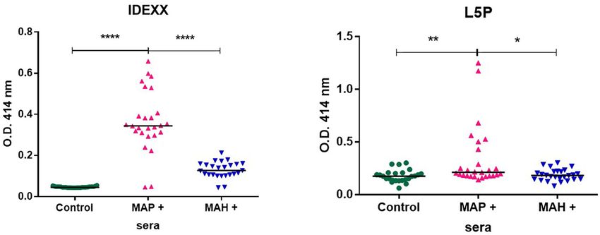
Bay et al.                                                                                                   Synthetic Lipopeptide for MAP Detection

(dd, 1H, 1CH2 β Phe), 3.23 (t, 2H, CH2 C or L, J = 6.86 Hz), 3.33          tested positive in commercial diagnostic ELISA IDEXX (IDEXX
(t, 2H, CH2 C or L, J = 6.84 Hz), 3.48–3.54 (m, 4H, CH2 E and J),          Paratuberculosis Screening Ab, Montpellier, France).
3.56–3.64 (m, 8H, CH2 F, G, H, and I), 4.04 (s, 2H, CH2 B), 4.06–
4.10 (m, 1H, CHα Ile), 4.18 (s, 2H, CH2 A), 4.23–4.28 (q, 1H,              ELISA Procedure
CHα Ala), 4.47 (d, 1H, CHα Val, J = 10.96 Hz), 4.61 (dt, 1H, CHα           ELISA using IDEXX commercial test (IDEXX Paratuberculosis
Phe), 4.68 (dt, 1H, CHα Phe), 7.16–7.19 (m, 2H, NH PEG), 7.21–             Screening Ab, Montpellier, France) were performed according to
7.38 (m, 10H, 2Ph), 7.97 (d, NH Ile), 8.13 (d, NH Phe). 13 C NMR           manufacturer’s instructions. ELISA experiments using synthetic
(MeOD): δ 11.34 (CH3 δ Ile), 15.75 (CH3 γ Ile), 18.34 (CH3 β Ala),         antigens were performed as described previously (8). Briefly,
19.87, 20.00 (2CH3 γ Val), 26.10 (CH2 γ Ile), 28.50 (CHβ Val),             L5P antigens were diluted at 25 µg/mL in ethanol. Water-soluble
30.35, 30.38 (CH2 D and K), 32.05 (NCH3 ), 37.69, 37.90 (CH2 C             L5P-Aq variant was diluted at 80 µg/mL in PBS (10 mM PBS,
and L), 38.14, 38.61 (2CH2 α Phe), 50.56 (CHα Ala), 53.35, 55.93           pH 7.4). Maxisorp microtiter plates (Nunc, Roskilde, Denmark)
(2CHα Phe), 59.50 (CHα Ile), 64.94 (CHα Val), 69.22 (CH2 A),               were coated with 50 µL of antigen preparation. Microtiter plates
69.82, 70.11 (CH2 E and J), 71.28, 71.31, 71.52, 71.58 (CH2 B, F, G,       coated with L5P were air-dried for 18 h at 37◦ C to allow the
H, and I), 127.87, 129.08, 129.56, 130.27, 130.35, 130.59, 135.26,         solvent to evaporate. Microtiter plates coated with L5P-Aq were
138.28 (Ph), 171.06, 171.92, 172.20, 172.85, 173.25, 173.63,               incubated for 18 h at 4◦ C. Following each incubation step, the
174.43 (CO).                                                               plates were washed three times with PBS supplemented with 0.1%
                                                                           (v/v) Tween 20 (PBST) (Sigma). After washing, the wells were
                                                                           blocked with 50 µL of Blocking Buffer (PBSTG: PBS-−0.05%
Animal Sera                                                                Tween 20, 0.5% Gelatin) (Gibco) at room temperature for 2 h.
A bank of serum samples of bovine origin, constituted by                   Fifty microliters of serum (diluted at 1:100 in PBSTG) were
CERVACODA, was provided by Dr. M. Govaerts. The status                     then added to each well and incubated for 1 h 30 min at 37◦ C.
of the sera was characterized by several diagnostic tests: fecal           Microtiter plates were washed five times with PBST and 50 µL
culture, Pourquier ELISA, and ID-Vet ELISA. The negative                   of solution of Recombinant Protein G Peroxidase Conjugated
sera came from farms known to be free from paratuberculosis,               (Thermo Scientific) diluted at 0.5 µg/mL in PBST, were added
this status was awarded after 3 consecutive years of whole-                to each well and incubated for 1 h at room temperature. After
herd negative serology. Sera were defined as being true positive           washing five times with PBST the reaction was visualized using
(n = 60) when they were positive in all three tests or as true             3,3′ ,5,5′ -tetramethylbenzidine (Sigma) and 0.5% v/v H2 O2 . The
negative (n = 50) when they were negative in all three tests. The          reaction was stopped with 1N sulfuric acid after 2 min of
same serum bank was used for the discovery of new antigenic                incubation in the dark and ODs were read at 450 nm on a
candidates in previous reports (22, 23). The sera were stored              Multiskan RC reader (Labsystems, Helsinki, Finland).
at −20◦ C and verified in IDEXX test (IDEXX Paratuberculosis
Screening Ab, Montpellier, France) (Supplementary Tables 1, 2              Data Analysis
and Supplementary Figure 2). In addition, a panel of sera were             The ELISA results of positive sera compared to negative sera
collected from cattle from a herd that was free of paratuberculosis        control, for each antigen were subjected to receiver–operator
but included in an official eradication campaign for bovine                characteristic (ROC) curve analysis. This method estimates the
tuberculosis. Eleven sera were collected from cattle from which            sensitivity and specificity of the ELISA, calculate likelihood ratios
M. bovis had been isolated and confirmed by IS6110 PCR and                 and provides an overall measure of test accuracy as area under
spoligotyping (24).                                                        the ROC curve (AUC) to evaluate the performance of the tests
    In the context of ovine paratuberculosis, 54 serum                     (32, 33). The optimal cut-off value is provided by ROC analysis
samples were carefully selected (25, 26). Fifteen negative                 as being associated with the maximal likelihood ratio.
sera were from unexposed sheep and 39 positive sera were                      Statistical differences between groups of goats unexposed or
from sheep experimentally infected with Telford 9.2 MAP                    goats experimentally infected either with MAP or MAH or
strain (25). The positive sera, verified by commercial test                between groups of cattle naturally infected by M. bovis or Map
(Supplementary Table 1 and Supplementary Figure 3) were                    was evaluated by Wilcoxon’s matched pairs test. Differences
classified in five groups according to the histopathological stage         were considered significant at p < 0.05. All statistical analyses
of the infection (27), (1) exposed but uninfected, (2) infected            were performed using statistical software (GRAPHPAD PRISM
with low grade lesions, (3) paucibacillary 3a, (4) multibacillary          version 6.07 for Windows, GraphPad software, San Diego,
3b, and (5) macroscopical lesions 3c.                                      CA, http://www.graphpad.com).
    Serum samples collected from unexposed goats (n = 25)
or goats experimentally infected either with MAP (n = 27)                  RESULTS
strain JII-1961 (28, 29) or with Mycobacterium avium subsp.
hominissuis (MAH) field isolate (n = 26) (30, 31) were provided            Chemical Synthesis of Pure L5P and
by Dr. Heike Koehler. Infection with MAP or MAH was                        Hydrosoluble Analog L5P-Aq
confirmed by cultural isolation of the respective strains from             The L5P is very hydrophobic and does behave very differently
tissues after necropsy and/or by histopathology (lesions) and              as compared to conventional protein antigens which are
immunohistochemistry (presence of mycobacteria) of tissue                  hydrophilic. It is soluble in DMSO, CHCl3 , CH2 Cl2 , MeOH, and
sections. Positive sera were from animals with severe lesions that         EtOH, but insoluble in water or aqueous buffers. Glass containers

Frontiers in Veterinary Science | www.frontiersin.org                  4                                        April 2021 | Volume 8 | Article 637841
Bay et al. Synthetic Lipopeptide for MAP Detection
have to be used, and contact with polypropylene surfaces must uninfected sheep. As discussed in detail below, this result is likely
be minimized. Material handling like dilution and transfer steps due to the minor lipopeptide differences present in the cell walls
also needs to be minimized. These properties of L5P would cause of sheep vs. cattle strains of MAP.
difficulties if a diagnostic test was based on the L5P antigen. To
circumvent these difficulties, we have designed a hydrosoluble Diagnostic Value of L5P in Goats Infected
derivative of L5P, L5P-Aq. The N-ter non-immunogenic lipidic With MAP or MAH
moiety of L5P was suppressed and the C-ter methyl ester was In their environment animals are naturally exposed to different
replaced by polyethylene glycol chain ending with a carboxyl mycobacteria. In the context of farm animals, it is recognized that
group (Figure 1A). this exposure can hamper diagnostic tests. To gain specificity,
The resulting L5P-Aq was obtained by solid-phase peptide commercial serologic screening tests for MAP use a serum
synthesis using 9-fluorenylmethoxycarbonyl chemistry. After adsorption step with antigens prepared from an environmental
cleavage from the resin, by contrast with its hydrophobic bacterium Mycobacterium phlei. Knowing that L5P is specific
counterpart, the crude product was actually extracted in to MAP we investigated its use without any pre-adsorption
aqueous phase. Purification by reverse-phase chromatography step against serum from animals experimentally infected with
then afforded 126 mg of L5P-Aq with a 88 % overall yield. To MAP, or MAH mycobacteria very close to MAP. As expected,
ensure both its purity and its identity, the L5P-Aq was analyzed the commercial IDEXX test makes it possible to distinctly
by RP-HPLC (Figure 1B, >99.5%), MS (Figure 1C), quantitative detect animals infected with MAP from animals infected
AAA, and NMR (Supplementary Figures 1A,B). with MAH and from non-infected animals. Compared to the
commercial test, L5P used alone without a pre-adsorption step
Antigenic Evaluation of L5P and L5P-Aq on with Mycobacterium phlei extracts, lacked sensitivity with MAP
a Bank of Bovine Sera positive sera but didn’t react with sera of animals infected with
In previous studies, the antibody response against L5P was MAH (Figure 4).
investigated with a limited panel of sera and it has not been
subjected to comprehensive, large scale evaluation. Synthetic L5P L5P as a Biomarker Able to Discriminate
and the hydrosoluble derivative L5P-Aq were therefore both MAP From/Between M. bovis Infection
assessed using a bank of well-defined sera including 60 sera Bovine tuberculosis (bTB) is a major zoonotic disease of cattle
from MAP-naturally infected cattle and 50 sera from healthy that is endemic in much of the world. The antemortem diagnostic
cattle (details in the method section). In receiver operating methods currently approved for use in cattle have limitations.
characteristic analysis (ROC analysis) of L5P, the area under The intradermal tuberculin test has suboptimal sensitivity and
the curve and its standard error were 0.97 (95% confidence inconsistent performance (34, 35). Nontuberculous mycobacteria
interval, 0.9418 to 0.9942) and 0.01336, respectively (Figure 2 (NTM) and MAP, in particular, have been repeatedly shown to
and Supplementary Table 1). The L5P-Aq was evaluated in interfere with the detection of M. bovis (36). There have been
ethanol and in PBS and the area under the curve and its standard a number of projects with aims to improve the diagnosis of
error were 0.937 (95 % confidence interval, 0.8909 to 0.9831) and bTB and JD by generating specific tools that do not compromise
0.02354, and 0.9427 (95% confidence interval, 0.9021 to 0.9832) sensitivity or specificity due to co-infections or testing regimes.
and 0.02068, respectively (Figure 2 and Supplementary Table 1). We therefore evaluated the L5P antigen against field sera from
The sensitivity of L5P and L5P-Aq in comparison to commercial cattle naturally infected with M. bovis. The results in Figure 5
test were 81.67, 61.67, and 98%, respectively. The specificity show that ELISA based on synthetic L5P did not react to field sera
of L5P and L5P-Aq were 98% identical to the commercial from M. bovis positive cattle in comparison to sera from MAP
test. When tested in same conditions, the acidic derivative positive cattle.
of the L5P lipid moiety (eicosanoic acid) was not antigenic,
showing that the serum response was not directed against
the L5P lipid (Supplementary Figure 4) according to previous DISCUSSION
report. These results confirm that both L5P and L5P-Aq have
The high lipid contents of mycobacterial cells has been
potential to discriminate MAP infected and uninfected cattle at
recognized for a long time and much effort has been devoted
a population level.
to the identification of the various types of lipids present,
many of which are glycolipids, unique to mycobacteria (37).
L5P Antigenic Response in the Context of M avium isolates are characterized by their production of
Ovine Paratuberculosis highly antigenic glycopeptidolipids or GPLs, which are suitable
Although ovine paratuberculosis is comparable to the disease for specific serodiagnosis (6, 38). Surprisingly, subspecies
in cattle, it is well-documented that MAP strains isolated from paratuberculosis isolates produce lipopeptides instead of GPLs;
sheep have host-dependent features. To investigate the immune these are characterized by the absence of sugars and the absence
response in MAP infected sheep, we used the L5P described as of both hydroxylation and double bonds in the fatty acid moiety
the native antigen in strains of MAP of subtype II isolated from (8). Attempts to develop serological assays based on the native
cattle. The results presented in Figure 3 show that synthetic L5P L5P structure are problematic due to the apolar nature of
is not able to significantly discriminate MAP positive sheep from the molecule and its lack of solubility in the aqueous buffers
Frontiers in Veterinary Science | www.frontiersin.org 5 April 2021 | Volume 8 | Article 637841Bay et al. Synthetic Lipopeptide for MAP Detection FIGURE 2 | ROC analysis of antibody response of bovine sera against L5P and it hydrosoluble variant L5P-Aq. Analysis performed on a bank of sera including 60 MAP positive (MAP +) and 50 control animals, using a L5P coated in ethanol, L5P-Aq in ethanol or L5P-Aq in PBS. All results are expressed as individual OD and were compared by ROC analysis. Serum samples were tested in triplicate. The horizontal bars indicate median. A, Area under the receiver operating characteristic curve. Significantly different when p < 0.05. FIGURE 3 | ROC analysis of antibody response of ovine sera against L5P. Analysis performed on a panel of sera from 39 MAP positive (MAP +) and 15 control animals, using L5P coated in ethanol. All results are expressed as individual OD and were compared by ROC analysis. The horizontal bars indicate median. Area under the receiver operating characteristic curve. Significantly different when p < 0.05. Not significantly different (NS). Frontiers in Veterinary Science | www.frontiersin.org 6 April 2021 | Volume 8 | Article 637841
Bay et al.                                                                                                                 Synthetic Lipopeptide for MAP Detection

 FIGURE 4 | Antibody response from goats uninfected (n = 27) or experimentally infected with MAP (n = 25) or MAH (n = 26), as analyzed by IDEXX test or using L5P
 antigen. Results are shown as OD450 nm of individual samples and medians are indicated as a black line. The statistical differences between the groups were
 determined using the non-parametric Mann–Whitney test. Significantly different when p < 0.05. *p < 0.05, **p < 0.01, and ****p < 0.0001.

                                                                                      typically used in ELISA. To overcome this issue, we engineered a
                                                                                      hydrophilic variant of the native lipopeptide, named L5P-Aq, and
                                                                                      we validated its antigenicity using a bank of MAP positive bovine
                                                                                      sera. Used alone without a pre-adsorption step, the L5P ELISA
                                                                                      test developed in this study can discriminate MAP infected goats
                                                                                      from MAH infected goats. Interestingly, the sera from MAP
                                                                                      infected sheep did not react with L5P, suggesting that lineage of
                                                                                      strains of MAP specific for the sheep enrolled in this study may
                                                                                      have another lipopeptide which has not yet been reported. This
                                                                                      report also shows that L5P may have utility as a reagent to assist
                                                                                      in the diagnosis of bTB.
                                                                                          Attempts to overcome the apolar nature of antigens in
                                                                                      serological assays have been performed on bacterial glycolipids
                                                                                      (39). For example, the Tween detergent has been used to optimize
                                                                                      the antigen coating. On the other hand, the use of Tween
                                                                                      was shown to be problematic as the detergent interacts with
                                                                                      the lipid portion of the molecule causing its detachment from
                                                                                      the plastic (39). From our results, 100% ethanol or methanol
                                                                                      solutions enabled the efficient coating of antigen onto microtiter
                                                                                      plate wells. Both our past and current studies show that the
                                                                                      critical L5P epitopes are located on the peptide portion (8).
                                                                                      Therefore, modifications to improve solubility were focused on
                                                                                      (i) suppressing the hydrophobic lipid and (ii) extending the
                                                                                      peptide with a hydrophilic chain. The resulting modification
                                                                                      of L5P will yield numerous advantages. The hydrosolubility of
                                                                                      the resulting L5P-Aq is an important benefit for its use in an
                                                                                      ELISA test, especially for high throughput formats. Furthermore,
                                                                                      safety issues associated with the use of organic solvents, including
 FIGURE 5 | L5P used to discriminate MAP infected cattle from M. bovis
                                                                                      alcohols, are avoided. Finally, handling of antigenic material is
 infected cattle. ELISA were performed on plates coated with L5P in ethanol.          much easier and reliable allowing repeatable procedures.
 The panel of sera tested included 3 MAP positive sera of reference, 3 negative           In most countries, the majority of routine screening for
 controls, and 11 M. bovis positive sera. Serum samples were tested in                MAP (control, surveillance, certification, control at introduction)
 triplicate. The statistical differences between the groups were determined           (99%) is carried out by serological ELISA tests (18, 40). The
 using the non-parametric Mann–Whitney test. Significantly different when p <
 0.05. ****p < 0.0001. Not significantly different (ns).
                                                                                      other methods such as PCR, bacteriological culture, direct
                                                                                      examination (Ziehl-Neelsen staining) are used for clinical case

Frontiers in Veterinary Science | www.frontiersin.org                             7                                           April 2021 | Volume 8 | Article 637841
Bay et al. Synthetic Lipopeptide for MAP Detection
confirmation. The final purpose of this project was to know if antigen if it is used alone. These results highlight the great need
synthetic lipopeptide antigen derivatives are able to specifically to characterize the antigens of MAP broadly across many strains,
discriminate sera from MAP-infected and uninfected cattle in a regardless of their protein or lipidic nature, and to consider
serological assay. Until now L5P (hydrophobic) was evaluated in the genetic diversity of this subspecies. Not only are lipids
just a few animals. We have thus assembled here a large panel different among MAP strain types, but they can change based
of thoroughly characterized sera, to test the recognition of these on the environment. For example, the L5P lipid was shown to
synthetic antigens. ROC analysis of results obtained with the be absent in MAP exposed to milk, but present in MAP cultured
L5P and its hydrosoluble variant, L5P-Aq, demonstrated a cut-off in Middlebrook media (42). Therefore, additional experiments
value corresponding to a relative sensitivity of 82 and 62% and a should be conducted to determine the extent to which L5P is
specificity of 98 and 98%, respectively (Supplementary Table 1 produced across many different environments, including feces
and Supplementary Figure 2). These results show that synthetic and milk. However, from a bovine diagnostics standpoint, it
L5P antigen, used alone, is able to discriminate MAP-infected is clear that MAP replicating inside cows do produce L5P
animals and controls at a population level although the lower as we and others have shown a bovine antibody response
sensitivity compared with the commercial test remains an issue to L5P.
to improve. Likewise, the lack of sensitivity of L5P observed with In conclusion, using an engineered synthetic antigen, we have
sera from experimentally infected goats suggests that there is a identified a potent hydrosoluble mimic of the native L5P and
“technical barrier” to be overcome to improve this parameter. established ELISA conditions for the specific diagnosis of bovine
Indeed to achieve the best assay performances, more research is paratuberculosis. Ongoing research into the characterization of
needed to improve not only on the ELISA procedure (antigen the non-protein antigens produced naturally by the different
presentation, buffer composition, coating process, secondary genetic lineages of MAP should identify new antigens that
reagent. . . ), but also on the target antigen(s). It is accepted that could contribute to achieve a diagnostic test with optimal
one universal antigen probably does not exist and that an efficient sensitivity and specificity. In addition, it would be important to
diagnostic test for paratuberculosis will require a cocktail of identify the L5P homolog in SI-type strains. New developments,
antigens. In this context, if used within an optimal combination, including chemical tailoring and formulation of these synthetic
such synthetic individual MAP antigens have potential to assist antigens need to be investigated to gain sensitivity observed
in the improvement of the antigens used commercially. In in this report. Other investigations should evaluate animal
addition, the use of synthetic antigen rather than crude protein responses according to the disease progression and excretory
extracts or culture-derived antigens has the advantage of avoiding status of the animal. These synthetic antigens could also be useful
the culture of slow growing pathogenic mycobacteria, such as for the improvement of existing commercial tests especially
MAP. The L5P-Aq synthesis can be easily performed by organic regarding the strong demand from diagnostic laboratories
chemistry that offers the possibility of having large quantities for no batch-to-batch variation and elimination of the
of pure material. Numerous other advantages also come from pre-absorption step.
the synthetic production of antigen, including the ability to
standardize the batches and also to modify the antigens and/or
their formulation, for example to graft fluorescent markers to DATA AVAILABILITY STATEMENT
monitor their handling. In the future, it would be interesting to
have access to a bovine serum panel built to cover all the stages The original contributions presented in the study are included
of JD infection and to be able to investigate the early stages of in the article/Supplementary Material, further inquiries can be
infection with these synthetic antigens. Preliminary data on the directed to the corresponding author/s.
use of L5P for the early diagnosis of MAP infection by detection
of interferon should be consolidated (15).
A very interesting issue of this study came from the “negative”
AUTHOR CONTRIBUTIONS
result obtained with the sera from MAP infected sheep. Our SB, RW, HK, JB, and FB conceived and designed the study.
current knowledge on the composition of lipids of the external All authors made substantial contributions to the analysis and
layer of the wall of the subspecies paratuberculosis allows us writing of the manuscript.
to understand this result. According to genomic analysis, the
S-type strains, which are more adapted in sheep, have evolved
in two distinct subtype S-I and S-III (41). In a recent report we FUNDING
described that the SIII-type strain S397, produced a unique lipid,
a tripeptide Phe-N-Methyl-Val-Ala with a N-ter lipid moiety, This study was funded by EMIDA-EraNet Mycobacdiagnosis
termed lipotripeptide (L3P) instead of the L5P detected in cattle (Convention: 2011-EMID-005-02).
strains (11). In addition, at the present time no L5P nor L3P
has been detected in the lipids extracted from the S-I strain
Telford that was used in the experimental sheep infections (data SUPPLEMENTARY MATERIAL
not shown). These observations may explain why L5P was not
significantly recognized by the sera of infected sheep. While most The Supplementary Material for this article can be found
MAP strains isolated from bovine are of C-type, infections with online at: https://www.frontiersin.org/articles/10.3389/fvets.
S type do occur, a situation which cannot be detected by this 2021.637841/full#supplementary-material
Frontiers in Veterinary Science | www.frontiersin.org 8 April 2021 | Volume 8 | Article 637841Bay et al. Synthetic Lipopeptide for MAP Detection
Supplementary Figure 1 | 1 H (A) and 13 C (B) NMR spectra of L5P-Aq. The IDEXX. All results are expressed as individual S/P (OD sample-OD negative
corresponding structural formula is depicted in panel (C). control/ meanOD positive control—Mean negative control) and were compared by
ROC analysis. Serum samples were tested in triplicate. Area under the receiver
Supplementary Figure 2 | ROC analysis of antibody response of a bank of operating characteristic curve (A).
bovine sera in the IDEXX test. ROC analysis was performed on a bank of sera
Supplementary Figure 4 | ROC analysis of antibody response of bovine sera
including 60 MAP positive and 50 control animals, using the commercial test
against lipid part of L5P. ROC analysis was performed on a bank of sera including
IDEXX. All results are expressed as individual OD and were compared by ROC
60 MAP positive and 50 control animals, using the lipid part of L5P coated in
analysis. Serum samples were tested in triplicate. Significantly different when
ethanol. All results are expressed as individual OD and were compared by ROC
p < 0.05. ∗∗∗∗ p < 0.0001. The horizontal bars indicate median. Not significantly
analysis. Serum samples were tested in triplicate. The horizontal bars indicate
different (ns).
median. Not significantly different (ns).
Supplementary Figure 3 | ROC analysis of antibody response of the ovine
Supplementary Table 1 | Statistical detail of ROC analysis.
serum panel in the IDEXX test. ROC analysis performed on a bank of sera
including 39 MAP positive and 15 control animals, using the commercial test Supplementary Table 2 | Detail of bank of sera.
REFERENCES paratuberculosis specific lipopentapeptide antigens. Res Vet Sci. (2013)
95:123–9. doi: 10.1016/j.rvsc.2013.03.002
1. Daffe M, Crick DC, Jackson M. Genetics of capsular polysaccharides 14. Verdier J, Deroche L, Allez M, Loy C, Biet F, Bodier CC, et al.
and cell envelope (Glyco)lipids. Microbiol Spectr. (2014) 2:1–2. Specific IgG response against Mycobacterium avium paratuberculosis in
doi: 10.1128/microbiolspec.MGM2-0021-2013 children and adults with Crohn’s disease. PLoS One. (2013) 8:e62780.
2. Marrakchi H, Laneelle MA, Daffe M. Mycolic acids: structures, biosynthesis, doi: 10.1371/journal.pone.0062780
and beyond. Chem Biol. (2014) 21:67–85. doi: 10.1016/j.chembiol.201 15. Holbert S, Branger M, Souriau A, Lamoureux B, Ganneau C, Richard G, et al.
3.11.011 Interferon gamma response to Mycobacterium avium subsp. paratuberculosis
3. Nakata N, Fujiwara N, Naka T, Yano I, Kobayashi K, Maeda S. Identification specific lipopentapeptide antigen L5P in cattle. Res Vet Sci. (2015) 102:118–21.
and characterization of two novel methyltransferase genes that determine the doi: 10.1016/j.rvsc.2015.07.017
serotype 12-specific structure of glycopeptidolipids of Mycobacterium 16. Niegowska M, Rapini N, Biet F, Piccinini S, Bay S, Lidano R, et al.
intracellulare. J Bacteriol. (2008) 190:1064–71. doi: 10.1128/JB.01 Seroreactivity against specific L5P antigen from Mycobacterium avium subsp.
370-07 paratuberculosis in children at risk for T1D. PLoS One. (2016) 11: e0157962.
4. Pourshafie M, Ayub Q, Barrow WW. Comparative effects of Mycobacterium doi: 10.1371/journal.pone.0157962
avium glycopeptidolipid and lipopeptide fragment on the function and 17. Barkema HW, Hesselink JW, McKenna SLB, Benedictus G, Groenendaal H.
ultrastructure of mononuclear cells. Clin Exp Immunol. (1993) 93:72–9. Global prevalence and economics of infection with Mycobacterium avium
doi: 10.1111/j.1365-2249.1993.tb06499.x subsp. paratuberculosis in ruminants. In: Behr MA, Collins DM, editors.
5. Ortalo-Magne A, Lemassu A, Laneelle MA, Bardou F, Silve G, Gounon Paratuberculosis: Organism, Disease, Control. Oxfordshire: Wallingford
P, et al. Identification of the surface-exposed lipids on the cell envelopes (2010). p. 10–21.
of Mycobacterium tuberculosis and other mycobacterial species. J Bacteriol. 18. Barkema HW, Orsel K, Nielsen SS, Koets AP, Rutten V, Bannantine JP, et al.
(1996) 178:456–61. doi: 10.1128/JB.178.2.456-461.1996 Knowledge gaps that hamper prevention and control of Mycobacterium avium
6. Kitada S, Yoshimura K, Miki K, Miki M, Hashimoto H, Matsui H, et al. subspecies paratuberculosis infection. Transbound Emerg Dis. (2018) 65(Suppl
Validation of a commercial serodiagnostic kit for diagnosing pulmonary 1):125–48. doi: 10.1111/tbed.12723
Mycobacterium avium complex disease. Int J Tuberc Lung Dis. (2015) 19:97– 19. Whittington R, Donat K, Weber MF, Kelton D, Nielsen SS, Eisenberg S, et
103. doi: 10.5588/ijtld.14.0564 al. Control of paratuberculosis: who, why and how. A review of 48 countries.
7. Rathnaiah G, Zinniel DK, Bannantine JP, Stabel JR, Grohn YT, Collins MT, et BMC Vet Res. (2019) 15:198. doi: 10.1186/s12917-019-1943-4
al. Pathogenesis, molecular genetics, and genomics of Mycobacterium avium 20. Bannantine JP, Bayles DO, Waters WR, Palmer MV, Stabel JR, Paustian
subsp. paratuberculosis, the Etiologic Agent of Johne’s Disease. Front Vet Sci. ML. Early antibody response against Mycobacterium avium subspecies
(2017) 4:187. doi: 10.3389/fvets.2017.00187 paratuberculosis antigens in subclinical cattle. Proteome Sci. (2008) 6:5.
8. Biet F, Bay S, Thibault VC, Euphrasie D, Grayon M, Ganneau C, et al. doi: 10.1186/1477-5956-6-5
Lipopentapeptide induces a strong host humoral response and distinguishes 21. Blankemeyer-Menge B, Nimtz M, Frank R. An efficient method for anchoring
Mycobacterium avium subsp. paratuberculosis from M. avium subsp avium. fmoc-anino acids to hydroxyl-functionalised solid supports. Tetrahedron Lett.
Vaccine. (2008) 26:257–68. doi: 10.1016/j.vaccine.2007.10.059 (1990) 31:1701–4. doi: 10.1016/S0040-4039(00)88858-9
9. Riviere M, Puzo G, Wright EL, Barrow WW. A unique phenylalanine- 22. Leroy B, Roupie V, Noel-Georis I, Rosseels V, Walravens K, Govaerts M, et
containing lipopeptide isolated from a rough-colony variant al. Antigen discovery: a postgenomic approach to paratuberculosis diagnosis.
of Mycobacterium avium. Eur J Biochem. (1996) 241:682–90. Proteomics. (2007) 7:1164–76. doi: 10.1002/pmic.200600988
doi: 10.1111/j.1432-1033.1996.00682.x 23. Leroy B, Viart S, Trinchero N, Roupie V, Govaerts M, Letesson JJ, et al. Use
10. Eckstein TM, Chandrasekaran S, Mahapatra S, McNeil MR, Chatterjee of Mycobacterium avium subsp. paratuberculosis specific coding sequences
D, Rithner CD, et al. A major cell wall lipopeptide of Mycobacterium for serodiagnosis of bovine paratuberculosis. Vet Microbiol. (2009) 135:313–9.
avium subspecies paratuberculosis. J Biol Chem. (2006) 281:5209–15. doi: 10.1016/j.vetmic.2008.09.065
doi: 10.1074/jbc.M512465200 24. Courcoul A, Moyen JL, Brugere L, Faye S, Henault S, Gares H, et al. Estimation
11. Bannantine JP, Etienne G, Laval F, Stabel JR, Lemassu A, Daffe M, et al. of sensitivity and specificity of bacteriology, histopathology and PCR for the
Cell wall peptidolipids of Mycobacterium avium: from genetic prediction to confirmatory diagnosis of bovine tuberculosis using latent class analysis. PLoS
exact structure of a nonribosomal peptide. Mol Microbiol. (2017) 105:525–39. One. (2014) 9:e90334. doi: 10.1371/journal.pone.0090334
doi: 10.1111/mmi.13717 25. Begg DJ, de Silva K, Di Fiore L, Taylor DL, Bower K, Zhong L, et al.
12. Costanzo G, Pinedo FA, Mon ML, Viale M, Gil A, Illia MC, Experimental infection model for Johne’s disease using a lyophilised, pure
et al. Accuracy assessment and screening of a dairy herd with culture, seedstock of Mycobacterium avium subspecies paratuberculosis. Vet
paratuberculosis by three different ELISAs. Vet Microbiol. (2012) 56:183–8. Microbiol. (2010) 141:301–11. doi: 10.1016/j.vetmic.2009.09.007
doi: 10.1016/j.vetmic.2011.10.029 26. Dukkipati VSR, Ridler AL, Thompson KG, Buddle BM, Hedgespeth BA,
13. Thirunavukkarasu S, Plain KM, Eckstein TM, de Silva K, Whittington Price-Carter M, et al. Experimental infection of New Zealand Merino sheep
RJ. Cellular and humoral immunogenicity of Mycobacterium avium subsp. with a suspension of Mycobacterium avium subspecies paratuberculosis (Map)
Frontiers in Veterinary Science | www.frontiersin.org 9 April 2021 | Volume 8 | Article 637841Bay et al. Synthetic Lipopeptide for MAP Detection
strain Telford: kinetics of the immune response, histopathology and Map antibodies in bovine tuberculosis. Clin Vaccine Immunol. (2017) 24:e00259–
culture. Vet Microbiol. (2016) 195:136–43. doi: 10.1016/j.vetmic.2016.09.018 17. doi: 10.1128/CVI.00259-17
27. Perez V, Garcia Marin JF, Badiola JJ. Description and classification of 36. Biet F, Boschiroli ML. Non-tuberculous mycobacterial infections
different types of lesion associated with natural paratuberculosis infection of veterinary relevance. Res Vet Sci. (2014) 97(Suppl):S69–77.
in sheep. J Comp Pathol. (1996) 114:107–22. doi: 10.1016/S0021-9975(96) doi: 10.1016/j.rvsc.2014.08.007
80001-6 37. Brennan PJ, Nikaido H. The envelope of mycobacteria. Annu Rev Biochem.
28. Kruger C, Kohler H, Liebler-Tenorio EM. Cellular composition of (1995) 64:29–63. doi: 10.1146/annurev.bi.64.070195.000333
granulomatous lesions in gut-associated lymphoid tissues of goats during 38. Jeong BH, Kim SY, Jeon K, Lee SY, Shin SJ, Koh WJ. Serodiagnosis
the first year after experimental infection with Mycobacterium avium of Mycobacterium avium complex and Mycobacterium abscessus complex
subsp. Paratuberculosis. Vet Immunol Immunopathol. (2015) 163:33–45. pulmonary disease by use of IgA antibodies to glycopeptidolipid core antigen.
doi: 10.1016/j.vetimm.2014.11.002 J Clin Microbiol. (2013) 51:2747–9. doi: 10.1128/JCM.00702-13
29. Mobius P, Nordsiek G, Holzer M, Jarek M, Marz M, Kohler H. Complete 39. Spencer JS, Brennan PJ. The role of Mycobacterium leprae phenolic glycolipid
genome sequence of JII-1961, a bovine Mycobacterium avium subsp. I (PGL-I) in serodiagnosis and in the pathogenesis of leprosy. Lepr Rev. (2011)
paratuberculosis field isolate from Germany. Genome Announc. (2017) 82:344–57. doi: 10.47276/lr.82.4.344
5:e00870–17. doi: 10.1128/genomeA.00870-17 40. Mikkelsen H, Aagaard C, Nielsen SS, Jungersen G. Review of Mycobacterium
30. Kohler H, Soschinka A, Meyer M, Kather A, Reinhold P, Liebler-Tenorio avium subsp. paratuberculosis antigen candidates with diagnostic potential.
E. Characterization of a caprine model for the subclinical initial phase of Vet Microbiol. (2011) 152:1–20. doi: 10.1016/j.vetmic.2011.03.006
Mycobacterium avium. subsp. paratuberculosis infection. BMC Vet Res. (2015) 41. Biet F, Sevilla IA, Cochard T, Lefrancois LH, Garrido JM, Heron I, et al. Inter-
11:74. doi: 10.1186/s12917-015-0381-1 and intra-subtype genotypic differences that differentiate Mycobacterium
31. Schinkothe J, Mobius P, Kohler H, Liebler-Tenorio EM. Experimental avium subspecies paratuberculosis strains. BMC Microbiol. (2012) 12:264.
infection of goats with Mycobacterium avium subsp. hominissuis: a Model doi: 10.1186/1471-2180-12-264
for Comparative Tuberculosis Research. J Comp Pathol. (2016) 155:218–30. 42. Alonso-Hearn M, Eckstein TM, Sommer S, Bermudez LE. A Mycobacterium
doi: 10.1016/j.jcpa.2016.06.008 avium subsp. paratuberculosis LuxR regulates cell envelope and virulence.
32. Bewick V, Cheek L, Ball J. Statistics review 13: receiver operating characteristic Innate Immun. (2010) 16:235–47. doi: 10.1177/1753425909339811
curves. Crit Care. (2004) 8:508–12. doi: 10.1186/cc3000
33. Gardner IA, Greiner M. Receiver-operating characteristic curves and Conflict of Interest: The authors declare that the research was conducted in the
likelihood ratios: improvements over traditional methods for the evaluation absence of any commercial or financial relationships that could be construed as a
and application of veterinary clinical pathology tests. Vet Clin Pathol. (2006) potential conflict of interest.
35:8–17. doi: 10.1111/j.1939-165X.2006.tb00082.x
34. Schiller I, Oesch B, Vordermeier HM, Palmer MV, Harris BN, Copyright © 2021 Bay, Begg, Ganneau, Branger, Cochard, Bannantine, Köhler,
Orloski KA, et al. Bovine tuberculosis: a review of current and Moyen, Whittington and Biet. This is an open-access article distributed under the
emerging diagnostic techniques in view of their relevance for disease terms of the Creative Commons Attribution License (CC BY). The use, distribution
control and eradication. Transbound Emerg Dis. (2010) 57:205–20. or reproduction in other forums is permitted, provided the original author(s) and
doi: 10.1111/j.1865-1682.2010.01148.x the copyright owner(s) are credited and that the original publication in this journal
35. Lyashchenko KP, Grandison A, Keskinen K, Sikar-Gang A, Lambotte P, is cited, in accordance with accepted academic practice. No use, distribution or
Esfandiari J, et al. Identification of novel antigens recognized by serum reproduction is permitted which does not comply with these terms.
Frontiers in Veterinary Science | www.frontiersin.org 10 April 2021 | Volume 8 | Article 637841You can also read