Comparison of walking variations during treadmill walking test between neurogenic and vascular claudication: a crossover study
←
→
Page content transcription
If your browser does not render page correctly, please read the page content below
Houle et al. Chiropractic & Manual Therapies              (2021) 29:24
https://doi.org/10.1186/s12998-021-00382-5

 RESEARCH                                                                                                                                             Open Access

Comparison of walking variations during
treadmill walking test between neurogenic
and vascular claudication: a crossover study
Mariève Houle1* , Julie O’Shaughnessy2, Charles Tétreau1, Claude-Édouard Châtillon3,
Andrée-Anne Marchand2 and Martin Descarreaux1

  Abstract
  Background: Lumbar spinal stenosis (LSS) and peripheral arterial disease (PAD) are two distinct conditions
  characterized by similar symptoms including leg pain and walking limitations due to claudication. Differentiation
  between both origins can be difficult and characteristics such as symptom manifestations, time to relief in rest
  position and pain localization should be considered when determining diagnosis and the treatment plan. The
  objectives of this study were to compare changes in walking time to symptom change during treadmill tests and
  self-reported outcomes measures related to claudication, kinesophobia and global health between individuals with
  LSS, PAD and non-specific low back pain (nLBP).
  Method: Fifty-five patients (23 with LSS, 14 with PAD and 18 with nLBP) were recruited from May 2018 to March
  2020 to complete a treadmill walking test involving two 5-min walking tasks (Upright and Forward Leaning Trunk
  (FLT) Walking tasks). The speed was set at 1.9 km/h (1.2 mph), and each task was followed by a 5-min rest period.
  Walking time to symptom change and Total walking time were recorded during each walking task. Patients were
  asked to complete four questionnaires related to the impact of claudication, walking impairment, kinesiophobia and
  global health. One-way ANOVAs were performed to compare walking time difference from the Upright to the FLT
  walking tasks and to compare questionnaires results between groups.
  Results: One-way ANOVAs showed a significant difference between groups regarding difference in Walking time to
  symptom change between both tasks (F = 4.12, p = 0.022). The LSS group improved its Walking time to symptom
  change from the Upright to the FLT walking tasks more than the PAD (p = 0.34) and the nLBP group (p = 0.12). The
  nLBP group was less impacted by claudication and less impaired during walking compared to the LSS and PAD
  groups (ps < 0.001). The nLBP group also had less kinesiophobia than the LSS one (p < 0.001), but was similar to the
  PAD group. The global health rating was not statistically different between groups (p = 0.118).
  Conclusion: The test was able to distinguish neurogenic from vascular or nLBP related claudication. However,
  further studies are needed to validate this new treadmill walking test.
  Trial registration: clinicaltrials.gov (NCT04058171), Registered August 15, 2019 –Registered during recruitment

* Correspondence: marieve.houle@uqtr.ca
1
 Département des Sciences de l’activité physique, Université du Québec à
Trois-Rivières, 3351, boul. des Forges, C.P. 500, Trois-Rivières, QC G8Z 4M3,
Canada
Full list of author information is available at the end of the article

                                          © The Author(s). 2021 Open Access This article is licensed under a Creative Commons Attribution 4.0 International License,
                                          which permits use, sharing, adaptation, distribution and reproduction in any medium or format, as long as you give
                                          appropriate credit to the original author(s) and the source, provide a link to the Creative Commons licence, and indicate if
                                          changes were made. The images or other third party material in this article are included in the article's Creative Commons
                                          licence, unless indicated otherwise in a credit line to the material. If material is not included in the article's Creative Commons
                                          licence and your intended use is not permitted by statutory regulation or exceeds the permitted use, you will need to obtain
                                          permission directly from the copyright holder. To view a copy of this licence, visit http://creativecommons.org/licenses/by/4.0/.
                                          The Creative Commons Public Domain Dedication waiver (http://creativecommons.org/publicdomain/zero/1.0/) applies to the
                                          data made available in this article, unless otherwise stated in a credit line to the data.
Houle et al. Chiropractic & Manual Therapies (2021) 29:24 Page 2 of 11 Keywords: Lumbar spinal stenosis, Peripheral artery disease, Low back pain, Treadmill test, Walking time, Walking posture Background Non-specific low back pain is a musculoskeletal condi- Intermittent claudication (IC) is defined as lameness due tion defined as pain located between the 12th ribs and to leg pain while standing or walking [1], with the leg the gluteal fold [18, 19], with or without referred pain in pain attenuating within seconds to a few minutes when one or both lower limbs [20]. For patients with nLBP, stopping activity or sitting [2–4], based on the under- pain in the lower limb is commonly due to referred pain lying health condition. Vascular and neurogenic claudi- into the buttock or thigh (above the knee) and there is cation represent both possible origins of IC, and their no neurological impairment [6]. Low back pain is a very symptoms are frequently described as pain, cramps, common symptom experienced by individuals of any age numbness and tingling in the lower limbs [5]. Vascular and particularly in people between 40 and 80 years old claudication is a common manifestation in individuals [18, 20], with a lifetime prevalence of 84% [21]. The term with peripheral arterial disease (PAD), while neurogenic nLBP describes LBP for which a specific cause of pain claudication occurs in lumbar musculoskeletal disorders cannot be identified [20]. Among people with nLBP, with neurological involvement such as specific low back some of the most common sources of referred pain into pain (LBP) conditions. One of these specific LBP condi- the lower limbs are sacroiliac joint syndrome, discogenic tions causing neurogenic claudication is lumbar spinal low back pain and facet joint pain [22]. In acute, sub- stenosis (LSS). In some cases, individuals with non- acute or chronic nLBP with referred pain into one or specific low back pain (nLBP) will experience referred both legs, patients can have difficulty with activities such pain into the lower limbs which will also cause difficulty as dressing, standing and walking [23]. during walking [6]. Even if these three conditions have distinct mecha- Peripheral arterial disease is a condition affecting the nisms, they can all affect walking capacity of patients blood vessels caused by a narrowing of the arteries, usu- through one of their main symptoms: intermittent clau- ally brought on by the accumulation of atheroma plates dication. Vascular and neurogenic claudication share (atherosclerosis) [1]. This accumulation leads to insuffi- similarities in their symptoms [2], but the posture of re- cient blood supply to the muscles which is accentuated lief differs between both types. In fact, in PAD, patients with increasing intensity of activities such as walking [7]. need to stop their activity while in LSS, patients need to Vascular claudication is the most common manifestation adopt a bending forward posture or to sit down. Add- of PAD [8] and its prevalence is estimated at 10 to 20% itionally, both claudication origins lead to several limita- [9] in 40-year-old individuals, whereas this number dou- tions in daily physical activities, such as a reduction of bles in individuals older than 60 years of age [10]. When walking time and walking distance [2, 17, 24, 25]. Be- PAD patients are walking the need in oxygen increases cause of their similarities, clinicians must establish their in muscles and the insufficient blood supply leads to diagnosis based on the reported symptoms and clinical pain into the leg(s) and subsequently to the need to stop manifestations in daily activities and combine the clinical or to sit to relieve the pain. history with diagnostic tests or medical imaging. Now- Lumbar spinal stenosis is a condition leading to mech- adays, the diagnosis of PAD is obtained using the stand- anical compression or ischemia of the nerve roots caus- ard Ankle-Brachial Index (ABI) [25, 26] which assess the ing neurogenic claudication [1, 11]. LSS is a systolic blood pressure ratio between the ankle and the degenerative musculoskeletal condition affecting up to brachial artery (ratio lower than 0.9 is defined as a sign 20% of the global population [12] with an increase in the of PAD) [27], while the diagnosis of LSS is commonly incidence with advancing age [13], and it affects mainly determined using magnetic resonance imaging (MRI), individuals 65 years and older [14]. The acquired central even if MRI is a diagnostic tool that presents important LSS form arises from the degenerative process of the limitations [28]. In fact, Boden showed that 21% of lumbar spine. Different osteoarthritic manifestations in- asymptomatic individuals over 60 years had lumbar cluding ligamentous hypertrophy (ligamentum flavum), spinal stenosis on MRI [29]. disc degeneration (bulging or hernia), spondylolisthesis Clinicians are currently facing an important challenge and/or facet osteoarthritis [3, 15, 16], may result in de- in the assessment and the diagnosis of the intermittent creased space in the vertebral canal, leading to central claudication origin because access to health care re- LSS. In LSS, leg pain occurs while walking or standing sources can be difficult. For example, the mean time be- for a moment and is relieved in seated position or by tween the referral by a general practitioner to the flexing the trunk forward [17]. consultation with a neurosurgeon was about 32.9 weeks
Houle et al. Chiropractic & Manual Therapies        (2021) 29:24                                                                         Page 3 of 11

in Canada in 2017 [30]. For patients with claudication,                      Participants
timely and accurate diagnosis are important aspects to                       Participants were recruited in collaboration with the
consider as they directly influence the patient trajectory                   Centre Intégré Universitaire de Santé et de Service
and clinical outcome. Knowing that trunk flexion, well                       Sociaux de la Mauricie et du Centre-du-Québec
known as the shopping cart sign, represents the hall-                        (CIUSSSMCQ) neurosurgeons, vascular surgeons and
mark in LSS claudication, the use of this specific clinical                  family doctors, as well as with clinicians from the UQTR
characteristic may be useful to rapidly distinguish be-                      university chiropractic outpatient clinic. To be included
tween vascular and neurogenic claudication. Indeed,                          in the study, participants had to have a main diagnosis
people with vascular claudication or nLBP should not be                      of degenerative LSS or PAD with intermittent claudica-
responsive to the modification of the trunk position                         tion, or of nLBP with referred pain in the lower limbs,
while walking compared to LSS. Using clinical manifes-                       and respect inclusion and exclusion criteria presented in
tations, such as the relief of leg pain with the bending                     Table 1. The main focus of the study was to compare
forward position, that are specific to neurogenic claudi-                    participants with claudication from central lumbar spinal
cation could be an additional resource to help clinicians                    stenosis and peripheral artery disease. Therefore, pa-
to rapidly distinguish between vascular and neurogenic                       tients with foraminal lumbar spinal stenosis were ex-
claudication which would also improve directing patients                     cluded from this first stage of treadmill walking test
to the most appropriate specialist.                                          development. Participants were enrolled and classified
  Therefore, the first objective of this study was to com-                   into one of the three study groups based on the referring
pare changes in Walking time to symptom change be-                           clinicians (neurosurgeons, vascular surgeons, family phy-
tween groups of individuals with LSS, PAD and nLBP.                          sicians or chiropractors) diagnosis and evaluation. The
The second objective was to compare self-reported out-                       nLBP group acted as a control group in this study.
come measures such as the impact of claudication, walk-                         This study was approved by the CIUSSSMCQ research
ing impairment, kinesiophobia and global health                              ethics committee (CER-2017-017) and by the Université
between groups. We hypothesized that participants in                         du Québec à Trois-Rivières ethics committee for re-
the LSS group would increase their Walking time to                           search with human beings (CER-18-244-10.01). All pa-
symptom change and their total walking time by bend-                         tients provided informed written consent prior to their
ing their trunk forward while walking compared to both                       participation in the study. The study was registered at
the PAD and nLBP groups. We also hypothesized that                           clinicaltrials.gov (NCT04058171).
participants in the nLBP group would report lower im-
pact of claudication, walking impairment and kinesio-                        Demographics data
phobia and that they would have higher global health                         Data collection began with a brief history to gather
score than the LSS and the PAD group.                                        demographic data as well as information regarding diag-
                                                                             nosis, number of years with claudication, time of diagno-
                                                                             sis, presence of comorbidities and perceived symptoms
Methods                                                                      (for example cramp, numbness, tingling and twinges).
This crossover study was completed at the Motor con-                         Mean and maximum leg pain intensity over the past
trol and neuromechanics laboratory at the Université du                      week and at the time of testing were assessed using a 10
Québec à Trois-Rivières (Canada). Recruitment and test-                      cm visual analog scale (VAS). All clinical and physical
ing of participants were conducted from May 2018 to                          outcomes were assessed during a single one-hour
March 2020.                                                                  session.

Table 1 Inclusion and exclusion criteria
                     Lumbar spinal stenosis                                           Peripheral arterial disease Non-specific low back pain
Inclusion criteria   - Central canal stenosis                                         - Claudication while walking - Referred pain in the lower limb(s)
                     - Pain in at least one leg                                       - Ankle-brachial index < 0.9 - Pain relieved by sitting
                     - Neurological signs in the lower limbs (numbness or tingling)   - Pain relieved by rest      - > 40 years old
                     - Perceived weaknesses in the lower limb                         - > 50 years old
                     - Pain relieved by sitting or bending the trunk
                     - > 50 years old
                     - Confirmed imaging of LSS
Exclusion criteria - Foraminal stenosis - Type 1 diabetes
                   - Spinal stenosis with predominant back pain - Knee or hip osteoarthritis
                   - Symptomatic disc herniation with nerve root irritation - Hip or knee arthroplasty
                   - Previous lumbar surgery - Inability to provide free and informed consent
                   - Previous vascular surgery
Houle et al. Chiropractic & Manual Therapies (2021) 29:24 Page 4 of 11
Clinical outcomes - questionnaires deemed inadequate for participants, they could walk at a
Four questionnaires were used to describe the impact of lower preferred walking speed. A handrail was added to
the claudication, walking impairment, kinesiophobia and the treadmill, and it was at the disposal of participants
perceived global health of participants in each group. during both walking tasks. However, the handrail height
The first questionnaire was the validated French- was adjusted to each participant during the Forward
Canadian adaptation of the Swiss Spinal Stenosis Ques- trunk lean walking task with the objective to mimic a
tionnaire (FR-SSSQ) [31]. This questionnaire is an LSS- shopping cart (see Fig. 1) and to ensure that they would
specific tool used to assess pain, function and satisfac- keep the trunk forward lean position while being com-
tion with care commonly used in spinal stenosis pa- fortable and secure. In both walking tasks, participants
tients. In this study, the part assessing satisfaction of were allowed to use the handrail, but they were asked
care in patients was removed because no patient had not to grip it with their hands. Each walking task was
undergone surgery. The pain subscale includes seven performed for a maximum of 5 min, because symptoms
questions which six are scored using a five-point Likert in both claudication types should occur or increase rap-
scale and the seventh question is scored using 1, 3 or 5 idly when walking and because of the time constraint in
points. The function subscale includes five questions clinics.
which are scored using a four-point Likert scale. For
each subscale, a higher score indicates greater disability. Upright Walking Task
The total score of the FR-SSSQ without the satisfaction During this walking task, participants were asked to walk
subscale was then 55. on the treadmill with an upright trunk position.
The second questionnaire, the French version of
Walking Impairment Questionnaire (WIQ) evaluated
patient-perceived walking performance. This validated FLT Walking Task
questionnaire provides estimates of walking distance, During this task, participants were asked to walk on the
walking speed and stair-climbing capacity [32, 33]. Two treadmill with a trunk flexion position. First, the ana-
of the three WIQ components are scored using a four- tomical position of each participant was used as the
point Likert scale. For walking distance score, zero rep- starting point and considered as 0° of trunk flexion.
resents the inability to walk the distance and four repre- Then participants put their hands on the handrail with-
sents the ability to walk the distance without difficulty. out putting weight on it and the digital inclinometer (±
For the walking speed, 0 represents the inability to walk 0.1° of precision, model 40–6067, Johnson Level & Tool
at the suggested speed and 4 represents the ability to jog Mfg. Co., Inc. Mequon, WI) was placed on the spinous
or run [32]. The third component, ability to climb stairs, process of L3. Finally, the patients were asked to bend
is measured using a three-point Likert scale. The total forward until 20° of flexion was reached. The FLT pos-
WIQ score ranges from 0 to 100 (transformation of each ition was monitored every 30 s during the task and cor-
subscale was completed using the mathematical formula rected when needed.
of the questionnaire). Each walking task was followed by a five-minute
Kinesiophobia was also assessed using the French- seated rest period to allow symptoms to alleviate or sub-
Canadian version of the Tampa Scale of Kinesiophobia side (i.e., return to their initial intensity according to
(TSK) [34], a 17-item questionnaire evaluating fear of participants symptoms before the walking task). Back
movement, with higher scores reflecting an increased and leg pain intensity was assessed halfway (2:30 min)
level of kinesiophobia [35]. Each TSK question is quanti- and at the end of the rest period (5 min). The two walk-
fied using a 4-point Likert scale (1 = strongly disagree ing tasks were randomized within each group using a
and 4 = strongly agree) with a total maximum score of computer-generated sequence. Prior to the beginning of
68 points. Finally, to evaluate the patients’ own global each walking task, patients were instructed to indicate
assessment of health, the 0–100 visual analog scale the onset or the increase of their symptoms (Walking
(VAS) of the EuroQol French version (EQ-5D) was used time to symptom change) and at least one reminder was
[36, 37]. made during the tasks. The total duration of each walk-
ing task was defined by either reaching 5 min or by the
Physical outcomes - treadmill walking test incapacity to continue the test because of leg symptoms,
Patients were invited to complete two different walking according to what occurred first.
tasks on a treadmill (the Upright walking task and the
Forward Leaning Trunk (FLT) walking task) at a con- Statistical analysis
stant speed of 1.9 km/h (1.2 mph) with zero degree of Statistical analyses were performed using SPSS statistical
inclination [38]. The walking speed was established to version 26 for Windows software, and the level of sig-
allow participants to walk comfortably, but if it was nificance was set at p < .05. The Kolmogorov–SmirnovHoule et al. Chiropractic & Manual Therapies (2021) 29:24 Page 5 of 11
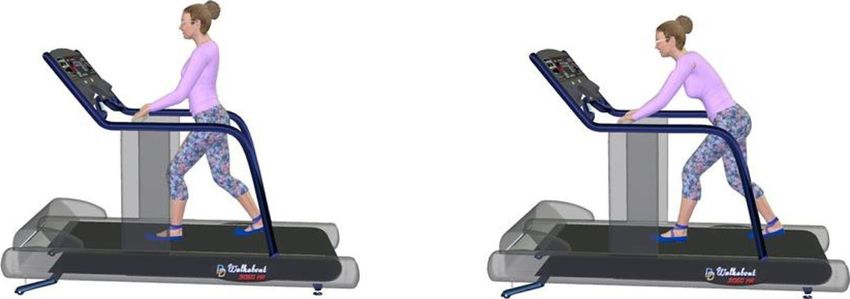
Fig. 1 Position of participants during (a) the Upright walking posture task and (b) the FLT walking task
test was used to assess each variable for normal Exploratory statistics
distribution. Preliminary statistics using receiver operating character-
istic curves (ROC) were used to assess the potential per-
Demographic data and clinical outcomes formance of the walking test to distinguish conditions
Pearson Chi-Square test was performed to compare gen- using Walking time to symptom change. The difference
der proportion between groups. One-way analyses of in Walking time to symptom change between the FLT
variance (ANOVAs) were conducted to assess if groups walking task and the Upright walking task was used in
were similar for age, height, and weight. One-way ANO- the ROC analyses to determine the sensibility and the
VAs were also conducted to compare the impact of clau- specificity of this new treadmill walking test.
dication, walking impairment, kinesiophobia and quality
of life between groups. Post hoc analyses were con- Results
ducted using Tukey’s post hoc test for pairwise compari- The sample size calculation indicated that in order to
sons whenever necessary. achieve a statistical power of 0.80 using an alpha value
of 0.05 and a 30% between-group difference in Δwalking
Treadmill walking test time to symptom change, groups of 24 participants were
The Pearson Chi-square was used to compare the pro- needed. However, due to the global COVID-19 pan-
portion of participants in each group who increased demic, this study had to be interrupted. Fifty-five partici-
their walking time between the Upright to the FLT walk- pants (23 with LSS, 18 with nLBP and 14 with PAD) had
ing tasks for both Walking time to symptom change and been enrolled at the time recruitment was halted for an
Total walking time. Then, T-test were performed to ver- indefinite period.
ify if there was not an order effect between walking
tasks. The order effect was first verified among all partic- Demographic data and clinical outcomes
ipants and it was then verified in all groups separately. Demographic data and baseline clinical characteristics
We compared Δwalking time to symptom change be- are presented in Table 2. For the gender proportion ana-
tween participants that began with the Upright walking lysis, Pearson Chi-square results showed that gender
task and participants that began with the FLT walking proportions were similar between groups (χ2 = 1.59, p =
task. 0.45). The one-way ANOVA showed a significant
The homoscedasticity of Walking time to symptom between-group difference for age (p < 0.001) and the
change was evaluated using the Levene test. One-way Tukey post hoc analysis showed that patients were youn-
ANOVA was performed to compare Walking time to ger in the nLBP group compared to the LSS (p < 0.001)
symptom change difference between groups (Δwalking and PAD groups (p < 0.001). However, patients were
time to symptom change = Walking time to symptom similar regarding weight, height, body mass index (BMI)
change during the FLT walking task – Walking time to and leg pain intensity in the previous week.
symptom change during the Upright walking task). Be- The ANOVAs for the other clinical outcomes showed
cause of the non-parametric distribution of the data re- significant between-group differences. Tukey post hoc
garding Total walking time in each group, Kruskal- tests revealed that patients in the LSS and PAD groups
Wallis test was conducted to assess Total walking time were similar for every clinical outcome and that they
difference between groups (Δtotal walking time = Total were both significantly different compared to the nLBP
walking time during the FLT walking task – Total walk- group, except for kinesiophobia scores. Detailed results
ing time during the Upright walking task). are presented in Table 2. Regarding results from theHoule et al. Chiropractic & Manual Therapies (2021) 29:24 Page 6 of 11
Table 2 Demographic data and participants’ results for clinical outcomes
LSS group PAD group nLBP group p-value
(n = 23) (n = 14) (n = 18)
F:M 9:14 5:9 10:8 N/A
Age (years) 70.00 ± 7.66 72.43 ± 9.41 52.00 ± 9.29 < 0.001*†
Weight (kg) 82.02 ± 14.76 83.20 ± 22.15 82.26 ± 9.95 0.975
Height (m) 1.68 ± 0.09 1.89 ± 0.80 1.69 ± 0.07 0.265
BMI (kg/m2) 29.02 ± 4.05 27.65 ± 9.02 28.85 ± 3.51 0.756
Leg pain intensity in the past week (/10) 5.63 ± 2.19 6.04 ± 2.06 3.74 ± 2.33 0.008*†
WIQdistance (%) 34.91 ± 30.30 24.34 ± 31.62 89.54 ± 17.97 0.001*†
WIQspeed (%) 31.00 ± 20.49 23.99 ± 24.33 75.24 ± 20.67 < 0.001* †
WIQstairs (%) 20.92 ± 14.51 15.19 ± 15.82 41.88 ± 10.66 < 0.001* †
WIQmean (%) 28.94 ± 20.52 21.17 ± 22.87 68.89 ± 14.20 < 0.001* †
FC-SSSQpain (/35) 20.87 ± 4.25 21.86 ± 3.46 14.17 ± 4.00 < 0.001* †
FC-SSSQfunction (/20) 11.96 ± 2.85 12.00 ± 4.22 7.39 ± 2.89 0.001* †
FC-SSSQtotal (/55) 33.26 ± 6.29 33.86 ± 6.01 21.56 ± 5.38 < 0.001* †
TSK (/68) 45.43 ± 8.39 38.46 ± 8.13 34.39 ± 7.91 < 0.001*#
EQ-VAS score (/100) 73.00 ± 18.38 66.79 ± 18.18 78.44 ± 6.78 0.118
* p < 0.001 between the LSS group and the nLBP group
† p < 0.001 between the PAD group and the nLBP group
# p < 0.05 between the LSS group and the PAD group
LSS: Lumbar Spinal Stenosis, PAD: Peripheral Arterial Disease, nLBP: non-specific Low Back Pain, F: female, M: male, BMI: Body Mass Index, WIQ: Walking
Impairment Questionnaire, FC-SSSQ: French-Canadian Swiss Spinal Stenosis Questionnaire, TSK: Tampa Scale of Kinesiophobia, EQ-VAS score: EQ-5D: European
Questionnaire– 5 dimensions Visual Analog Scale
WIQ questionnaire, the Tukey post hoc showed that pa- Upright walking task and 83% reported symptoms in the
tients in the nLBP group were able to walk for a longer FLT walking task. Regarding vascular claudication, 79%
period, were able to walk faster and had less impairment of patients in the PAD group reported symptoms during
during stairs climbing than the LSS and the PAD groups. the Upright walking task and 86% during the Forward
Regarding pain and functional assessments (FC-SSSQ), trunk lean walking task. Patients in the nLBP group
the ANOVA showed significant between-group differ- showed little variability for both treadmill walking condi-
ences for pain, function and total score. FC-SSSQ scores tions. In fact, this group did not respond to the treadmill
for pain and function subscales were significantly lower walking test, as 28% of the patients reported symptoms
in the previous month for the nLBP group than for the during the Upright walking task and 33% during the FLT
LSS and PAD groups. For the total score of the FC- walking task. The Pearson Chi-square showed a
SSSQ, the analysis revealed a significant difference be- between-group significant difference for the Walking
tween the nLBP group and the LSS group, as well as be- time to symptom change increased (sec) between the Up-
tween the nLBP group and the PAD group. In each case, right walking task and the FLT walking task (x2 = 7.88;
the nLBP group showed a lower total score. Regarding p = 0.02). This difference between group is mainly due to
kinesiophobia, the Tukey post hoc showed a significant the nLBP which had much lower chance of responding
difference between the nLBP group and the LSS group, to the walking tasks.
indicating that the nLBP group had less kinesiophobia. Regarding Total walking time, even if Walking time to
The Tukey post hoc also showed a difference between symptom change was sometimes brief (minimum = 19 s),
the LSS and the PAD group (p = 0.045). There was no several patients were able to complete the 5-min of
significant difference between the nLPB and the PAD walking in both tasks in the LSS group. In fact, 61% of
groups. As for the visual analog scale (VAS) section of patients in the LSS group, compared to 50% in the PAD
the EQ-5D questionnaire, results for global self-reported group were able to walk the entire 5 min during both
health status showed that all groups were similar (p = the Upright walking task and the FLT walking task. Re-
0.118). garding the nLBP group, 94% of patients were able to
complete both the Upright walking task and the FLT
Treadmill walking test walking task (see Fig. 2). Kruskal-Wallis results also
Regarding Walking time to symptom change, 87% of pa- showed no significant difference between groups regard-
tients in the LSS group reported symptoms in the ing Δtotal walking time (p = 0.298).Houle et al. Chiropractic & Manual Therapies (2021) 29:24 Page 7 of 11 Fig. 2 Median, maximum and minimum for Total walking time Results from the T-test showed no significative order analysis also showed a significant difference between effect when considering all participants (p = 0.14), and the PAD group and the nLBP group (see Fig. 3). within each group (for the LSS group p = 0.40, for the Considering that the mean duration for Δwalking PAD group p = 0.17 and for the nLBP group p = 0.26). time to symptom change was 40.43 s and that the One-way ANOVA showed that there was a signifi- mean duration for Walking time to symptom change cant difference between groups regarding Δwalking during the Upright walking task was 118 s, partici- time to symptom change (F = 4.12, p = 0.022). Post pants increased their walking time to symptom hoc analysis showed that participants from the LSS change of 34.26%. group increased their walking time to symptom change from the Upright position to the Forward lean Exploratory statistics trunk position compared to the PAD group (p = The preliminary results of ROC curve analysis consider- 0.034) and the nLBP group (p = 0.012). Post hoc ing Walking time to symptom change for LSS and PAD Fig. 3 Comparison of Δwalking time to symptom change between the three groups. LSS = lumbar spinal stenosis, PAD = peripheral arterial disease, nLBP = non-specific low back pain
Houle et al. Chiropractic & Manual Therapies   (2021) 29:24                                                    Page 8 of 11

groups showed a sensitivity of 60.9% and a specificity of     diameter and dural sac cross-sectional area to increase
78.6% while sensitivity and specificity considering Walk-     [41]. On the contrary, and as expected, the inclined
ing time to symptom change for LSS and nLBP were re-          walking posture had no positive effect on Walking time
spectively 60.9 and 88.9%.                                    to symptom change in patients with PAD and nLBP. Leg
                                                              symptoms in PAD are caused by the partial occlusion of
Discussion                                                    the blood vessels, which causes a decrease in oxygen
The objectives of the present study were (1) to compare       supply to muscles, and time to leg symptom change can
changes in walking time to symptom change and (2) to          be influenced by the intensity of the task, which will
compare self-reported outcome measures such as the            generate higher needs in oxygen rather than the position
impact of claudication, walking impairment, kinesiopho-       of the trunk while walking [10]. During the treadmill
bia and global health between groups of individuals with      walking test, walking intensity was the same for both
LSS, PAD and nLBP. As hypothesized, patients with             walking tasks, which means that there was no expected
spinal stenosis were able to walk longer in the forward       change in Walking time to symptom change and Total
trunk lean walking task than in the Upright walking task      walking time for patients with PAD. The nLBP group
before the symptom change appeared, while no differ-          was included in this study to highlight the differences in
ence between positions was observed for patients with         leg pain arising from nLBP and LSS.
PAD and with nLBP. Our second hypothesis was also                The preliminary sensitivity and specificity analysis sug-
confirmed as participants in the nLBP group reported          gests that the treadmill walking test can be a helpful tool
lower claudication impact and walking impairment than         for health care professionals who wish to assess the ori-
the two other groups.                                         gin of claudication and differentiate LSS from nLBP. In
                                                              fact, the treadmill test has shown to be moderately sensi-
Treadmill walking test                                        tive (60.9%) and moderately to highly specific (78.6–
Results from the treadmill walking test used in this study    88.9%) to the neurogenic claudication present in lumbar
showed that the FLT walking posture increases the             spinal stenosis. In addition, the increased walking time
Walking time to leg symptom change compared to the            to symptom change observed in the LSS group was
Upright walking posture for patients with LSS. This find-     higher than the minimally clinically important difference
ing was coherent with previous studies showing that leg       (MCID) reported in a previous study [42].
pain is decreased when patients with LSS bend their
trunk forward while walking [39, 40]. Even if claudica-       Clinical outcomes
tion impact was similar for the LSS and the PAD groups,       With regard to patients’ characteristics, results showed
patients in the PAD group did not increase their Walk-        that LSS and PAD groups were similar for a majority of
ing time to symptom change from the Upright to the FLT        clinical outcomes. Overall, patients in both groups
walking task as much as patients in the LSS group. How-       showed similar mean scores for leg pain intensity in the
ever, Total walking time was the same for both LSS and        previous week, important walking impairment and mod-
PAD groups. The fact that trunk position impacts Walk-        erate pain and function impact on their daily activities.
ing time to symptom changed in the LSS group as op-           For their part, patients with nLBP presented significant
posed to the PAD group suggests that it was a specific        differences from LSS and PAD patients for several char-
characteristic of LSS patients. As initially hypothesized,    acteristics such as kinesiophobia and walking impair-
such differences between the two walking tasks were not       ment, as assessed with the WIQ and FC-SSSQ
observed in patients with PAD or nLBP because of the          questionnaires. In the present study, the kinesiophobia
different mechanisms that generate claudication. Claudi-      scores were different for the LSS and PAD groups, which
cation in LSS is normally caused by pain due to nerve         is consistent with the results reported in a previous
root compression, which is accentuated with upright           study that compared different characteristics, including
posture, while claudication in PAD is caused by a nar-        kinesiophobia, between three groups (neurogenic claudi-
rowing of the arteries; symptoms therefore increase with      cation, vascular claudication and asymptomatic) [43]. In
the increasing oxygen demand from lower limb muscles.         fact, in the study by Woods et al. (2012), patients with
For patients with nLBP, referred pain to the lower limbs      neurogenic claudication had a higher score on the
is not caused by nerve root compression or oxygen de-         Tampa Scale of Kinesiophobia Questionnaire than pa-
mand. Referred pain to the lower limbs will be present        tients with vascular claudication. Regarding quality of
regardless of the trunk position. For patients with LSS,      life, the present study suggests that patients with neuro-
the leaned forward posture is well known to decrease leg      genic or vascular claudication still consider having a
symptoms [3, 14]. In fact, patients in LSS see their legs     good health status, according to their rating of health on
symptoms decrease in a seated position or by the trunk        the EQ-5D VAS scale. In addition, according to the
leaned forward position, which causes the vertebral canal     WIQ and FC-SSSQ questionnaires, patients from the
Houle et al. Chiropractic & Manual Therapies (2021) 29:24 Page 9 of 11
current study with LSS or PAD were impacted by claudi- referral to the best health care specialist based on the
cation; this impact was also observable during the tread- origin of the claudication.
mill walking test, as only 55% of patients with LSS and
50% of patients with PAD were able to walk for the en- Conclusion
tire 5 min. As the results regarding self-reported mea- Preliminary results for this new treadmill walking test
sures were similar between LSS and PAD, the treadmill showed that patients with LSS increase their walking
walking test was able to distinguish between these two time to symptom change compared to PAD and nLBP
distinct conditions. groups when walking with a leaned forward trunk pos-
ition. Exploratory results also showed that the test was
Limitations moderately sensitive and moderately to highly specific to
The study is not without limitations, since we assumed symptoms manifestations of claudication in LSS. This
that diagnosis was unequivocal to a neurosurgeon and/ treadmill walking test seems to be an interesting tool to
or a vascular surgeon regarding patients with LSS or help health care workers distinguish the origin of the
PAD and to a chiropractor regarding patients with claudication and establish a diagnostic while awaiting
nLBP. We did not have access to medical files nor to the further medical investigation. However, future studies
clinical test results done by the neurosurgeon and/or the with larger sample sizes are needed to confirm the cap-
vascular surgeon, which means that some patients could acity of this test to distinguish neurogenic claudication
have originally been incorrectly classified or have had from vascular claudication.
coexisting vascular and neurogenic claudication. The co-
Abbreviations
existence of both claudication types in one patient may IC: Intermittent claudication; PAD: Peripheral arterial disease; LBP: Low back
have limited the treadmill walking test discriminating pain; LSS: Lumbar spinal stenosis; nLBP: non-specific low back pain;
performance. In fact, clinicians should not assume that a ABI: Ankle-Brachial Index; MRI: Magnetic resonance imaging; CIUSSS
MCQ: Centre intégré universitaire de santé et de services sociaux de la
negative treadmill test definitively rules out LSS. In Mauricie-et-du-Centre-du-Québec; VAS: Visual analog scale; FR-SSSQ: French-
addition, due to the modest sample size, results should Canadian adaptation of the Swiss Spinal Stenosis Questionnaire;
be interpreted with caution. In fact, results may be over- WIQ: Walking impairment questionnaire; TSK: Tampa Scale of Kinesiophobia;
EQ-5D: EuroQol; ANOVA: Analysis of variance; ROC: Receiver operating
estimated due to the low variability between the patients characteristic; BMI: Body mass index; MIC: Minimal important change;
of the three groups concerning their walking capacity FLT: Forward Leaning Trunk
and the severity of their pathology. The fixed speed walk
Acknowledgements
was another limitation, as it sometimes made patients Not applicable.
uncomfortable during the walking tasks. Another limita-
tion of this study was that the reliability of the treadmill Authors’ contributions
Research area and study design: MH, CEC and MD; recruitment: MH, JO and
walking test was not measured. For future studies, self- CEC; data acquisition: MH and CT; statistical analysis: MH and MD; supervision
pace walking task should be considered to assess leg and mentorship: MD and AAM. Manuscript writing: MH, AAM, JO, CT and
symptoms, since it may better represent the daily life ac- CEC. MD takes responsibility that this study was been reported, transparently
and honestly. All authors have read and approved the manuscript.
tivities of patients.
Funding
Clinical implications This study was supported by the Fonds Institutionnel de Recherche (FIR) de
l’Université du Québec à Trois-Rivières, the Chaire de recherche internation-
Interestingly, 95% of participants were able to complete ale en santé neuromusculosquelettique and its partner, the Centre intégré
the treadmill test at the predefined walking speed. In universitaire de santé et de services sociaux de la Mauricie-et-du-Centre-du-
addition, all patients were back to their baseline leg pain Québec.
intensity within the 5-min rest period that followed both Availability of data and materials
treadmill walking tasks. This study showed that a short The datasets used and/or analyzed during the current study are available
treadmill walking test can help health care professionals from the corresponding author on a reasonable request.
to discriminate the neurogenic claudication from the Declarations
vascular claudication. The next stages of the treadmill
test development should include patients with other spe- Ethics approval and consent to participate
This study was approved by the CIUSSS MCQ research ethics committee
cific LBP that are associated with leg pain. To our know- (CER-2017-017) and by the Université du Québec à Trois-Rivières ethics com-
ledge, this is the first treadmill walking test to assess the mittee for research with human beings (CER-18-244-10.01). All patients pro-
difference between neurogenic and vascular claudication vided informed written consent prior to their participation in the study. The
study was registered at clinicaltrials.gov (NCT04058171).
in a quantitative way. The approach, when fully vali-
dated, could contribute to the early detection of claudi- Consent for publication
cation origin and consequently improve care pathways. Not applicable.
This early identification should contribute to speeding Competing interests
up the establishment of a treatment plan and allow early The authors declare that they have no competing interests.Houle et al. Chiropractic & Manual Therapies (2021) 29:24 Page 10 of 11
Author details 19. Hurwitz EL, Randhawa K, Yu H, Côté P, Haldeman S. The global spine care
1
Département des Sciences de l’activité physique, Université du Québec à initiative: a summary of the global burden of low back and neck pain
Trois-Rivières, 3351, boul. des Forges, C.P. 500, Trois-Rivières, QC G8Z 4M3, studies. Eur Spine J. 2018;27(S6):796–801. https://doi.org/10.1007/s00586-01
Canada. 2Département de Chiropratique, Université du Québec à 7-5432-9.
Trois-Rivières, 3351, boul. des Forges, C.P. 500, Trois-Rivières, QC G8Z 4M3, 20. Hartvigsen J, Hancock MJ, Kongsted A, Louw Q, Ferreira ML, Genevay S,
Canada. 3Centre Intégré Universitaire de Santé et de Services Sociaux de la et al. What low back pain is and why we need to pay attention. Lancet.
Mauricie-et-du-Centre-du-Québec (CIUSSS MCQ), 1991 Boulevard du Carmel, 2018;391(10137):2356–67. https://doi.org/10.1016/S0140-6736(18)30480-X.
Trois-Rivières, QC G8Z 3R9, Canada. 21. Sundell CG, Bergström E, Larsén K. Low back pain and associated disability
in Swedish adolescents. Scand J Med Sci Sports. 2019;29(3):393–9. https://
Received: 12 February 2021 Accepted: 9 June 2021 doi.org/10.1111/sms.13335.
22. Maher C, Underwood M, Buchbinder R. Non-specific low back pain. Lancet.
2017;389(10070):736–47. https://doi.org/10.1016/S0140-6736(16)30970-9.
23. Hoy D, March L, Brooks P, Woolf A, Blyth F, Vos T, et al. Measuring the
References global burden of low back pain. Best Pract Res Clin Rheumatol. 2010;24(2):
1. Nadeau M, Rosas-Arellano MP, Gurr KR, Bailey SI, Taylor DC, Grewal R, et al. 155–65. https://doi.org/10.1016/j.berh.2009.11.002.
The reliability of differentiating neurogenic claudication from vascular 24. McDermott MM, Guralnik JM, Tian L, Liu K, Ferrucci L, Liao Y, et al.
claudication based on symptomatic presentation. Can J Surg. 2013;56(6): Associations of borderline and low normal ankle-brachial index values with
372–7. https://doi.org/10.1503/cjs.016512. functional decline at 5-year follow-up: the WALCS (walking and leg
2. Jeon CH, Han SH, Chung NS, Hyun HS. The validity of ankle-brachial index circulation study). J Am Coll Cardiol. 2009;53(12):1056–62. https://doi.org/1
for the differential diagnosis of peripheral arterial disease and lumbar spinal 0.1016/j.jacc.2008.09.063.
stenosis in patients with atypical claudication. Eur Spine J. 2012;21(6):1165– 25. Tomkins-Lane CC, Holz SC, Yamakawa KS, Phalke VV, Quint DJ, Miner J, et al.
70. https://doi.org/10.1007/s00586-011-2072-3. Predictors of walking performance and walking capacity in people with
3. Genevay S, Atlas SJ. Lumbar spinal stenosis. Best Pract Res Clin Rheumatol. lumbar spinal stenosis, low back pain, and asymptomatic controls. Arch
2010;24(2):253–65. https://doi.org/10.1016/j.berh.2009.11.001. Phys Med Rehabil. 2012;93(4):647–53. https://doi.org/10.1016/j.apmr.2011.09.
4. Conte SM, Vale PR. Peripheral arterial disease. Heart Lung Circ. 2018;27(4): 023.
427–32. https://doi.org/10.1016/j.hlc.2017.10.014. 26. Aboyans V, Ricco J-B, Bartelink M-LE, Björck M, Brodmann M, Cohnert T,
5. Messiah S, Tharian AR, Candido KD, Knezevic NN. Neurogenic claudication: a Collet J-P, Czerny M, De Carlo M, Debus S: 2017 ESC Guidelines on the
review of current understanding and treatment options. Curr Pain Diagnosis and Treatment of Peripheral Arterial Diseases, in collaboration
Headache Rep. 2019;23(5):32. https://doi.org/10.1007/s11916-019-0769-x. with the European Society for Vascular Surgery (ESVS) Document covering
6. Koes BW, Van Tulder M, Lin C-WC, Macedo LG, McAuley J, Maher C. An atherosclerotic disease of extracranial carotid and vertebral, mesenteric,
updated overview of clinical guidelines for the management of non-specific renal, upper and lower extremity arteries Endorsed by: the European Stroke
low back pain in primary care. Eur Spine J. 2010;19(12):2075–94. https://doi. Organization (ESO) The Task Force for the Diagnosis and Treatment of
org/10.1007/s00586-010-1502-y. Peripheral Arterial Diseases of the European Society of Cardiology (ESC) and
7. Criqui MH, Aboyans V. Epidemiology of peripheral artery disease. Circ Res. of the European Society for Vascular Surgery (ESVS). Eur Heart J. 2017, 39:
2015;116(9):1509–26. https://doi.org/10.1161/CIRCRESAHA.116.303849. 763–816.
8. Sawlani NN, Kinlay S. Claudication: pay for structured exercise or go take a 27. Selvin E, Erlinger TP. Prevalence of and risk factors for peripheral arterial
hike. JACC Cardiovasc Interv. 2017;10(7):725–7. https://doi.org/10.1016/j.jcin.2 disease in the United States: results from the National Health and nutrition
017.02.036. examination survey, 1999-2000. Circulation. 2004;110(6):738–43. https://doi.
9. Hamburg NM, Creager MA. Pathophysiology of intermittent claudication in org/10.1161/01.CIR.0000137913.26087.F0.
peripheral artery disease. Circ J. 2017;81(3):281–9. https://doi.org/10.1253/ 28. De Schepper EI, Overdevest GM, Suri P, Peul WC, Oei EH, Koes BW, et al.
circj.CJ-16-1286. Diagnosis of lumbar spinal stenosis: an updated systematic review of the
10. Dua A, Lee CJ. Epidemiology of peripheral arterial disease and critical limb accuracy of diagnostic tests. Spine. 2013;38(8):E469–81. https://doi.org/10.1
ischemia. Tech Vasc Interv Radiol. 2016;19(2):91–5. https://doi.org/10.1053/j. 097/BRS.0b013e31828935ac.
tvir.2016.04.001. 29. Boden SD, Davis DO, Dina TS, Patronas NJ, Wiesel SW. Abnormal magnetic-
11. Uesugi K, Sekiguchi M, Kikuchi S, Kanayama M, Takahashi K, Chiba K, et al. resonance scans of the lumbar spine in asymptomatic subjects. A
Lumbar spinal stenosis associated with peripheral arterial disease: a prospective investigation. J Bone Joint Surg Am. 1990;72(3):403–8. https://
prospective multicenter observational study. J Orthop Sci. 2012;17(6):673–81. doi.org/10.2106/00004623-199072030-00013.
https://doi.org/10.1007/s00776-012-0311-z. 30. Barua B: Wait Times for Health Care in Canada, 2017 Report fraserinstituteorg 2017.
12. Kalichman L, Cole R, Kim DH, Li L, Suri P, Guermazi A, et al. Spinal stenosis 31. Marchand AA, Tetreau C, O'Shaughnessy J, Descarreaux M. French-Canadian
prevalence and association with symptoms: the Framingham study. Spine J. adaptation and validation of the Swiss spinal stenosis questionnaire for
2009;9(7):545–50. https://doi.org/10.1016/j.spinee.2009.03.005. patients with lumbar spinal stenosis. Spine (Phila Pa 1976). 2019;44(8):E487–
13. Lurie J, Tomkins-Lane C. Management of lumbar spinal stenosis. BMJ 93. https://doi.org/10.1097/BRS.0000000000002896.
(Clinical research ed) 2016, 352:h6234. 32. McDermott MM, Liu K, Guralnik JM, Martin GJ, Criqui MH, Greenland P.
14. Comer CM, Redmond AC, Bird HA, Conaghan PG. Assessment and Measurement of walking endurance and walking velocity with
management of neurogenic claudication associated with lumbar spinal questionnaire: validation of the walking impairment questionnaire in men
stenosis in a UK primary care musculoskeletal service: a survey of current and women with peripheral arterial disease. J Vasc Surg. 1998;28(6):1072–81.
practice among physiotherapists. BMC Musculoskelet Disord. 2009;10(1):121. https://doi.org/10.1016/S0741-5214(98)70034-5.
https://doi.org/10.1186/1471-2474-10-121. 33. Nicolai SP, Kruidenier LM, Rouwet EV, Graffius K, Prins MH, Teijink JA. The
15. Kobayashi S. Pathophysiology, diagnosis and treatment of intermittent walking impairment questionnaire: an effective tool to assess the effect of
claudication in patients with lumbar canal stenosis. World J Orthopedics. treatment in patients with intermittent claudication. J Vasc Surg. 2009;50(1):
2014;5(2):134–45. https://doi.org/10.5312/wjo.v5.i2.134. 89–94. https://doi.org/10.1016/j.jvs.2008.12.073.
16. Kim HJ, Chun HJ, Han CD, Moon SH, Kang KT, Kim HS, et al. The risk 34. French DJ, Roach PJ, Mayes S. Peur du mouvement chez des accidentés du
assessment of a fall in patients with lumbar spinal stenosis. Spine (Phila Pa travail: L'Échelle de Kinésiophobie de Tampa (EKT). Can J Behavioural
1976). 2011;36(9):E588–92. https://doi.org/10.1097/BRS.0b013e3181f92d8e. Science/Revue canadienne des sciences du comportement. 2002;34(1):28–
17. Ammendolia C, Stuber K, Tomkins-Lane C, Schneider M, Rampersaud YR, 33. https://doi.org/10.1037/h0087152.
Furlan AD, et al. What interventions improve walking ability in neurogenic 35. French DJ, France CR, Vigneau F, French JA, Evans RT. Fear of movement/
claudication with lumbar spinal stenosis? A systematic review. Eur Spine J. (re) injury in chronic pain: a psychometric assessment of the original English
2014;23(6):1282–301. https://doi.org/10.1007/s00586-014-3262-6. version of the Tampa scale for kinesiophobia (TSK). Pain. 2007;127(1):42–51.
18. Hoy D, Bain C, Williams G, March L, Brooks P, Blyth F, et al. A systematic https://doi.org/10.1016/j.pain.2006.07.016.
review of the global prevalence of low back pain. Arthritis Rheum. 2012; 36. Perneger TV, Combescure C, Courvoisier DS. General population reference
64(6):2028–37. https://doi.org/10.1002/art.34347. values for the French version of the EuroQol EQ-5D health utilityHoule et al. Chiropractic & Manual Therapies (2021) 29:24 Page 11 of 11
instrument. Value Health. 2010;13(5):631–5. https://doi.org/10.1111/j.1524-4
733.2010.00727.x.
37. Rabin R, Charro Fd: EQ-SD: a measure of health status from the EuroQol
group. Ann Med 2001, 33:337–343, 5, DOI: https://doi.org/10.3109/07853
890109002087.
38. Deen HG Jr, Zimmerman RS, Lyons MK, McPhee MC, Verheijde JL, Lemens
SM. Test-retest reproducibility of the exercise treadmill examination in
lumbar spinal stenosis. Mayo Clin Proc. 2000;75(10):1002–7. https://doi.org/1
0.4065/75.10.1002.
39. Penning L. Functional pathology of lumbar spinal stenosis. Clin Biomech.
1992;7(1):3–17. https://doi.org/10.1016/0268-0033(92)90002-L.
40. Kuwahara W, Deie M, Fujita N, Tanaka N, Nakanishi K, Sunagawa T, et al.
Characteristics of thoracic and lumbar movements during gait in lumbar
spinal stenosis patients before and after decompression surgery. Clin
Biomech. 2016;40:45–51. https://doi.org/10.1016/j.clinbiomech.2016.10.016.
41. Madsen R, Jensen TS, Pope M, Sørensen JS, Bendix T. The effect of body
position and axial load on spinal canal morphology: an MRI study of central
spinal stenosis. Spine. 2008;33(1):61–7. https://doi.org/10.1097/BRS.0b013e31
815e395f.
42. Ammendolia C, Côté P, Southerst D, Schneider M, Budgell B, Bombardier C,
et al. Comprehensive nonsurgical treatment versus self-directed care to
improve walking ability in lumbar spinal stenosis: a randomized trial. Arch
Phys Med Rehabil. 2018;99(12):2408–19 e2402. https://doi.org/10.1016/j.a
pmr.2018.05.014.
43. Wood DW, Haig AJ, Yamakawa KS. Fear of movement/(re) injury and activity
avoidance in persons with neurogenic versus vascular claudication. Spine J.
2012;12(4):292–300. https://doi.org/10.1016/j.spinee.2012.02.015.
Publisher’s Note
Springer Nature remains neutral with regard to jurisdictional claims in
published maps and institutional affiliations.You can also read