UCLA UCLA Previously Published Works
←
→
Page content transcription
If your browser does not render page correctly, please read the page content below
UCLA
UCLA Previously Published Works

Title
PRIMA subretinal wireless photovoltaic microchip implantation in non-human primate and
feline models.

Permalink
https://escholarship.org/uc/item/4k66m9pf

Journal
PloS one, 15(4)

ISSN
1932-6203

Authors
Muqit, Mahiul MK
Hubschman, Jean Pierre
Picaud, Serge
et al.

Publication Date
2020

DOI
10.1371/journal.pone.0230713

Peer reviewed

 eScholarship.org                                Powered by the California Digital Library
                                                                 University of California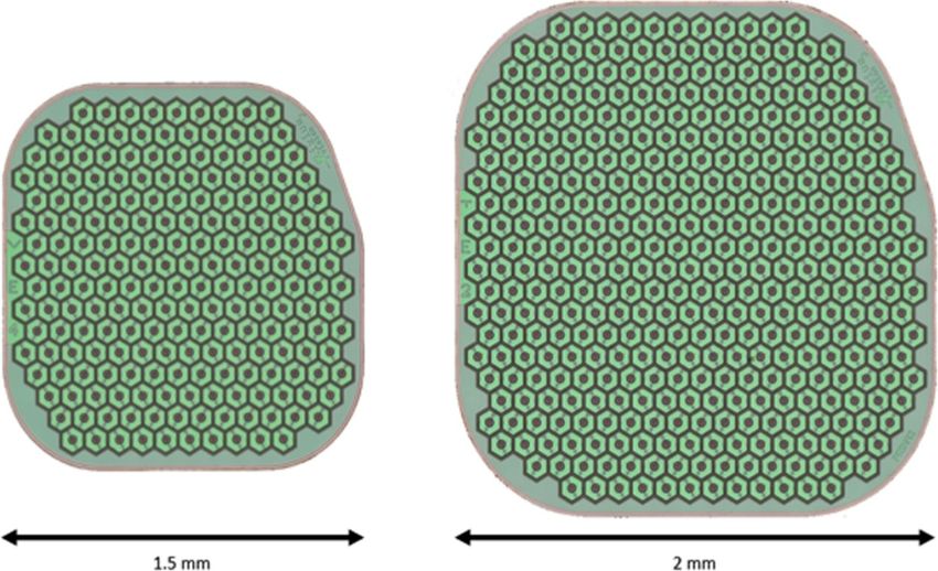
PLOS ONE

                                                     RESEARCH ARTICLE

                                                     PRIMA subretinal wireless photovoltaic
                                                     microchip implantation in non-human
                                                     primate and feline models
                                                     Mahiul M. K. Muqit ID1,2☯*, Jean Pierre Hubschman ID3☯, Serge Picaud4☯, Douglas
                                                     B. McCreery5☯, Jan C. van Meurs6,7‡, Ralf Hornig8☯, Guillaume Buc8☯, Martin Deterre ID8☯,
                                                     Céline Nouvel-Jaillard4‡, Elodie Bouillet8☯, Claire-Maelle Fovet ID9‡, Philippe Hantraye9‡,
                                                     José Sahel4,10,11,12☯, Joseph N. Martel11‡, Yannick Le Mer ID12☯
                                                     1 Vitreoretinal Service, Moorfields Eye Hospital, London, United Kingdom, 2 Institute of Ophthalmology,
a1111111111
                                                     University College London, London, United Kingdom, 3 Stein Eye Institute, University of California Los
a1111111111                                          Angeles, Los Angeles, CA United States of America, 4 Sorbonne Université, INSERM, CNRS, Institut de la
a1111111111                                          Vision, Paris, France, 5 Huntington Medical Research Institutes, Pasadena, CA, United States of America,
a1111111111                                          6 Rotterdam Eye Hospital, Rotterdam, The Netherlands, 7 ErasmusMC, Rotterdam, The Netherlands,
a1111111111                                          8 Pixium Vision, Paris, France, 9 Molecular Imaging Research Center (MIRCen), CEA, Fontenay aux Roses,
                                                     France, 10 Hopital des Quinze Vingts, Paris, France, 11 Retina and Vitreous Service, University of
                                                     Pittsburgh Medical School, Pittsburgh, PA, United States of America, 12 Fondation Ophtalmologique A. De
                                                     Rothschild, Paris, France

                                                     ☯ These authors contributed equally to this work.
   OPEN ACCESS                                       ‡ These authors also contributed equally to this work.
                                                     * Mahi.Muqit1@nhs.net
Citation: Muqit MMK, Hubschman JP, Picaud S,
McCreery DB, van Meurs JC, Hornig R, et al.
(2020) PRIMA subretinal wireless photovoltaic
microchip implantation in non-human primate and      Abstract
feline models. PLoS ONE 15(4): e0230713. https://
doi.org/10.1371/journal.pone.0230713

Editor: Demetrios G. Vavvas, Massachusetts Eye &     Purpose
Ear Infirmary, Harvard Medical School, UNITED
STATES                                               To evaluate the surgical technique for subretinal implantation of two sizes of PRIMA photo-
                                                     voltaic wireless microchip in two animal models, and refine these surgical procedures for
Received: June 17, 2019
                                                     human trials.
Accepted: March 5, 2020

Published: April 8, 2020                             Methods
Copyright: © 2020 Muqit et al. This is an open       Cats and Macaca fascicularis primates with healthy retina underwent vitrectomy surgery
access article distributed under the terms of the
Creative Commons Attribution License, which
                                                     and were implanted with subretinal wireless photovoltaic microchip at the macula/central
permits unrestricted use, distribution, and          retina. The 1.5mm PRIMA chip was initially studied in feline eyes. PRIMA implant
reproduction in any medium, provided the original    (2mm,1.5mm sizes) arrays were studied in primates. Feasibility of subretinal chip implanta-
author and source are credited.
                                                     tion was evaluated with a newly-developed surgical technique, with surgical complications
Data Availability Statement: All relevant data are   and adverse events recorded.
within the manuscript and its Supporting
Information files.
                                                     Results
Funding: The studies were funded in part by the
SightAgain project under the Structural R&D          The 1.5mm implant was placed in the central retina of 11 feline eyes, with implantation dura-
Projects for Competitiveness (PSPC) and              tion 43–106 days. The 1.5mm implant was correctly positioned into central macula of 11 pri-
Investment for the Future (PIA) funding, managed
                                                     mate eyes, with follow-up periods of minimum 6 weeks (n = 11), 2 years (n = 2), and one eye
by Bpifrance. They were partly conducted at the
Institut de la Vision within the framework of the    for 3 years. One primate eye underwent multi-chip 1.5mm implantation using two 1.5mm
LABEX LIFESENSES [ANR-10-LABX-65] and IHU            chips. The 2mm implant was delivered to 4 primate eyes. Optical coherence tomography

PLOS ONE | https://doi.org/10.1371/journal.pone.0230713 April 8, 2020                                                                                   1 / 16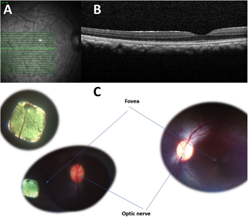
PLOS ONE                                                                                                      PRIMA Subretinal Wireless Photovoltaic Microchip

FOReSIGHT [ANR-18-IAHU-0001] supported by               confirmed correct surgical placement of photovoltaic arrays in the subretinal space in all 26
French state funds managed by the Agence                eyes. Intraoperative complications in primate eyes included retinal tear, macular hole, retinal
Nationale de la Recherche within the
Investissements d’Avenir program. The funders
                                                        detachment, and vitreous hemorrhage that resolved spontaneously. Postoperatively, there
provided support in the form of funding for the         was no case of significant ocular inflammation in the 1.5mm implant group.
hospital/institutional projects but did not have any
additional role in the study design, data collection
and analysis, decision to publish, or preparation of    Conclusions
the manuscript.                                         We report subretinal implantation of 1.5mm and 2mm photovoltaic arrays in the central ret-
Competing interests: MMK Muqit, (Consultant for         ina of feline and central macula of primate eyes with a low rate of device-related complica-
Pixium Vision); JP Hubschman, (Consultant for           tions. The in vivo PRIMA implantation technique has been developed and refined for use for
Pixium Vision); Serge Picaud (Consultant for
Pixium Vision and Gensight Biologics; Financial
                                                        a 2mm PRIMA implant in ongoing human trials.
interests in Pixium Vision, Gensight Biologics); DB
McCreery, None; JC van Meurs, (Consultant for
Pixium Vision); R Hornig, Pixium Vision (Employee
for Pixium Vision); G Buc, Pixium Vision (Employee
for Pixium Vision); M Deterre, Pixium Vision            Introduction
(Employee for Pixium Vision); C Nouvel-Jaillard,
                                                        The PRIMA implant is a wireless photovoltaic microchip array for subretinal stimulation [1].
None; E Bouillet, Pixium Vision (Employee for
Pixium Vision); C-M Fovet, None; P Hantraye,
                                                        In this optoelectronic prosthetic system, each pixel of the subretinal array photovoltaically con-
None; J Sahel, Pixium (Investor in Pixium Vision,       verts patterned pulsed near-infrared (NIR) light projected from video glasses into pulses of bi-
Consultant for Pixium Vision); Y Le Mer,                phasic electric current to stimulate the inner retinal cells in front of it [2–4]. The interface with
(Consultant for Pixium Vision). The SightAgain          the visual environment is achieved through a mini camera integrated in a pair of glasses that
project was managed by Bpifrance and Pixium             captures the overall field of view ("visual scene"). The visual information is then processed and
Vision. French state funds were managed by the
                                                        converted to stimulation information which is used to activate the implanted retinal prostheses
Agence Nationale de la Recherche within the
Investissements d’Avenir program. Any                   ("Implants"). Stimulation waveforms of infrared light are projected into the eye, through a
commercial affiliation for the authors does not alter   near-to-eye digital mirror projection system. When the gaze direction is such that some part of
our adherence to PLOS ONE policies on sharing           the implant is illuminated by part of the stimulation data, the photovoltaic retinal implant con-
data and materials.                                     verts that part of the signal into electrical current that stimulates the retina accordingly (Fig 1).
                                                           Different types of photovoltaic arrays have been studied in rat and pig eye models by Ade-
                                                        kunle and coworkers, and the biocompatibility and subretinal integration did not show any

                                                        Fig 1. Schematic of PRIMA system.
                                                        https://doi.org/10.1371/journal.pone.0230713.g001

PLOS ONE | https://doi.org/10.1371/journal.pone.0230713 April 8, 2020                                                                                   2 / 16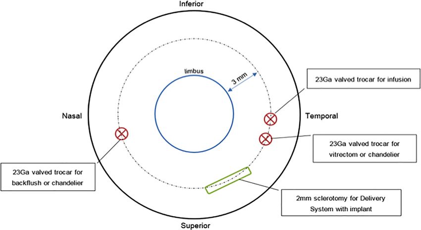
PLOS ONE                                                                                        PRIMA Subretinal Wireless Photovoltaic Microchip

                                            significant adverse issues [5]. Lorach and Palanker’s group have clearly demonstrated the
                                            direct electrical relationship and interaction between natural visually evoked signals and pros-
                                            thetic responses in a photovoltaic subretinal implant in Long Evans rat models [6]. Improve-
                                            ments in visual acuity have been demonstrated in blind animal models compared to healthy
                                            rat models [7]. In 2018, Ho and co-workers compared prosthetic stimulation in blind retinal
                                            degenerate Royal College of Surgeon rats implanted with subretinal 1mm and 2mm photovol-
                                            taic arrays compared to healthy Long Evan rats [8]. Near infrared light stimulated the blind
                                            animals with startling responses noted in behaviour patterns at pulsed electrical thresholds.
                                                The safety of a similar implant has been shown in a human clinical trial [9]. The artificial
                                            silicon retina (ASR) microchip (Optobionics Corp, Naperville, Illinois, USA) was a 2mm
                                            diameter semi-conductor microphotodiode array of 25μm thickness designed for subretinal
                                            implantation. In a pilot 18-month study, the ASR was safely implanted in patients with retinitis
                                            pigmentosa with no significant adverse chip-related or surgical complications, and improved
                                            visual function was reported [9]. The biocompatibility, electrical safety and targeted location
                                            of the ASR within the subretinal space have been reported in animal studies, with optimal
                                            stimulation resolution from the subretinal ASR [10–12].
                                                The translation of this photovoltaic retinal implant evaluation to human clinical trials with
                                            defined surgical protocols and assessment of functional outcomes is the next step. Our initial
                                            study aimed at evaluating the feasibility of subretinal 1.5mm PRIMA chip implantation in a
                                            feline model. Two different sizes of PRIMA implants were then studied in non-human primate
                                            retinal model. The goal of these in vivo studies were to further refine surgery protocols, and to
                                            evaluate complications, chip integrity, and chip stability when placed in the subretinal space
                                            before translation to human trial.

                                            Materials and methods
                                            Study design
                                            The feline study was conducted in 2015. Surgical procedures and follow-up were performed at
                                            Huntington Medical Research Institutes (HMRI) who are accredited by the American Associa-
                                            tion for Laboratory Animal Care (AALAC). All animals were followed-up throughout the
                                            study by a veterinary physician. The study was approved by the institutional review board of
                                            HMRI (Protocol Number: R15017). The animals were managed according to standard internal
                                            policy on animal welfare and care at HMRI. The studies followed the WMA statement on ani-
                                            mal use in biomedical research.
                                               The sequential non-human primate animal study was conducted between 2015 and 2018.
                                            Surgical procedures and follow-up were performed at the Molecular Imaging Research Center
                                            (MIRCen- CEA, Fontenay aux Roses), in cooperation with Institut de la Vision, Paris, and
                                            institutional review board approval was in place. The study was approved by the institutional
                                            review board of Institut de la Vision and MIRCen-CEA (Protocol Number: R16014; R16025;
                                            R16050; and, R16064;). The animals were managed according to standard internal policy on
                                            animal welfare and care at MIRCEN. The studies followed the WMA statement on animal use
                                            in biomedical research.
                                               The primary aims of the study were to establish an implantation procedure of two sizes of
                                            PRIMA implant in feline and non-human primate models. The secondary aims were to assess
                                            implant delivery at the appropriate location at the macula, with the electrode side up, and with-
                                            out implant debris left in the eye. We aimed to assess for surgical complications related to the
                                            implant and to the surgery procedure itself including significant inflammations, retinal dam-
                                            age, choroidal damage, and/or other adverse effects on the eye tissues. Adverse events (AE)
                                            and serious adverse events (SAE) were defined by standard BS EN ISO 14155:2011.

PLOS ONE | https://doi.org/10.1371/journal.pone.0230713 April 8, 2020                                                                     3 / 16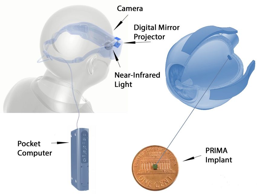
PLOS ONE                                                                                       PRIMA Subretinal Wireless Photovoltaic Microchip

                                            Animal models
                                                Feline. The cats were housed in Huntington Medical Research Institutes (HMRI). The
                                            HMRI follows national standards for animal welfare according to ARRIVE guidelines. The
                                            Animal Care and Use program of HMRI is accredited by the Association for Advancement
                                            and Accreditation of Laboratory Animal Care (AAALAC). Facilities and records are inspected
                                            regularly by a representative from the United States Department of Agriculture. All animal
                                            studies were performed in accordance with the standards set forth in the Guide for the Care
                                            and Use of Laboratory Animals (8th edition) and are approved by the HMRI Animal Care and
                                            Use Committee. All cats were purchased from Liberty Research. All cats received fresh water
                                            and food daily according to their nutritional needs. They were fed a standard Feline Labora-
                                            tory diet. They resided in the outdoor group housing exercise areas on most days and were
                                            adapted to outdoor temperatures through daily exposure, except in inclement weather. A daily
                                            exercise log was maintained for the outdoors exercise area, including the minimum and maxi-
                                            mum daily temperature. Aseptic surgical procedures, including implantation of the PRIMA
                                            device were performed in a dedicated surgical suite. After the surgery the cat was visually mon-
                                            itored and vital signs recorded every 15 minutes until the animal was able to maintain sternal
                                            recumbency, at which time they received an injection of bupronorphine for pain relief. When
                                            recovered from anesthesia the cats were returned to the primary animal care facility. During
                                            the first 4 days after the implant surgery they were housed in a large individual cage (16 ft2)
                                            and were not released into the outside exercise area so that they may be observed more closely.
                                            At night the cats were housed and fed in their individual cages, each of which is 4 ft2, with an
                                            elevated perch. To encourage resumption of eating after the implant surgery they are offered
                                            moist commercial cat food in addition to the standard feline laboratory diet. All animals were
                                            subject to an eye examination by the Study Veterinary Physician after 1 day then 8 days post-
                                            operatively. Fundus photography and OCT images were performed after surgery. Slitlamp
                                            examination of the anterior chamber was performed in order to detect any sign of inflamma-
                                            tion, and eye pressure was recorded. At the end of this study, the disposition of the animals
                                            was by euthanasia. Animals were sacrificed by overdose of pentobarbital.
                                                The feline model was selected for this study because the feline retina approximates the
                                            human retina. In both cat and human, the retina contains rods and cones and have a central
                                            region of high cone density. The cats used for this study were healthy, domestic, males with
                                            normal vision and aged between eleven [11] and sixteen [16] months at the time of interven-
                                            tion. A 1.5mm implant was delivered into 11 eyes, and follow up was between 43 and 106 days.
                                            All surgeries were performed under general anesthesia.
                                                Non-human primate. The animal model was the Macaca fascicularis and MIRCen-CEA
                                            follows national standards for animal welfare according to ARRIVE guidelines.
                                                All animal experimentations were performed by experienced veterinary surgeons, in con-
                                            formity with national and European laws. The non-human primate species macaca fascicularis
                                            (common name cynomolgus monkey), male or female, originate from a breeder in Mauritius.
                                            The animal facility is authorized for animal experimentation on non-human primates (n˚ D
                                            92 032 02) by local authorities and complies with the EU directive requirements regarding the
                                            use of animal in research (2010/63/EU). Animals are subjected to daily controls, which include
                                            clinical and behavioural assessments. Trained staff was responsible for care and housing proce-
                                            dures (animal caretakers, veterinarians and neuroscientists). Housing facility is equipped with
                                            an HVAC (heating, ventilation and air conditioning) secured system. Animals are housed
                                            socially in caging systems that allow vertical movement and space to rest. The cage’s orienta-
                                            tion within the room allows the animals to see each other and to maintain social contact (facial
                                            mimics and vocalizations). All cages are equipped with a squeeze back to restrain the animal

PLOS ONE | https://doi.org/10.1371/journal.pone.0230713 April 8, 2020                                                                    4 / 16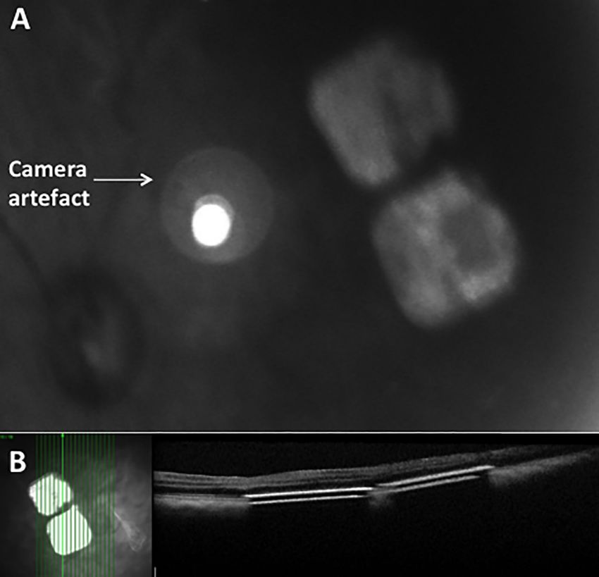
PLOS ONE                                                                                         PRIMA Subretinal Wireless Photovoltaic Microchip

                                            gently and to minimize the physical contact. Animals are housed in groups (minimum pair)
                                            when the scientific goals of the study allow it. If not, the isolated animal is given specific envi-
                                            ronmental enrichments and the visual contact with a social partner is maintained. Positive
                                            reinforcement is used in the everyday procedures.
                                                Animals received fresh water and food daily according to their nutritional needs. Extruded
                                            pellets are specifically manufactured to fulfil macaque’s nutritional needs (Altromin, ND).
                                            Animals are provided with environmental enrichment to express their natural behaviours that
                                            minimize environmental change and enhance their homeostasis. The environmental enrich-
                                            ment program is standardized and includes food enrichment (daily fruits, weekly access to
                                            seeds boxes/bags) and manipulating tools (mirrors, triangles, dental toys). Smooth music is
                                            played during working hours to cover unexpected sounds and to provide an acoustic enrich-
                                            ment. Lights turn on and off under a progressive program that mimics dawn and sunset.
                                                General anesthesia was used for all surgery and longitudinal examinations. The induction
                                            of anaesthesia used intramuscular ketamine 10mg/kg and xylazine 0.5mg/kg after fasting the
                                            day before the surgery. Maintenance of anesthesia used intravenous propofol 1ml/kg/h with
                                            oxybuprocaine for local anesthesia of the eye. Intubation of the animal was performed as a pre-
                                            cautionary measure during surgery
                                                Humane endpoints were followed with non-human primates only receiving an implant in
                                            one eye. Procedure specific humane endpoints were implemented in case of problems as indi-
                                            cated below. In order to quantify the impact of experimental procedures on the animals, we
                                            proposed a graded classification of ocular problem severity that included blindness, conjuncti-
                                            vitis, orbital swelling, and cataract. Nonspecific Humane endpoints were applied according to
                                            the “Humane Endpoint Guidelines for Nonhuman Primates in Biomedical Research” (Associ-
                                            ation of Primate Veterinarians). Specific humane endpoints for this procedure included: Bin-
                                            ocular blindness grade 2 for more than 15 days; glaucoma grade 2 non-responsive to treatment
                                            (15 days); conjunctivitis grade 2 non-responsive to treatment (15 days); non-specific pain
                                            symptoms; and, anorexia for 3 days/ lost weight above 15% of initial weight. Special care was
                                            given to avoid rank fights that might arise from impaired vision in one eye. non-human pri-
                                            mates having received implant were observed closely (each day after surgery) for the procedure
                                            specific impact. If social interactions between non-human primates undergoing the study
                                            changed, they were housed in separate cages while ensuring social interaction across the cages
                                            (animals were still able to see each other).
                                                Close-monitoring of the animals was conducted, and follow-up was conducted in the
                                            1.5mm implantation group. Fundus photography and OCT images were performed: 1 day, 2, 4,
                                            6 weeks after surgery; and, at 1–3 years in some eyes after surgery. Slitlamp examination of the
                                            anterior chamber was performed in order to detect any sign of inflammation, and eye pressure
                                            was recorded. If required, the disposition of the non-human primate was through euthanasia. If
                                            euthanasia was performed, animals were initially calmed in their cages by intramuscular injec-
                                            tion of a mixture of Ketamine at 10 mg/kg and xylazine (0.5mg/kg). A barbiturate was then
                                            injected intravenously (pentobarbital at 180mg/kg), and the eyes were enucleated.
                                                The diameter of the eye in this species is approximately 18 to 20mm. Anatomy is very close
                                            to human, and the retina and eye examination was normal in each eye studied. All surgeries
                                            were performed in sterile conditions under general anesthesia by veterinary anesthesiologists.
                                            The 1.5mm implant (Fig 2) was delivered in total 11 eyes.
                                                In one animal, the feasibility of the surgical procedure to deliver multi-chip implantation of
                                            two 1.5mm arrays was evaluated. The follow-up period was 4 weeks (n = 11) 2 years (n = 2),
                                            and 3 years (n = 1) for the 1.5mm implanted animals. The 2mm implant (Fig 2) was delivered
                                            to 4 eyes of four animals. All animals implanted with a 2mm implant were euthanized after the
                                            intervention.

PLOS ONE | https://doi.org/10.1371/journal.pone.0230713 April 8, 2020                                                                      5 / 16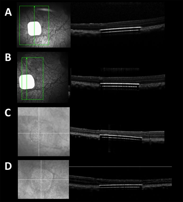
PLOS ONE                                                                                         PRIMA Subretinal Wireless Photovoltaic Microchip

                                            Fig 2. Design of PRIMA retinal implants.
                                            https://doi.org/10.1371/journal.pone.0230713.g002

                                            Surgery procedure
                                            The PRIMA surgery was performed according to a defined surgical protocol by the authors
                                            (YlM, JH) who both underwent a standardized briefing and training with the original surgical
                                            protocols for the studies. In this way, the different steps of the surgical technique could be
                                            assessed in different surgical conditions/models and using two independent operators. There
                                            is a time lag between studies due to the time for different country regulatory approvals to be
                                            completed. The surgery technique was identical in both the feline and primate studies and is
                                            detailed here in different steps:
                                            Step 1: Three 23-gauge (23G) sclerotomies were placed 3mm posterior to the limbus, and stan-
                                               dard pars plana vitrectomy (PPV) was performed using a Synergetics Versavit (Synergetics
                                               USA Inc) for the cat study, and Alcon Constellation (Alcon Laboratories Inc, Fort Worth,
                                               TX) for the primate study. The Resight 500 wide-angle viewing system and Zeiss Lumera
                                               700 microscope (Carl Zeiss Meditec AG, Jena, Germany) was used for primate surgery
                                               under general anesthesia (Fig 3).
                                            Step 2: Triamcinolone acetonide (Kenalog-40, Bristol Myers Squibb, Princeton, NJ) was
                                               injected at the beginning of the vitrectomy through the 23G trocar to ensure complete pos-
                                               terior vitreous detachment and vitrectomy.
                                            Step 3: A subretinal injection of balanced salt solution was delivered to create a bleb in the tem-
                                               poral part of the posterior pole using a 41-gauge subretinal cannula. Several injections
                                               could be used to enable detachment of the central macula (primate) /central retina (feline).
                                            Step 4: A 3mm area of the temporal macula, near or between the upper retinal vessels was
                                               coagulated with endodiathermy. This coagulated area marks the entry point of the implant,
                                               and was aligned parallel to the sclerotomy.
                                            Step 5: A retinotomy was made with vertical subretinal scissors, at the superior temporal part
                                               of the subretinal fluid bleb. The size of the retinotomy was calculated using a scaling
                                               method of preoperative optic nerve imaging. On average, the length of the retinotomy was
                                               2 times the longest dimension of the optic nerve. The size of this retinotomy was enlarged
                                               for the 2mm implant insertion compared to the 1.5mm implant.

PLOS ONE | https://doi.org/10.1371/journal.pone.0230713 April 8, 2020                                                                      6 / 16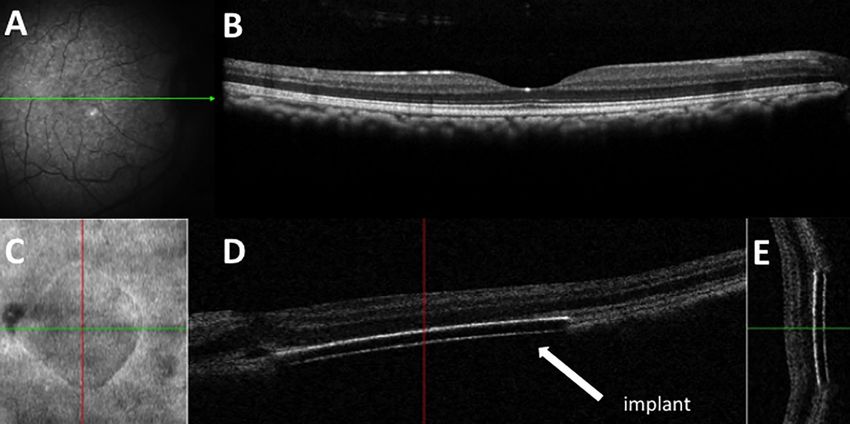
PLOS ONE                                                                                                 PRIMA Subretinal Wireless Photovoltaic Microchip

 Fig 3. Diagram of vitrectomy instrumentation entry ports.
 https://doi.org/10.1371/journal.pone.0230713.g003

                                                     Step 6: A new 2.4mm sclerotomy was created 3mm from the limbus, and the choroid cauter-
                                                        ized behind the incision. This would minimize potential intraocular hemorrhage during
                                                        implant insertion. Sutures of 6/0 vicryl were preplaced at the large sclerotomy site, and a
                                                        chandelier trocar inserted for bimanual surgery.
                                                     Step 7: The implant was introduced with special silicone-tipped forceps through the sclerot-
                                                        omy, and the sclerotomy temporarily closed by the assistant by pulling on the 6/0 vicryl
                                                        sutures to reduce intravitreal turbulence and fluid leakage.
                                                     Step 8: The tip of the forceps holding the implant was passed through the retinotomy, and the
                                                        implant was docked 1mm from the center of the fovea (primate)/central retina (feline). The
                                                        larger sclerotomy for implant insertion was completely closed with 6/0 vicryl.
                                                     Step 9: A bubble of perfluorocarbon liquid (PFCL) was then injected over the retina to stabilize
                                                        the implant positioned under the retina and flatten the macula.
                                                     Step 10: A laser photocoagulation at power 100-200mW was applied around the retinotomy,
                                                        and PFCL removed during an air-fluid exchange.
                                                     Step 11: The macula (primate) and central retina (feline) were checked to ensure the absence
                                                        of subretinal fluid. In the non-human primate study intraoperative OCT (iVue, Optovue,
                                                        Fremont, CA, USA) was performed to confirm the absence of fluid under the macula.
                                                     Step 12: Either intraocular sulfhexafluoride gas or 1000 centistokes silicone oil was injected
                                                        into the vitreous cavity. Subconjunctival kenalog-40 was injected, and the three sclero-
                                                        tomies closed with 8/0 vicryl.

PLOS ONE | https://doi.org/10.1371/journal.pone.0230713 April 8, 2020                                                                              7 / 16
PLOS ONE PRIMA Subretinal Wireless Photovoltaic Microchip
During the study, a number of surgery-related adverse events were noted. The following
modifications to the surgery protocol were made during the studies. The retinotomy size
revised to a minimum two times the length of the optic disc to avoid iatrogenic retinal tear
(Step 5). A change of incision entry to at least 3mm from the limbus (Step 6) was amended on
the surgery protocol to minimize the risk of postoperative retinal detachment complications.
During creation of the subretinal bleb at the posterior pole (Step 3), the surgeon would con-
sequently limit the intraocular subretinal fluid injection pressure to 20 psi to prevent the risk
of perforating the fovea and/or macular hole development.
An overview of the key surgical PRIMA implantation steps in primate is shown in S1
Video.
Results
Anatomical safety and stability of the implanted PRIMA
Feline. All PRIMA implants were delivered below the central retina within the subretinal
space in 11 feline eyes (S1 Fig), with the PRIMA microchip electrodes in contact with the ret-
ina. Immediately after the subretinal implantation, the retina was flat on top of the implants
with no signs of intraretinal or subretinal fluid. There were no intraoperative complications
during the follow-up period that ranged from 43–106 days.
Primate. Eleven primate eyes were implanted with the 1.5mm implant at the central mac-
ula (S2 Fig). In two other eyes, the retina could not be detached to allow implant delivery.
There were no adverse events in these two primates at 6 weeks to 12 months follow-up period.
Four primate eyes were implanted with the 2mm implant at the central macula. (S3 Fig).
The average surgical duration for implantation of the 2mm implant in Primate eyes was 1.6
hours (range 1.5 to 2 hours) and was equivalent to the average implantation time observed for
the implantation of 1.5mm implants in the study (range 2 to 3 hours including intraoperative
OCT imaging, Fig 4).
Using the 2mm implant, one subject was implanted in the left eye, three in the right eye
(Fig 5).
In addition, it has been verified that the implant was well supported by silicone oil after 2, 4
weeks, and the implant remained stable in the eye up to 1 year following implantation (Fig 6).
Immediately after the subretinal implantation, the retina was flat on top of the implants
with no signs of intraretinal or subretinal fluid. In order to confirm that the surgical procedure
was appropriate for the implantation of PRIMA implant in primates, the acceptance criteria
listed below were defined in the protocol. (1) The implant does not break in the surgical proce-
dure: No implant breakage occurred during all implantations, and any implant lost in the flux;
(2) The implant is delivered subretinally under the fovea with a tolerance of one implant width
(3) The implant did not migrate from its original position by more than one implant width:
Follow up retinal and OCT images show that no implant migration occurred. (4) There was
no significant retinal injury or eye damage beyond the mild AEs caused by the implantation
surgery: there was no unexpected surgical complication above the standard risks of vitrectomy
surgery.
In this study, all 1.5 and 2mm implants have been delivered at the target location with an
off-centering of less than one implant width: either under the fovea or in the parafoveal area
for two of the 1.5mm implants as this was the target area for a behavioral study conducted
with these two primates [13]. In one eye, there was implant migration noted following the sur-
gery. During PRIMA implantation, the surgeon noted implant breakage could occur if surgical
handling was incorrect or excessive.
PLOS ONE | https://doi.org/10.1371/journal.pone.0230713 April 8, 2020 8 / 16PLOS ONE PRIMA Subretinal Wireless Photovoltaic Microchip
Fig 4. Retinal imaging of a subretinal 1.5mm PRIMA chip implantation in Primate eye before and immediately
after surgery. A: Preoperative fundus photograph of macula in Macaca fascicularis; B: Preoperative optical coherence
scan of macula; C: Fundus photograph of right eye 1.5mm subretinal implant with comparative left eye.
https://doi.org/10.1371/journal.pone.0230713.g004
In one primate eye, multi-chip 1.5mm arrays were delivered subretinally close to the fovea
at a distance of respectively 0.4 and 1.5mm. The two 1.5mm implants did not touch or overlap,
and there was a gap of approximately 0.2mm that remained stable (Fig 7).
Adverse events
PRIMA 1.5mm implant feline surgery. In the feline study, four vitrectomy surgery-
related AEs were reported in four animals at 1 and 8 days following surgery. This included two
Fig 5. Retinal imaging of a subretinal 2mm PRIMA chip implantation in Primate eye before and immediately
after surgery. A: Preoperative fundus photograph of macula in Macaca fascicularis; B: Preoperative optical coherence
scan of macula; C: Enface image of the 2mm retinal implant; D: Horizontal OCT scan showing subretinal location of
implant; E: Vertical OCT scan showing subretinal location of implant.
https://doi.org/10.1371/journal.pone.0230713.g005
PLOS ONE | https://doi.org/10.1371/journal.pone.0230713 April 8, 2020 9 / 16PLOS ONE PRIMA Subretinal Wireless Photovoltaic Microchip
Fig 6. Retinal imaging of a subretinal 1.5mm PRIMA chip implantation in Primate eye at 2 weeks, 4 weeks, 8
months, and 12 months after surgery. A: 2 week postoperative optical coherence scan of macula in Macaca
fascicularis; B: 4 week postoperative optical coherence scan of macula; C: 8 month postoperative optical coherence scan
of macula; D: 12 month postoperative optical coherence scan of macula showing implant stability.
https://doi.org/10.1371/journal.pone.0230713.g006
cases of corneal ulcers developing later into corneal scars, and two cases of cataract. These AE are
associated risks of general intraocular surgery procedures, and were not PRIMA device-related.
There were no additional AE during the follow-up period that ranged from 43–106 days.
PRIMA 2mm implant primate surgery. There was one case of a mild vitreous cavity
hemorrhage that resolved with no complication. There was retinal tear near the retinotomy
site in one animal but the retina remained attached. In one case, there was a minor area of sub-
macular hemorrhage, with minimum hemorrhage lying on the implant surface. There was no
significant postoperative inflammation, and no case of retinal detachment. A summary of
non-device-related, surgical AEs is shown in Table 1.
PRIMA 1.5mm implant primate urgery. A summary of non-device-related, surgical AEs
is shown in Table 1. There was one case of an intraoperative macular hole associated with a ret-
inal detachment (Fig 8).
At 6 weeks follow-up, there was no significant retinal scar and the retina was attached. One
case of a small bubble of PFCL retained in the subretinal space outside the zone of PRIMA
implantation. There was no significant postoperative inflammation. In one eye, a retinal
detachment was detected at the 6 week control. A revision vitrectomy surgery with silicone oil
was performed that successfully treated this complication. In one eye, an iatrogenic tear was
made at the edge of the retinotomy during implant delivery, but the implant remained cen-
tered at the fovea postoperatively (Fig 9).
PLOS ONE | https://doi.org/10.1371/journal.pone.0230713 April 8, 2020 10 / 16PLOS ONE PRIMA Subretinal Wireless Photovoltaic Microchip
Fig 7. Intraoperative colour fundus photograph and OCT of 1.5mm multichip in macula of primate. A: Fundus
photograph of two 1.5mm chips; B: OCT scan of 1.5mm chips placed under the macula.
https://doi.org/10.1371/journal.pone.0230713.g007
After the end of 6-week follow-up period, as this study was terminated, the silicone oil was
removed. Subject 1 was examined 7 days following the silicone oil removal and 17 days follow-
ing oil removal surgery. At the final visit, a localised retinal detachment was detected in the
posterior pole. An iatrogenic retinal tear occurred during the silicone oil removal surgery and
Table 1. Summary of adverse events for 1.5mm and 2mm PRIMA surgery in primates.
Complication Surgery timing 1.5mm PRIMA 2mm PRIMA Postoperative, Final Ocular Status
Implant Implant
Macular hole and retinal During subretinal injection 1 0 Minor foveal scar with attached retina
detachment
Minor submacular haemorrhage Noted after chip insertion 0 1 Normal
Retinal tear at retinotomy with During PFCL injection 1 0 Retinal tear with subretinal PFCL† Retina attached
subretinal PFCL†
Retinal tear at retinotomy During retinotomy creation 1 1 Retina attached
Minor vitreous cavity Postoperative 0 1 Normal
haemorrhage
Postoperative Retinal Postoperative due to entry site break 1 0 Retinal detachment treated with silicone oil. Retina
detachment attached after secondary oil removal.
Retinotomy edge tear During creation of retinotomy 1 1 Normal
Non-surgery related retinal Postoperative after sequential oil 1 0 Retina attached
detachment removal
No implantation of PRIMA Not possible to surgically detach the 2 0 Normal
central retina/macula
† PFCL, perfluorocarbon liquid
https://doi.org/10.1371/journal.pone.0230713.t001
PLOS ONE | https://doi.org/10.1371/journal.pone.0230713 April 8, 2020 11 / 16PLOS ONE PRIMA Subretinal Wireless Photovoltaic Microchip
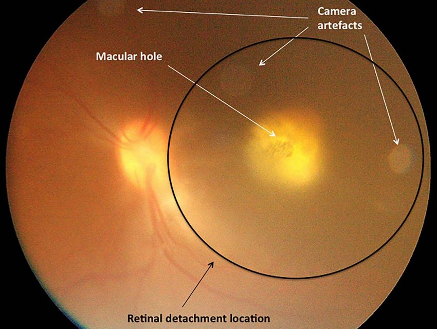
Fig 8. Intraoperative colour fundus photograph in macula of primate. Subfoveal 1.5mm chip placement with
overlying macular hole and localised retinal detachment at macula.
https://doi.org/10.1371/journal.pone.0230713.g008
resulted in this rhegmatogenous retinal detachment. The implant was still under the retina
and close to the fovea but was not attached by the retina. A retinal detachment is a recognized
complication following vitrectomy surgery and this was designated as a SAE. Three eyes were
followed up for 2 years with no additional AE reported.
Fig 9. Red-free fundus photograph of in Macaca fascicularis. The 1.5mm implant remains in the central macula at 2
weeks postoperatively.
https://doi.org/10.1371/journal.pone.0230713.g009
PLOS ONE | https://doi.org/10.1371/journal.pone.0230713 April 8, 2020 12 / 16PLOS ONE PRIMA Subretinal Wireless Photovoltaic Microchip
Discussion
We report subretinal implantation of 1.5mm and 2mm PRIMA implants in central retina of
cat eyes and central macula of Macaca fascicularis primate eyes. This study demonstrates that
the surgical procedure is appropriate for both 1.5mm and 2mm PRIMA retinal implants, with
slight adjustments of sclerotomy location, and the retinotomy size due to the additional
0.5mm in size.
The main purpose of this study was to assess the surgical implantation technique of two
sizes of PRIMA wireless photovoltaic microchip implants in different animal models that have
similar architecture to the human retina. During the studies, the surgery protocol was
amended following any procedure-related AEs, and thereafter, there were no additional sur-
gery-related AEs observed. Surgical implantation endpoints were reached in all cases. The
chips were placed within the subretinal space with a single case of postoperative retinal implant
migration away from the primary implantation site. The retinal implants remained stable
under silicone oil in all cases with no cases of significant ocular inflammation.
We observed no device-related AE in the feline study. In the primate study, there were sev-
eral minor AEs that resolved without complications at 6 weeks. One SAE of retinal detachment
developed after secondary vitrectomy with silicone oil removal, but this SAE was not device-
related, not primary procedure-related, nor related to the presence of the implant in the eye. A
second SAE of retinal detachment occurred at the 6-week control in a different primate’s eye,
and this was not related to silicone oil removal. A retinal detachment is a recognized risk in
vitreoretinal surgery, and retinal detachment in similar subretinal surgery models occurs at a
rate of 6 to 9% [14]. In the event of a retinal detachment, a revision vitrectomy surgery may
involve either intraocular gas tamponade or silicone oil re-injection. The root cause of this ini-
tial SAE has been determined and effective counter measures are implemented to minimize
future occurrence of the event. Close follow-up of patients would be instructed after silicone
oil removal to detect early signs of a retinal detachment. The second SAE retinal detachment
occurred in an eye where the sclerotomy incision was made at 3mm from the limbus.
Early removal of silicone oil will be planned for patients in a clinical trial as per this proto-
col. Silicone oil will only be intended for use as a short-term tamponade in human trials, and
will be removed as soon as the retinotomy is closed with formation of a chorioretinal adhesion.
The final aim of the procedure in humans is to have a subretinal chip without silicone oil. The
refractive changes made by silicone oil make focus of stimulating infrared beam out of range.
Silicone oil will therefore be used as a short-term tamponade and removed as soon as the reti-
notomy was closed.
Further analysis of the foveal perforation/macular hole AE during the study led to addi-
tional refinements of the surgery protocol. In one case, an intraoperative foveal perforation
occurred at injection pressure greater than 20psi. For the remaining cases, we did not encoun-
ter any cases of foveal perforation at an injection pressure of 20psi or less [15]. In the one case
of macular hole observed during chip implantation, the PRIMA microchip was implanted in
the subretinal space.
In previous studies of human subretinal delivery of a retinal implant, there were no signifi-
cant complications reported [9, 10]. In other in vivo subretinal interventional safety studies,
there have been no significant surgical complications reported during subretinal injection
delivery [16–18]. A recent in vivo safety study of subretinal gene therapy reported choroidal
haemorrhage, macular hole, retinal haemorrhage, and retinal tear [19].
Considering the eye size, 18 to 20mm in the study primates versus 24mm on average in
humans, a 1.5mm implant in a primate eye is equivalent to a 2mm implant in human. The study
implants are otherwise equivalent to the implants intended to be implanted in human patients.
PLOS ONE | https://doi.org/10.1371/journal.pone.0230713 April 8, 2020 13 / 16PLOS ONE PRIMA Subretinal Wireless Photovoltaic Microchip
Conclusions
This study demonstrated that both 1.5mm and 2mm PRIMA implants can be implanted safely
into the subretinal space of the macula/central retina in the eye of cats and primates. A multi-
chip subretinal implantation is also feasible. Based on the results of our study, modifications of
the implantation surgery procedure were adopted to minimize and prevent complications. As
the study models are very close to the human eye, it can be concluded that the surgical proce-
dure is appropriate for implantation in human. The next studies of PRIMA subretinal implan-
tation are in progress for human feasibility clinical trials.
Supporting information
S1 Fig. Fundus photographs and optical coherence scans in feline eyes. Fundus photographs
and optical coherence scans of 10 feline eyes.
(TIF)
S2 Fig. Fundus imaging and OCT scanning of 1.5mm chip placement in primate eyes. Fun-
dus photographs and optical coherence scans of the implanted macula of Macaca fascicularis.
(TIF)
S3 Fig. Fundus imaging and OCT scanning of 2mm chip placement in primate eyes. Fun-
dus photographs and optical coherence scans of the implanted macula of Macaca fascicularis.
(TIF)
S1 Video. Video clip of PRIMA implantation under macula of Macaca fascicularis.
(MP4)
Acknowledgments
The authors are grateful to Prof Daniel Palanker (Stanford University) who is the inventor of
PRIMA system. Pixium Vision designed, fabricated the PRIMA implants and sponsored the
studies. Authors would like to also extend their gratitude to all the scientific, R&D, medical
and clinical research team involved in the preparation and care of the animals in these studies
especially Joanna Demilly and Elena Brazhnikova, and staff involved at Molecular Imaging
Research Center (MIRCen). Academic and Institutional Support for the NIHR London Bio-
medical Research Centre is acknowledged.
Author Contributions
Conceptualization: Serge Picaud, Ralf Hornig, Guillaume Buc, Martin Deterre, Céline Nou-
vel-Jaillard, Elodie Bouillet, Philippe Hantraye, José Sahel.
Data curation: Jean Pierre Hubschman, Douglas B. McCreery, Ralf Hornig.
Formal analysis: Mahiul M. K. Muqit, Douglas B. McCreery, Jan C. van Meurs, Martin
Deterre, Elodie Bouillet, Yannick Le Mer.
Funding acquisition: Serge Picaud, Philippe Hantraye, José Sahel.
Investigation: Jean Pierre Hubschman, Douglas B. McCreery, Ralf Hornig, Céline Nouvel-
Jaillard, Yannick Le Mer.
Methodology: Jean Pierre Hubschman, Douglas B. McCreery, Ralf Hornig, Guillaume Buc,
Martin Deterre, José Sahel, Yannick Le Mer.
PLOS ONE | https://doi.org/10.1371/journal.pone.0230713 April 8, 2020 14 / 16PLOS ONE PRIMA Subretinal Wireless Photovoltaic Microchip
Project administration: Jean Pierre Hubschman, Ralf Hornig, Martin Deterre, Céline Nou-
vel-Jaillard, Elodie Bouillet, Claire-Maelle Fovet, Philippe Hantraye, Yannick Le Mer.
Resources: Douglas B. McCreery, Claire-Maelle Fovet, Philippe Hantraye, José Sahel, Yannick
Le Mer.
Software: Douglas B. McCreery.
Supervision: Mahiul M. K. Muqit, Jean Pierre Hubschman, Serge Picaud, Douglas B.
McCreery, Ralf Hornig, Guillaume Buc, Claire-Maelle Fovet, Philippe Hantraye, Yannick
Le Mer.
Validation: Mahiul M. K. Muqit, Jean Pierre Hubschman, Serge Picaud, Douglas B. McCreery,
Ralf Hornig, Guillaume Buc, Martin Deterre, Elodie Bouillet, Philippe Hantraye, Yannick
Le Mer.
Visualization: Jean Pierre Hubschman, Guillaume Buc, Martin Deterre, Elodie Bouillet, José
Sahel, Joseph N. Martel, Yannick Le Mer.
Writing – original draft: Mahiul M. K. Muqit, Jean Pierre Hubschman, Douglas B. McCreery,
Jan C. van Meurs, Ralf Hornig, Guillaume Buc, Martin Deterre, Elodie Bouillet, Claire-
Maelle Fovet, José Sahel, Joseph N. Martel, Yannick Le Mer.
Writing – review & editing: Mahiul M. K. Muqit, Jean Pierre Hubschman, Serge Picaud,
Douglas B. McCreery, Jan C. van Meurs, Ralf Hornig, Guillaume Buc, Martin Deterre,
Céline Nouvel-Jaillard, Elodie Bouillet, Philippe Hantraye, José Sahel, Joseph N. Martel,
Yannick Le Mer.
References
1. Palanker D, Vankov A, Huie P, Baccus S. Design of a high-resolution optoelectronic retinal prosthesis.
Neural Eng. 2005; 2(1): S105–120.
2. Loudin JD, Simanovskii DM, Vijayraghavan K, Sramek CK, et al. Optoelectronic retinal prosthesis: sys-
tem design and performance. J Neural Eng. 2007; 4: S72–S84. https://doi.org/10.1088/1741-2560/4/1/
S09 PMID: 17325419
3. Loudin JD, Cogan SF, Mathieson K, Sher A, Palanker DV. Photodiode circuits for retinal prostheses.
IEEE Trans Biomed Circuits Syst. 2011; 5(5): 468–480. https://doi.org/10.1109/TBCAS.2011.2144980
PMID: 23852178
4. Wang L, Mathieson K, Kamins TI, et al. Photovoltaic retinal prosthesis: implant fabrication and perfor-
mance. J Neural Eng. 2012; 9(4): 046014. https://doi.org/10.1088/1741-2560/9/4/046014 PMID:
22791690
5. Adekunle AN, Adkins A, Wang W, et al. Integration of perforated subretinal prostheses with retinal tis-
sue. Transl Vis Sci Technol. 2015; 14: 4(4): 5. https://doi.org/10.1167/tvst.4.4.5 PMID: 26290776
6. Lorach H, Lei X, Galambos L, Kamins T, et al. Interactions of prosthetic and natural vision in animals
with local retinal degeneration. Invest Ophthalmol Vis Sci. 2015; 56(12): 7444–7450. https://doi.org/10.
1167/iovs.15-17521 PMID: 26618643
7. Lorach H, Goetz G, Smith R, et al. Photovoltaic restoration of sight with high visual acuity. Nat Med.
2015; 21(5): 476–482. https://doi.org/10.1038/nm.3851 PMID: 25915832
8. Ho E, Lorach H, Goetz G, et al. Temporal structure in spiking patterns of ganglion cells defines percep-
tual thresholds in rodents with subretinal prosthesis. Sci Rep. 2018; 16; 8(1): 3145. https://doi.org/10.
1038/s41598-018-21447-1 PMID: 29453455
9. Chow AY, Chow VY, Packo KH, Pollack JS, Peyman GA, Schuchard R. The artificial silicon retina
microchip for the treatment of vision loss from retinitis pigmentosa. Arch of Ophthalmol. 2004; 122(4):
460–469.
10. Chow AY, Bittner AK, Pardue MT. The artificial silicon retina in retinitis pigmentosa patients (An Ameri-
can Ophthalmological Association Thesis). Trans Am Ophthalmol Soc. 2010; 108: 120–154. PMID:
21212852
PLOS ONE | https://doi.org/10.1371/journal.pone.0230713 April 8, 2020 15 / 16PLOS ONE PRIMA Subretinal Wireless Photovoltaic Microchip
11. Chow AY, Pardue MT, Chow Vy, et al. Implantation of silicone chip microphotodiode arrays into the cat
subretinal space. IEEE Trans Neural Syst Rehabil Eng. 2001; 9(1): 86–95. https://doi.org/10.1109/
7333.918281 PMID: 11482368
12. Chow AY, Pardue MT, Perlman JI, et al. Subretinal implantation of semiconductor-based photodiodes:
Durability of novel implant designs. J Rehabil Res Dev. 2002; 39(3): 313–321. PMID: 12173752
13. Prévot P-H, Gehere F, Arcizet H, et al. Behavioural responses to a photovoltaic subretinal prosthesis
implanted in non-human primates. Nat Biomed Eng. 2019; 2019 Dec 2. https://doi.org/10.1038/s41551-
019-0484-2 PMID: 31792423
14. Rainer G, Schrader W. Longterm results in surgical removal of subfoveal choroidal neovascularization
in age-related macular degeneration. Acta Ophthalmol. 2004; 82(6): 686–690.
15. Takahashi K, Morizane Y, Hisatomi Tet al. The influence of subretinal injection pressure on the micro-
structure of the monkey retina. PLoS One. 2018 Dec 31; 13(12):e0209996. https://doi.org/10.1371/
journal.pone.0209996 PMID: 30596769
16. Maguire AM, Simonelli F, Pierce EAet al. Safety and efficacy of gene transfer for Leber’s congenital
amaurosis. N Engl J Med. 2008; 358(21): 2240–2248. https://doi.org/10.1056/NEJMoa0802315 PMID:
18441370
17. Bainbridge JW, Smith AJ, Barker SSet al. Effect of gene therapy on visual function in Leber’s congenital
amaurosis. N Engl J Med. 2008; 358(21): 2231–2239. https://doi.org/10.1056/NEJMoa0802268 PMID:
18441371
18. MacLaren RE, Groppe M, Barnard AR et al. Retinal gene therapy in patients with choroideremia: initial
findings from a phase 1/2 clinical trial. Lancet. 2014; 383(9923): 1129–1137. https://doi.org/10.1016/
S0140-6736(13)62117-0 PMID: 24439297
19. Maguire AM, Russell S, Wellman JA et al. Efficacy, Safety, and Durability of Voretigene Neparvovec-
rzyl in RPE65 Mutation-Associated Inherited Retinal Dystrophy: Results of Phase 1 and 3 Trials. Oph-
thalmology. 2019; 126(9): 1273–1285. https://doi.org/10.1016/j.ophtha.2019.06.017 PMID: 31443789
PLOS ONE | https://doi.org/10.1371/journal.pone.0230713 April 8, 2020 16 / 16You can also read