Abnormal regional homogeneity and its relationship with symptom severity in cervical dystonia: a rest state fMRI study
←
→
Page content transcription
If your browser does not render page correctly, please read the page content below
Wei et al. BMC Neurology (2021) 21:55
https://doi.org/10.1186/s12883-021-02079-x
RESEARCH ARTICLE Open Access
Abnormal regional homogeneity and its
relationship with symptom severity in
cervical dystonia: a rest state fMRI study
Shubao Wei1, Chunhui Lu2, Xiuqiong Chen1, Lu Yang2, Jing Wei2, Wenyan Jiang2, Yang Liu2, Hui Hui Li2,
Yuhong Qin3, Yiwu Lei3, Chao Qin2, Caiyou Hu1* and Shuguang Luo2*
Abstract
Background: Although several brain networks play important roles in cervical dystonia (CD) patients, regional
homogeneity (ReHo) changes in CD patients have not been clarified. We investigated to explore ReHo in CD
patients at rest and analyzed its correlations with symptom severity as measured by Tsui scale.
Methods: A total of 19 CD patients and 21 gender-, age-, and education-matched healthy controls underwent fMRI
scans at rest state. Data were analyzed by ReHo method.
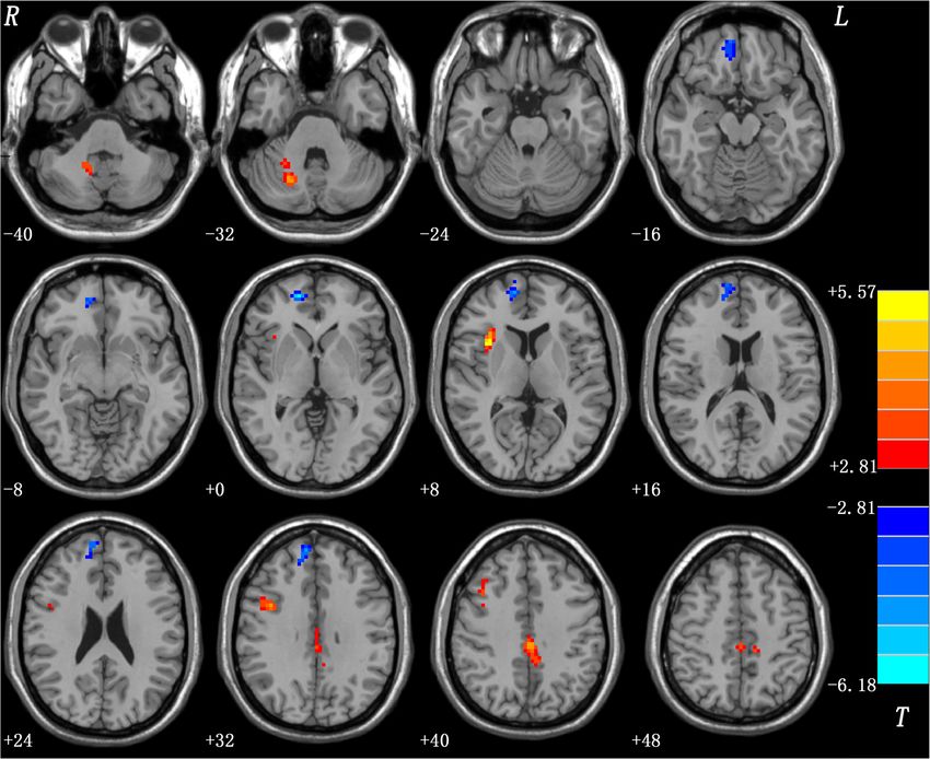
Results: Patients showed increased ReHo in the right cerebellum crus I and decreased ReHo in the right superior
medial prefrontal cortex (MPFC). Moreover, the right precentral gyrus, right insula, and bilateral middle cingulate
gyrus also showed increased ReHo values. A significantly positive correlation was observed between ReHo value in
the right cerebellum crus I and symptom severity (p < 0.05).
Conclusions: Our investigation suggested abnormal ReHo existed in brain regions of the “pain matrix” and salience
network (the right insula and bilateral middle cingulate gyrus), the motor network (the right precentral gyrus), the
cerebellum and MPFC and further highlighted the significance of these networks in the pathology of CD.
Keywords: Cervical dystonia, Resting-state functional magnetic resonance, Regional homogeneity, Default mode
network
Background disorder is frequently accompanied by head tremor and
Cervical dystonia (CD) is a neurologic disorder charac- chronic neck pain [17]. Analysis of the influence of CD
terized by involuntary sustained contractions of the cer- on work productivity has confirmed the substantial
vical musculature, causing the head to rotate abnormally negative influence of CD on employment, with CD-
or tilt in a particular directions [1]. The head may typic- related pain as a particularly important driver [27].
ally turn to a specific direction resulting in torticollis, Moreover, CD patients sustain significantly psychosocial
laterocollis, anterocollis or retrocollis [19]. CD is the disability and decline of life quality. Thus, it is important
most common form among the focal dystonia and fre- to identify CD patients and provide them with effective
quently starting at later ages of 40–60 years. The treatment. However, the pathophysiology underlining
the disorder is only partly understood.
* Correspondence: cyhu.hua@163.com; luoshuguang@stu.gxmu.edu.cn
1
Developments in neuroimaging techniques opened
Department of Rehabilitation Medicine, Jiangbin Hospital of Guangxi
new avenues for detailed investigation of structural
Zhuang Autonomous Region, Nanning 530021, Guangxi, China
2
Department of Neurology, The First Affiliated Hospital of Guangxi Medical changes and regional activities in the brain involved in
University, Nanning 530021, Guangxi, China the pathophysiology of CD. Some CD patients show
Full list of author information is available at the end of the article
© The Author(s). 2021 Open Access This article is licensed under a Creative Commons Attribution 4.0 International License,
which permits use, sharing, adaptation, distribution and reproduction in any medium or format, as long as you give
appropriate credit to the original author(s) and the source, provide a link to the Creative Commons licence, and indicate if
changes were made. The images or other third party material in this article are included in the article's Creative Commons
licence, unless indicated otherwise in a credit line to the material. If material is not included in the article's Creative Commons
licence and your intended use is not permitted by statutory regulation or exceeds the permitted use, you will need to obtain
permission directly from the copyright holder. To view a copy of this licence, visit http://creativecommons.org/licenses/by/4.0/.
The Creative Commons Public Domain Dedication waiver (http://creativecommons.org/publicdomain/zero/1.0/) applies to the
data made available in this article, unless otherwise stated in a credit line to the data.Wei et al. BMC Neurology (2021) 21:55 Page 2 of 8
structural alterations in the basal gangli, thalamus, cere- therapy in the three recent months; and (4) any history
bellum, motor cortex, and supplementary motor cortices of neurological or psychiatric disorders.
[9, 10, 29, 34, 35, 47]. In addition, a negative correlation Healthy controls were simultaneously recruited from
between putamen volume and symptom severity in CD the community. Each healthy control was right-handed
patients has been reported [8]. Functional magnetic res- and group-matched in gender, and age,. Exclusion cri-
onance imaging (fMRI) results are consistent with that teria for the control group were as follows: (1) any his-
of structural neuroimaging. Results from fMRI re- tory of serious medical or neurological illness; (2) any
searches demonstrate aberrant activation in basal gan- history of severe neuropsychiatric diseases; and (3) any
glia, premotor, and motor-related areas [5, 6]. In general, family history of neurological or psychiatric disorders in
growing evidences indicated that not only the basal gan- their first-degree relatives. Participants who had any
glia but also the cerebellum and sensorimotor cortices contraindications for MRI or shown changes under con-
may be conducive to the pathology of CD. However, re- ventional MRI scans were excluded.
sults are not merely contradictory, and whether these Every patients were evaluated using Tsui scale [43] to
changes are causative or compensatory is still uncertain. measure symptom severity of CD. All participants were
Therefore, the pathology of CD is remain unclear. showed the information related to the study procedures
Independent component analysis (ICA) and seed- and signed the consent forms before evaluations. The
based region of interest (ROI) are two methods most ethics committee of the First Affiliated Hospital,
widely employed for analyzing the resting-state data. Guangxi Medical University approved our study. All par-
Both methods present several significant benefits and ticipants were given information regarding study proce-
disadvantages that have stated previously [44]. Given the dures and subsequently provided written informed
shortcomings of both methods, the present study uses a consent.
method called regional homogeneity (ReHo) to examine
the regional homogeneity in CD patients. In addition, Image acquisition
asymmetric activity patterns have observed in CD pa- Resting-state scans were captured by using a Siemens
tients [4, 6], but these patterns were remain uncertain. 3.0 T scanner (Erlangen, Germany). Earplugs were ap-
Thus, a measure of regional homogeneity might give a plied for reducing scanner noise and foam padding for
better insight on this aspect. ReHo is a measurement of minimizing head movement. All participants were re-
similarity or synchronicity of the time series of nearest quested to lie still, stay awake with their eyes closed and
neighboring voxels. A lower ReHo may imply hypoactive relax. Each participant was asked to avoid thinking of
in the regional area, and vice versa [46]. Aberrant ReHo anything in particular during the image acquisition.
could indicate the disturbance of temporal aspects of After the session, each subject was asked whether they
neural activity and be related to pathophysiology under- had fallen asleep during the scanning. The one who
lining disorder [37]. So far, ReHo has been well applied responded positively or ambiguously was excluded.
in study of schizophrenia, depression and somatization Echo-planar imaging sequence was acquired from each
disorder [12, 13, 24, 37]. subject by using the following parameters: repetition
In order to investigate the regional homogeneity in time/echo time (TR/TE) = 2000/30 ms, slice thickness =
CD patients, we used ReHo approach to analyze fMRI 4 mm, gap = 0.4 mm, matrix = 64 × 64, flip angle = 90°,
data at rest state. We hypothesize that CD patients FOV = 24 cm × 24 cm, number of volumes = 250.
would show abnormal regional homogeneity, particularly
the motor-related areas. Image pre-processing
The resting-state fMRI (DPARSF) [2] data processing as-
Methods sistant was used for image preprocessing in MATLAB
Subjects [26]. We discarded the first ten images of each run to
The study is conducted in an outpatient setting of the consider the equilibration of the signal. We first cor-
Department of Neurology, the First Affiliated Hospital, rected for slice timing, and then conducted head motion.
Guangxi Medical University, China. A total of 21 right- Afterward, participants with a maximum displacement
handed CD patients were originally recruited. CD was of more than 2° motion and 2 mm in x, y, z (rotation
diagnosed by 2011EFNS (European Federation of Neuro- and translation parameters) were excluded. Subse-
logical Societies) guidelines on diagnosis and treatment quently, fMRI images were normalized to the standard
of primary dystonias. Exclusion criteria for CD group Montreal Neurological Institute (MNI) EPI space and re-
were as follows: (1) secondary spasmodic torticollis that sampling to 3 mm × 3 mm × 3 mm. Acquired images
is definitely diagnosed; (2) any history of serious medical were temporally band-pass filtered (0.01–0.08 Hz) and
or neurological illness; (3) any history of botulinum linear detrended for the reduction of low-frequency drift
toxin treatment, related medical treatment, or operation and respiratory and cardiac noises.Wei et al. BMC Neurology (2021) 21:55 Page 3 of 8
ReHo analyses no significant differences in age, and sex ratio between
We performed ReHo analyses by MATLAB [26] (Math- the patient group and the control group. Information on
works) using software REST (http://www.resting-fmri. the demographic and clinical characteristics of included
sourceforge.net). Kendall’s coefficient of concordance subjects is detailedly given in Table 1. In addition, a total
(KCC) was calculated to represent the ReHo of a speci- of 17 patients reported neck pain; 7 patients were left-
fied voxel. In voxel-wise analysis, measurements of each side affected and the others were bilateral-side affected;
subject’s ReHo were obtained by calculating the KCC of a total of 18 of the 19 patients reported sensory tricks,
the time series of a particular voxel and its nearest that is the abnormal posture and involuntary movement
neighbor (26 voxels). The calculation formula of KCC of the head and neck can be temporarily improved from
value have already expounded by Zang et al. [46]. For re- light touching the lower part of the cheek, jaw and pos-
ducing the confounding of individual variations in KCC terior neck and leaning against the wall, as well as keep-
value, the normalization of ReHo maps was performed ing something in the mouth and carrying a weight
by dividing KCC among each voxel by the averaged backpack on the back. Only one patient has never re-
KCC of the whole brain. Generated ReHo maps were ported sensory tricks.
then spatially smoothed with a Gaussian kernel of 8 mm
full-width at half-maximum. ReHo: between-group comparison
Significant differences between ReHo values of the pa-
Statistical analyses tient and control groups were observed within the whole
Clinical and demographic information was calculated in- brain based on the two-sample t-tests by voxelwise
cluding age, sex between the patients and the control cross-subject comparisons. Compared with healthy con-
group and the average of illness duration, symptom se- trols, CD patients had higher ReHo in the right cerebel-
verity in the patients group. Quantitative variables were lum crus I, right insula, right precentral gyrus, and
compared by the two-sample t-test, whereas qualitative bilateral middle cingulate gyrus but lower ReHo in the
variables were compared by the χ2 test (p < 0.05). ReHo right superior medial prefrontal cortex (MPFC) (Fig. 1
analyses were performed using REST through the two- and Table 2).
sample t-tests. The level of significance was set at the
corrected p < 0.005 using the Gaussian random field Correlations between ReHo and clinical variables
(GRF) correction method at the cluster level (voxel sig- A significant positive correlation was observed between
nificance: p < 0.001, cluster significance: p < 0.005). We ReHo value in the right cerebellum crus I and symptom
also respectively performed a voxel-based Pearson cor- severity at p < 0.05 (Fig. 2). Linear correlations in the pa-
relative analysis between mean ReHo values of clusters tient group were evaluated between ReHo and illness
and the patients’ age, and Spearman correlative analysis duration, and age; no significant correlations were de-
between illness duration and Tsui total score. The level tected, p > 0.05.
of significant correlation was set at p < 0.05.
Discussion
Results ReHo is a measure to detect the similarity or
Subjects synchronization of the time series of nearest neighboring
There were no subject excluded due to any contraindica- voxels (usually 27 voxels) with the calculation of KCC.
tions for MRI or shown changes under conventional This approach was used with fMRI data at rest to inves-
MRI scans. No subjects were excluded due to falling tigate the regional homogeneity in CD. Compared with
asleep during image acquisition. Two patients with ex- controls, CD patients showed higher ReHo in the right
cessive head movement and excluded from further ana- insula and the bilateral middle cingulate gyrus, the right
lysis. Consequently, a total of 19 patients and 21 healthy precentral gyrus, and the right cerebellum crus I but
controls were included in further analysis. There were lower ReHo in the right superior MPFC.. Furthermore, a
Table 1 Demographics and clinical characteristics of the patients and the controls
Variables (mean ± standard deviation) Patients Controls p value
Gender (female/male) 10/9 15/6 0.220a
Age, years 38.74 ± 10.71 39.62 ± 6.62 0.759b
Illness duration, months 24.29 ± 31.26
Tsui 16.32 ± 4.45
Tsui: Tsui scale
a
The p value for gender distribution in the two groups was obtained by chi-square test
b
The p values were obtained by two sample t-testsWei et al. BMC Neurology (2021) 21:55 Page 4 of 8
Fig. 1 ReHo differences between patients with CD and controls. Red and blue denote higher and lower ReHo respectively and the color bars
represent the t values from two-sample t-test of the group analysis. ReHo = regional homogeneity; CD = cervical dystonia
significant positive correlation was observed between the
ReHo value in the right cerebellum crus I and the symp-
Table 2 Brain regions with significant ReHo differences in the tom severity.
patients By usage, the dystonia that covered the cervical dys-
Brain regions Peak (MNI) Number of T value tonia has been attributed to the dysfunction of the basal
voxels
x y z ganglia. However, the cerebellum has been recently sug-
Patients > Controls gested as a probable originate region. Traditionally, the
Right Cerebellum Crus I 24 −66 −33 54 4.5063 cerebellum is identified as a region engaged in motor co-
Right Insula 33 15 9 46 5.5657
ordination. It plays a pivotal role in modulation of pre-
motor, sensorimotor and posterior parietal regions for
Right Precentral Gyrus 39 6 33 68 4.4075
fine-tuning motor control. Pong et al. have demon-
Bilateral Middle Cingulate 0 −27 42 125 5.0219 strated that the output of the cerebellum and the output
Gyrus
of from the basal ganglia work together to participate in
Patients < Controls
movement control of the head and face [33]. Moreover,
Right Superior MPFC 15 54 3 193 −6.1797 the cerebellum has been suggested as a processor of sen-
x, y, z, coordinates of primary peak locations in the MNI space; T statistical sory information, integrating descending visual input
value of peak voxel showing ReHo differences between the patients with CD
and the controls; CD: cervical dystonia; ReHo regional homogeneity, MNI
from the parietal cortex and ascending input from the
Montreal Neurological Institute, MPFC medial prefrontal cortex spinocerebellar pathway for promoting a forward model,Wei et al. BMC Neurology (2021) 21:55 Page 5 of 8 Fig. 2 Positive correlation between ReHo in Right Cerebellum Crus I and symptom severity in the patient group. ReHo = regional homogeneity. The correlation between ReHo in Right Cerebellum Crus I and symptom severity was obtained by Pearson correlation analysis as well as for predicting sensory consequences of an ac- many higher executive functions, involving in emotion, tion [45]. Given these findings, we have reason to believe decision-making, goal-directed behavior, working mem- that the abnormality in the right cerebellum crus I with ory and attention [25]. Therefore, we proposed that the CD could influenced the accuration of the movement decreased ReHo value of the right superior MPFC pos- control of the head and face impaired because the ab- sibly influence this region’s function and result in losing normal modulation. top-down regulation, which is suggested as the founda- It is noteworthy that a significant positive correlation tion in the pathology of changes of cognitive, emotional was observed between ReHo value in the right cerebel- processing, and behavior in CD. In our other study, we lum crus I and symptom severity in the present study. have found patients with CD exhibit significantly de- Previously, an animal studies observed that pharmaco- creased VMHC in superior MPFC [18]. Besides, changes logical exciting of the cerebellum leads to dystonia [32]. in the cognitive processing of movement have been pre- These findings supported the conjecture that dystonia is viously observed in idiopathic dystonia patients [15, 23]. caused by aberrant, distorted functional output [20]. In other studies, in addition to organization and execu- Based on these notions, the result of a higher ReHo tion of movement, aberrant motor cognition consisting value in the right cerebellum crus I in CD patients here of a mental rotation of body parts, temporal processing consistently indicated that the dysfunction of this region and alterations of movement, and body representation can be a key factor in the occurrence of motor symp- have been observed in CD [7]. These findings strengthen toms in CD, which highlighted the importance of the our conjecture. cerebellum for motor modulation in the pathology of Additionally, in animal study, MPFC was confirmed cervical dystonia. receiving afferent projections representing all sensory Anatomically, MPFC composed of discrete and modalities, which originated from widespread areas of cytoarchitectonically areas receiving large-scale of sen- the cortex (and associated thalamic nuclei). The dorsal sory information from the external environment and the MPFC presumably integrated and utilized this informa- body [39]. Specifically, the lobule Crus I of the cerebel- tion for goal directed actions [16]. Thus, abnormal lum closely associated with MPFC [28], agrees with a neuro-activity in MPFC may influence goal directed ac- prominent role of both regions in non-motor functions tions. On this basis, the “sensory trick”, which we sug- [21]. Functionally, MPFC was proposed to be involved in gested as a goal directed action, may have a correlation
Wei et al. BMC Neurology   (2021) 21:55                                                                           Page 6 of 8

with the changes of MPFC. In the present study, the             causative factors of CD-related pain are still a matter of
sensory trick phenomenon was observed in 18 patients,           debate whether excessive muscle contractions or alter-
which was consistent with previous report [11]. These           ations of transmission and processing of nociceptive
patients usually acquire attenuation of abnormal head           stimuli [41]. Study has released cutaneous nociceptive
movements by slightly touching a particular area of the         pathway function in CD patients is normal [42]. This
face or head. It has previously been proposed to use sen-       generates a hypothesis of the crucial note of CD-related
sory techniques to influence proprioceptive input to bal-       pain may correlate to the centre nervous system.
ance the inhibition ratio to facilitation [36]. Recently, the     It is worthy to note that the right insula and the bilat-
sensory trick was suggested as a modulation of abnormal         eral middle cingulate gyrus exhibited an increased ReHo
connections between sensory input and motor output              in CD patients. Both two regions have been confirmed
[11]. The inconsistent standpoint renders the mechan-           to be related to pain perception [31], so that they were
ism latent the sensory trick phenomenon still intricate.        recognized as brain regions belonging to the “pain
Our findings probably supported the latter. Conse-              matrix”. In masses of previous findings, CD was linked
quently, we suggested MPFC involved in the mechanism            to multiple brain regions but rarely to the insula and the
of the sensory trick.                                           cingulate gyrus. In fact, it is well known that the differ-
   Increasing evidence emphasizes dystonia as a disorder        ent brain areas constituting the “pain matrix”, including
of motor organization, programming, sensorimotor, and           SII, the insula and the anterior cingulate cortex. The
execution [7]. the primary motor cortex (as the M1),            same areas are part of the salience network circuit which
that is the precentral gyrus, received projections from         is linked to pain [22]. Moreover, the insular and cingu-
BA2 and BA5 areas that contain the contralateral cuta-          late cortex play crucial roles in integrating multimodal
neous, muscular and articular information, and subse-           information significant for several functions, such as
quently corrected movement. Meanwhile, as the target            sensorimotor, allostatic/homeostatic, emotional, and
of projections from frontal cortical regions and subcor-        cognitive functions [40]. On these basis, the abnormal
tical regions, precentral gyrus is a probable site conver-      ReHo values in these brain areas might be of important
ging mechanisms of the selection, initiation and                significance. The increased ReHo in the right insula and
inhibition movement [38]. This connectional architec-           the bilateral middle cingulate gyrus may influenced the
ture induced the precentral gyrus to play a crucial role        functions of the “pain matrix” and/or the salience net-
in the mechanism of generated CD. The abnormality in            work circuit consequently resulted the generation or im-
motor system physiology of patients with dystonia was           pair processing of CD-related pain and induce an
exhibited by reduced surround inhibition resulting in           abnormal sensorimotor function in CD patients. Based
unnecessary contractions of more muscles than what is           on our results and the reasoning aforementioned, we
required for specified motor behavior [5]. Therefore, the       speculated that CD-related might caused by alterations
increased ReHo in the right precentral gyrus in CD pa-          of transmission and processing of nociceptive stimuli.
tients could lead to impaired selection, initiation or in-      Thus, the observation of abnormal activities in these
hibition of movement though impairing the cortices -            brain areas extended the understandings of the path-
basal ganglia – cortices circuit. In other words, it was in     ology underlying CD.
line with the notion that the cervical dystonia has been          We observed asymmetric activity patterns in CD that
attributed to not only the dysfunction of the basal gan-        were strongly involved in the right-hemispheric
glia but also the cortices. In our other study, patients        dystonia-related connectivity pattern. This phenomenon
with CD showed abnormal activities with decreasing              has been previously reported, for instance, during finger
GFC in the the M1-SMA motor network, including right            movements [4], and at resting-state [6]. It was possibly
supplementary motor area and right precentral gyrus.            associated to the larger number of contralateral-side
Moreover, the GFC values in the right precentral gyrus          muscles affected patients. However, patients in our study
of CD patients was significantly negative correlated to         were primary bilateral-side muscles affected. A possible
the symptomatic severity [30]. Hence, abnormal regional         explanation for the laterality of the right-hemispheric
homogeneity in this region is important for the path-           may be due to its dominance of position control, that is
ology of CD.                                                    the right hemisphere determine response modification
   Pain is the most common and disabling non-motor              so that it is dominant for position control [14].
symptom and contributes significantly to patient disabil-         Aside from the small sample size, several limitations
ity and low quality of life in CD. The incidence of pain is     must be stated in this study. First, all of the patients’
reported up to 88.9% and most of them rated their pain          dystonic posturing was minimal in the supine position
as moderate or severe [3]. In the present study, a total of     or absent during scanning. To confirm whether this is a
17 patients (89.4%) reported painful neck muscles. The          specific sensory trick is difficult. Therefore, influences of
ratio was close to that reported in previous study. So far,     sensory trick could not be easily eliminated. Besides,
Wei et al. BMC Neurology             (2021) 21:55                                                                                                              Page 7 of 8

physiological noises including heart rhythm and respira-                               3.    Charles PD, Manack Adams A, Davis T, Bradley K, Schwartz M, Brin MF, Patel
tory cannot be completely eliminated even though a                                           AT. Neck pain and cervical dystonia: treatment outcomes from CD PROBE
                                                                                             (cervical dystonia patient registry for observation of OnabotulinumtoxinA
relatively low sampling rate (TR = 2 s) is used. Lastly,                                     efficacy). Pain Pract. 2016. https://doi.org/10.1111/papr.12408.
with a small sample size, patients were not subdivided                                 4.    de Vries PM, Johnson KA, de Jong BM, Gieteling EW, Bohning DE, George
further into different groups according to head rotation.                                    MS, Leenders KL. Changed patterns of cerebral activation related to
                                                                                             clinically normal hand movement in cervical dystonia. Clin Neurol
Hence, research using a larger sample size is required to                                    Neurosurg. 2008;110(2):120–8. https://doi.org/10.1016/j.clineuro.2007.09.020.
expand our results.                                                                    5.    Delnooz CC, Pasman JW, Beckmann CF, van de Warrenburg BP. Task-free
                                                                                             functional MRI in cervical dystonia reveals multi-network changes that
                                                                                             partially normalize with botulinum toxin. PLoS One. 2013;8(5):e62877.
Conclusions                                                                                  https://doi.org/10.1371/journal.pone.0062877.
Our investigation suggested abnormal ReHo existed in                                   6.    Delnooz CC, Pasman JW, Beckmann CF, van de Warrenburg BP. Altered
brain regions of the “pain matrix” and salience network                                      striatal and pallidal connectivity in cervical dystonia. Brain Struct Funct.
                                                                                             2015;220(1):513–23. https://doi.org/10.1007/s00429-013-0671-y.
(the right insula and bilateral middle cingulate gyrus),                               7.    Delorme C, Roze E, Grabli D, Mayer JM, Degos B, Vidailhet M, Worbe Y.
the motor network (the right precentral gyrus), the cere-                                    Explicit Agency in Patients with cervical dystonia: altered recognition of
bellum and MPFC and further highlighted the signifi-                                         temporal discrepancies between motor actions and their feedback. PLoS
                                                                                             One. 2016;11(8):e0162191. https://doi.org/10.1371/journal.pone.0162191.
cance of these networks in the pathology of CD.                                        8.    Draganski B, Schneider SA, Fiorio M, Kloppel S, Gambarin M, Tinazzi M, et al.
                                                                                             Genotype-phenotype interactions in primary dystonias revealed by
Acknowledgments                                                                              differential changes in brain structure. Neuroimage. 2009;47(4):1141–7.
We give special thanks to Dr. GWB managed and analyzed the imaging data.                     https://doi.org/10.1016/j.neuroimage.2009.03.057.
The authors thank all individuals who served as the research participants.             9.    Draganski B, Thun-Hohenstein C, Bogdahn U, Winkler J, May A. "motor
                                                                                             circuit" gray matter changes in idiopathic cervical dystonia. Neurology. 2003;
Authors’ contributions                                                                       61(9):1228–31.
Dr. LSG and HCY conceived and designed the study. Drs. WSB, Dr. LCH, CXQ,              10.   Egger K, Mueller J, Schocke M, Brenneis C, Rinnerthaler M, Seppi K, et al. Voxel
YL, WJ, JWY, LY, LHH, QYH, LYW, and QC collected the original imaging data.                  based morphometry reveals specific gray matter changes in primary dystonia.
Dr. HCY directed statistical analysis and the manuscript writing, and Drs. WSB               Mov Disord. 2007;22(11):1538–42. https://doi.org/10.1002/mds.21619.
and Dr. LCH wrote the first draft of the manuscript. The author(s) read and            11.   Filip P, Sumec R, Balaz M, Bares M. The clinical phenomenology and
approved the final manuscript.                                                               associations of trick maneuvers in cervical dystonia. J Neural Transm
                                                                                             (Vienna). 2016;123(3):269–75. https://doi.org/10.1007/s00702-015-1488-z.
Funding                                                                                12.   Guo WB, Liu F, Xue ZM, Yu Y, Ma CQ, Tan CL, et al. Abnormal neural
This study was supported by grants from the National Natural Science                         activities in first-episode, treatment-naive, short-illness-duration, and
Foundation of China (Grant No. 81460203), Guangxi Appropriate Technology                     treatment-response patients with major depressive disorder: a resting-state
for Medical and Health Research and Development Project (Grant No.                           fMRI study. J Affect Disord. 2011a;135(1–3):326–31. https://doi.org/10.1016/j.
S201415–05).                                                                                 jad.2011.06.048.
                                                                                       13.   Guo WB, Sun XL, Liu L, Xu Q, Wu RR, Liu ZN, et al. Disrupted regional
Availability of data and materials                                                           homogeneity in treatment-resistant depression: a resting-state fMRI study.
The datasets used and analyzed during the current study available from the                   Prog Neuro-Psychopharmacol Biol Psychiatry. 2011b;35(5):1297–302. https://
corresponding author on reasonable request.                                                  doi.org/10.1016/j.pnpbp.2011.02.006.
                                                                                       14.   Haaland KY, Prestopnik JL, Knight RT, Lee RR. Hemispheric asymmetries for
Ethics approval and consent to participate                                                   kinematic and positional aspects of reaching. Brain. 2004;127(Pt 5):1145–58.
The ethics committee of the First Affiliated Hospital, Guangxi Medical                       https://doi.org/10.1093/brain/awh133.
University approved our study. All participants were given information                 15.   Hebb AO, Zhang JJ, Mahoor MH, Tsiokos C, Matlack C, Chizeck HJ, Pouratian
regarding study procedures and subsequently provided written informed                        N. Creating the feedback loop: closed-loop neurostimulation. Neurosurg
consent.                                                                                     Clin N Am. 2014;25(1):187–204. https://doi.org/10.1016/j.nec.2013.08.006.
                                                                                       16.   Hoover WB, Vertes RP. Anatomical analysis of afferent projections to the
Consent for publication                                                                      medial prefrontal cortex in the rat. Brain Struct Funct. 2007;212(2):149–79.
Not applicable.                                                                              https://doi.org/10.1007/s00429-007-0150-4.
                                                                                       17.   Jankovic J, Leder S, Warner D, Schwartz K. Cervical dystonia: clinical findings
Competing interests                                                                          and associated movement disorders. Neurology. 1991;41(7):1088–91.
None.                                                                                  18.   Jiang W, Lei Y, Wei J, Yang L, Wei S, Yin Q, et al. Alterations of
                                                                                             Interhemispheric functional connectivity and degree centrality in cervical
Author details                                                                               dystonia: a resting-state fMRI study. Neural Plast. 2019;2019:7349894. https://
1
 Department of Rehabilitation Medicine, Jiangbin Hospital of Guangxi                         doi.org/10.1155/2019/7349894.
Zhuang Autonomous Region, Nanning 530021, Guangxi, China. 2Department                  19.   Jinnah HA, Berardelli A, Comella C, Defazio G, Delong MR, Factor S, et al.
of Neurology, The First Affiliated Hospital of Guangxi Medical University,                   The focal dystonias: current views and challenges for future research. Mov
Nanning 530021, Guangxi, China. 3Department of Radiology, the First                          Disord. 2013;28(7):926–43. https://doi.org/10.1002/mds.25567.
Affiliated Hospital, Guangxi Medical University, Nanning 530021, Guangxi,              20.   Jinnah HA, Hess EJ. A new twist on the anatomy of dystonia: the basal
China.                                                                                       ganglia and the cerebellum? Neurology. 2006;67(10):1740–1. https://doi.org/
                                                                                             10.1212/01.wnl.0000246112.19504.61.
Received: 2 July 2020 Accepted: 26 January 2021                                        21.   Krienen FM, Buckner RL. Segregated fronto-cerebellar circuits revealed by
                                                                                             intrinsic functional connectivity. Cereb Cortex. 2009;19(10):2485–97. https://
                                                                                             doi.org/10.1093/cercor/bhp135.
References                                                                             22.   Legrain V, Iannetti GD, Plaghki L, Mouraux A. The pain matrix reloaded: a
1. Chan J, Brin MF, Fahn S. Idiopathic cervical dystonia: clinical characteristics.          salience detection system for the body. Prog Neurobiol. 2011;93(1):111–24.
    Mov Disord. 1991;6(2):119–26. https://doi.org/10.1002/mds.870060206.                     https://doi.org/10.1016/j.pneurobio.2010.10.005.
2. Chao-Gan Y, Yu-Feng Z. DPARSF: a MATLAB toolbox for "pipeline" data                 23.   Little S, Pogosyan A, Neal S, Zavala B, Zrinzo L, Hariz M, et al. Adaptive deep
    analysis of resting-state fMRI. Front Syst Neurosci. 2010;4:13. https://doi.org/         brain stimulation in advanced Parkinson disease. Ann Neurol. 2013;74(3):
    10.3389/fnsys.2010.00013.                                                                449–57. https://doi.org/10.1002/ana.23951.
Wei et al. BMC Neurology (2021) 21:55 Page 8 of 8
24. Liu H, Liu Z, Liang M, Hao Y, Tan L, Kuang F, et al. Decreased regional 46. Zang Y, Jiang T, Lu Y, He Y, Tian L. Regional homogeneity approach to fMRI
homogeneity in schizophrenia: a resting state functional magnetic data analysis. Neuroimage. 2004;22(1):394–400. https://doi.org/10.1016/j.
resonance imaging study. Neuroreport. 2006;17(1):19–22. neuroimage.2003.12.030.
25. Martin-Cortecero J, Nunez A. Sensory responses in the medial prefrontal 47. Zoons E, Booij J, Nederveen AJ, Dijk JM, Tijssen MA. Structural, functional
cortex of anesthetized rats. Implications for sensory processing. and molecular imaging of the brain in primary focal dystonia--a review.
Neuroscience. 2016;339:109–23. https://doi.org/10.1016/j.neuroscience.2016. Neuroimage. 2011;56(3):1011–20. https://doi.org/10.1016/j.neuroimage.2011.
09.045. 02.045.
26. MATLAB. 2012b. https://www.mathworks.com.
27. Molho ES, Stacy M, Gillard P, Charles D, Adler CH, Jankovic J, et al.
Impact of cervical dystonia on work productivity: an analysis from a
Publisher’s Note
Springer Nature remains neutral with regard to jurisdictional claims in
patient registry. Mov Disord Clin Pract. 2016;3(2):130–8. https://doi.org/
published maps and institutional affiliations.
10.1002/mdc3.12238.
28. Nitschke MF, Arp T, Stavrou G, Erdmann C, Heide W. The cerebellum in the
cerebro-cerebellar network for the control of eye and hand movements--an
fMRI study. Prog Brain Res. 2005;148:151–64. https://doi.org/10.1016/s0079-
6123(04)48013-3.
29. Obermann M, Yaldizli O, De Greiff A, Lachenmayer ML, Buhl AR, Tumczak F,
et al. Morphometric changes of sensorimotor structures in focal dystonia.
Mov Disord. 2007;22(8):1117–23. https://doi.org/10.1002/mds.21495.
30. Pan P, Wei S, Ou Y, Jiang W, Li W, Lei Y, et al. Reduced Global-Brain
Functional Connectivity and Its Relationship With Symptomatic Severity in
Cervical Dystonia. Front Neurol. 2020;10. https://doi.org/10.3389/fneur.2019.
01358.
31. Peyron R, Laurent B, Garcia-Larrea L. Functional imaging of brain responses
to pain. A review and meta-analysis (2000). Neurophysiol Clin. 2000;30(5):
263–88.
32. Pizoli CE, Jinnah HA, Billingsley ML, Hess EJ. Abnormal cerebellar signaling
induces dystonia in mice. J Neurosci. 2002;22(17):7825–33.
33. Pong M, Horn KM, Gibson AR. Pathways for control of face and neck
musculature by the basal ganglia and cerebellum. Brain Res Rev. 2008;58(2):
249–64. https://doi.org/10.1016/j.brainresrev.2007.11.006.
34. Prell T, Peschel T, Kohler B, Bokemeyer MH, Dengler R, Gunther A,
Grosskreutz J. Structural brain abnormalities in cervical dystonia. BMC
Neurosci. 2013;14:123. https://doi.org/10.1186/1471-2202-14-123.
35. Ramdhani RA, Simonyan K. Primary dystonia: conceptualizing the disorder
through a structural brain imaging lens. Tremor Other Hyperkinet Mov (N
Y). 2013;3. https://doi.org/10.7916/d8h70dj7.
36. Ramos VF, Karp BI, Hallett M. Tricks in dystonia: ordering the complexity. J
Neurol Neurosurg Psychiatry. 2014;85(9):987–93. https://doi.org/10.1136/
jnnp-2013-306971.
37. Song Y, Su Q, Jiang M, Liu F, Yao D, Dai Y, et al. Abnormal regional
homogeneity and its correlations with personality in first-episode,
treatment-naive somatization disorder. Int J Psychophysiol. 2015;97(2):108–
12. https://doi.org/10.1016/j.ijpsycho.2015.05.012.
38. Stinear CM, Coxon JP, Byblow WD. Primary motor cortex and movement
prevention: where stop meets go. Neurosci Biobehav Rev. 2009;33(5):662–
73. https://doi.org/10.1016/j.neubiorev.2008.08.013.
39. Su Q, Yao D, Jiang M, Liu F, Jiang J, Xu C, et al. Dissociation of regional
activity in default mode network in medication-naive, first-episode
somatization disorder. PLoS One. 2014;9(7):e99273. https://doi.org/10.1371/
journal.pone.0099273.
40. Taylor KS, Seminowicz DA, Davis KD. Two systems of resting state
connectivity between the insula and cingulate cortex. Hum Brain Mapp.
2009;30(9):2731–45. https://doi.org/10.1002/hbm.20705.
41. Tinazzi M, Squintani GM, Bhatia KP, Segatti A, Donato F, Valeriani M, Erro R.
Pain in cervical dystonia: evidence of abnormal inhibitory control.
Parkinsonism Relat Disord. 2019;65:252–5. https://doi.org/10.1016/j.parkreldis.
2019.06.009.
42. Tinazzi M, Valeriani M, Squintani G, Corra F, Recchia S, Defazio G, Berardelli
A. Nociceptive pathway function is normal in cervical dystonia: a study
using laser-evoked potentials. J Neurol. 2012;259(10):2060–6. https://doi.org/
10.1007/s00415-012-6454-1.
43. Tsui JK, Eisen A, Stoessl AJ, Calne S, Calne DB. Double-blind study of
botulinum toxin in spasmodic torticollis. Lancet. 1986;2(8501):245–7.
44. Wei S, Su Q, Jiang M, Liu F, Yao D, Dai Y, et al. Abnormal default-mode
network homogeneity and its correlations with personality in drug-naive
somatization disorder at rest. J Affect Disord. 2016;193:81–8. https://doi.org/
10.1016/j.jad.2015.12.052.
45. Wolpert DM, Goodbody SJ, Husain M. Maintaining internal representations:
the role of the human superior parietal lobe. Nat Neurosci. 1998;1(6):529–33.
https://doi.org/10.1038/2245.You can also read