The Diagnosis of Gastroesophageal Reflux Disease
←
→
Page content transcription
If your browser does not render page correctly, please read the page content below
REVIEW

The Diagnosis of Gastroesophageal Reflux Disease
Brian E. Lacy, PhD, MD,a Kirsten Weiser, MD, MPh,a Jocelyn Chertoff, MD, MS,b Ronnie Fass, MD,c
John E. Pandolfino, MD,d Joel E. Richter, MD,e Richard I. Rothstein, MD,a Chad Spangler, MD,a
Michael F. Vaezi, MD, PhD, MScf
a
  Division of Gastroenterology and Hepatology, bDivision of Radiology, Dartmouth-Hitchcock Medical Center, Lebanon, NH;
c
  Southern Arizona VA Health Care System, Tucson; dNorthwestern University Feinberg School of Medicine, Chicago, Ill;
e
  Temple University School of Medicine, Philadelphia, Pa; fVanderbilt University Medical Center, Nashville Tenn.

                    ABSTRACT

                   BACKGROUND: Gastroesophageal reflux disease is a highly prevalent condition that imposes a significant
                   economic impact on the US health care system. The utility of commonly used tests for the diagnosis of
                   gastroesophageal reflux disease has not been adequately reviewed.
                   METHODS: A comprehensive review of the literature was undertaken to provide an evidence-based
                   approach to the diagnosis of gastroesophageal reflux disease. EMBASE (1980-December 2008), OVID
                   MEDLINE, and PubMed, (1966-December 2008) were searched using “gastroesophageal reflux” and
                   “adults” with other terms, including medications, diagnostic tests, symptoms, and epidemiologic terms.
                   Studies were limited to human trials, English language, and full articles.
                   RESULTS: Heartburn is a reasonably sensitive symptom for the diagnosis of gastroesophageal reflux
                   disease, although it does not reliably predict esophagitis. Standardized questionnaires have limited
                   specificity, whereas the double-contrast barium swallow has a low sensitivity to diagnose gastroesophageal
                   reflux. The role of esophageal manometry is limited to accurate placement of a pH-measuring device. pH
                   testing has reasonable sensitivity and specificity for the diagnosis of gastroesophageal reflux disease. The
                   sensitivity of upper endoscopy to diagnose gastroesophageal reflux is lower than that of pH tests.
                   CONCLUSION: The diagnosis of gastroesophageal reflux disease remains difficult. In the absence of alarm
                   symptoms, empiric treatment with acid suppression is warranted. pH testing provides valuable information in
                   many patients, although the clinical utility of newer tests needs to be determined. Endoscopy should not be the
                   first test used to diagnose gastroesophageal reflux.
                   © 2010 Elsevier Inc. All rights reserved. • The American Journal of Medicine (2010) 123, 583-592

                    KEYWORDS: Acid reflux; Bravo pH capsule; Esophageal manometry; Esophagus; Gastroesophageal reflux disease;
                    Impedance; pH probe; Proton pump inhibitor

Gastroesophageal reflux disease is the most common out-                            validated.10-14 The major physiologic causes of gastro-
patient gastroenterology diagnosis in the United States, with                      esophageal reflux include an increased number of transient
a prevalence of 10% to 20% in the western world and an                             lower esophageal sphincter relaxations, ineffective esophageal
annual incidence of 0.38% to 0.45%.1-9 Currently, several                          motility, and reduced lower esophageal sphincter tone.15,16
definitions are used to diagnose gastroesophageal reflux                           Risk factors for gastroesophageal reflux disease include obe-
disease (Table 1), although none have been prospectively

     Funding: None.
     Conflict of Interest: Drs Weiser, Spangler, Vaezi, and Chertoff do not        Dr Rothstein has received investigator-initiated research funds for studies
have any potential conflicts of interest to disclose. Dr Lacy has received         involving the endoscopic treatment of reflux disease from Ethicon, Olympus,
investigator-initiated, unrestricted funding for clinical research projects from   and Bard.
AstraZeneca, Novartis, and Takeda. Dr Fass has received investigator-initiated         Authorship: All authors had access to the data and played a role in
research funds from AstraZeneca, Wyeth, and Takeda. He serves as a consul-         writing this manuscript.
tant to Takeda, AstraZeneca, Eisai, GlaxoSmithKline, Vecta, Procter & Gam-             Reprint requests should be addressed to Brian E. Lacy, PhD, MD,
ble, Pfizer, Xenoport, and Addex. Dr Richter has served on the speaker’s           Division of Gastroenterology and Hepatology, Dartmouth-Hitchcock Med-
bureaus for AstraZeneca and TAP. Dr Pandolfino has received investigator-          ical Center, 1 Medical Center Drive, Area 4C, Lebanon, NH 03756.
initiated research funds from AstraZeneca, Santarus, Medtronics, and Crospon.          E-mail address: brian.lacy@hitchcock.org

0002-9343/$ -see front matter © 2010 Elsevier Inc. All rights reserved.
doi:10.1016/j.amjmed.2010.01.007
584 The American Journal of Medicine, Vol 123, No 7, July 2010
sity; the presence of a hiatal hernia; and the use of estrogen, and “esophagogastroduodenoscopy.” Similar search terms
nitrates, anticholinergics, and tobacco products.16-18 were used for EMBASE (1980 to December 2008). Results
Gastroesophageal reflux disease reduces health-related quality were limited to human trials, English language, adults, and full
of life19,20 and imposes a significant economic burden on the US articles. References within studies that met selection criteria
health care system.21,22 Esophageal complications of untreated or were manually searched for other potentially relevant studies.
undertreated gastroesophageal reflux
are listed in Table 2.16,23 Utility of Symptoms to
Although diagnostic tests are Diagnose
CLINICAL SIGNIFICANCE
used routinely to evaluate patients
Gastroesophageal Reflux
with suspected gastroesophageal ● Gastroesophageal reflux disease is
reflux disease, considerable con- Disease
highly prevalent, with rates approach- Heartburn and regurgitation are
troversy exists over how best to ing 20% in the western world.
diagnose this prevalent disorder. the cardinal symptoms of gastro-
The goal of this monograph is to ● Gastroesophageal reflux reduces pa- esophageal reflux disease. Heart-
provide a comprehensive review tients’ quality of life and imposes a sig- burn describes the sensation of
on the diagnosis of gastroesopha- nificant economic impact on the US discomfort or burning behind the
geal reflux disease, including an sternum rising up into the neck,
health care system.
evaluation of the clinical utility of made worse after meals or on re-
commonly used tests. ● A number of tests are now available to clining, and eased by antacids.13
help diagnose gastroesophageal reflux; Regurgitation is defined as the
however, their utility is unclear. perception of flow of refluxed gas-
MATERIALS AND METHODS tric contents into the mouth or hy-
popharynx.13 Symptoms often oc-
Literature Search cur in clusters, and patients
A search of the published literature using OVID MEDLINE, frequently cannot define a predominant symptom.24
PubMed, and EMBASE databases was performed. For Ovid The accuracy of heartburn and regurgitation in the diag-
MEDLINE and PubMed (1966 to December 2008), “gastro- nosis of gastroesophageal reflux disease is difficult to de-
esophageal reflux” (English language) was combined (using fine, limited by the lack of a gold standard for the diagnosis
the “AND” operator) with “adults,” followed by additional of gastroesophageal reflux disease. Furthermore, many lan-
search terms, including “diagnosis,” “reflux,” “heartburn,” guages do not have a direct translation for the word “heart-
“questionnaires,” “diagnosis,” “symptoms,” “medications,” burn,” and studies are not available examining unselected
“esophageal manometry,” “pH-metry,” “Bravo pH capsule,” populations with heartburn and correlating symptoms with
“impedance,” “barium studies,” “radiology,” “endoscopy,” both endoscopy and pH monitoring.13 A recent systematic
review identified 7 studies (n ⫽ 5134) that assessed the
accuracy of reflux symptoms in the diagnosis of esophagi-
Table 1 Gastroesophageal Reflux Disease Defined tis.25 The sensitivity (30%-76%) and specificity (62%-96%)
Genval Workshop Report (1999)10 of reflux symptoms were generally disappointing in diag-
Individuals exposed to the risk of physical complications nosing endoscopically proven esophagitis. Other studies
from gastroesophageal reflux or those who experience confirm these results.26-28
clinically significant impairment of health-related well-being Commonly, chest pain, chronic cough, symptoms of
(quality of life) because of reflux-related symptoms, after chronic laryngitis, and asthma are observed among patients
adequate reassurance of the benign nature of their
symptoms.
Canadian Consensus Conference (2004)11 Table 2 Complications of Gastroesophageal Reflux
The reflux of gastric contents into the esophagus causing
Esophageal Extra-esophageal
symptoms sufficient to reduce quality of life or cause injury.
Endoscopy negative reflux disease applies to individuals who Esophagitis Hoarseness
have gastroesophageal reflux disease and normal endoscopy Esophageal ulcers Laryngitis
findings. Peptic stricture Laryngeal nodules
American College of Gastroenterology (2005)12 Barrett’s esophagus Laryngeal cancer
Symptoms or mucosal damage produced by the abnormal Adenocarcinoma Globus
reflux of gastric contents into the esophagus. Asthma Chronic cough
Montreal Definition (2006)13 Chronic bronchitis
A condition that develops when reflux of gastric contents Pulmonary fibrosis
causes troublesome symptoms or complications. Pneumonitis
American Gastroenterological Association (2008)14 Chest pain (noncardiac)
Montreal definition adopted. Dental erosionsLacy et al The Diagnosis of Gastroesophageal Reflux Disease 585
with esophagitis or reflux symptoms.2,29-31 The reported endoscopy or a pH study.38,40-44,46 Many have limited sen-
odds ratios for these extraesophageal symptoms among pa- sitivity and specificity.26,48-50 Although a questionnaire
tients with gastroesophageal reflux disease ranged from 1.2 would seem to be the perfect instrument to diagnose gas-
to 3.0, with nocturnal cough and chest pain having the troesophageal reflux disease, no questionnaire has become
strongest association. Meta-analyses find a high probability the gold standard because of complexities in symptom de-
of chest pain responding to aggressive acid suppression,32 scription, symptom breadth, and cross-cultural differences.
thereby proving causality, but this is not the case for similar In addition, poor specificity hampers diagnostic accuracy.
analyses for asthma, hoarseness, or cough.33,34
The symptom of heartburn is reasonably sensitive in that UTILITY OF RADIOLOGIC STUDIES
it is expressed by a majority of patients defined as having
Fluoroscopic studies are considered positive for the diagno-
gastroesophageal reflux disease on the basis of an abnormal
sis of gastroesophageal reflux disease if reflux is witnessed
pH study result or the finding of esophagitis on endoscopy.
during the examination or there is morphologic evidence of
However, heartburn does not reliably predict esophagitis
reflux esophagitis (ie, a finely nodular or granular-appearing
and cannot consistently distinguish gastroesophageal reflux
mucosa). Overall, the double-contrast esophagram is
disease from dyspepsia, another highly prevalent disorder of
thought to have a limited role in detecting gastroesophageal
the upper gastrointestinal tract.28,35
reflux.51,52 A non-systematic review of 10 fluoroscopic
studies using different techniques (n ⫽ 587) found that gas-
DIAGNOSING GASTROESOPHAGEAL REFLUX troesophageal reflux was observed in just 35% of symptom-
DISEASE WITH A STANDARDIZED atic patients.53 Provocative maneuvers, such as the water-
QUESTIONNAIRE siphon test, increased the sensitivity of the barium test from
26% to 70%.54
Researchers have developed multiple questionnaires to im-
Two studies have compared the double-contrast esopha-
prove the accuracy of diagnosing gastroesophageal reflux;
gram to ambulatory pH monitoring in patients with reflux
however, many have limitations that preclude routine use
symptoms. One small study (n ⫽ 11) found that all patients
(Table 3).26,36-50 Some have been validated in only 1 lan-
with reflux at or above the thoracic inlet had pathologic
guage, whereas others have not been directly compared with
reflux on recumbent pH monitoring,55 whereas a larger
study (n ⫽ 112) found that 30% of patients with an abnor-
mal ambulatory transnasal pH study result had radiograph-
Table 3 Questionnaires Used to Diagnose Gastroesophageal ically diagnosed esophagitis, compared with 10% with a
Reflux Disease normal pH study result (P ⬍ .05).56
No. of Double-contrast barium studies of the esophagus are
Author Year Questions Sensitivity Specificity useful if the goal of the study is to define the anatomy of the
esophagus or to identify complications of gastroesophageal
Greatorex and 1983 6 NR NR
reflux.52,57-59 The sensitivity of a barium swallow to detect
Thorpe37
Locke et al38,a 1994 80 NR NR
gastroesophageal reflux is low if a provocative maneuver is
Carlsson et al39 1998 7 92% 19% not used.
Shaw et al40,b 2001 22 NR NR
Elola-Olasu et al41c 2002 80 NR NR ROLE OF UPPER ENDOSCOPY
Numans and de 2003 7 48%-73% 50%-73%
Endoscopic findings in patients with gastroesophageal re-
Wit26,d
Wong et al42,e 2003 20 82% 84% flux disease include esophagitis, erosions and ulcers, stric-
Wang et al43,e 2004 3 79%-96% 35%-69% tures, and Barrett’s esophagus. However, most individuals
Chinese Study 2004 4 94% 50% with gastroesophageal reflux disease symptoms have nor-
Group44,e mal endoscopic examination results and are considered to
Zimmerman45 2004 5 89% 94% have either nonerosive reflux disease or a condition other
Shimoyama et al46,f 2005 9 80% 54% than reflux (ie, functional dyspepsia).60,61 Although the sen-
Horowitz et al47 2007 15 70%-75% 63%-78% sitivity of esophagogastroduodenoscopy for the diagnosis of
Ho et al48,g 2008 6 77% 51% gastroesophageal reflux disease is lower than that of 24-
NR ⫽ not reported. hour pH-metry, the specificity for diagnosing mucosal in-
a
b
This questionnaire is commonly referred to as “GERQ.” jury is excellent.62
Study distilled initial 22 questions to 12.
c
Many patients with uncomplicated gastroesophageal re-
Validated in a Spanish population.
d
Carlsson-Dent questionnaire used and compared with esophagogas- flux symptoms undergo treatment with an acid suppressant
troduodenoscopy. before endoscopic evaluation. A recent study randomized
e
Validated in a Chinese population. patients with uncomplicated gastroesophageal reflux disease
(n ⫽ 612) to either empiric proton pump inhibitor therapy or
f
Validated in a Japanese population.
g
Modified Carlsson-Dent questionnaire-reported sensitivity and spec- endoscopy, followed by treatment based on mucosal find-
ificity are for the English version.
ings.63 Empiric therapy was more cost-effective without586 The American Journal of Medicine, Vol 123, No 7, July 2010
negatively affecting patient health-related quality of life. ROLE OF ESOPHAGEAL MANOMETRY
Acid suppression before esophagogastroduodenoscopy may Esophageal manometry is used commonly during the eval-
significantly limit the sensitivity of endoscopy as a diagnos- uation of patients with dysphagia, chest pain, and gastro-
tic tool. esophageal reflux disease. Manometry assesses peristalsis
Interobserver agreement on the endoscopic assessment and contractile pressures in the body of the esophagus, in
of reflux esophagitis has been shown to be acceptable, and addition to measuring resting tone and relaxation of both the
the extent of esophageal acid contact time seems to be lower and upper esophageal sphincters (Figure 1).
related to the grade of esophagitis and the presence of Esophageal manometry is clinically indicated to diag-
complications.64 Although endoscopic determination of the nose achalasia, assist in the placement of pH probes, eval-
grade of esophagitis can predict the expected healing re- uate patients with symptoms of dysphagia, and evaluate
sponse to antisecretory agents and the need for effective patients with chest pain after sufficient empiric treatment for
maintenance regimens,65 the treatment of gastroesophageal gastroesophageal reflux disease.68 Esophageal manometry
reflux disease is typically guided by symptoms, and thus should not be used to make or confirm a diagnosis of
determination of the grade of esophagitis for most clinical gastroesophageal reflux.
situations is not necessary.14 One study evaluated the utility of esophageal manome-
The role of newer endoscopic technologies—including try.69 In this prospective study (n ⫽ 286), esophageal ma-
narrow band imaging, chromoendoscopy, confocal endomi- nometry was most likely to change patient diagnosis in
croscopy, magnification and high-resolution endoscopy, those referred for dysphagia (51%) compared with those
capsule endoscopy, and ultra-thin, unsedated transnasal en- referred for noncardiac chest pain (38%) or reflux symp-
doscopy—for the diagnosis of gastroesophageal reflux dis- toms (25%; P ⬍ .05).
ease is controversial, primarily because of a lack of com- The role of esophageal manometry in patients with reflux
parison with other validated tests.66,67 symptoms is to assist in the placement of a pH-measuring
Upper endoscopy should not be the first test used for the device.68 As a diagnostic tool, esophageal manometry can-
diagnosis of gastroesophageal reflux disease. A recent ther- not make a diagnosis of gastroesophageal reflux disease for
apeutic trial of a proton pump inhibitor makes it even more the simple reason that it does not measure acid reflux. The
unlikely to find erosive changes consistent with a diagnosis clinical utility of high-resolution manometry has not been
of gastroesophageal reflux disease. studied in patients with gastroesophageal reflux.70
Figure 1 Tracing of normal esophageal motility study using a solid-state catheter. The patient swallows water, and a peristaltic wave
develops in the proximal esophagus and propagates through the mid and lower esophagus (top 3 panels). The lower esophageal sphincter
(bottom) relaxes normally with the water swallow. The x-axis is time (in seconds), and the y-axis is amplitude (in millimeters of
mercury). WS ⫽ water; LES ⫽ lower esophageal sphincter.Lacy et al The Diagnosis of Gastroesophageal Reflux Disease 587
UTILITY OF TRANSNASAL pH PROBES AND role of diagnostic testing in gastroesophageal reflux dis-
IMPEDANCE-pH ease.14,76,77 Objective testing is now used to identify pa-
Esophageal pH monitoring was first used in 1969, and the tients who do not respond to acid suppressants and to verify
first systematic analysis of esophageal acid exposure was the diagnosis of gastroesophageal reflux disease before
published in 1974.71,72 Food and Drug Administration fundoplication.14
clearance followed in 1984. Most ambulatory pH probes Ambulatory 24-hour pH monitoring has acceptable sen-
contain a small antimony electrode attached to an external sitivity (77%-100%) and specificity (85%-100%) in patients
digital data-logger. The electrode is passed transnasally and with endoscopically proven esophagitis; however, the test is
positioned 5 cm above the upper border of the lower esoph- rarely indicated in this situation. The sensitivity (0%-71%)
ageal sphincter (Figure 2). The sensitivity and reproducibil- and specificity (85%-100%) are more varied in those with
ity of the 24-hour test (81%) are better than in tests of endoscopy-negative gastroesophageal reflux disease.77 On
shorter duration.73 the basis of a consensus statement for impedance monitor-
Ambulatory impedance monitoring was first introduced ing,78 and by using both manometry and pH monitoring as
in 1991 and approved by the Food and Drug Administration comparators, this test has a sensitivity and specificity of
in 2002. The impedance probe uses a series of electrode greater than 90% and is considered to be the best tool
rings positioned along the catheter to measure the electrical available to test reflux–symptom association. However, the
conductance of refluxed material.74 This device measures clinical utility of either device in those refractory to empiric
esophageal exposure to gastroduodenal contents and when proton pump inhibitor therapy remains elusive.
combined with a pH probe (impedance-pH) can determine Ambulatory pH testing is safe, inexpensive, and fairly
whether gastric refluxate is acidic (pH ⬍ 4), weakly acidic accurate at diagnosing esophageal acid reflux. The sensitiv-
(pH 4-7), or non-acidic (pH ⬎ 7) in nature.74 The test seems ity for diagnosing endoscopy negative reflux disease, which
to produce valid and reproducible data.75 is thought to represent an acid-sensitive esophagus, is lower.
The original role of esophageal pH testing was to diag- On or off proton pump inhibitor therapy pH testing for a
nose gastroesophageal reflux disease in patients with reflux patient refractory to therapy is currently controversial.79,80
symptoms but normal upper endoscopy findings. However, Positive test findings off therapy suggest the presence of
the empiric use of proton pump inhibitors has changed the reflux but do not reliably answer why the patient is refrac-
Figure 2 Twenty-four hour pH probe recording from a patient with reflux symptoms. This study was performed off of proton pump
inhibitor therapy. The pH probe is positioned 5 cm above the mid-portion of the lower esophageal sphincter. The x-axis is time, and the
y-axis shows pH levels. The faint gray block background (4 individual sections) indicates the time when the patient ingested a meal; this
portion of the study is excluded from data analysis. The white background indicates upright time, and the dark gray background (⬃1
hour on the left side of the diagram and ⬃8 hours on the right side of the diagram) indicates supine time. This study shows acid exposure
predominantly in the upright position. Note the near absence of reflux in the nocturnal supine position (indicated by the dark gray area
on the right side of the diagram).588 The American Journal of Medicine, Vol 123, No 7, July 2010
the squamocolumnar junction using endoscopy or 5 cm
above the proximal aspect of the lower esophageal sphincter
using manometry.82 pH data (Figures 3 and 4) are transmit-
ted to an external receiver via a radiofrequency signal. The
performance of the Bravo wireless pH electrode in measur-
ing distal esophageal acid exposure has been validated and
found to be a useful substitute for conventional transnasal
catheter-based pH systems.81,83 Sedation does not seem to
affect test characteristics.82
Tolerability is better with the wireless system compared
with catheter-based pH monitoring in both randomized and
uncontrolled comparison studies.84,85 Studies can now be
performed off and then on therapy during extended time
periods.86 Extending the study period also has the potential
benefit of increasing the yield of symptom reflux correla-
tion. As an example, with the use of distal esophageal acid
exposure time, 12.4% of patients would have been miscat-
egorized as not having acid reflux if only 24 hours of
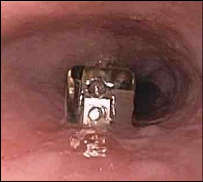
Figure 3 Bravo pH capsule (Givens; Yoqneam, Israel) recording were analyzed instead of 48 hours. The clinical
placed in the distal esophagus, 5 cm proximal to the gastro- utility of the Bravo pH capsule (n ⫽ 309 patients) seems to
esophageal junction. be high because results of the Bravo pH capsule frequently
changed both patient management (64%) and diagnosis
(22%).87 A decision model analysis of a hypothetic man-
aged care organization found that timely use of the Bravo
tory to therapy. Conversely, positive test findings on therapy
pH capsule reduced unnecessary proton pump inhibitor use
do not imply symptom correlation given the lack of out-
and medication costs.88
come studies. Negative findings using either device in a
The wireless Bravo pH monitoring system is a safe,
patient refractory to therapy has a higher value.
readily available, validated alternative to catheter-based pH
monitoring. It is the diagnostic test of choice for patients
UTILITY OF THE BRAVO PH CAPSULE who cannot tolerate traditional pH-catheter placement and
The Bravo pH monitoring system (Givens; Yoqneam, Is- those who require a longer duration of pH monitoring.
rael) was developed to circumvent limitations of transnasal Forty-eight hours of recording increases the diagnostic ac-
pH monitoring by substituting a wireless radiotelemetry pH curacy of identifying patients with acid reflux who are
recording capsule (6 ⫻ 5.5 ⫻ 25 mm) that attaches to the mistakenly classified as normal using only 24 hours of pH
esophageal mucosa.81 The capsule is positioned 6 cm above recording. Limitations include the cost of the capsule, the
Figure 4 Bravo pH capsule recording. The Bravo pH capsule (shown in Figure 3) is positioned 5 cm above the squamocolumnar
junction. The x-axis is time (in hours), and the y-axis denotes pH levels. Upright time is shown as a white background, and supine time
is shown as a dark gray background. Symptoms of heartburn, regurgitation, and chest pain are indicated by vertical lines and were
recorded by the patient so that symptoms can be correlated with acid reflux. Note that most acid reflux events occur in the upright position
in this patient, whereas no acid reflux occur in the supine (dark gray background) position.Lacy et al The Diagnosis of Gastroesophageal Reflux Disease 589
ability to only measure acid reflux, and the rare need for gastroesophageal reflux disease symptoms demonstrated
upper endoscopy to remove the capsule because of severe that short-term treatment with a proton pump inhibitor (1-4
chest pain.85 Considerable controversy exists whether weeks) does not confidently establish the diagnosis of gas-
Bravo pH testing should be performed on or off proton troesophageal reflux disease.100 The sensitivity and speci-
pump inhibitor therapy. ficity of the proton pump inhibitor therapeutic trial were
78% and 54%, respectively. The low specificity may be due
to a therapeutic response by some patients diagnosed with
UTILITY OF EMPIRIC ACID SUPPRESSION IN functional heartburn. Also, a subset of these patients may
THE DIAGNOSIS OF GASTROESOPHAGEAL respond to a proton pump inhibitor therapeutic trial because
REFLUX DISEASE their underlying mechanism for heartburn is hypersensitiv-
Many clinicians currently use the response to a proton pump ity to normal levels of gastroesophageal reflux.36,92
inhibitor therapeutic trial as evidence for the presence or ab- A proton pump inhibitor therapeutic trial is a safe, sim-
sence of gastroesophageal reflux disease. The accuracy of a ple, and noninvasive diagnostic tool for patients thought to
proton pump inhibitor therapeutic trial in diagnosing gastro- have gastroesophageal reflux disease-related noncardiac
esophageal reflux disease is similar to that of 24-hour pH chest pain. It can be a powerful diagnostic tool for clinicians
monitoring.89 A variety of proton pump inhibitor doses have when evaluating and treating patients with different mani-
been studied in patients with symptoms suggestive of gastro- festations of gastroesophageal reflux disease and offers sig-
esophageal reflux disease or noncardiac chest pain.36,63,90-95 In nificant cost-savings when compared with other diagnostic
patients with laryngeal manifestations of gastroesophageal tools.36,90 Patients with persistent symptoms of gastro-
reflux disease, the doses ranged from 40 to 80 mg omepra- esophageal reflux who do not respond to a proton pump
zole daily.96-98 The most commonly used proton pump inhibitor trial should be evaluated for gastroesophageal re-
inhibitor has been omeprazole, which led to the term “ome- flux using pH testing as described.
prazole test.”36,90-93 However, studies using other proton
pump inhibitors have demonstrated that they are equally as
efficacious.94,95 CONCLUSIONS
An important factor in determining the sensitivity of a This review highlights the fact that the diagnosis of gastro-
proton pump inhibitor therapeutic trial is the definition of a esophageal reflux disease remains problematic. The high
positive test. In most studies, a symptom score cutoff was prevalence of this disorder, combined with its significant
used. If the symptom assessment score for heartburn, chest negative economic impact on the health care system, man-
pain, or other symptoms improved by more than 50% to dates that we become better equipped to diagnose gastro-
75% relative to baseline (depending on the study), the test esophageal reflux disease. To begin, a global consensus
was considered positive. Studies rarely calculated the re- must be reached on how to define gastroesophageal reflux
ceiver operator curve.36,90,99 disease. Next, a simple and reliable questionnaire that can
Assessment of the diagnostic accuracy of the proton accurately diagnose gastroesophageal reflux needs to be
pump inhibitor therapeutic trial in patients with symptom- developed. In addition, a large prospective multinational
atic gastroesophageal reflux disease or nonerosive reflux study is needed to evaluate and compare the utility of
disease is limited by the lack of a gold standard for diag- diagnosing gastroesophageal reflux disease with question-
nosing gastroesophageal reflux disease. The proton pump naires, upper endoscopy, impedance-pH probes, and the
inhibitor therapeutic trial has minimal utility in patients with Bravo pH capsule. Finally, all diagnostic studies need to be
erosive esophagitis, whereas its value increases in patients critically evaluated with regard to their clinical utility.
in whom the likelihood of a specific syndrome being attrib-
uted to reflux is low (ie, hoarseness). The proton pump References
inhibitor therapeutic trial has been shown to be fairly sen- 1. Shaheen NJ, Hansen RA, Morgan DR, et al. The burden of gastro-
sitive (68%-92%) and specific (36%-100%) in diagnosing intestinal and liver diseases, 2006. Am J Gastroenterol. 2006;101:
2128-2138.
gastroesophageal reflux disease-related noncardiac chest 2. Locke GR, Talley NJ, Fett SL, et al. Prevalence and clinical spectrum
pain.90,92,93 Two separate meta-analyses concluded that the of gastroesophageal reflux: a population-based study in Olmsted
proton pump inhibitor therapeutic trial reduces chest symp- County, Minnesota. Gastroenterology. 1997;112:1448-1456.
toms and is useful as a diagnostic tool in identifying gas- 3. Locke GR, Talley NJ, Fett SL, et al. Risk factors associated with
symptoms of gastroesophageal reflux. Am J Med. 1999;106:642-649.
troesophageal reflux disease-related noncardiac chest pain
4. Mohammed I, Cherkas LF, Riley SA, et al. Genetic influences in
with an overall sensitivity of 80% (95% confidence interval gastro-oesophageal reflux disease: a twin study. Gut. 2003;52:1085-
[CI], 71%-87%) and a specificity of 74% (95% CI, 1089.
64%-83%).32 5. Fujiwara Y, Higuchi K, Watanabe Y, et al. Prevalence of gastro-
In contrast, the specificity of the proton pump inhibitor esophageal reflux disease and gastroesophageal reflux disease symp-
toms in Japan. J Gastroenterol Hepatol. 2005;20:26-29.
therapeutic trial for patients with reflux symptoms (but
6. Wong WM, Lai KC, Hui WM, et al. Prevalence, clinical spectrum
without chest pain) was found to be relatively low. A meta- and health care utilization of gastro-oesophageal reflux disease in a
analysis of 15 studies that evaluated the value of the proton Chinese population: a population-based study. Aliment Pharmacol
pump inhibitor therapeutic trial in patients with typical Ther. 2003;18:595-604.590 The American Journal of Medicine, Vol 123, No 7, July 2010
7. Ruigomez A, Garcia-Rodriguez LA, Wallander M-A, et al. Natural 29. Garcia-Rodriguez LA, Wallender M, Johansson S, et al. Natural
history of gastro-oesophageal reflux disease diagnosed in general history of chest pain in GERD. Gut. 2005;54(Suppl VII):A75, OP-
practice. Aliment Pharmacol Ther. 2004;20:751-760. G-325.
8. Kotzan J, Wade W, Yu HH. Assessing NSAID prescription use as a 30. El-Serag HB, Sonnenberg A. Comorbid occurrence of laryngeal or
predisposing factor for gastroesophageal reflux disease. Pharm Res. pulmonary disease with esophagitis in US military veterans. Gastro-
2001;18:1367-1372. enterology. 1997;113:755-760.
9. Wong WM, Lai KC, Hui WM, et al. Onset and disappearance of 31. Gislason T, Janson C, Vermiere P, et al. Respiratory symptoms and
reflux symptoms in a Chinese population: a 1-year follow-up study. nocturnal gastro-oesophageal reflux: a population based study of
Aliment Pharmacol Ther. 2004;20:803-812. young adults in three European countries. Chest. 2002;121:158-163.
10. Dent J, Brun J, Fendrick AM, et al. An evidence-based appraisal of 32. Cremonini F, Wise J, Moayyedi P, Talley N. Diagnostic and thera-
reflux disease management-the Genval Workshop Report. Gut. 1999; peutic use of PPIs in non-cardiac chest pain: a meta analysis. Am J
44:S1-S16. Gastroenterol. 2005;100:1226-1232.
11. Armstrong D, Marshall JK, Chiba N, et al. Canadian Consensus 33. Gibson PG, Henry RL, Coughlan JL. Gastro-oesophageal reflux treat-
Conference on the management of gastroesophageal reflux disease in
ment for asthma in adults and children. Cochrane Database Syst Rev.
adults-update 2004. Can J Gastroenterol. 2005;19:15-35.
2003(1):CD001496.
12. DeVault KR, Castell DO. Updated guidelines for the diagnosis and
34. Chang AB, Lasserson TJ, Gaffney J, et al. Gastro-oesophageal reflux
treatment of gastroesophageal reflux disease. Am J Gastroenterol.
treatment for prolonged non-specific cough in children and adults.
2005;100:190-200.
Cochrane Database Syst Rev. 2006(4):CD004823.
13. Vakil N, van Zanten SV, Kahrilas P, et al. The Montreal definition
35. Lacy BE, Cash BD. A 32-year-old woman with chronic abdominal
and classification of gastro-esophageal reflux disease (GERD)-a
pain. JAMA. 2008;299:555-565.
global evidence-based consensus. Am J Gastroenterol. 2006;101:
1900-2000. 36. Fass R, Ofmann JJ, Gralnek IM, et al. Clinical and economic assess-
14. Kahrilas PJ, Shaheen NJ, Vaezi MF, et al. American Gastroenter- ment of the omeprazole test in patients with symptoms suggestive of
ological Association medical position statement on the management gastroesophageal reflux disease. Arch Intern Med. 1999;159:2161-
of gastroesophageal reflux disease. Gastroenterology. 2008;135: 2168.
1383-1391. 37. Greatorex RA, Thorpe JA. Clinical assessment of gastro-oesophageal
15. Dent J, Dodds WJ, Friedman RH, et al. Mechanisms of gastroesoph- reflux by questionnaire. Br J Clin Pract. 1983;37:133-135.
ageal reflux in recumbent asymptomatic human subjects. J Clin 38. Locke GR, Talley NJ, Weaver AL, Zinsmeister AR. A new ques-
Invest. 1980;65:256-267. tionnaire for gastroesophageal reflux disease. Mayo Clin Proc. 1994;
16. Kahrilas PJ. Gastroesophageal reflux disease. New Engl J Med. 2008; 69:539-547.
359:1700-1707. 39. Carlsson R, Dent Y, Bolling-Sternevald E, et al. The usefulness of a
17. Hampel H, Abraham NS, El-Serag HB. Meta-analysis: obesity and structured questionnaire in the assessment of symptomatic gastro-
the risk for gastroesophageal reflux disease and its complications. esophageal reflux disease. Scand J Gastroenterol. 1998;33:1023-
Ann Intern Med. 2005;143:199-211. 1029.
18. Zheng Z, Margolis KL, Liu S, et al. Effects of estrogen with and 40. Shaw M, Talley NJ, Beebe T, et al. Initial validation of a diagnostic
without progestin and obesity on symptomatic gastroesophageal re- questionnaire for gastroesophageal reflux disease. Am J Gastroen-
flux. Gastroenterology. 2008;135:72-81. terol. 2001;96:52-57.
19. Dimenas E, Glise H, Hallerback B, et al. Quality of life in patients 41. Elola-Olaso M, Rey C, Rodriguez-Artalejo E, et al. Adaptation and
with upper gastrointestinal symptoms. An improved evaluation of validation of a gastroesophageal reflux questionnaire for use on a
treatment regimens? Scand J Gastroenterol. 1993;28:681-687. Spanish population. Revista Espanola de Enfermedades Digestivas.
20. Revicki DA, Wood M, Maton PN, Sorensen S. The impact of gas- 2002;94:745-758.
troesophageal reflux disease on health-related quality of life. Am J 42. Wong WM, Lam KF, Lai KC, et al. A validated symptoms question-
Med. 1998;104:252-258. naire (Chinese GERDQ) for the diagnosis of gastro-esophageal reflux
21. Sandler RS, Everhart JE, Donowitz M, et al. The burden of selected disease in the Chinese population. Aliment Pharmacol Ther. 2003;
digestive diseases in the United States. Gastroenterology. 2002;122: 17:1407-1413.
1500-1511. 43. Wang JH, Luo JY, Dong L, et al. Composite score of reflux symp-
22. Levin TR, Schmittdiel JA, Kunz K, et al. Costs of acid-related
toms in diagnosis of gastroesophageal reflux disease. World J Gas-
disorders to a health maintenance organization. Am J Med. 1997;103:
troenterol. 2004;10:3332-3335.
520-528.
44. Chinese Study Group. Value of reflux diagnostic questionnaire in the
23. Lagergren J, Bergstrom R, Lindgren A, Nyren O. Symptomatic gas-
diagnosis of gastroesophageal reflux disease. Chinese J Dig Dis.
troesophageal reflux as a risk factor for esophageal adenocarcinoma.
2004;5:51-55.
New Engl J Med. 1999;340:825-831.
45. Zimmerman J. Validation of a brief inventory for diagnosis and
24. Thomson AB, Barkun AN, Armstrong D, et al. The prevalence of
monitoring of symptomatic gastroesophageal reflux. Scand J Gastro-
clinically significant endoscopic findings in primary care patients
with uninvestigated dyspepsia: The Canadian Adult Dyspepsia Em- enterol. 2004;39:212-216.
piric treatment-prompt endoscopy (CADET-PE) study. Aliment 46. Shimoyama Y, Kusano M, Sugimoto S, et al. Diagnosis of gastro-
Pharmacol Ther. 2003;17:1481-1491. esophageal reflux disease using a new questionnaire. J Gastroenterol
25. Moayyedi P, Talley NJ, Fennerty MB, Vakil N. Can the clinical Hepatol. 2005;20:643-647.
history distinguish between organic and functional dyspepsia? JAMA. 47. Horowitz N, Moshkowitz M, Halpern Z, Leshno M. Applying data
2006;295:1566-1576. mining techniques in the development of a diagnostic questionnaire
26. Numans ME, de Wit NJ. Reflux symptoms in general practice: for GERD. Dig Dis Sci. 2007;52:1871-1878.
diagnostic evaluation of the Carlsson-Dent gastroesophageal reflux 48. Ho KY, Gwee KA, Khor, JL, et al. Validation of a graded response
disease questionnaire. Aliment Pharmacol Ther. 2003;17:1049-1055. questionnaire for the diagnosis of gastroesophageal reflux disease in
27. Klauser AG, Schindlbeck NE, Muller-Lissner SA. Symptoms in an Asian primary care population. J Clin Gastroenterol. 2008;42:
gastro-oesophageal reflux disease. Lancet. 1990;335:205-208. 680-686.
28. Moayyedi P, Axon ATR. The usefulness of the likelihood ratio in the 49. Kinekawa F, Kubo F, Matsuda K, et al. Is the questionnaire for the
diagnosis of dyspepsia and gastro-oesphageal reflux disease. Am J assessment of gastroesophageal reflux useful for diabetic patients?
Gastroenterol. 1999;94:3122-3125. Scand J Gastroenterol. 2005;40:1017-1020.Lacy et al The Diagnosis of Gastroesophageal Reflux Disease 591
50. Hung CS, Lee CL, Yang JN, et al. Clinical application of Carlsson’s 73. Bianchi Porro G, Pace F. Comparison of three methods of intraesoph-
questionnaire to predict erosive GERD among healthy Chinese. J ageal pH recordings in the diagnosis of gastroesophageal reflux.
Gastro Hepatol. 2005;20:19001095. Scand J Gastroenterol. 1988;23:743-750.
51. Creteur V, Thoeni RF, Federle MP, et al. The role of single and 74. Hong SK, Vaezi MF Gastroesophageal reflux monitoring: pH (cath-
double-contrast radiography in the diagnosis of reflux esophagitis. eter & capsule) and impedance. Gastrointest Endoscopy Clin N Am.
Radiology. 1983;147:71-75. 2009;19:1-22.
52. Dibble C, Levine MS, Rubesin SE, et al. Detection of reflux esoph- 75. Bredenoord AJ, Weusten BLAM, Timmer R, Smouth AJPM. Repro-
agitis on double-contrast esophagrams and endoscopy using the his- ducibility of multichannel intraluminal electrical impedance monitor-
tologic findings as the gold standard. Abdom Imaging. 2004;29:421- ing of gastroesophageal reflux. Am J Gastroenterol. 2005;100:265-
425. 269.
53. Ott DJ. Gastroesophageal reflux: what is the role of barium studies? 76. Hirano I, Richter JE. ACG practice guidelines: esophageal reflux
AJR Am J Roentgenol. 1994;162:627-629. testing. Am J Gastroenterol. 2007;102:668-685.
54. Thompson JK, Koehler RE, Richter JE. Detection of gastroesopha- 77. Kahrilas PJ, Quigley EMM. American Gastroenterological Associa-
geal reflux: value of barium studies compared with 24-hr pH moni- tion medical position statement: guidelines on the use of esophageal
toring. Am J Roentgenol. 1994;162:621-626. pH recording. Gastroenterology. 1996;110:1981-1996.
55. Pan JJ, Levine MS, Redfern RO, et al. Gastroesophageal reflux: 78. Sifrim D, Castell D, Dent J, Kahrilas PJ. Gastroesophageal reflux
comparison of barium studies with 24-h pH monitoring. Eur J Radiol. monitoring: review and consensus report on detection and definitions
2003;47:149-153. of acid, non-acid and gas reflux. Gut. 2004;53:1024-1031.
56. Chen MY, Ott DJ, Sinclair JW, et al. Gastroesophageal reflux dis- 79. Hemmink GJM, Bredenoord AJ, Weusten BLAM, et al. Esophageal
ease: correlation of esophageal pH testing and radiographic findings. pH-impedance monitoring in patients with therapy-resistant reflux
Radiology. 1992;185:483-486. symptoms: “on” or “off” proton pump inhibitor? Am J Gastroenterol.
57. Levine MS, Chu P, Furth EE, et al. Carcinoma of the esophagus and 2008;103:2446-2453.
esophagogastric junction: sensitivity of radiographic diagnosis. Am J 80. Pritchett JM, Aslam M, Slaughter JC, et al. Efficacy of esophageal
Roentgenol. 1997;168:1423-1426. impedance/pH monitoring in patients with refractory gastroesopha-
58. Levine MS, Rubesin SE, Laufer I. Barium esophagography: a study geal reflux disease, on and off therapy. Clin Gastroenterol Hepatol.
for all seasons. Clin Gastroenterol Hepatol. 2008;6:11-25. 2009;7:743-748.
59. Baker ME, Einstein DM, Herts BR, et al. Gastroesophageal reflux 81. Schneider JH, Kramer KM, Konigsrainer A, Granderath FA. Ambu-
disease: integrating the barium esophagram before and after antire- latory pH: monitoring with a wireless system. Surg Endosc. 2007;
flux surgery. Radiology. 2007;243:329-339. 21:2076-2080.
60. Lind T, Havelund T, Carlsson R, et al. Heartburn without esophagitis: 82. Lacy BE, O’Shana T, Hynes M, et al. Safety and tolerability of
efficacy of omeprazole therapy and features determining therapeutic transoral Bravo capsule placement after transnasal manometry using
response. Scand J Gastroenterol. 1997;32:974-979. a validated conversion factor. Am J Gastroenterol. 2007;102:24-32.
61. Mantynen T, Farkkila M, Kunnamo I, et al. The impact of upper GI 83. Pandolfino JE, Schreiner MA, Lee TJ, et al. Comparison of the Bravo
endoscopy referral volume on the diagnosis of gastroesophageal wireless and Digitrapper catheter-based pH monitoring systems for
reflux disease and its complications: a 1-year cross-sectional study in measuring esophageal acid exposure. Am J Gastroenterol. 2005;100:
a referral area with 260,000 inhabitants. Am J Gastroenterol. 2002; 1466-1476.
97:2524-2529. 84. Wong WM, Bautista J, Dekel R, et al. Feasibility and tolerability of
62. Richter JE. Severe reflux esophagitis. Gastrointest Endosc Clin N transnasal/per-oral placement of the wireless pH capsule vs. tradi-
Am. 1994;4:677-697. tional 24-h oesophageal pH monitoring—a randomized trial. Aliment
63. Giannini EG, Zentilin P, Dulbecco P, et al. Management strategy for Pharmacol Ther. 2005;21:155-163.
patients with gastroesophageal reflux disease: a comparison between 85. Pandolfino JE, Richter JE, Ours T, et al. Ambulatory esophageal pH
empirical treatment with esomeprazole and endoscopy-oriented treat- monitoring using a wireless system. Am J Gastroenterol. 2003;98:740-
ment. Am J Gastroenterol. 2008;103:267-275. 749.
64. Lundell LR, Dent J, Bennett JR, et al. Endoscopic assessment of 86. Scarpulla G, Camilleri S, Galante P, et al. The impact of prolonged pH
oesophagitis: clinical and functional correlates and further validation measurements on the diagnosis of gastroesophageal reflux disease: 4-day
of the Los Angeles classification. Gut. 1999;45:172-180. wireless pH studies. Am J Gastroenterol. 2007;102:2642-2647.
65. Vigneri S, Termini R, Leandro G, et al. A comparison of five 87. Lacy BE, Dukowicz AC, Paquette L, et al. Clinical utility of the
maintenance therapies for reflux esophagitis. N Engl J Med. 1995; Bravo capsule. Am J Gastroenterol. 2007;102:S144-S145.
333:1106-1110. 88. Lee WC, Yeh YC, Lacy BE, et al. Timely confirmation of gastro-
66. Sharma P, Wani S, Bansai A, et al. A feasibility trial of narrow band esophageal reflux disease via pH monitoring: estimating budget im-
imaging endoscopy in patients with gastroesophageal reflux disease. pact on managed care organizations. Curr Med Res Opin. 2008;24:
Gastroenterology. 2007;133:454-464. 1317-1327.
67. Amano Y, Yamashita H, Koshino K, et al. Does magnifying endos- 89. Fass R, Ofman JJ, Sampliner RE, et al. The omeprazole test is as
copy improve the diagnosis of erosive esophagitis? J Gastroenterol sensitive as 24-h oesophageal pH monitoring in diagnostic gastro-
Hepatol. 2008;23:1063-1068. oesophageal reflux disease in symptomatic patients with erosive oe-
68. Pandolfino JE, Kahrilas PJ. AGA technical review: clinical use of sophagitis. Aliment Pharmacol Ther. 2000;14:389-396.
esophageal manometry. Gastroenterology. 2005;128:209-224. 90. Fass R, Fennerty MB, Ofman JJ, et al. The clinical and economic
69. Lacy BE, Paquette L, Robertson DJ, et al. The clinical utility of value of a short course of omeprazole in patients with noncardiac
esophageal manometry. J Clin Gastroenterol. 2009 March 17 [Epub chest pain. Gastroenterology. 1998;115:42-49.
ahead of print]. 91. Bate CM, Riley SA, Chapman RWG, et al. Evaluation of omeprazole
70. Kahrilas PJ, Sifrim D. High-resolution manometry and impedance- as a cost-effective diagnostic test for gastro-oesophageal reflux dis-
pH/manometry: valuable tools in clinical and investigational esoph- ease. Aliment Pharmacol Ther. 1999;13:59-66.
agology. Gastroenterology. 2008;135:756-769. 92. Juul-Hansen P, Rydning A, Jacobsen CD, Hansen T. High-dose
71. Spencer J. Prolonged pH recording in the study of gastro-oesophageal proton-pump inhibitors as a diagnostic test of gastro-esophageal re-
reflux. Br J Surg. 1969;56:912-914. flux disease in endoscopic-negative patients. Scand J Gastroenterol.
72. Johnson LF, DeMeester TR. Twenty-four hour pH monitoring of the 2001;36:806-810.
distal esophagus. A quantitative measure of gastroesophageal reflux. 93. Pandak WM, Arezo S, Everett S, et al. Short course of omeprazole:
Am J Gastroenterol. 1974;62:325-332. a better first diagnostic approach to noncardiac chest pain than en-592 The American Journal of Medicine, Vol 123, No 7, July 2010
doscopy, manometry, or 24-hour esophageal pH monitoring. J Clin 97. Ours T, Kavuru MS, Schilz RJ, Richter JE. A prospective evaluation
Gastroenterol. 2002;35:307-314. of esophageal testing and a double-blind, randomized study of ome-
94. Bautista J, Fullerton H, Briseno M, et al. The effect of an empirical prazole in a diagnostic and therapeutic algorithm for chronic cough.
trial of high-dose lansoprazole on symptom response of patients with Am J Gastroenterol. 1999;94:3131-3138.
non-cardiac chest pain—a randomized, double-blind, placebo-con- 98. Kiljander TO, Salomaa ERM, Heitanen EK, Terho EO. Chronic
trolled, crossover trial. Aliment Pharmacol Ther. 2004;19:1123-1130. cough and gastro-oesophageal reflux: a double-blind placebo-con-
95. Dickman R, Emmons S, Cui H, et al. The effect of a therapeutic trial trolled study with omeprazole. Eur Respir J. 2000;16:633-638.
of high-dose rabeprazole on symptom response of patients with 99. Fass R. Empirical trials in treatment of gastroesophageal reflux dis-
non-cardiac chest pain: a randomized, double-blind, placebo-con- ease. Dig Dis. 2000;18:20-26.
trolled, crossover trial. Aliment Pharmacol Ther. 2005;22:547-555. 100. Numans ME, Lau J, de Wit NJ, Bonis PA. Short-term treatment with
96. Metz D, Childs ML, Ruiz C, Weinstein GS. Pilot study of the oral proton-pump inhibitors as a test for gastroesophageal reflux disease:
omeprazole test of reflux laryngitis. Otolaryngol Head Neck Surg. a meta-analysis of diagnostic test characteristics. Ann Intern Med.
1997;116:41-46. 2004;140:518-527.You can also read