Mycoplasma agassizii disease risk assessment for Australia - Zoo and Aquarium Association
←
→
Page content transcription
If your browser does not render page correctly, please read the page content below
Mycoplasma agassizii disease
risk assessment for Australia
Version 2.0 May 2020
Dr Andrea Reiss
BVSc (Hons), MVS (Zoo and wildlife medicine), MANZCVS (Medicine of Australasian wildlife)
Regional Veterinary Officer, Zoo and Aquarium Association Australasia,
andrea@zooaquarium.org.au
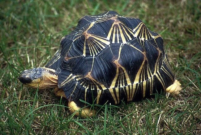
Image 1: Radiated tortoise (Astrochelys radiata) [credit Vladimír Motyčka]Contents
1. Executive summary ......................................................................................................................... 3
2. Purpose of this document ............................................................................................................... 4
Risk questions addressed: ............................................................................................................... 4
3. Background and context ................................................................................................................. 4
Scope and limitations of this risk assessment .................................................................................... 5
4. Introduction .................................................................................................................................... 6
Taxonomy, terminology, biology and ecology of chelonians ............................................................. 6
Chelonians housed in Australian zoos ................................................................................................ 6
A note on upper respiratory tract disease in chelonians .................................................................... 7
5. Epidemiology of M. agassizii........................................................................................................... 7
Aetiologic agent .................................................................................................................................. 7
Agent properties ................................................................................................................................. 8
Transmission (disease dynamics) ........................................................................................................ 8
Host response ..................................................................................................................................... 9
Clinical signs and pathology .............................................................................................................. 10
Diagnosis ........................................................................................................................................... 10
Treatment ......................................................................................................................................... 10
Prevention and control ..................................................................................................................... 11
Host species ...................................................................................................................................... 11
Significance of infection .................................................................................................................... 13
Reports of M. agassizii outside family Testudinidae ........................................................................ 14
Global distribution ............................................................................................................................ 15
Occurrences and testing in Australia ................................................................................................ 15
6. Risk summary ................................................................................................................................ 16
7. Risk assessment ............................................................................................................................ 17
8. Risk management measures ......................................................................................................... 20
9. Conclusions ................................................................................................................................... 20
10. References ................................................................................................................................ 21
Appendix 1: Taxonomy of chelonids mentioned in this document .................................................. 25
Appendix 2: Testudinidae in Zoo and Aquarium Association member zoos (Jan 2019) .................. 26
Appendix 3: Likelihood and consequence categories and definitions .............................................. 27
Appendix 4: Risk assessment matrix – overall risk ........................................................................... 28
Mycoplasma agassizii risk assessment for Australia – Zoo and Aquarium Association v 2.0 May 2020
21. Executive summary
Mycoplasma agassizii is a known pathogen of land tortoises (family Testudinidae), in particular the
host genus Gopherus (gopher and desert tortoises, native to southern USA). The pathogen causes
upper respiratory tract (URT) disease in susceptible hosts, with persistent infection resistant to
antimicrobial treatment. There is extensive published literature on M. agassizii, and it is perhaps the
best studied pathogen of Testudinidae.
Mycoplasma agassizii is host specific, requires close contact for transmission and does not survive
for long outside the host. A number of exotic Testudinidae, housed in Australian zoos, are known to
be positive for M. agassizii by PCR test.
An extensive literature review found reports of infection with M. agassizii (with possible associated
URT disease) in several species of Testudinidae. There are two unpublished reports of infection in
the closely related family Emydidae in the USA. Other than these two reports, there are no reports
and no evidence of infection with M. agassizii or M. testudineum occurring outside the Testudinidae.
There is no evidence that chelonians other than Testudinidae (and possibly Emydidae) are
susceptible to infection with M. agassizii. Neither of the families Testudinidae and Emydidae occur
naturally in Australia and Australian native chelonians are distantly related to these taxa.
This risk assessment determined that there is a negligible risk that M. agassizii could be a threat to
native chelonian species in Australia. No risk mitigation is considered necessary.
The risk to non-chelonians, including other native species, humans and domestic animals, is
considered negligible. No risk mitigation is considered necessary.
This risk assessment determined that there is a potential risk of transfer of M. agassizii infection to
currently uninfected Testudinidae within Australian zoos, with a risk of moderate consequences to
the individual. Australian zoos may wish to adopt internal risk assessment and risk management
measures to mitigate this potential risk.
In Australia, only licensed zoos are permitted to keep exotic reptiles. Notwithstanding, some exotic
reptiles, likely including Testudinidae, are held illegally in Australia. Risk to (or from) exotic
Testudinidae present in Australia, but held outside the zoo compartment, was not assessed during
this process.
Increased testing, including sequential testing, and/ or inclusion of serological tests, may better
inform the status of current infection and exposure in Testudinidae in Australia.
As a general recommendation, active disease surveillance, along with opportunistic and targeted
collection of baseline health and disease data, is encouraged for all Australian free-ranging wildlife
populations. This, along with full investigation of morbidity and mortality events in wildlife, will help
to better understanding and preparedness for response to all and any emerging diseases.
Mycoplasma agassizii risk assessment for Australia – Zoo and Aquarium Association v 2.0 May 2020
32. Purpose of this document
This document is a desktop disease risk assessment to assess the potential risk of the bacterial
pathogen Mycoplasma agassizii (or the closely related M. testudineum) to Australia.
Risk questions addressed:
1) a) What is the likelihood of native Australian fauna, domestic animals or humans outside the zoo
compartment in Australia being exposed to M. agassizii (or the closely related M. testudineum)
with the subsequent establishment and spread of the pathogen throughout Australia?
b) What is the likely consequence of a species of native Australian fauna or domestic animal (or
human), outside the zoo compartment in Australia, being infected by M. agassizii (or the closely
related M. testudineum), following exposure to the pathogen?
2) a) What is the likelihood of exotic or native fauna (or zoo workers), present within the zoo
compartment in Australia, being exposed to M. agassizii (or the closely related M. testudineum)
with the subsequent establishment and spread of the pathogen?
b) What is the likely consequence of exotic or native fauna (or zoo workers), present within the
zoo compartment in Australia, being infected by M. agassizii (or the closely related M.
testudineum) following exposure to the pathogen?
3. Background and context
Mycoplasma agassizii is a bacterial pathogen of land tortoises in the family Testudinidae.
Mycoplasma testudineum is a closely related species of bacterium, which has been associated with
disease in only two species of land tortoises (Gopherus spp.).
Testudinidae are a family of land tortoises (see front page image). Testudinidae are not native to
Australia. Small numbers of individual Testudinidae, of a number of different species, reside in
several Australian zoos, in almost all states and territories of Australia. Many of these individuals
have been present within Australian zoos for years, or even decades. Only licenced exhibitors (zoos)
are permitted to keep exotic reptiles in Australia. Notwithstanding, some exotic reptiles, likely
including Testudinidae, are held illegally in Australia, by private entities.
A PCR test was recently developed by Murdoch University for this pathogen. Subsequent testing
revealed M. agassizii infection in several individual Testudinidae resident in zoos in Australia (see
Occurrence in Australia).
Very limited testing for Mycoplasma spp. has been undertaken in native chelonians (free-living or
captive) in Australia. The susceptibility of Australian native chelonians to M. agassizii (or M.
testudineum) has not been previously assessed.
Radiated tortoises (Astrochelys radiata) fall in the family Testudinidae. A group of radiated tortoises
was imported into Australia in 2018 and underwent post-arrival quarantine in approved
Mycoplasma agassizii risk assessment for Australia – Zoo and Aquarium Association v 2.0 May 2020
4arrangements in a registered zoo in Qld. During this time, several of the tortoises underwent PCR
testing for M. agassizii, with positive results.
Animal Health Committee (AHC; www.agriculture.gov.au/animal/health/committees/ahc)
determined that a risk assessment should be undertaken to investigate and document the potential
disease risks associated with the presence, and possible establishment or spread of M. agassizii in
Australia. They requested the Zoo and Aquarium Association (the Association; ZAA;
www.zooaquarium.org.au/) to lead this project.
Dr Andrea Reiss, Regional Veterinary Officer with the ZAA, has extensive experience undertaking
biosecurity import risk assessments (www.agriculture.gov.au/biosecurity/risk-analysis) associated
with importation of zoo animals, and wildlife disease risk assessments associated with wildlife
translocations and management (Jakob-Hoff et al. 2014). Wildlife disease risk experts at Wildlife
Health Australia (www.wildlifehealthaustralia.com.au/) reviewed and provided comment on a draft
of this document. The document was also provided to Animal Biosecurity at the Commonwealth
Department of Agriculture and Water Resources (now Department of Agriculture, Water and the
Environment) for review and comment.
Scope and limitations of this risk assessment
1. Scope is outlined in Section 2 “Risk questions addressed”.
2. No entry risk assessment has been undertaken as M. agassizii is already present in Australia
(in zoo chelonians).
3. Disease risk has only been assessed for M. agassizii. (Where relevant, information is
provided on the closely related M. testudineum). Disease risk associated with other
Mycoplasma species has not been assessed.
4. The information on captive chelonians in Australia is drawn from data held by the Zoo and
Aquarium Association and is specific to ZAA-member zoos.
5. No data is available from non-ZAA zoos in Australia. There is no method by which the Zoo
and Aquarium Association can access information on non-member zoos, either on their
collections or their practices and there is no established avenue to collect data from non-
ZAA member zoos in Australia. State and territory licensing authorities maintain information
on licensed premises within their jurisdictions, but they do not provide this information to
ZAA.
6. No data is available on captive chelonians held (illegally) outside zoos in Australia; there is no
method by which the Zoo and Aquarium Association can access this information.
7. Disease risk to (or from) exotic species of chelonians illegally present in Australia (i.e. outside
of licensed zoos) is unknown and has not been assessed.
8. It is assumed that all licensed zoo operations are compliant with existing biosecurity and
containment regulations (which differ between jurisdictions).
9. It is assumed that existing containment regulations for exotic chelonians within Australian
zoos (i.e. that only licensed zoos are permitted to hold exotic chelonians) will not be relaxed
in the future.
Mycoplasma agassizii risk assessment for Australia – Zoo and Aquarium Association v 2.0 May 2020
54. Introduction
Taxonomy, terminology, biology and ecology of chelonians
(See also Appendix 1).
Chelonian is the term used to refer to all turtles and tortoises. The terms turtle and tortoise are
generally interchangeable when discussing Australian species. The term terrapin is used outside
Australia to refer to fresh-water species of Chelonian. All living turtles/ tortoises/ terrapins are in the
class Reptilia, order Testudines. There are two suborders of chelonians: Cryptodira (‘hidden-necked
turtles’) and Pleurodia (‘side-necked’ turtles), characterised by how they withdraw their neck into
their shell.
The Cryptodira suborder has three living superfamilies, the Chelonioidea (sea turtles), Testudinoidea
(land tortoises and pond turtles) and Trionychoidea (soft-shell turtles and relatives).
Land tortoises (or “true” tortoises) all fall in the family Testudinidae, suborder Cryptodira,
having a typical appearance of domed carapace and elephantine hind limbs (see front page
image). There are no Testudinidae native to Australia. The nearest endemic land tortoises
are those native to south-east Asia.
Emydidae is the most closely related chelonian family to Testudinidae (in the same
superfamily). This family comprises terrapins and pond turtles. Emydidae are native to the
western hemisphere and there are no Emydidae native to Australia.
All marine turtles fall into the suborder Cryptodira, within the families Chelonidae and
Dermochelyidae.
Australia’s native chelonians are 23 species of freshwater turtle and six species of marine turtle. All
but one of Australia’s native freshwater species belong to the family Chelidae in the suborder
Pleurodia; this family is found only in Australasia and South America (see
www.environment.nsw.gov.au/topics/animals-and-plants/native-animals/native-animal-
facts/freshwater-turtles). There are no Testudinidae or Emydidae native to Australia.
The only Australian freshwater turtle species in the suborder Cryptodira is Carettochelys insculpta
(pig-nosed or pit-shelled turtle), which is the monotypic member of the family Carettochelydidae
(see http://reptilesofaustralia.com/turtles/turtles.htm#.W44U5egzaM8).
Summary: Australian native chelonians are taxonomically very distantly related to land tortoises
(Testudinidae).
Chelonians housed in Australian zoos
In Australia, only licensed zoos are permitted to keep exotic reptiles. A large number of native and
exotic chelonian species are housed in Australian zoos (29 different genera [10 native; 19 exotic] in
ZAA zoos, 15 Jan 2019); this includes a number of exotic land tortoise species (family Testudinidae).
The Zoo and Aquarium Association maintains studbook information on all species of chelonians held
in ZAA-member zoos in Australia. ZAA-member zoos hold 51 individual land tortoises, across 16
species (data compiled 15 Jan 2019; see Appendix 2). Land tortoises are held in ZAA zoos in every
Mycoplasma agassizii risk assessment for Australia – Zoo and Aquarium Association v 2.0 May 2020
6state/ territory other than Tasmania. All land tortoises in ZAA-member zoos have either been
imported from overseas or are the descendants of imported individuals.
There is no reliable information available on the number or species of exotic land tortoises housed in
Australian zoos outside ZAA membership (see “Scope and limitations of this risk assessment, dot
point 5”). Enquiries made by ZAA in late 2019 via the reptile-keeping “community” across Australia
did not reveal, to the best of our collective knowledge, the presence of tortoises of the family
Testudinidae held in Australian zoos outside ZAA membership.
Although only licensed zoos are permitted to keep exotic reptiles in Australia, it is widely accepted
that a number of exotic reptiles are held (illegally) and without permit, by private individuals in
Australia. The generally accepted knowledge is that the majority of these illegally held exotic reptiles
are either snakes or lizards. Reptiles in Australian zoos are all obtained through approved permit and
there is no unregulated passage of illegally held reptiles in Australia into the zoo compartment.
In summary it is likely that there may be land tortoises resident in Australia, outside ZAA-member
zoos, however no data on this has been made available (see “Scope and limitations of this risk
assessment; dot point 6”).
A note on upper respiratory tract disease in chelonians
Upper respiratory tract (URT) disease is a commonly reported syndrome in land tortoises
(Testudinidae) in the northern hemisphere. There is a proven association (causation) between
infection with M. agassizii (and/or M. testudineum) and URT disease in North American desert
tortoises (also called Agassiz’s desert tortoise; Gopherus agassizii - see detailed information below)
and other hosts of the genus Gopherus. Infection with M. agassizii has not been shown to cause URT
disease in land tortoise species outside the genus Gopherus.
There are also many reports of URT disease in other species of land tortoises, where there is no
accompanying evidence of Mycoplasma spp. infection (e.g. Blahak et al. 2004). Due to biology,
physiology and environmental conditions, it appears that land tortoises often exhibit URT disease.
Across the spectrum of all cases, it is likely there are multiple contributory and causal factors.
Pasteurella testudines, iridovirus, herpesvirus, Chlamydia sp., several different fungi, nutritional
deficiencies, and environmental irritants and allergens have all be reported as factors in URT disease
in chelonians (Berry et al. 2002; Blahak et al. 2004; Boyer and Boyer 2006; Wendland et al. 2006).
Mycoplasma agassizii is has been shown to be associated with (but not necessarily the cause of)
some, but not all, cases of URT disease in Testudinidae. Outside the Gopherus host species where
these pathogens have fulfilled infection trial requirements, there is little evidence that M. agassizii
(or M. testudineum) consistently results in URTD in other Testudinidae hosts.
5. Epidemiology of M. agassizii
Aetiologic agent
Mycoplasma spp. are members of the class Mollicutes and the order Mycoplasmatales. They are the
smallest free-living organisms. They have no cell wall but are bounded by a membrane (Stalheim
1990). Mycoplasmas are obligate parasites that only occur within a strict host association due to
their requirement for many nutrients (Citti and Blanchard 2013).
Mycoplasma agassizii risk assessment for Australia – Zoo and Aquarium Association v 2.0 May 2020
7Mycoplasma microorganisms are found in a wide range of living hosts. Many species of Mycoplasma
are considered non-pathogenic and many have a commensal role in the mucosal surfaces of the host
(Frey 2002). This is considered to be the case for many of the Mycoplasma species infecting
chelonians (Berry et al. 2002).
Pathogenic Mycoplasma spp. have a predilection for mucosal surfaces where they localize and
persist, protected by the fibrinous tissue reactions that characterize mycoplasmosis [disease due to
mycoplasma infection] (Stalheim 1990). Pathogenic mycoplasma infections are often chronic in
nature, with high morbidity and low mortality in the host. Infected hosts that do not display clinical
signs (i.e. asymptomatic carriers) may be important in transmission of the pathogen. Pathogenic
Mycoplasma spp. generally have a narrow host range and are adapted to a specific host species (one
or sometimes several species) in which clinical disease is seen (Citti and Blanchard 2013).
Occasionally, secondary or “atypical” host species are colonised by a species of Mycoplasma. In the
secondary host, signs of disease are rare, and if seen, are generally very mild (Frey 2002).
Virulence factors may result in differing levels of expression of disease from a Mycoplasma sp. within
a given host species; this is believed to be the case for M. agassizii (McLaughlin 1997; Wendland et
al. 2006; Citti and Blanchard 2013).
Mycoplasma agassizii (and M. testudineum) are known bacterial pathogens of the Gopherus genus
of land tortoises (desert or ‘’gopher” tortoises, native to southern USA). In these host species,
infection has been shown to cause upper respiratory tract disease (“tortoise mycoplasmosis”)
(Jacobson et al. 2014). The pathogens were first described by Brown et al. (1994) and Brown et al.
(1995) when disease was seen in free-ranging desert tortoises (Gopherus agassizii and G. morafkai)
in the United States. Mycoplasma agassizii has also been found in a wider range of land tortoise
species (family Testudinidae), including species native to Europe. Its role as a potential pathogen in
these species is far less clear (see further information below).
Mycoplasma agassizii is perhaps the best studied pathogen of Testudinidae and published literature
is extensive. The pathogens, associated disease, investigation and epidemiology have recently been
reviewed by Jacobson et al. (2014).
Agent properties
Mycoplasma agassizii (and M. testudineum) is a fastidious organism that grows slowly (2–6 weeks)
at 30 °C in SP4 broth or agar (Brown et al. 1995; Brown et al. 2004; Citti and Blanchard 2013).
Mycoplasma sp. lack a cell wall and are highly susceptible to desiccation in natural environments
(Wendland et al. 2006). The organisms do not survive well outside the host.
Transmission (disease dynamics)
Mycoplasma agassizii is transmitted horizontally through respiratory secretions. Transmission
pathways in the primary hosts (Gopherus spp.) have been well established by infection studies in
desert tortoises and gopher tortoises (see Brown et al. 1994; Brown et al. 1995; McLaughlin 1997;
Brown et al. 1999a; Wendland 2007). Transmission is via direct contact, notably when nasal
discharge is present (Wendland and Brown 2019). Although transmission is considered more likely to
occur when an individual has clinical disease, it has been suggested that, under appropriate
Mycoplasma agassizii risk assessment for Australia – Zoo and Aquarium Association v 2.0 May 2020
8conditions, subclinically infected individuals may be able to transmit Mycoplasma spp. (Jacobson et
al. 2014).
Close and prolonged contact between individuals, such as those that occur during courtship, mating,
and agonistic behaviours, are considered requirements for transmission of M. agassizii and M.
testudineum, in the primary hosts (Wendland et al. 2010).
Aerosol transmission is considered to be unlikely (Berry et al. 2002). A study in gopher tortoises
showed that control animals, housed in pens adjacent to clinically affected tortoises, did not become
clinically diseased nor did they seroconvert, suggesting that M. agassizii did not travel even relatively
short distances over low (0.7 m) barriers (McLaughlin 1997).
Environmental transmission is considered very unlikely because of the susceptibility of Mycoplasma
spp. to desiccation in natural environments (Berry et al. 2002). It is considered possible that in
captive conditions, factors such as increased host density and higher loads of organic material could
facilitate the persistence of the organism outside the host (Wendland et al. 2006). These risk factors
can also be managed by appropriate stocking densities, hygiene regimes and by management of
environmental conditions (such as temperature, humidity and pH) in the animals’ enclosure. Factors
which increase physiological stress (such as crowding, inappropriate temperature, poor nutrition and
concurrent infections) may exacerbate expression of clinical disease in infected, captive tortoises
(Wendland and Brown 2019). There is no evidence for vertical transmission and it is considered to be
an unlikely mode of transmission (Jacobson et al. 2014).
Host response
A range of responses to Mycoplasma infection are known to occur in susceptible host species, from
absence of disease to severe disease. In general, host immunological response plays a key role in the
development of disease and expression of clinical signs across a wide range of taxa.
In Testudinidae, it is believed that differing strains of M. agassizii have differing pathogenicity,
however there is currently no method available to assess the virulence of strains (Weitzman et al.
2017). It appears that that not all individuals from susceptible host species respond to M. agassizii
infection with a severe inflammatory response, either because of differing strains of M. agassizii
with variable pathogenicity or because of differing immune responses related to different host
genotypes (McLaughlin 1997; Jacobson et al. 2014).
Development of an antibody response to Mycoplasma infection in experimental studies generally
occurred 8 weeks post-infection (Brown et al. 2002). Recent studies indicate that an antibody
response may not occur for more than 400 days post-exposure in susceptible species (Drake et al.
2019).
Infection with M. agassizii or M. testudineum in susceptible species (i.e. Testudinidae), once present,
appears to be persistent (Jacobson et al. 2014; Wendland and Brown 2019).
Mycoplasma agassizii risk assessment for Australia – Zoo and Aquarium Association v 2.0 May 2020
9Clinical signs and pathology
In susceptible species (Testudinidae, Gopherus spp.), infection can cause upper respiratory tract
(URT) disease: nasal discharge, chronic rhinitis, conjunctivitis, ocular discharge and palpebral
oedema (Wendland et al. 2006; Jacobson et al. 2014).
Infection may be present in susceptible species without clinical signs. Signs, when present, may
occur intermittently, over periods of months or years (Wendland and Brown 2019). Subclinically
infected animals may shed the organism and may show changes at a histopathological level
(Wendland and Brown 2019).
Pathology primarily occurs within the nasal cavity, although pneumonia has occasionally been
reported. Lesions in the nasal cavity may be focal or diffuse and range from mild to severe
inflammatory changes, which include mucosal and submucosal hyperplasia with heterophilic and
histiocytic infiltrates (Jacobson et al. 2014).
Infection with M. testudineum in susceptible species appears to cause similar but less severe, and
more focal, pathology than M. agassizii (Jacobson and Berry 2012; Jacobson et al. 2014).
Diagnosis
Evidence of infection includes consistent clinical signs in known or suspected susceptible individuals
of susceptible species. Typical histopathological changes are seen in samples collected during post
mortem examination (Jacobson et al. 2014).
Two laboratory tests have been developed in an aid to diagnosis of mycoplasmosis in live chelonians.
ELISAs have been developed in the USA to detect antibodies against M. agassizii and M. testudineum
in plasma and serum of Gopherus spp. tortoises (Brown et al. 1999a; Brown et al. 1999b; duPre et al.
2011). The ELISA test is not available in Australia and will not be discussed further in this risk
assessment.
Conventional and qPCR (specific for M. agassizii) have been developed in the USA and are now
available in Australia (Brown et al. 1995; Brown et al. 2004; duPre et al. 2011). Samples for PCR
include swabs of choana, nasal discharge or nasal flushes. The PCR test is considered highly specific.
Sensitivity can vary as there is a risk of false negative results if the individual is not shedding or if
sampling is inadequate (Jacobson et al. 2014).
Culture of the organism is difficult due to the fastidious nature of Mycoplasma spp. and can take up
to 6 weeks for primary isolation. Nasal lavage is the most commonly used sample (Jacobson et al.
2014).
Treatment
Successful treatment of tortoises with mycoplasmosis is problematic, due to the characteristic agent
properties and involvement of the host immune response in the expression of clinical disease. A
range of systemic antibiotics suitable for Mycoplasma therapy have been used, along with
supportive care and housing in the upper ranges of the individual’s preferred body temperature
range. There are no reports of confirmed long term success of antimicrobial treatment for
mycoplasmosis in tortoises. Complete eradication of infection is considered unlikely in infected
individuals, and relapse of shedding or clinical signs is often reported, some period after treatment
ceases (even with prolonged courses of antibiotics) (Cowan 2018). At best, antimicrobial therapy
may suppress clinical expression of disease and may suppress shedding of the organism by the host.
Mycoplasma agassizii risk assessment for Australia – Zoo and Aquarium Association v 2.0 May 2020
10Once infection is confirmed in an individual, it should be considered to be persistently infected and a
potential ongoing source of infection.
Prevention and control
Mycoplasma spp. do not survive for long outside the host. Standard infection control practices,
including appropriate hygiene and disinfection, should be followed in all cases and in particular if
animals are known or suspected to be infected.
Individual animals may be tested to determine their exposure status. Repeat PCR testing is advisable
as shedding may be intermittent over weeks, months or years (Jacobson et al. 2014).
In susceptible species (Gopherus spp.), it is advisable to house known-infected animals separately
from known-naive individuals in captivity, and to practice excellent hygiene when working with and
between the two groups.
Captive animals should be maintained with excellent husbandry to reduce any physiological stress
that may contribute either to acquisition of infection, or expression of clinical signs (which is also
likely to increase their infectivity). Overcrowding, poor nutrition, inappropriate ambient conditions
and high organic loads should be avoided in a captive environment (Wendland and Brown 2019).
Host species
There are numerous published reports of infection with Mycoplasma spp., or unspecified
“mycoplasmosis” in chelonians. In many of these, the species of Mycoplasma has not been
identified, or is not reported. Reports of infection include those associated with clinical disease and
those where clinical disease is not present or is not reported. This risk assessment focuses on
confirmed reports of M. agassizii in chelonians.
Mycoplasma agassizii infection has been reported in a wide range of free-living and captive land
tortoise species in North America and Europe (Jacobson et al. 2014), Table 1.
Mycoplasma agassizii risk assessment for Australia – Zoo and Aquarium Association v 2.0 May 2020
11Table 1: List of tortoise species in which M. agassizii has been detected (modified from Wendland et al. 2006),
compiled March 2019, modified January 2020.
Common name Species Family Reference
Desert tortoise Gopherus agassizii Testudinidae Jacobson et al. (1991); Brown et
(Agassiz’s) al. (1994); Jacobson et al. (1995);
Lederle et al. (1997)
Desert tortoise Gopherus morafkai Testudinidae Dickinson et al. (2001); Jacobson
(Morafka’s) et al. (2014); Berry et al. (2015)
Texas tortoise Gopherus berlanderi Testudinidae Guthrie et al. (2013)
Gopher tortoise Gopherus polyphemus Testudinidae Beyer (1993); McLaughlin (1997);
Brown et al. (1999a); Wendland
(2007); Berish et al. (2010)
Leopard tortoise Stigmochelys (Geo.) Testudinidae Jacobson et al. (1991); McArthur
pardalis et al. (2002); Blahak et al. (2004)
Indian star Geochelone elegans Testudinidae Jacobson et al. (1991), Blahak et
tortoise al. (2004)
Radiated Astrochelys (Geo.) Testudinidae Jacobson et al. (1991); Blahak et
tortoise radiata al. (2004)
African spurred Geochelone sulcata Testudinidae Blahak et al. (2004)
tortoise
Hinge-back Kinixys sp. Testudinidae Blahak et al. (2004)
tortoise
Spider tortoise Pyxis arachnoides Testudinidae Blahak et al. (2004)
Hermann’s Testudo hermanni Testudinidae Brown et al. (1999a); Blahak et al.
tortoise (2004); Soares et al. (2004)
Afghan (Russian) Testudo (Agrionemys) Testudinidae McArthur et al. (2002); Blahak et
tortoise horsfieldii al. (2004); Soares et al. (2004)
Spur-thighed Testudo graeca Testudinidae Jacobson et al. (1991); Blahak et
tortoise al. (2004); Soares et al. (2004)
Marginated Testudo marginata Testudinidae Blahak et al. (2004); Soares et al.
tortoise (2004)
Egyptian tortoise Testudo kleinmanni Testudinidae Blahak et al. (2004)
Red-footed Chelonoidis Testudinidae Jacobson et al. (1991), Blahak et
tortoise (Geochelone) carbonaria al. (2004)
Forsten’s Indotestudo forstenii Testudinidae Brown et al. (2001); Blahak et al.
tortoise (2004)
Elongated Indotestudo elongata Testudinidae Reported after testing zoo
tortoise tortoises in Australia
Florida box Terrapene carolina bauri Emydidae Siefkas et al. (1998); Rossell et al.
turtle* (2002)
Red-eared Trachemys scripta Emydidae Jacobson et al. (2014); E. Jacobson
slider* elegans pers. comm. Jan 2019
* see further explanation below
Mycoplasma testudineum infection has only been reported in desert and gopher tortoises (Gopherus
spp.) (Brown et al. 2004; Wendland 2007; Jacobson and Berry 2012; Jacobson et al. 2014; Wendland
and Brown 2019). There is no evidence that M. testudineum has spread outside these two host
species (Jacobson et al. 2014).
Mycoplasma agassizii risk assessment for Australia – Zoo and Aquarium Association v 2.0 May 2020
12Significance of infection
Situation in North America
Mycoplasma agassizii infection has been confirmed to be the cause of disease only in a small
number of land tortoise species. All these species are native to North America and all fall within the
same genus:
• Agassiz’s desert tortoises (Gopherus agassizii)
• Morafka’s desert tortoises (G. morafkai, formerly falling within G. agassizii)
• gopher tortoises (G. polyphemus)
• Texas tortoises (G. berlanderi) [association only, not causality, shown].
Infection (with variable disease expression) has been detected regularly in free-living populations of
these North American species. The origin of the pathogen is not known. Clinical signs are generally
mild but infection with M. agassizii in these species is suspected to increase individual morbidity and
mortality by hindering the host’s ability to forage (due to a reduced sense of smell) and causing
altered behaviours, possibly making a tortoise more susceptible to predation (Jacobson et al. 2014).
Despite extensive study, there is no clear demonstration of a population level effect of
mycoplasmosis in free-living Gopherus spp. (Sandmeier et al. 2009; Weitzman et al. 2017). We note
it can be extremely difficult to demonstrate population level effects from disease in free-living
wildlife species. Population level effects of M. agassizii have not been suggested in host species
outside Gopherus spp.
Mycoplasma testudineum (in gopher and desert tortoises only) is associated with less severe and
more focal pathology than M. agassizii (Wendland and Brown 2019).
Many earlier reports make poor distinction between infection with Mycoplasma spp. (which may be
asymptomatic) and “mycoplasmosis” (which implies a disease state is present) (see for instance
Brown D page 9 in Berry et al. 2002). In addition, in many reports, “Mycoplasma” is reported as
being present, but the species of Mycoplasma is not identified. Subsequent publications have
erroneously attributed infection to M. agassizii, when the original publication only identified
Mycoplasma spp. e.g. Wendland et al. (2006) citing Berry et al. (2002). In some reports, a
Mycoplasma species other than M. agassizii or M. testudineum has been identified e.g. novel
Mycoplasma sp. identified in box turtles (Feldman et al. 2006; Farkas and Gál 2009).
Situation in Europe and other areas outside Australia
Several studies have reported M. agassizii infection in captive or wild Testudinidae in Europe (i.e.
other than in Gopherus spp.) (McArthur et al. 2002; Blahak et al. 2004; Soares et al. 2004; Salinas et
al. 2011). However, in many of the European reports of M. agassizii infection in Testudinidae, there
was no evidence of clinical disease associated with infection, and/ or no causal link shown between
infection with Mycoplasma and clinical signs exhibited.
One study of captive (privately held) Testudo spp. (family Testudinidae) in the UK confirmed
presence of M. agassizii but found no statistically significant correlation between infection and
clinical disease (although more infected animals had clinical signs than non-infected animals and
signs were comparable with those seen with M. agassizii infection in North American Testudinidae)
(Soares et al. 2004). Another study found European Testudo spp. with clinical signs of URT disease
had low prevalence of both chelonian herpesvirus (chHV) and Mycoplasma spp. (including M.
agassizii) and no correlation between infection and signs of URT disease (Salinas et al. 2011).
Mycoplasma agassizii risk assessment for Australia – Zoo and Aquarium Association v 2.0 May 2020
13Another study examined three species of free-living European Testudinidae (Testudo spp.) that were
temporarily held in captive arrangements (but sampled on arrival). They found strains of
Mycoplasma spp. which were phylogenetically very close to (but not the same as) M. agassizii or M.
testudineum. There was some evidence of clinical disease associated with infection but only in a
relative small number of positive animals (n=3), making analysis difficult (Lecis et al. 2011).
Conclusion: Although there is some evidence that M. agassizii infection may be associated with URT
disease in European and Asian species of Testudinidae, the association between infection and clinical
disease (or pathological changes) remains far less clear than that demonstrated for American desert
tortoises (Gopherus spp.) (Blahak et al. 2004). In the European and Asian species of Testudinidae,
infection with M. agassizii may be one possible contributory factor in a multifactorial process
resulting in signs of URT disease.
Reports of M. agassizii outside family Testudinidae
There are only two (both old) reports of M. agassizii infection outside the Testudinidae family,
despite considerable study and testing for the pathogen across a wide range of chelonian species
globally. There is one brief report of M. agassizii identified from a wild Florida box turtle (Terrapene
carolina bauri; family Emydidae, closely related to Testudinidae) with URT disease (Siefkas et al.
1998). In the 20 years since this one report, there have been no further reports of M. agassizii
infection in box turtles, despite ongoing testing. Recent studies encompassing wild and captive box
turtles found a range of different Mycoplasma sp., but no evidence of M. agassizii (Feldman et al.
2006; Farkas and Gál 2009; Palmer et al. 2016; Jarred et al. 2018). At least some of the Mycoplasma
sp. reported were considered likely to be commensal (Ossiboff et al. 2015). The original report of M.
agassizii in the Florida box turtle is now considered likely to have been erroneous (and probably due
to presence of a similar species of Mycoplasma) as no confirmatory sequencing was undertaken (J.
Wellehan, Assoc. Professor Zoological medicine and microbiology, Uni. of Florida, pers. comm. Feb
2019).
There is one unpublished report of identification of M. agassizii by PCR in the lungs of red-eared
sliders (Trachemys scripta elegans; family Emydidae) with pneumonia from Louisiana, USA (J.
Roberts and E. Jacobson, unpublished data cited in Jacobson et al. 2014). Email correspondence with
Elliott Jacobson (Professor, College of Veterinary Medicine, University of Florida; 9 January 2019)
provided further information: the red-eared sliders were from a large turtle breeding facility (i.e.
captive) and individuals had severe diffuse pneumonia. Multiple potential pathogens, including M.
agassizii, were identified. In this instance, the role of Mycoplasma in disease expression is unclear,
given the finding of several other pathogens, and the fact that M. agassizii is usually associated with
URT disease, not pneumonia. It is not known if sequencing was undertaken in this instance to
confirm the species of Mycoplasma detected. More recent studies in red-eared (and other) sliders
have found other species of Mycoplasma, but have not found M. agassizii, despite testing
(Silbernagel et al. 2013; Jarred et al. 2018).
To the best of our knowledge, there are no other reports of M. agassizii infection outside the family
Testudinidae, despite extensive testing globally for this pathogen in a wide range of chelonian
species. Given the numerous reports of new and novel Mycoplasma spp. in a range of chelonian
species globally (including in Australia) e.g. Ossiboff et al. (2015), it is considered likely that many
different Mycoplasma spp., are associated with different species of chelonians, including several
Mycoplasma agassizii risk assessment for Australia – Zoo and Aquarium Association v 2.0 May 2020
14that are yet to be described. Many Mycoplasma spp. are likely to be commensal in chelonians, whilst
some few may be shown to be associated with disease (Berry et al. 2002). Further work is required
to better understand the implications of these findings in these hosts, however this is outside the
scope of this risk assessment.
In conclusion, M. agassizii infection has been almost exclusively reported in chelonians from the
family Testudinidae. Two unvalidated reports in the family Emydidae are not recent and are likely
inaccurate as they have not been substantiated in more recent studies.
Only members of the Gopherus genus within family Testudinidae appear to be highly susceptible
to M. agassizii infection. Koch’s postulates have been fulfilled for M. agassizii infection and URT
disease in two species of Gopherus, desert and gopher tortoises, and other members of this host
genus (Gopherus) are clearly susceptible to disease as a result of infection with M. agassizii. There is
limited evidence for an association between disease and infection in Testudinidae outside Gopherus
spp. There is very little evidence that infection is significant in any species of chelonian outside the
family Testudinidae (Jacobson et al. 2014; Wendland and Brown 2019).
Global distribution
Mycoplasma agassizii was originally reported from free-living Gopherus sp. of land tortoises in the
USA. Subsequently, M. agassizii has also been described in several reports in captive and free-living
land tortoises in Europe. It is likely that these pathogens moved from North America to Europe, and
now Australia, due to human-assisted movement of reptiles around the globe. In Europe, studies
indicate the likely presence of these pathogens in free-living Testudinidae, possibly as a result of
practices of keeping mixed species of chelonians in the same housing and subsequently releasing
animals into the wild without sufficient disease risk management.
Occurrences and testing in Australia
Captive Testudinidae in Australian zoos
Mycoplasma agassizii in known to be present in captive Testudinidae in a number of Australian zoos.
Until recently, there was no ability to undertake molecular testing specifically for M. agassizii in
Australia. A PCR test was developed (2016) by Dr Tim Hyndman at Murdoch University, WA
(T.Hyndman@murdoch.edu.au). Use of this PCR revealed the presence of M. agassizii infection in
several individual Testudinidae at least four licensed zoos in Australia. Testing has been limited. It is
possible that the pathogen is present in a wider number of captive Testudinidae species, including
ones held in other locations in Australia.
A group of radiated tortoises (Astrochelys radiata; family Testudinidae) were imported into Australia
and underwent post-arrival quarantine in approved arrangements in a registered zoo in Qld during
2018. During this time, several of the tortoises underwent PCR testing for M. agassizii, with positive
results.
Native Australian chelonians
To the best of our knowledge, the susceptibility of Australian native chelonians to M. agassizii (or M.
testudineum) has not previously been assessed. Very limited testing for M. agassizii (or Mycoplasma
Mycoplasma agassizii risk assessment for Australia – Zoo and Aquarium Association v 2.0 May 2020
15sp. in general) has been undertaken recently in native chelonians (free-living or captive) in Australia.
A small number of captive western swamp tortoises (Pseudemydura umbrina; family Chelidae),
native to Western Australia have been tested (negative results) for M. agassizii via PCR (Simone
Vitali, Senior Vet Perth Zoo, pers. comm., Dec 2018). Bellinger River snapping turtles (Myuchelys
georgesi; family Chelidae), endemic to NSW, were also tested (negative results) for M. agassizii via
PCR, during investigations of a fatal disease outbreak (later attributed to a novel nidovirus) (Zhang et
al. 2018).
It is known that Mycoplasma are, in general, highly host specific. Infection has not been confirmed in
any chelonian species outside family Testudinidae1. M. agassizii appears to follow typical known
epidemiology for pathogenic Mycoplasma spp., in that it is host-specific, with resulting mild or no
disease in “secondary” hosts. All Australian native chelonians are taxonomically and genetically very
distantly related to Testudinidae. Given this taxonomic distance and the high host-specificity of
Mycoplasma in general and M. agassizii in particular, it would appear very unlikely that native
chelonians would be susceptible to disease caused by infection with M. agassizii (or M.
testudineum). We can find no reports of disease in Australian native chelonians similar to that seen
in Gopherus spp. infected with M. agassizii.
6. Risk summary
• M. agassizii (and the closely related M. testudineum) are microorganisms known to cause upper
respiratory tract disease in Gopherus spp. tortoises (land tortoises; family Testudinidae) in the
USA.
• M. agassizii has only been detected in chelonians of the taxonomic family Testudinidae2 . Neither
Testudinidae nor Emydidae occur naturally in Australia. Representatives of both families are held
in Australian zoos.
• M. agassizii appears to follow typical known epidemiology for pathogenic Mycoplasma spp., in
that it is host-specific, with mild or no disease in “secondary” hosts.
• Infections in susceptible hosts tend to be chronic or life-long, with intermittent expression of
disease.
• Transmission is via respiratory secretions and there is probably a higher risk of transmission
during times when the host displays clinical signs and nasal discharge.
• The organism only survives for a limited time outside the host and close contact is necessary for
transmission.
• Physiological stress likely plays a role in expression of clinical signs in susceptible individuals.
• Diagnosis in the living host is by PCR (available in Australia) and ELISA (not available in Australia),
supported by known species susceptibility and clinical signs. Asymptomatic individuals may be
infectious, but shedding is often intermittent, and negative PCR results should be interpreted
with caution. Culture of nasal discharges is possible, but problematic.
• There is no vaccination available.
1,2
Other than two unproven reports in Emydidae, discussed in detail in Section 5: “Reports of M. agassizii
outside family Testudinidae”
Mycoplasma agassizii risk assessment for Australia – Zoo and Aquarium Association v 2.0 May 2020
16• Treatment (with antimicrobials) has been described, but due to the nature of the disease, is
problematic. There is no evidence that even extended antimicrobial therapy has permanently
eradicated infection in susceptible individuals.
• There has been extensive testing of wild and captive chelonians in the northern hemisphere for
Mycoplasma spp., including M. agassizii and M. testudineum.
• There is no substantive evidence that either M. agassizii3 or M. testudineum can infect, or be
carried by, chelonians outside the family Testudinidae.
• There is no evidence of M. agassizii or M. testudineum infection or carriage in any other
taxonomic group (including other taxa of chelonians) and there is no evidence that either M.
agassizii or M. testudineum are zoonotic.
• There has been limited testing of native Australian chelonians for Mycoplasma spp. including M.
agassizii. There is no evidence of M. agassizii infection or carriage in native Australian chelonians.
• Native Australian chelonian species are all are distantly related to the known susceptible host
species.
• M. agassizii is a well-studied pathogen and there have been numerous studies undertaken in
northern hemisphere chelonians. Only members of the Testudinidae family appear to be
genuinely susceptible to M. agassizii infection with resultant URT disease.
• In many reports in Testudinidae other than Gopherus spp., there is little association between
infection and disease. Other than in Gopherus spp. hosts, it is likely that M. agassizii is only mildly
pathogenic and/ or requires synergistic action from other pathogens/ causal factors for disease to
occur.
• URT disease in chelonians (in particular Testudinidae) is common. There may be multiple and
sometimes multifactorial causes, not clearly related to Mycoplasma infection, of URT disease.
• Mycoplasma agassizii is known to be present in multiple animals of different species of land
tortoises (Testudinidae) in at least four different registered zoos in Australia, across three
jurisdictions.
• Exotic reptiles may only be held under permit in licensed zoos in Australia. However, it is
accepted that a number of exotic reptiles (primarily snakes and lizards) are held illegally by
private entities in Australia. There is no unregulated movement of illegally held reptiles into
Australian zoos.
7. Risk assessment
The likelihood of entry into Australia of M. agassizii was not assessed as the pathogen is already
present in several Australian zoos. The likelihood of entry of M. testudineum was not assessed. As
there is no evidence that M. testudineum is present in Australia, this hazard was not considered
further in this risk assessment.
The likelihood of establishment of M. agassizii (across the two different compartments defined in
the risk questions) was assessed against a six-point system. Consequence of establishment and
spread was also assessed against a six-point system (see Appendix 3 for categories used and
definitions). Establishment and consequence scores were entered into a standard risk assessment
matrix to determine the overall risk (see Appendix 4).
3
Other than two unproven reports in Emydidae, discussed in detail in Section 5: “Reports of M. agassizii
outside family Testudinidae”
Mycoplasma agassizii risk assessment for Australia – Zoo and Aquarium Association v 2.0 May 2020
171) a) What is the likelihood of native Australian fauna, domestic animals or humans outside the zoo
compartment in Australia being exposed to M. agassizii (or the closely related M. testudineum) with
the subsequent establishment and spread of the pathogen throughout Australia?
b) What is the likely consequence of a species of native Australian fauna or domestic animal (or
humans) outside the zoo compartment in Australia being infected by M. agassizii (or the closely
related M. testudineum) following exposure to the pathogen?
The likelihood of native Australian fauna, domestic animals or humans outside the zoo compartment
being exposed to M. agassizii (or M. testudineum) with the subsequent establishment and spread of
the pathogen is assessed as extremely low or negligible.
Along with the extremely low or negligible likelihood of exposure, the consequence for native
Australian fauna, domestic animals or humans outside the zoo compartment is assessed as
negligible.
Using the table supplied in Appendix 3, the overall risk to animals and humans outside the zoo
compartment is therefore assessed as negligible.
Rationale: Mycoplasma sp. are known to be host-specific. Transmission of this pathogen requires
close contact between susceptible individuals. Fomite, environmental or aerosol spread are unlikely
to be significant in transmission. Only chelonians in the two families Testudinidae (and, rarely,
Emydidae) have been shown to be infected with these pathogens, despite extensive studies. Neither
of these families occur naturally in Australia, and Australian native chelonians are distantly related to
these taxa. There is no evidence to suggest that Australian native chelonian species would be
susceptible to M. agassizii (or M. testudineum). Australian native chelonian species are not typically
housed with Testudinidae species in Australian zoos, as their physiology, husbandry and
environmental requirements are extremely different.
Exotic chelonians housed in Australian registered zoos are held in secure, purpose-build enclosures
within secure facilities. Individual exotic chelonians may be purposefully moved between registered
zoos but do not leave the zoo “compartment”. Due to the closed nature of zoo operations in
Australia, there is no opportunity for exotic chelonians housed in Australian zoos to have direct
contact with free-living Australian native chelonians (see AUSVETPLAN Zoos Enterprise Manual
www.animalhealthaustralia.com.au/wp-content/uploads/2015/09/ZOO3.0-11-FINAL1Oct14-1.pdf).
Following this rationale, no other taxa (native fauna, feral or domestic species or humans) are
considered at risk in any way from M. agassizii (or M. testudineum).
2) a) What is the likelihood of exotic or native fauna (or zoo workers) present within the zoo
compartment in Australia being exposed to M. agassizii (or the closely related M. testudineum) with
the subsequent establishment and spread of the pathogen?
b) What is the likely consequence of exotic or native fauna (or zoo workers) present within the zoo
compartment in Australia being infected by M. agassizii (or the closely related M. testudineum)
following exposure to the pathogen?
Mycoplasma agassizii risk assessment for Australia – Zoo and Aquarium Association v 2.0 May 2020
18The likelihood of exotic or native fauna (or zoo workers) within the zoo compartment in Australia
being exposed to M. agassizii (or M. testudineum) with the subsequent establishment and spread of
the pathogen is dependent on the exposed taxon and the opportunities for direct contact with
infected Testudinidae (see below). The consequences of exposure for species within the zoo
compartment will vary according to the exposed taxon and their assumed susceptibility to disease
from this pathogen.
For individuals in the family Testudinidae, housed in close contact with infected and shedding
individuals, it is moderate to highly likely that some non-infected, susceptible individuals will be
exposed to and acquire infection. The consequences of infection for a susceptible individual are
likely to vary with the individual from very minor to moderate.
Rationale: Severe disease has only been clearly associated with M. agassizii in hosts of the genus
Gopherus. No members of this genus are currently held in ZAA-member zoos and no ZAA-member
zoos indicate an interest in acquiring this genus. Different strains of M. agassizii appear to be related
to differing levels of clinical expression of disease. There is currently no method to determine the
pathogenicity of strains M. agassizii present in Australian zoo Testudinidae and different strains of
differing virulence may be present. There is a low likelihood of infected hosts shedding large
amounts of infectious organism (given the lack of Gopherus spp. hosts in Australian zoos4), and
therefore the exposure risk is lower than it might be if infected Gopherus spp. hosts were present in
Australian zoos. Nevertheless, overall there is a moderate to high likelihood that some individual
Testudinidae will acquire infection and subsequently develop disease primarily associated with M.
agassizii (if no risk management measures are implemented). The consequence of infection for the
individual Testudinidae are likely to be insignificant to moderate (for example a range from minor,
intermittent URT signs to moderate URT impacting individual welfare and overall health). The
consequences to the population, zoo, species or wider community) are likely to be minor. The
overall risk to individuals in the family Testudinidae within zoos was assessed as low5.
For species within the family Emydidae within zoos the likelihood of establishment and spread of
M. agassizii was assessed as extremely low. The consequence for individuals and populations in
family Emydidae within zoos was assessed as very minor. The overall risk for individuals and
populations within the family Emydidae within zoos was assessed as negligible.
For all other species outside the families Testudinidae and Emydidae (including humans) the
likelihood of establishment and spread of M. agassizii was assessed as negligible (as there is no
evidence that infection can occur in other chelonian taxa). In addition, any consequence of exposure
in this group was assessed as insignificant (as the risk of disease occurring in species not closely
related to the primary host is considered very unlikely in Mycoplasma spp.) The overall risk for all
other species outside the families Testudinidae and Emydidae (including humans) was assessed as
negligible, following the rationale outlined above.
4
Based on information available to the Zoo and Aquarium Association
5
Taking the highest scoring across the different scenarios discussed in this section
Mycoplasma agassizii risk assessment for Australia – Zoo and Aquarium Association v 2.0 May 2020
19You can also read