Prediction Medicine: Biomarkers, Risk Calculators and Magnetic Resonance Imaging as Risk Stratification Tools in Prostate Cancer Diagnosis - MDPI
←
→
Page content transcription
If your browser does not render page correctly, please read the page content below
International Journal of
           Molecular Sciences

Review
Prediction Medicine: Biomarkers, Risk Calculators
and Magnetic Resonance Imaging as Risk
Stratification Tools in Prostate Cancer Diagnosis
Daniël F. Osses 1,2, *, Monique J. Roobol 2                  and Ivo G. Schoots 1
 1    Department of Radiology & Nuclear Medicine, Erasmus University Medical Center, 3015 GD Rotterdam,
      The Netherlands; i.schoots@erasmusmc.nl
 2    Department of Urology, Erasmus University Medical Center, 3015 GD Rotterdam, The Netherlands;
      m.roobol@erasmusmc.nl
 *    Correspondence: d.osses@erasmusmc.nl; Tel.: +31-010-703-2239; Fax: +31-010-703-5315

 Received: 1 March 2019; Accepted: 29 March 2019; Published: 2 April 2019

 Abstract: This review discusses the most recent evidence for currently available risk stratification
 tools in the detection of clinically significant prostate cancer (csPCa), and evaluates diagnostic
 strategies that combine these tools. Novel blood biomarkers, such as the Prostate Health Index (PHI)
 and 4Kscore, show similar ability to predict csPCa. Prostate cancer antigen 3 (PCA3) is a urinary
 biomarker that has inferior prediction of csPCa compared to PHI, but may be combined with other
 markers like TMPRSS2-ERG to improve its performance. Original risk calculators (RCs) have the
 advantage of incorporating easy to retrieve clinical variables and being freely accessible as a web
 tool/mobile application. RCs perform similarly well as most novel biomarkers. New promising risk
 models including novel (genetic) markers are the SelectMDx and Stockholm-3 model (S3M). Prostate
 magnetic resonance imaging (MRI) has evolved as an appealing tool in the diagnostic arsenal with
 even stratifying abilities, including in the initial biopsy setting. Merging biomarkers, RCs and MRI
 results in higher performances than their use as standalone tests. In the current era of prostate MRI,
 the way forward seems to be multivariable risk assessment based on blood and clinical parameters,
 potentially extended with information from urine samples, as a triaging test for the selection of
 candidates for MRI and biopsy.

 Keywords: prostate cancer detection; risk stratification; biomarker; risk calculator; magnetic
 resonance imaging; cost-effective diagnostic pathways

1. Introduction
      Although the European Randomized study of Screening for Prostate Cancer (ERSPC) and the
recent analyses from the Prostate, Lung, Colorectal, and Ovarian (PLCO) Cancer Screening Trial show
evidence that prostate-specific antigen (PSA)-based screening significantly reduces prostate cancer
(PCa)-specific mortality, screening for PCa remains a controversial issue [1–5]. False positive PSA tests
in patients with benign prostatic hyperplasia (BPH) and/or prostatitis result in unnecessary testing
(performance of unnecessary systematic transrectal ultrasound [TRUS]-guided prostate biopsy [SBx]).
In addition, PSA-based screening can lead to the overdiagnosis, and potentially overtreatment, of PCa
which will never become clinically significant. These harms have a significant effect on the quality of
life and therefore diminish the number of quality-adjusted life years (QALYs) due to PSA-based PCa
screening. Refinements to the PCa diagnostic pathway, focusing on detecting only those cancers that
are potentially life-threatening, are needed to make the pathway less burdensome to patients and as
such more cost-effective and acceptable to the general population and health care providers [6].

Int. J. Mol. Sci. 2019, 20, 1637; doi:10.3390/ijms20071637                          www.mdpi.com/journal/ijms
Int. J. Mol. Sci. 2019, 20, 1637 2 of 19
International guidelines propose such refinements in men requesting their physician to
“early detect” PCa by recommending an individualized opportunistic PCa screening policy [7,8].
This opportunistic screening goes along with shared informed decision-making, taking into account
the individual potential advantages and damages related to PSA testing [7,8]. Furthermore, guidelines
recommend the use of risk stratification tools, such as novel biomarkers, risk calculators (RCs) and
magnetic resonance imaging (MRI), for the prediction of a positive prostate biopsy as reflex tests after
an elevated PSA level [9–17]. This may support the process of shared informed decision-making,
reduce the number of unnecessary biopsies by better identification of those men at risk of PCa, and
better differentiate aggressive from non-aggressive cancers.
While these risk stratification tools have additional value within the diagnostic pathway,
physicians should ask themselves if these tools are necessary in every man with an elevated PSA level,
taking into account the height of its additional diagnostic and predictive information, the burden for the
patient, the availability and costs for society. Risk stratification could be based on one tool. Performing
additional tests only in those men considered to be at high-risk of having clinically significant PCa
(csPCa) (defined as Gleason score [GS] ≥ 3 + 4 or ≥ International Society of Urological Pathology
(ISUP) grading group 2) could be an acceptable option [18,19]. The risk of promising “easy-to-perform”
tools is extensively (and unnecessary) testing all men (not only the high-risk men), which could result
in the opposite effect than intended; to be specific and cost-effective [20]. Clear and explicit directions
for diagnostic pathways that combine risk stratification tools after an elevated PSA level in order to
potentially reduce the number of tests without missing csPCa are currently lacking.
The aim of this review is to discuss the most recent advancements of state-of-the-art risk
stratification tools in the detection of csPCa, and their application in contemporary practice.
Furthermore, we evaluated diagnostic pathways that combine several stratification tools to potentially
realize a high csPCa detection rate together with a high cost-effectiveness.
2. Novel Biomarkers and Risk Calculators in Prostate Cancer Diagnosis
Several PSA derivatives have been proposed as PCa biomarkers to improve the specificity of the
PSA test. The percentage of free PSA (fPSA) to total PSA (tPSA) was introduced three decades ago
but this test improved clinical judgment only when levels reached extreme values [21]. More recently,
fPSA has been found to include the isoforms benign PSA (bPSA), proPSA (with its most stable form
[−2]proPSA) and intact PSA (iPSA) with usefulness in the detection of PCa [22]. Combining these
isoforms has resulted in the Prostate Health Index (PHI) and four-kallikrein (4K) panel. Furthermore,
molecular biology has allowed the study of genes associated with PCa. Next to novel biomarkers
many RCs have been developed to predict biopsy outcome. In addition, novel biomarkers have
been incorporated into existing RCs and new PCa risk models including novel biomarkers have been
developed (e.g. SelectMDx, Stockholm-3 [S3M]) (Table 1).Int. J. Mol. Sci. 2019, 20, 1637 3 of 19
Table 1. Summary table with the performances of currently available risk stratification tools (as standalone tests and merged together) and the diagnostic pathways
that combine the tools in the detection of clinically significant prostate cancer (csPCa) (all on average; results can differ between populations).
Reduced Low-Risk
Indication (Biopsy Reduced Biopsies Missed csPCa (≥GS
Risk Stratification Tool Reduced MRIs (%) PCa Diagnoses Costs ($ or €) *
Setting) (= SBx and/or TBx) (%) 3 + 4 or ≥GG 2) (%)
(= GS 6 or GG 1) (%)
Blood-based biomarkers:
PHI (cut-off ≥25) –> SBx Initial and repeat N/A 40 25 5 $80
4Kscore (cut-off ≥9% csPCa) –> SBx Initial and repeat N/A 43 ND 2 $500
Urine-based biomarkers:
PCA3 (cut-off ≥35) –> SBx Repeat N/A 67 40 21 $300
PCA3 (cut-off ≥25) plus TMPRSS2-ERG (cut-off ≥10) –> SBx Initial and repeat N/A 35 19 10 ND
Original risk calculators (including PSA and standard clinical data):
ERSPC RPCRC (cut-off ≥4% csPCa) –> SBx Initial and repeat N/A 32 25 5 Free of charge
PCPT 2.0 (cut-off ≥4% csPCa) –> SBx Initial and repeat N/A 16 15 3 Free of charge
Sunnybrook (cut-off ≥4% csPCa) –> SBx Initial and repeat N/A 25 22 5 Free of charge
New risk calculators (including novel biomarkers):
4Kscore-ERSPC RPCRC combined (cut-off ≥5% csPCa) –> SBx Initial N/A 66 14 2 $500
PCA3-based nomogram Hansen (cut-off ≥30% PCa) –> SBx Initial N/A 55 ND 2 $300
MiPS-PCPT RC (cut-off ≥40% PCa) –> SBx Initial and repeat N/A 47 10 2 $700
SelectMDx (cut-off ≥-2.8 risk score) –> SBx Initial and repeat N/A 42 ND 2 € 300
S3M (cut-off ≥10% csPCa) –> SBx Initial N/A 38 17 6 ND
Magnetic Resonance Imaging:
Upfront MRI + TBx Initial 0 32 37 4 $1000
After previous negative SBx –> MRI + TBx Repeat 0 32 38 2 $1000
Novel biomarkers and MRI merged together:
PHI (cut-off ≥35) + MRI suspicion score –> TBx + SBx Repeat 0 42 13 5 $1080
PHI-density (cut-off ≥0.44) + MRI suspicion score –> TBx + SBx Repeat 0 35 ND 8 $1080
4Kscore (cut-off TBx + SBx Initial and repeat 0 15 ND 2 $1500
Risk calculators including MRI data:
MRI-ERSPC RPCRC 3 (cut-off ≥10% csPCa) –> TBx + SBx Initial 0 14 13 10 $1000
MRI-ERSPC RPCRC 4 (cut-off ≥10% csPCa) –> TBx + SBx Repeat 0 36 15 4 $1000
Van Leeuwen model (cut-off ≥10% csPCa) –> TBx + SBx Initial 0 28 13 3 $1000
Truong model (cut-off TBx Repeat 0 29 14 8 $1000
Mehralivand model (cut-off ≥20% csPCa) –> TBx + SBx Initial and repeat 0 38 ND 11 $1000
Diagnostic strategies combining tools:
Initial 4Kscore (cut-off ≥7.5% csPCa) –> MRI + TBx Initial and repeat 25 83 75 33 $500–$1500
Initial PCA3 (cut-off ≥35) –> MRI + TBx Initial 52 76 87 48 $300–$1300
Initial ERSPC RPCRC 3 –> MRI + TBx + SBx Initial 37 37 23 6 $0–$1000
Initial ERSPC RPCRC 4 –> MRI + TBx Repeat 37 55 66 17 $0–$1000
Initial SelectMDx (cut-off ≥10% csPCa) –> MRI + TBx + SBx Initial and repeat 35 35 52 2 €300–€1300
Initial S3M (cut-off ≥10% csPCa) –> MRI + TBx + SBx Initial and repeat 38 38 42 8 ND
* Including only the estimated costs of the risk stratification tool(s); excluding the costs of biopsy procedures, consultations etc. MRI: magnetic resonance imaging; SBx: systematic biopsy;
TBx: MRI-targeted biopsy; PCa: prostate cancer; GS: gleason score; GG: grade group; csPCa: clinically significant prostate cancer; PHI: Prostate Health Index; N/A: not applicable; ND: not
determined; 4K: four-kallikrein; PSA: prostate-specific antigen; ERSPC: European Randomized study of Screening for Prostate Cancer; RPCRC: Rotterdam Prostate Cancer Risk Calculator;
PCPT: Prostate Cancer Prevention Trial; MiPS: MiProstate Score; S3M: Stockholm-3 model. Red = disadvantage, Orange = neutral, Green = advantage.Int. J. Mol. Sci. 2019, 20, 1637 4 of 19
2.1. Blood-Based Biomarkers: Prostate Health Index and Four-Kallikrein Panel
√
The PHI test result is based on the following mathematical formula: ([–2]proPSA/fPSA x PSA)
and is developed to predict the probability of any PCa and csPCa at prostate biopsy. PHI is the least
expensive ($80 in the USA) of currently available commercial multiplex biomarkers and is suggested
in the initial and repeat biopsy settings [8,23,24]. On average, using the PHI with a cut-off of ≥25
to biopsy could avoid 40% of biopsies and reduce 25% of GS 6 diagnoses at the cost of missing 5%
csPCa [25]. Recently, Chiu et al. compared the performance of PHI in different ethnic groups from nine
sites (1688 Asian and 800 European men), concluding that PHI was more effective in safely reducing
biopsies in Asian men compared to European men (56% versus 40% biopsy reduction) [26].
The 4Kscore is based on serum biomarkers (i.e. the 4K-panel = tPSA, fPSA, iPSA and human
kallikrein 2 [hK2]) and includes clinical variables like age, digital rectal examination (DRE) and prior
biopsy results to predict the risk of csPCa on biopsy. The 4Kscore is a commercially available assay,
it is not available in Europe and costs around $500 in the USA [27]. Its use is recommended in
patients undergoing initial and repeat biopsy [28]. A systematic review to evaluate the performance
of the 4Kscore in the pre-biopsy setting showed a pooled area under the curve (AUC) above 0.80
for the discrimination of csPCa, which was highly consistent across 11 studies involving over 10,000
subjects [29]. The AUC of the receiver operating characteristic (ROC)-curve summarizes the value of a
test. The higher the AUC of the ROC-curve is, the more combinations of high sensitivity and specificity
are available, thus the better the test performs. On average, using the 4Kscore with a cut-off risk of
9% csPCa to indicate systematic biopsy (SBx) could avoid 43% biopsies at the cost of missing 2.4%
csPCa [12,30,31]. In a comparative study including 531 men undergoing first-time biopsy, Nordström
et al. found that the PHI test and 4Kscore showed similar ability to predict the detection of csPCa (AUC
0.71 versus 0.72) [32]. In summary, the serum-based biomarkers PHI and 4Kscore show comparable
performance but are substantially different in price.
2.2. Urine-Based Biomarkers: PCA3, TMPRSS2-ERG, HOXC6, TDRD1 and DLX1 Genes
Prostate cancer antigen 3 (PCA3) is a gene that transcribes a long non-coding messenger RNA
(mRNA) that is overexpressed in PCa tissue and is detectable in urine after DRE. The PCA3 score is
calculated measuring the concentration of PCA3 mRNA in relation to PSA mRNA and costs around
$300 in the USA [33]. Guidelines recommend using a cut-off of 35 in men with moderately elevated
PSA for whom repeat biopsy is being considered [8,28]. Numerous studies indicate that the PCA3
score has greater accuracy for overall PCa detection in the repeat biopsy setting compared to tPSA and
fPSA [34–36]. Data about the association of the PCA3 score with csPCa are, however, conflicting [37–40].
In recent years, comparative studies have demonstrated that PHI outperforms PCA3 for the prediction
of csPCa on biopsy [41,42]. As the current paradigm emphasizes detection of csPCa, the potential of
PCA3 as a reflex test is questionable.
Another gene associated with PCa and detectable in urine after DRE is TMPRSS2-ERG fusion.
Studies demonstrated that the TMPRSS2-ERG fusion gene has a greater diagnostic accuracy than tPSA,
with a high specificity (93%) and positive predictive value (PPV) (94%) for the detection of PCa [43,44].
Unlike PCA3, TMPRSS2-ERG levels were associated with csPCa. However, its low sensitivity reduces
its value as a standalone test. Combining PCA3 with TMPRSS2-ERG can improve the prediction
of csPCa [15,43,44]. A commercial test, the MiProstate Score (MiPS), incorporates PSA, PCA3 and
TMPRSS2-ERG to predict the risk of PCa and csPCa. MiPS costs around $700 in the USA and is a
promising test following PSA screening, but has not yet been validated in prospective studies and
directly compared with other biomarkers [45,46].
Microarray analysis of mRNA from PCa tissue compared with normal prostate tissue revealed 39
potential biomarker candidates [40]. Among them, eight mRNAs were upregulated in precipitates of
urine obtained after DRE from men with PCa. From these eight genes a panel (HOXC6, TDRD1 and
DLX1) was selected for the detection of PCa and in particular csPCa [47,48]. This urinary three-geneInt. J. Mol. Sci. 2019, 20, 1637 5 of 19
panel showed higher accuracy (AUC 0.77) to predict csPCa in biopsies compared with the PCA3 score
or serum PSA.
2.3. Combinations of Biomarkers and Clinical Data = Risk Calculators
2.3.1. Risk Calculators Including Only Standard Clinical Parameters
RCs have the advantage of incorporating easy to retrieve clinical variables. A systematic review
identified 127 existing RCs in the field of PCa [9]. Only six RCs to predict biopsy outcome have been
externally validated in more than five study populations other than the development population:
the ERSPC Rotterdam Prostate Cancer Risk Calculator (RPCRC), the Finne model, the Chun model,
the Karakiewicz model, the Prostate Cancer Prevention Trial (PCPT) model and the ProstataClass
model [10,49–53]. Besides PSA, the DRE was the most common predictor variable to be included in
the risk models, followed by age, fPSA and transrectal ultrasound (TRUS) prostate volume (PV). In a
recent head-to-head comparison, RCs incorporating PV were shown to be superior in identifying men
at risk of csPCa [54]. Therefore, the incorporation of PV into RCs is recommended [54–56]. The same
study showed that the above-mentioned RCs and the so-called Sunnybrook RC have a moderate to
well discriminatory ability when predicting any PCa (AUCs from 0.64 to 0.72) [54,57]. The ERSPC
RPCRC was shown to be slightly superior in predicting men at risk of csPCa. On average, using the
ERSPC RPCRC with biopsy at a cut-off of 4% csPCa risk could avoid 32% of biopsies and reduce 25%
of GS 6 diagnoses while keeping a 95% sensitivity for detecting csPCa [54].
Another advantage of RCs using only readily available clinical data is that they are available as
web tool and mobile applications (Apps), making (most of) them freely-accessible for everyone [58]. A
recent systematic review assessing the everyday functionality and utility of the currently available RC
Apps showed that based on the Mobile Application Rating Scale, the ERSPC RPCRC App performed
well [59].
2.3.2. Risk Calculators Including Novel Biomarkers next to Clinical Parameters
The original RCs were virtually all developed in the 1990s. That means that they do not include
later-developed biomarkers. The addition of PHI to the ERSPC RPCRC 3 (initial biopsy) and four
(repeat biopsy) significantly improved the prediction of csPCa [60,61]. More recently, Loeb et al.
confirmed the added value of PHI when incorporated into the PCPT RC and ERSPC RPCRC, and
created a new PHI-based prediction model with an AUC of 0.75 [62].
The 4Kscore is in fact a risk prediction model combining novel biomarkers (i.e. the 4K-panel) and
standard clinical data. Verbeek et al. recently investigated in a cohort of 2872 men (initial biopsy) the
clinical impact of the 4Kscore, ERSPC RPCRC and the combination of both for predicting csPCa [63].
In this study the 4Kscore and ERSPC RPCRC had similar AUCs (0.88 versus 0.87). The 4Kscore-ERSPC
RPCRC combination significantly improved the AUC to 0.89 [64]. Gains in net benefit must, however,
be weighed against additional costs and the availability of tests.
The PCA3 score has also been investigated in conjunction with other variables. Hansen et al.
designed a PCA3-based nomogram specifically to predict initial prostate biopsy results [65]. This model
could lead to the avoidance of 55% biopsies while missing 2% of patients with csPCa. PCA3 has also
been incorporated into existing prediction tools for men undergoing initial or repeat biopsy, such as
the ERSPC RPCRC, PCPT RC (updated in 2018 with TMPRSS2-ERG added) and Chun model [66–70].
Incorporation of PCA3 improved the diagnostic accuracy of all RCs, which is perhaps the most
appropriate application of PCA3 [71]. Similarly, the addition of MiPS to the PCPT RC was superior to
a base model [46]. Using various cut-offs, the MiPS-PCPT RC model would avoid 35–47% of biopsies
while missing 6–10% low-risk PCa and 1.0–2.3% csPCa.
Based on the high predictive accuracy for csPCa of the urinary three-gene panel—HOXC6, TDRD1
and DLX1—Van Neste et al. developed a new risk model combining HOXC6 and DLX1 with clinical
parameters (age, PSA, DRE, PV and family history). This model is available as the SelectMDx test andInt. J. Mol. Sci. 2019, 20, 1637 6 of 19
costs around €300 in Europe [40,72]. The European Association of Urology (EAU) guidelines suggest
considering the use of SelectMDx in deciding whether to take an initial or repeat biopsy [8]. The model
demonstrated an AUC of 0.86 for csPCa and outperformed the base model without mRNA markers
and the PCPT RC. Decision curve analysis suggested that SelectMDx could reduce 42% of biopsies
while missing 2% csPCa. Recently, analyses showed that with SelectMDx quality-adjusted life years
(QALYs) could be gained while saving healthcare costs in the initial diagnosis of PCa, making the use
of SelectMDx before proceeding to biopsy potentially a cost-effective strategy [73–75]. As stated by Van
Neste et al. the SelectMDx model is mainly driven by the strong predictive value of PSA-density [72].
Another new risk model is the S3M. This model is based on plasma protein biomarkers (PSA,
fPSA, iPSA, hK2, MSMB, MIC1) combined with genetic polymorphisms (232 single nucleotide
polymorphisms) and clinical variables (age, DRE, PV, family and biopsy history). The model was
created using data from the Stockholm-3 study, with PSA-density being once more the strongest
predictor [76]. The S3M is not available outside of Sweden and it is difficult to judge its exact price [77].
The S3M is proposed to be used in the initial biopsy setting. In a screening cohort, the S3M performed
significantly better than PSA alone for the detection of csPCa (AUC 0.74 versus 0.56) [76]. At the
same level of sensitivity as the PSA test using a cut-off of ≥3.0 ng/mL to diagnose csPCa, use of
the S3M could reduce the number of biopsies by 32% and avoid 17% GS 6 diagnoses [78]. Recently,
the S3M was updated and showed a slightly improved AUC [77]. In a contemporary independent
cohort, the S3M also performed well (38% biopsy avoidance at the cost of missing 6% csPCa) [79].
The S3M’s performance characteristics should be compared with other biomarkers and RCs before
wide incorporation in daily practice.
3. Magnetic Resonance Imaging (MRI) as Clinical “Biomarker” in Prostate Cancer Diagnosis
With the technological advancements in recent years and increasing experience among technicians,
radiologists, urologists and pathologists, MRI has evolved as an appealing tool in the diagnostic
arsenal [8]. MRI has shown to be the preferred imaging modality for detecting areas suspicious for
csPCa and allowing guidance for targeted biopsy (TBx), with a total cost of $700–$3000 depending
on regional differences in healthcare systems outside of Europe [80,81]. In Europe, the costs of a
prostate MRI is estimated to be €300–€500 [81]. TBx can be performed using in-bore MR-guided biopsy,
cognitive fusion biopsy and software fusion biopsy, without significant differences in the detection
rate of csPCa among the three techniques [82]. TBx is most often performed in combination with
SBx. Guidelines for standardized prostate MR image acquisition and reporting are published [83].
The Prostate Imaging—Reporting and Data System (PI-RADS) describes the assessment of MRI lesions,
judged on a likelihood scale from 1 to 5. A PI-RADS assessment score of 3 to 5 is mostly used as
definition for a suspected lesion on MRI [83]. Strategies incorporating MRI as a (subjective) ‘biomarker’
in different clinical settings have been undergoing investigation or are still being investigated. In
addition, to better identify those men who would benefit from TBx and/or additional SBx after an
MRI scan, MRI data have been combined with (objective) novel biomarkers and incorporated into
existing and new developed risk models (Table 1).
3.1. Initial Biopsy Setting
Although MRI with or without TBx (MRI strategy), in addition to or as a replacement of SBx, is
increasingly investigated in the initial biopsy setting, guidelines do not yet recommend a pre-biopsy
MRI or an upfront MRI-directed biopsy management in biopsy-naïve men [8,28]. Over the last years,
studies have shown that MRI in combination with TBx significantly improved the detection rate of
csPCa in the repeat biopsy setting but not (yet) in biopsy-naïve men [80,84]. High-level evidence
for csPCa detection by the MRI strategy as compared to SBx in biopsy-naïve men has been scarce
until 2018.
Recently, two multicenter randomized controlled trials (RCTs) in biops- naïve men investigated
the performance of the MRI strategy versus SBx [17,85]. The PRECISION trial showed that MRI inInt. J. Mol. Sci. 2019, 20, 1637 7 of 19
combination with TBx detected 12% more csPCa and 13% less low-risk PCa (=GS 6 PCa or ISUP grading
group 1) than SBx, while a 28% reduction of biopsies was realized. Porpiglia et al. also concluded that
the MRI strategy outperformed SBx. Furthermore, two prospective multicenter studies investigating
the agreement of PCa detection between the MRI strategy (i.e. without additional SBx) and SBx in
biopsy-naïve men have been published recently [86,87]. In the 4M-study and MRI-FIRST trial the
proportion of detected csPCa by MRI with or without TBx (25%–32%) was similar to the proportion
csPCa detected by SBx (23%–30%). However, the MRI strategy detected significantly less low-risk PCa
compared to SBx and MRI could have avoided 18%–49% of biopsy procedures at the cost of missing
5% csPCa. Lastly, a Cochrane review determined in a mixed biopsy population (initial and repeat) that
at a prevalence of 30% csPCa, the negative predictive value (NPV) for MRI, MRI-TBx, MRI strategy
and SBx was 90%, 93%, 90% and 87% (using template biopsy as reference standard), respectively [88].
An additional agreement analysis showed an equivalent proportion of detected csPCa by MRI with
or without TBx (22%) and SBx (20%) in biopsy-naïve men. However, the MRI strategy beneficially
avoided the detection of a significant proportion (37%) of low-risk PCa and reduced 32% of biopsy
procedures (negative MRI) at the cost of missing 4% csPCa, across 20 included studies involving over
5000 biopsy-naïve subjects.
3.2. Repeat Biopsy Setting
Guidelines recommend the use of MRI and TBx in the setting of persistent clinical suspicion of
PCa after previous negative SBx [8,28,89]. Studies have shown that the MRI strategy can significantly
improve the detection of csPCa while reducing the detection of low-risk PCa and number of performed
biopsy procedures in comparison to repeat SBx [80,84,90–93]. The NPV of the MRI strategy in this
setting is, however, also not 100% [94–96].
The Cochrane review from Drost et al. included 10 studies involving over 1500 subjects
to determine the agreement of PCa detection between the MRI strategy and SBx in the repeat
biopsy setting [88]. The analysis showed that the MRI strategy detected 44% more csPCa than
SBx. Furthermore, the MRI strategy avoided the detection of a significant proportion (38%) of low-risk
PCa and reduced 32% of biopsies at the cost of missing 2% csPCa.
3.3. Novel Biomarkers and MRI Merged Together
Gnanapragasam et al. showed in 279 men requiring a repeat biopsy that adding PHI to the MRI
suspicion score improved csPCa prediction (AUC 0.75) compared to PSA + MRI alone (AUC 0.69).
Using a PHI cut-off ≥35, 13% of low-risk PCa and 5% of csPCa was missed while 42% of men potentially
spared a repeat biopsy [97]. Recently, Druskin et al. showed in men with previous negative biopsy that
PHI-density and PI-RADS score were complementary, with a PI-RADS score ≥3 or, if PI-RADS score
≤2, a PHI-density ≥0.44, being 100% sensitive for csPCa. Using 0.44 as a threshold for PHI-density
combined with MRI, 35% of biopsies could have been avoided at the cost of missing 8% csPCa [98].
In a population of 300 men (initial and repeat biopsy) the combined use of 4K and prostate
MRI showed to be superior in the prediction of csPCa (AUC 0.82) and patient’s selection for biopsy,
compared to using the 4Kscore (AUC 0.70) or PI-RADS score (AUC 0.74) individually [99]. If one
was to defer a biopsy in men with a negative MRI and a 4KscoreInt. J. Mol. Sci. 2019, 20, 1637 8 of 19
observed only beyond the 10% risk threshold for csPCa. Recently, Alberts et al. improved the ERSPC
RPCRCs. They used a multicenter cohort of 961 men who underwent SBx with or without TBx, and
added next to PI-RADS v1 score age as parameter to the ERSPC RPCRCs [102]. For the MRI-ERSPC
RPCRC 3 net benefit was only observed above a 10% risk threshold for csPCa, which would result
in 14% biopsies avoided while missing low-risk PCa in 13% and csPCa in 10% of biopsy-naïve men.
The MRI-ERSPC RPCRC 4 would have avoided 36% of repeat biopsies, missing low-risk PCa in 15%
and csPCa in 4% of men.
Other groups developed new MRI-based prediction models. Van Leeuwen et al. constructed
a model based on the data of 393 biopsy-naïve men undergoing template biopsy with or without
TBx incorporating the same parameters as used in the MRI-ERSPC RPCRCs. Using a csPCa risk
threshold of 10% would have avoided 28% of biopsies in their cohort, missing 13% low-risk PCa and
3% csPCa [103]. Truong et al. developed a nomogram for predicting benign pathology on TBx in the
setting of an abnormal MRI after previous negative biopsy [104,105]. The model (PSA, age, PV and
PI-RADS v2 score) had an AUC ranging from 0.77 to 0.80. At a benign pathology risk threshold of
70% to biopsy, 29% of biopsies could be avoided with 14% low-risk PCa and 8% csPCa being missed.
Recently, Mehralivand et al. constructed a RC to differentiate among patients with positive MRI
findings who would benefit from TBx and SBx from those who would not [106]. At a csPCa risk
threshold of 20% to biopsy, 38% of biopsies could have been avoided while identifying 89% of csPCa.
Again, we are close to being confronted with dozens of RCs predicting biopsy outcome using
amongst others MRI results. To avoid this, it is strongly advised that the publication of yet another
model should only be pursued after performance is compared with already available models that
have shown good discriminative capability. Calibration to a particular setting is relatively easy to
do (provided that the predictive effects of other covariates are similar between the development and
designated clinical setting), as now is stated in the new MRI-ERSPC RPCRC App. In that way we will
create a situation where the best-performing model (both with respect to discrimination and calibration)
will be used and that results can be compared that may potentially lead to further refinement.
4. Diagnostic Pathways that Combine Risk Stratification Tools in Prostate Cancer Diagnosis
Prostate MRI seems to be the most useful risk stratification tool because of its ability to detect
suspicious lesions and guide for TBx, next to inform one about the risk at csPCa (PI-RADS score).
However, in a considerable proportion of patients the MRI will not show any abnormalities making it
thereby potentially a redundant test. In addition, some patients will have false positive abnormalities
on MRI (i.e. benign pathology or low-risk PCa) resulting in unnecessary TBx. The state-of-the-art
challenge in the current MRI era is to identify those men who will benefit from an MRI with TBx, for
maximum csPCa detection while reducing the number of unnecessary MRIs, biopsies and diagnoses
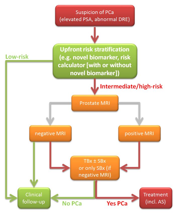
of low-risk PCa. An option could be upfront risk stratification with a novel biomarker or RC (with or
without novel biomarker(s) included), and if indicated subsequent MRI with if indicated subsequent
biopsy (Table 1 and Figure 1).Int. J. Mol. Sci. 2019, 20, 1637 9 of 19
Int. J. Mol. Sci. 2018, 19, x FOR PEER REVIEW 6 of 19
Figure 1. Flowchart of men with elevated prostate-specific antigen (PSA) and/or abnormal digital
Figure 1. Flowchart of men with elevated prostate-specific antigen (PSA) and/or abnormal digital
rectal examination (DRE), with the combination of upfront risk stratification and if indicated prostate
rectal examination (DRE), with the combination of upfront risk stratification and if indicated prostate
MRI and biopsy. PSA: prostate-specific antigen; DRE: digital rectal examination; PCa: prostate
MRI and biopsy. PSA: prostate-specific antigen; DRE: digital rectal examination; PCa: prostate
cancer; MRI: magnetic resonance imaging; PI-RADS: MRI suspicion score; TBx: MRI-targeted biopsy;
cancer; MRI: magnetic resonance imaging; PI-RADS: MRI suspicion score; TBx: MRI-targeted biopsy;
SBx: systematic biopsy; AS: active surveillance.
SBx: systematic biopsy; AS: active surveillance.
4.1. Upfront Novel Biomarker and If Indicated Subsequent MRI and Biopsy
4.1. Upfront Novel Biomarker and If Indicated Subsequent MRI and Biopsy
PHI has been tested as a predictor of a positive MRI in men requiring repeat biopsy [97]. PHI
scoresPHI hasgenerally
were been tested as a predictor
higher in men with of aan
positive MRI in However,
MRI lesion. men requiringusingrepeat biopsy
PHI only [97]. PHI
marginally
scores were generally higher in men with an MRI lesion. However, using
increased predictive value compared to PSA in this study suggesting that PHI is unlikely to be useful PHI only marginally
increased
as a triagingpredictive value compared
test in deciding if an MRI will to PSA in this study
be positive. Punnen suggesting thatatPHI
et al. looked is unlikely
different to be
sequencing
useful as a triaging test in deciding if an MRI will be positive. Punnen
strategies to combine the 4Kscore and MRI in a mixed biopsy population (initial and repeat) [99]. et al. looked at different
sequencing
A strategy ofstrategies
doing an to combine
initial 4Kscore,the followed
4Kscore and by anMRI
MRIinifathemixed biopsy
4Kscore waspopulation
greater than (initial
7.5% andand
repeat) [99]. A strategy of doing an initial 4Kscore, followed by an MRI
a subsequent TBx if the MRI was positive showed a 25%, 83% and 75% reduction in the number of if the 4Kscore was greater
than 7.5%
MRIs, and aand
biopsies subsequent
low-risk PCaTBx diagnoses,
if the MRI was positive showed
respectively. However, a 25%, 83% andresulted
this strategy 75% reduction
in 33% of in
the number
csPCa of MRIs,
being missed. biopsiespathway
A similar and low-risk PCa diagnoses,
using PCA3 score ≥35 as respectively.
threshold wouldHowever,
resultthis
in 52%strategy
MRI
resulted in 33% of csPCa being missed. A similar pathway using PCA3 score ≥35
reduction, 76.4% reduction of biopsies and 86.6% less diagnoses of low-risk PCa, at the cost of missing as threshold would
result csPCa
47.5% in 52%[107].
MRI reduction,
All studies76.4%
concludereduction of biopsies
that optimized and 86.6%ofless
sequencing diagnoses
novel biomarkers of low-risk
and MRI PCa,
is
at the cost of missing 47.5% csPCa [107]. All studies conclude that optimized
the other way around, i.e. an initial MRI followed by a novel biomarker among only those men with a sequencing of novel
biomarkers
low to moderateand suspicion
MRI is the other
score way around,
on MRI. However,i.e. anstill
that initial
wouldMRI followed
mean at leastbyanaMRI
novel biomarker
in every man
among only those
with a suspicion of PCa. men with a low to moderate suspicion score on MRI. However, that still would
mean at least an MRI in every man with a suspicion of PCa.
4.2. Upfront Risk Calculator Including only Standard Clinical Parameters and If Indicated Subsequent MRI
and
4.2. Biopsy
Upfront Risk Calculator Including only Standard Clinical Parameters and If Indicated Subsequent MRI
and Biopsy
Alberts et al. studied whether upfront risk stratification with the ERSPC RPCRC could be used
beforeAlberts
the decision to perform
et al. studied an MRI
whether in menrisk
upfront confronted with with
stratification a previous negative
the ERSPC SBx while
RPCRC couldhaving
be used a
persistent
before thesuspicion
decision to of perform
csPCa [19]. an The
MRIanalysis
in men was restricted
confronted to TBx
with outcomes.
a previous In their
negative SBx cohort,
whileupfront
having
a persistent suspicion of csPCa [19]. The analysis was restricted to TBx outcomes. In their cohort,Int. J. Mol. Sci. 2019, 20, 1637 10 of 19
ERSPC RPCRC-based patient selection for MRI would have avoided 51% of MRIs, 69% of biopsies
and 25% of low-risk PCa diagnoses, while missing 10% csPCa. In a repeat biopsy setting Drost et al.
found that upfront use of the ERSPC RPCRC to select men for MRI with TBx could diagnose most of
the csPCa (83%), while saving 37% of MRIs, 55% of biopsies and 66% of low-risk PCa diagnoses [108].
Recently, Mannaerts et al. showed in a retrospective biopsy-naïve cohort of 200 men that a
pathway of initial ERSPC RPCRC, followed by an MRI if the ERSPC RPCRC advised to perform biopsy
and subsequent SBx with additional TBx in case of a positive MRI, would reduce 37% of MRIs and
biopsies, 23% of low-risk PCa diagnoses while missing 6% csPCa [109]. A TBx-only strategy after
ERSPC RPCRC would have missed 27% of csPCa in this cohort. Currently, a Dutch prospective study
(MR PROPER) evaluating the MRI strategy versus SBx in biopsy naïve men (3000 inclusions aimed),
both after initial risk stratification with the ERSPC RPCRC, is ongoing and will provide more clarity
about the value of the ERSPC RPCRC-MRI pathway [110]. In any case, results obtained till now argue
for an ERSPC RPCRC-based selection for MRI with performance of only MRI with or without TBx
in repeat biopsy men considered to be at high-risk of csPCa according to the ERSPC RPCRC, while
biopsy-naïve men considered to be at high-risk should undergo both MRI with or without TBx and SBx.
4.3. Upfront Risk Calculator Including Novel Biomarker(s) and If Indicated Subsequent MRI and Biopsy
In a retrospective study the SelectMDx score was significantly higher in patients with a suspicious
lesion on MRI compared to patients with a negative MRI. For the prediction of MRI outcome, the AUC
of SelectMDx was 0.83 compared to 0.66 for PSA and 0.65 for PCA3, suggesting a positive association
between SelectMDx and the final PI-RADS v2 score [111]. Trooskens et al. presented data on the
use of SelectMDx (including TRUS PV) to exclude low-risk patients from undergoing an MRI [112].
A strategy of doing upfront SelectMDx, followed by an MRI if the risk for csPCa was greater than
10% and subsequent SBx with additional TBx if the MRI was positive (defined as PI-RADS score ≥4),
would reduce 35% of MRIs and biopsies, 52% of low-risk PCa diagnoses while missing 2% csPCa.
Grönberg et al. recently investigated the combination of S3M and MRI in a cohort of 532 men who
were referred for PCa workup (initial and repeat biopsy). Performing MRI with or without TBx and
additional SBx only in men with a risk >10% for csPCa using the S3M would reduce the number of
MRIs and biopsies with 38%, while diagnosing 42% less low-risk PCa at the cost of missing 8% csPCa
cases [113]. The strategy of performing only MRI with or without TBx for men with a positive S3M
test would save even more biopsies (42%) and low-risk PCa diagnoses (46%), however, at the cost of
missing 19% csPCa.
On average, the value of upfront risk stratification with one of the new risk models seems similar
to the upfront use of the ERSPC RPCRC to select candidates for MRI. Taking into account the costs
and availability of the tests, the ERSPC RPCRC might be preferable. However, to determine the most
cost-effective diagnostic pathway in PCa diagnosis, ideally a large prospective cohort study of men
biopsied irrespective of risk stratification tool outcome and retrospectively compared performance of
all relevant stratification tools should become available for both the initial and repeat biopsy setting.
5. Conclusions
There are numerous risk stratification tools available that can help increase the specificity of PSA
for the detection of csPCa in the initial and repeat biopsy setting. These tools may thereby refine the
PCa diagnostic pathway, improving diagnostic outcome, reducing the burden for patients and making
it more cost-effective and acceptable to the general population and health care providers. All risk
stratification tools result in a considerable decrease in unnecessary testing and carry a generally small
risk of missing csPCa.
Taking into account the costs, RCs using PSA and clinical parameters which perform similarly
well as novel, most often more expensive, biomarkers seem to be the preferred choice. However,
head-to-head-comparisons of all biomarkers and RCs are necessary. Pre-biopsy prostate MRI
has been shown to have more added value in men requiring repeat biopsy than in biopsy-naïveInt. J. Mol. Sci. 2019, 20, 1637 11 of 19
men. Recent studies show evidence for an MRI-directed biopsy management in all men, including
biopsy-naïve men.
Merging novel biomarkers, RCs and MRI results in higher diagnostic accuracies and net benefit
than the use of these risk stratification tools as standalone test. However, in state-of-the-art clinical
decision-making, the patient should benefit from further testing and treatment, even when the
diagnostic test is ‘easy-to-perform’. Therefore, the way forward in the current era of prostate MRI is to
have an accurate predictive low-cost risk stratification tool. This risk stratification tool as triaging test
for the selection of candidates for further testing (e.g. MRI, biopsy) seems to be a multivariable risk
assessment based on blood and clinical parameters, potentially extended with information from urine
samples, which is free to use, available everywhere, extensively externally validated, and calibrated
for different populations. Large prospective and comparative studies remain, however, necessary to
fully assess the potentials and risks of these combined strategies.
Author Contributions: Conceptualization: D.F.O., I.G.S.; Investigation: D.F.O.; Writing—Original Draft
Preparation: D.F.O.; Writing—Review & Editing: D.F.O., M.J.R., I.G.S.; Visualization: D.F.O., M.J.R., I.G.S.;
Supervision: M.J.R., S I.G.S.; Project Administration: D.F.O., I.G.S.
Funding: This research received no external funding.
Conflicts of Interest: The authors declare no conflict of interest.
Abbreviations
4K four-kallikrein
Apps mobile applications
AUC area under the curve
bPSA benign PSA
csPCa clinically significant prostate cancer
DRE digital rectal examination
EAU European Association of Urology
ERSPC European Randomized study of Screening for Prostate Cancer
fPSA free PSA
GG Grade group
GS Gleason score
hK2 human kallikrein 2
iPSA intact PSA
ISUP International Society of Urological Pathology
MiPS MiProstate Score
MRI magnetic resonance imaging
mRNA messenger RNA
NPV negative predictive value
PCa prostate cancer
PCA3 prostate cancer antigen 3
PCPT Prostate Cancer Prevention Trial
PHI Prostate Health Index
PI-RADS Prostate Imaging—Reporting and Data System
PLCO Prostate, Lung, Colorectal, and Ovarian Cancer Screening Trial
PPV positive predictive value
PSA prostate-specific antigen
PV prostate volume
QALYs quality-adjusted life years
RC risk calculator
RCT randomized controlled trial
RPCRC Rotterdam Prostate Cancer Risk CalculatorInt. J. Mol. Sci. 2019, 20, 1637 12 of 19
S3M Stockholm-3 model
SBx systematic biopsy
TBx targeted biopsy
tPSA total PSA
TRUS transrectal ultrasound
USA United States of America
References
1. Schroder, F.H.; Hugosson, J.; Roobol, M.J.; Tammela, T.L.; Zappa, M.; Nelen, V.; Kwiatkowski, M.; Lujan, M.;
Maattanen, L.; Lilja, H.; et al. Screening and prostate cancer mortality: Results of the european randomised
study of screening for prostate cancer (erspc) at 13 years of follow-up. Lancet 2014, 384, 2027–2035. [CrossRef]
2. Pinsky, P.F.; Miller, E.; Prorok, P.; Grubb, R.; Crawford, E.D.; Andriole, G. Extended follow-up for prostate
cancer incidence and mortality among participants in the prostate, lung, colorectal and ovarian randomized
cancer screening trial. BJU Int. 2018. [CrossRef] [PubMed]
3. Tsodikov, A.; Gulati, R.; Heijnsdijk, E.A.M.; Pinsky, P.F.; Moss, S.M.; Qiu, S.; de Carvalho, T.M.; Hugosson, J.;
Berg, C.D.; Auvinen, A.; et al. Reconciling the effects of screening on prostate cancer mortality in the erspc
and plco trials. Ann. Intern. Med. 2017, 167, 449–455. [CrossRef] [PubMed]
4. de Koning, H.J.; Gulati, R.; Moss, S.M.; Hugosson, J.; Pinsky, P.F.; Berg, C.D.; Auvinen, A.; Andriole, G.L.;
Roobol, M.J.; Crawford, E.D.; et al. The efficacy of prostate-specific antigen screening: Impact of key
components in the erspc and plco trials. Cancer 2018, 124, 1197–1206. [CrossRef] [PubMed]
5. Osses, D.F.; Remmers, S.; Schroder, F.H.; van der Kwast, T.; Roobol, M.J. Results of prostate cancer screening
in a unique cohort at 19yr of follow-up. Eur. Urol. 2018, 75, 374–377. [CrossRef]
6. Heijnsdijk, E.A.; de Carvalho, T.M.; Auvinen, A.; Zappa, M.; Nelen, V.; Kwiatkowski, M.; Villers, A.; Paez, A.;
Moss, S.M.; Tammela, T.L.; et al. Cost-effectiveness of prostate cancer screening: A simulation study based
on erspc data. J. Natl. Cancer Inst. 2015, 107. [CrossRef] [PubMed]
7. Siegel, R.L.; Jemal, A.; Wender, R.C.; Gansler, T.; Ma, J.; Brawley, O.W. An assessment of progress in cancer
control. CA Cancer J. Clin. 2018, 68, 329–339. [CrossRef] [PubMed]
8. Mottet, N.; Bellmunt, J.; Bolla, M.; Briers, E.; Cumberbatch, M.G.; De Santis, M.; Fossati, N.; Gross, T.;
Henry, A.M.; Joniau, S.; et al. Eau-estro-siog guidelines on prostate cancer. Part 1: Screening, diagnosis, and
local treatment with curative intent. Eur. Urol. 2017, 71, 618–629. [CrossRef] [PubMed]
9. Louie, K.S.; Seigneurin, A.; Cathcart, P.; Sasieni, P. Do prostate cancer risk models improve the predictive
accuracy of psa screening? A meta-analysis. Ann. Oncol. 2015, 26, 848–864. [CrossRef]
10. Roobol, M.J.; Steyerberg, E.W.; Kranse, R.; Wolters, T.; van den Bergh, R.C.; Bangma, C.H.; Schroder, F.H.
A risk-based strategy improves prostate-specific antigen-driven detection of prostate cancer. Eur. Urol. 2010,
57, 79–85. [CrossRef]
11. Tosoian, J.J.; Druskin, S.C.; Andreas, D.; Mullane, P.; Chappidi, M.; Joo, S.; Ghabili, K.; Mamawala, M.;
Agostino, J.; Carter, H.B.; et al. Prostate health index density improves detection of clinically significant
prostate cancer. BJU Int. 2017, 120, 793–798. [CrossRef] [PubMed]
12. Punnen, S.; Pavan, N.; Parekh, D.J. Finding the wolf in sheep’s clothing: The 4kscore is a novel blood test
that can accurately identify the risk of aggressive prostate cancer. Rev. Urol. 2015, 17, 3–13.
13. Dani, H.; Loeb, S. The role of prostate cancer biomarkers in undiagnosed men. Curr. Opin. Urol. 2017,
27, 210–216. [CrossRef] [PubMed]
14. Anceschi, U.; Tuderti, G.; Lugnani, F.; Biava, P.M.; Malossini, G.; Luciani, L.; Cai, T.; Marsiliani, D.;
Filianoti, A.; Mattevi, D.; et al. Novel diagnostic biomarkers of prostate cancer: An update. Curr. Med. Chem.
2018, 25, 1–14. [CrossRef] [PubMed]
15. Leyten, G.H.; Hessels, D.; Jannink, S.A.; Smit, F.P.; de Jong, H.; Cornel, E.B.; de Reijke, T.M.; Vergunst, H.;
Kil, P.; Knipscheer, B.C.; et al. Prospective multicentre evaluation of pca3 and tmprss2-erg gene fusions as
diagnostic and prognostic urinary biomarkers for prostate cancer. Eur. Urol. 2014, 65, 534–542. [CrossRef]
16. Ahmed, H.U.; El-Shater Bosaily, A.; Brown, L.C.; Gabe, R.; Kaplan, R.; Parmar, M.K.; Collaco-Moraes, Y.;
Ward, K.; Hindley, R.G.; Freeman, A.; et al. Diagnostic accuracy of multi-parametric mri and trus biopsy in
prostate cancer (promis): A paired validating confirmatory study. Lancet 2017, 389, 815–822. [CrossRef]Int. J. Mol. Sci. 2019, 20, 1637 13 of 19
17. Kasivisvanathan, V.; Rannikko, A.S.; Borghi, M.; Panebianco, V.; Mynderse, L.A.; Vaarala, M.H.; Briganti, A.;
Budaus, L.; Hellawell, G.; Hindley, R.G.; et al. Mri-targeted or standard biopsy for prostate-cancer diagnosis.
N. Engl. J. Med. 2018, 378, 1767–1777. [CrossRef]
18. Schoots, I.G.; Rouviere, O. Mri and mri-targeted biopsy take precedence over systematic biopsy in primary
prostate cancer diagnosis. BMJ Evid. Based Med. 2018. [CrossRef]
19. Alberts, A.R.; Schoots, I.G.; Bokhorst, L.P.; van Leenders, G.J.; Bangma, C.H.; Roobol, M.J. Risk-based
patient selection for magnetic resonance imaging-targeted prostate biopsy after negative transrectal
ultrasound-guided random biopsy avoids unnecessary magnetic resonance imaging scans. Eur. Urol.
2016, 69, 1129–1134. [CrossRef]
20. Walz, J. The “promis” of magnetic resonance imaging cost effectiveness in prostate cancer diagnosis?
Eur. Urol. 2018, 73, 31–32. [CrossRef]
21. Lee, R.; Localio, A.R.; Armstrong, K.; Malkowicz, S.B.; Schwartz, J.S.; Free PSA Study Group. A meta-analysis
of the performance characteristics of the free prostate-specific antigen test. Urology 2006, 67, 762–768.
[CrossRef] [PubMed]
22. Mikolajczyk, S.D.; Marks, L.S.; Partin, A.W.; Rittenhouse, H.G. Free prostate-specific antigen in serum is
becoming more complex. Urology 2002, 59, 797–802. [CrossRef]
23. Loeb, S.; Lilja, H.; Vickers, A. Beyond prostate-specific antigen: Utilizing novel strategies to screen men for
prostate cancer. Curr. Opin. Urol. 2016, 26, 459–465. [CrossRef] [PubMed]
24. Boegemann, M.; Stephan, C.; Cammann, H.; Vincendeau, S.; Houlgatte, A.; Jung, K.; Blanchet, J.S.;
Semjonow, A. The percentage of prostate-specific antigen (psa) isoform [−2]propsa and the prostate health
index improve the diagnostic accuracy for clinically relevant prostate cancer at initial and repeat biopsy
compared with total psa and percentage free psa in men agedInt. J. Mol. Sci. 2019, 20, 1637 14 of 19
35. Haese, A.; de la Taille, A.; van Poppel, H.; Marberger, M.; Stenzl, A.; Mulders, P.F.; Huland, H.; Abbou, C.C.;
Remzi, M.; Tinzl, M.; et al. Clinical utility of the pca3 urine assay in European men scheduled for repeat
biopsy. Eur. Urol. 2008, 54, 1081–1088. [CrossRef]
36. Gittelman, M.C.; Hertzman, B.; Bailen, J.; Williams, T.; Koziol, I.; Henderson, R.J.; Efros, M.; Bidair, M.;
Ward, J.F. Pca3 molecular urine test as a predictor of repeat prostate biopsy outcome in men with previous
negative biopsies: A prospective multicenter clinical study. J. Urol. 2013, 190, 64–69. [CrossRef] [PubMed]
37. Merola, R.; Tomao, L.; Antenucci, A.; Sperduti, I.; Sentinelli, S.; Masi, S.; Mandoj, C.; Orlandi, G.; Papalia, R.;
Guaglianone, S.; et al. Pca3 in prostate cancer and tumor aggressiveness detection on 407 high-risk patients:
A national cancer institute experience. J. Exp. Clin. Cancer Res. 2015, 34, 15. [CrossRef]
38. Chevli, K.K.; Duff, M.; Walter, P.; Yu, C.; Capuder, B.; Elshafei, A.; Malczewski, S.; Kattan, M.W.; Jones, J.S.
Urinary pca3 as a predictor of prostate cancer in a cohort of 3,073 men undergoing initial prostate biopsy.
J. Urol. 2014, 191, 1743–1748. [CrossRef] [PubMed]
39. Hessels, D.; van Gils, M.P.; van Hooij, O.; Jannink, S.A.; Witjes, J.A.; Verhaegh, G.W.; Schalken, J.A. Predictive
value of pca3 in urinary sediments in determining clinico-pathological characteristics of prostate cancer.
Prostate 2010, 70, 10–16. [CrossRef] [PubMed]
40. Leyten, G.H.; Hessels, D.; Smit, F.P.; Jannink, S.A.; de Jong, H.; Melchers, W.J.; Cornel, E.B.; de Reijke, T.M.;
Vergunst, H.; Kil, P.; et al. Identification of a candidate gene panel for the early diagnosis of prostate cancer.
Clin. Cancer Res. 2015, 21, 3061–3070. [CrossRef]
41. Seisen, T.; Roupret, M.; Brault, D.; Leon, P.; Cancel-Tassin, G.; Comperat, E.; Renard-Penna, R.; Mozer, P.;
Guechot, J.; Cussenot, O. Accuracy of the prostate health index versus the urinary prostate cancer antigen 3
score to predict overall and significant prostate cancer at initial biopsy. Prostate 2015, 75, 103–111. [CrossRef]
[PubMed]
42. Loeb, S. Predicting prostate biopsy results—Pca3 versus phi. Nat. Rev. Urol. 2015, 12, 130. [CrossRef]
[PubMed]
43. Tomlins, S.A.; Aubin, S.M.; Siddiqui, J.; Lonigro, R.J.; Sefton-Miller, L.; Miick, S.; Williamsen, S.; Hodge, P.;
Meinke, J.; Blase, A.; et al. Urine tmprss2:Erg fusion transcript stratifies prostate cancer risk in men with
elevated serum psa. Sci. Transl. Med. 2011, 3, 94ra72. [CrossRef] [PubMed]
44. Hessels, D.; Smit, F.P.; Verhaegh, G.W.; Witjes, J.A.; Cornel, E.B.; Schalken, J.A. Detection of tmprss2-erg
fusion transcripts and prostate cancer antigen 3 in urinary sediments may improve diagnosis of prostate
cancer. Clin. Cancer Res. 2007, 13, 5103–5108. [CrossRef]
45. Stephan, C.; Cammann, H.; Jung, K. Re: Scott a. Tomlins, john r. Day, robert j. Lonigro, et al. Urine
tmprss2:Erg plus pca3 for individualized prostate cancer risk assessment. Eur. Urol. 2015, 68, e106–e107.
[CrossRef]
46. Tomlins, S.A.; Day, J.R.; Lonigro, R.J.; Hovelson, D.H.; Siddiqui, J.; Kunju, L.P.; Dunn, R.L.; Meyer, S.;
Hodge, P.; Groskopf, J.; et al. Urine tmprss2:Erg plus pca3 for individualized prostate cancer risk assessment.
Eur. Urol. 2016, 70, 45–53. [CrossRef]
47. Hamid, A.R.; Hoogland, A.M.; Smit, F.; Jannink, S.; van Rijt-van de Westerlo, C.; Jansen, C.F.; van
Leenders, G.J.; Verhaegh, G.W.; Schalken, J.A. The role of hoxc6 in prostate cancer development. Prostate
2015, 75, 1868–1876. [CrossRef]
48. Liang, M.; Sun, Y.; Yang, H.L.; Zhang, B.; Wen, J.; Shi, B.K. Dlx1, a binding protein of beta-catenin, promoted
the growth and migration of prostate cancer cells. Exp. Cell Res. 2018, 363, 26–32. [CrossRef] [PubMed]
49. Finne, P.; Finne, R.; Bangma, C.; Hugosson, J.; Hakama, M.; Auvinen, A.; Stenman, U.H. Algorithms based on
prostate-specific antigen (psa), free psa, digital rectal examination and prostate volume reduce false-positive
psa results in prostate cancer screening. Int. J. Cancer 2004, 111, 310–315. [CrossRef] [PubMed]
50. Chun, F.K.; Steuber, T.; Erbersdobler, A.; Currlin, E.; Walz, J.; Schlomm, T.; Haese, A.; Heinzer, H.;
McCormack, M.; Huland, H.; et al. Development and internal validation of a nomogram predicting the
probability of prostate cancer gleason sum upgrading between biopsy and radical prostatectomy pathology.
Eur. Urol. 2006, 49, 820–826. [CrossRef] [PubMed]
51. Karakiewicz, P.I.; Benayoun, S.; Kattan, M.W.; Perrotte, P.; Valiquette, L.; Scardino, P.T.; Cagiannos, I.;
Heinzer, H.; Tanguay, S.; Aprikian, A.G.; et al. Development and validation of a nomogram predicting the
outcome of prostate biopsy based on patient age, digital rectal examination and serum prostate specific
antigen. J. Urol. 2005, 173, 1930–1934. [CrossRef] [PubMed]Int. J. Mol. Sci. 2019, 20, 1637 15 of 19
52. Ankerst, D.P.; Hoefler, J.; Bock, S.; Goodman, P.J.; Vickers, A.; Hernandez, J.; Sokoll, L.J.; Sanda, M.G.;
Wei, J.T.; Leach, R.J.; et al. Prostate cancer prevention trial risk calculator 2.0 for the prediction of low- vs
high-grade prostate cancer. Urology 2014, 83, 1362–1367. [CrossRef]
53. Stephan, C.; Cammann, H.; Semjonow, A.; Diamandis, E.P.; Wymenga, L.F.; Lein, M.; Sinha, P.; Loening, S.A.;
Jung, K. Multicenter evaluation of an artificial neural network to increase the prostate cancer detection rate
and reduce unnecessary biopsies. Clin. Chem. 2002, 48, 1279–1287.
54. Pereira-Azevedo, N.; Verbeek, J.F.M.; Nieboer, D.; Bangma, C.H.; Roobol, M.J. Head-to-head comparison of
prostate cancer risk calculators predicting biopsy outcome. Transl. Androl. Urol. 2018, 7, 18–26. [CrossRef]
[PubMed]
55. Roobol, M.J.; Schroder, F.H.; Hugosson, J.; Jones, J.S.; Kattan, M.W.; Klein, E.A.; Hamdy, F.; Neal, D.;
Donovan, J.; Parekh, D.J.; et al. Importance of prostate volume in the european randomised study of
screening for prostate cancer (erspc) risk calculators: Results from the prostate biopsy collaborative group.
World J. Urol. 2012, 30, 149–155. [CrossRef]
56. Nordstrom, T.; Akre, O.; Aly, M.; Gronberg, H.; Eklund, M. Prostate-specific antigen (psa) density in the
diagnostic algorithm of prostate cancer. Prostate Cancer Prostatic Dis. 2018, 21, 57–63. [CrossRef]
57. Nam, R.K.; Toi, A.; Klotz, L.H.; Trachtenberg, J.; Jewett, M.A.; Appu, S.; Loblaw, D.A.; Sugar, L.; Narod, S.A.;
Kattan, M.W. Assessing individual risk for prostate cancer. J. Clin. Oncol. 2007, 25, 3582–3588. [CrossRef]
[PubMed]
58. Pereira-Azevedo, N.; Osorio, L.; Fraga, A.; Roobol, M.J. Rotterdam prostate cancer risk calculator:
Development and usability testing of the mobile phone app. JMIR Cancer 2017, 3, e1. [CrossRef]
59. Adam, A.; Hellig, J.C.; Perera, M.; Bolton, D.; Lawrentschuk, N. ‘Prostate cancer risk calculator’ mobile
applications (apps): A systematic review and scoring using the validated user version of the mobile
application rating scale (umars). World J. Urol. 2018, 36, 565–573. [CrossRef] [PubMed]
60. Roobol, M.J.; Vedder, M.M.; Nieboer, D.; Houlgatte, A.; Vincendeau, S.; Lazzeri, M.; Guazzoni, G.; Stephan, C.;
Semjonow, A.; Haese, A.; et al. Comparison of two prostate cancer risk calculators that include the prostate
health index. Eur. Urol. Focus 2015, 1, 185–190. [CrossRef]
61. Foley, R.W.; Maweni, R.M.; Gorman, L.; Murphy, K.; Lundon, D.J.; Durkan, G.; Power, R.; O’Brien, F.;
O’Malley, K.J.; Galvin, D.J.; et al. European randomised study of screening for prostate cancer (erspc)
risk calculators significantly outperform the prostate cancer prevention trial (pcpt) 2.0 in the prediction of
prostate cancer: A multi-institutional study. BJU Int. 2016, 118, 706–713. [CrossRef] [PubMed]
62. Loeb, S.; Shin, S.S.; Broyles, D.L.; Wei, J.T.; Sanda, M.; Klee, G.; Partin, A.W.; Sokoll, L.; Chan, D.W.;
Bangma, C.H.; et al. Prostate health index improves multivariable risk prediction of aggressive prostate
cancer. BJU Int. 2017, 120, 61–68. [CrossRef]
63. Roobol, M.J.; Verbeek, J.F.M.; van der Kwast, T.; Kummerlin, I.P.; Kweldam, C.F.; van Leenders, G. Improving
the rotterdam european randomized study of screening for prostate cancer risk calculator for initial prostate
biopsy by incorporating the 2014 international society of urological pathology gleason grading and cribriform
growth. Eur. Urol. 2017, 72, 45–51. [CrossRef]
64. Verbeek, J.F.M.; Bangma, C.H.; Kweldam, C.F.; van der Kwast, T.H.; Kummerlin, I.P.; van Leenders, G.;
Roobol, M.J. Reducing unnecessary biopsies while detecting clinically significant prostate cancer including
cribriform growth with the erspc rotterdam risk calculator and 4kscore. Urol. Oncol. 2019, 37, 138–144.
[CrossRef]
65. Hansen, J.; Auprich, M.; Ahyai, S.A.; de la Taille, A.; van Poppel, H.; Marberger, M.; Stenzl, A.; Mulders, P.F.;
Huland, H.; Fisch, M.; et al. Initial prostate biopsy: Development and internal validation of a biopsy-specific
nomogram based on the prostate cancer antigen 3 assay. Eur. Urol. 2013, 63, 201–209. [CrossRef] [PubMed]
66. Wei, J.T.; Feng, Z.; Partin, A.W.; Brown, E.; Thompson, I.; Sokoll, L.; Chan, D.W.; Lotan, Y.; Kibel, A.S.;
Busby, J.E.; et al. Can urinary pca3 supplement psa in the early detection of prostate cancer? J. Clin. Oncol.
2014, 32, 4066–4072. [CrossRef] [PubMed]
67. Chun, F.K.; de la Taille, A.; van Poppel, H.; Marberger, M.; Stenzl, A.; Mulders, P.F.; Huland, H.; Abbou, C.C.;
Stillebroer, A.B.; van Gils, M.P.; et al. Prostate cancer gene 3 (pca3): Development and internal validation of a
novel biopsy nomogram. Eur. Urol. 2009, 56, 659–667. [CrossRef] [PubMed]
68. Ankerst, D.P.; Groskopf, J.; Day, J.R.; Blase, A.; Rittenhouse, H.; Pollock, B.H.; Tangen, C.; Parekh, D.;
Leach, R.J.; Thompson, I. Predicting prostate cancer risk through incorporation of prostate cancer gene 3.
J. Urol. 2008, 180, 1303–1308; discussion 1308. [CrossRef] [PubMed]You can also read