Coronary Physiology in the Cardiac Catheterization Laboratory - MDPI
←
→
Page content transcription
If your browser does not render page correctly, please read the page content below
Journal of
           Clinical Medicine

Review
Coronary Physiology in the Cardiac Catheterization
Laboratory
Samit M. Shah 1, *        and Steven E. Pfau 1,2
 1    Yale-New Haven Hospital, Yale School of Medicine, New Haven, CT 06510, USA; steven.pfau@yale.edu
 2    VA Connecticut Healthcare System, West Haven, CT 06516, USA
 *    Correspondence: samit.shah@yale.edu

 Received: 10 December 2018; Accepted: 14 February 2019; Published: 18 February 2019

 Abstract: Coronary angiography has been the principle modality for assessing the severity of
 atherosclerotic coronary artery disease for several decades. However, there is a complex relationship
 between angiographic coronary stenosis and the presence or absence of myocardial ischemia. Recent
 technological advances now allow for the assessment of coronary physiology in the catheterization
 laboratory at the time of diagnostic coronary angiography. Early studies focused on coronary flow
 reserve (CFR) but more recent work has demonstrated the physiologic accuracy and prognostic value
 of the fractional flow reserve (FFR) and instantaneous wave free ratio (iFR) for the assessment of
 coronary artery disease. These measurements have been validated in large multi-center clinical trials
 and have become indispensable tools for guiding revascularization in the cardiac catheterization
 laboratory. The physiological assessment of chest pain in the absence of epicardial coronary
 artery disease involves coronary thermodilution to obtain the index of microcirculatory resistance
 (IMR) or Doppler velocity measurement to determine the coronary flow velocity reserve (CFVR).
 Physiology-based coronary artery assessment brings “personalized medicine” to the catheterization
 laboratory and allows cardiologists and referring providers to make decisions based on objective
 findings and evidence-based treatment algorithms. The purpose of this review is to describe the
 theory, technical aspects, and relevant clinical trials related to coronary physiology assessment for an
 intended audience of general medical practitioners.

 Keywords: coronary artery disease; cardiovascular physiology; microvascular angina

1. Introduction
     The first coronary angiogram nearly 60 years ago provided a simplified glimpse of a dynamic
and highly regulated process—the delivery of blood to working myocardium. Sones’ selective
coronary angiography launched a new field of diagnosis and treatment for atherosclerotic coronary
obstruction [1]. Since that time our understanding of the physiology underlying myocardial ischemia
has increased rapidly [2], and while invasive coronary angiography remains the “gold standard” its
limitations are well-documented [3,4]. There is a complex relationship between the anatomic severity
of luminal narrowing and the presence of myocardial ischemia or risk of subsequent myocardial
infarction [5,6] and angiography alone is unable to determine whether a luminal stenosis causes
ischemia or anginal symptoms. Technological advances in the last 30 years now allow direct, real
time measurement of coronary flow and pressure in individual patients and this data can be applied
directly at the time of coronary angiography to guide treatment of individual atherosclerotic lesions.
Most recently, real time assessment of coronary physiology is being applied to conditions that affect
coronary blood flow outside the realm of atherosclerotic luminal obstruction, providing insight into a
variety of disease states such as endothelial dysfunction and microvascular disease. The purpose of

J. Clin. Med. 2019, 8, 255; doi:10.3390/jcm8020255                               www.mdpi.com/journal/jcm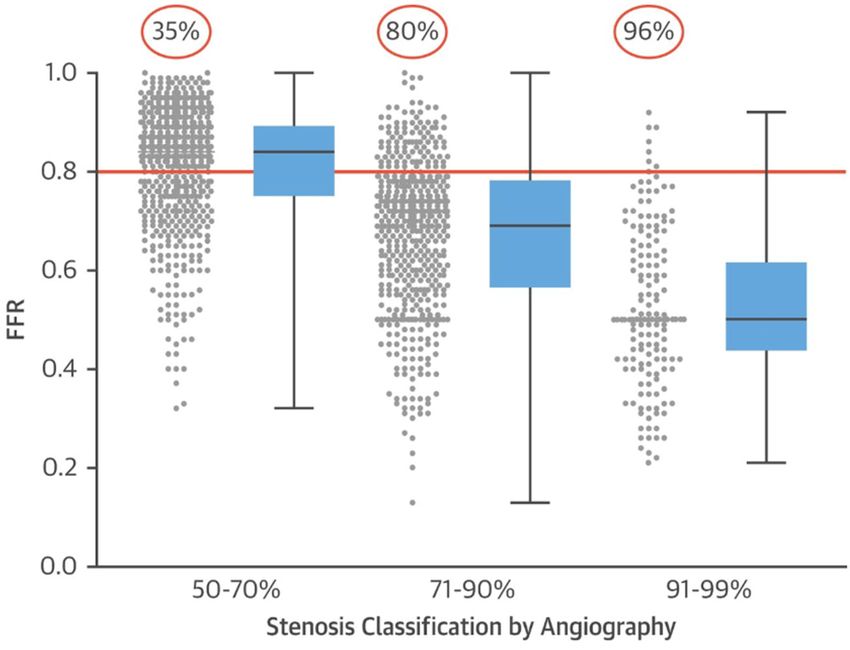
J. Clin. Med. 2019, 8, 255 2 of 14
this review is to describe the theory, technical aspects, and relevant clinical trials related to coronary
physiology assessment for an intended audience of general medical practitioners.
2. Historical Basis for Assessing Coronary Physiology in the Catheterization Laboratory
J. Clin. Med. 2019, 8, x FOR PEER 2 of 14
With exertion, coronary blood flow can increase to 300–400% of resting levels [7], both in animal
models2.and in humans during treadmill exercise [8]. In 1974 Gould
Historical Basis for Assessing Coronary Physiology in the Catheterization Laboratory et al. described the concept
of coronary flow reserve (CFR), a ratio of maximal flow to resting flow in response to increases in
With exertion, coronary blood flow can increase to 300–400% of resting levels [7], both in animal
myocardial oxygen demand [5]. Using an in vitro animal model, the authors demonstrated that resting
models and in humans during treadmill exercise [8]. In 1974 Gould et al. described the concept of
coronary blood flow
coronary is not significantly
flow reserve (CFR), a ratio affected
of maximalby flow
a stenosis untilflow
to resting theinlumen is attoleast
response 85% in
increases occluded.
However, during hyperemia
myocardial oxygen demand (achieved withana in
[5]. Using vasodilatory
vitro animalcontrast
model, thedyeauthors
bolus)demonstrated
the maximalthat achievable
coronaryresting
blood coronary
flow was blood flow isby
limited nota significantly affected
luminal stenosis asby a stenosis
low until (Figure
as 30–40% the lumen 1).isThis
at least 85% study
seminal
occluded. However, during hyperemia (achieved with a vasodilatory
demonstrated that the significance of a coronary stenosis is directly related to coronary blood contrast dye bolus) the flow,
maximal achievable coronary blood flow was limited by a luminal stenosis as low as 30–40% (Figure
and that the magnitude of the reduction in blood flow is most clinically relevant during maximal
1). This seminal study demonstrated that the significance of a coronary stenosis is directly related to
coronary bloodblood
coronary flowflow,(or hyperemia). Questions
and that the magnitude of theregarding
reduction inthe clinical
blood flow iscorrelations
most clinicallyand prognostic
relevant
implications
duringofmaximal
the physiological
coronary bloodassessment of an anatomic
flow (or hyperemia). coronary
Questions stenosis
regarding set thecorrelations
the clinical stage for decades
of further
andinvestigation.
prognostic implications of the physiological assessment of an anatomic coronary stenosis set the
stage for decades of further investigation.
Figure 1. Effect
Figure of coronary
1. Effect stenosis
of coronary stenosisononcoronary flowreserve.
coronary flow reserve. Reprinted
Reprinted withwith permission
permission from Gould
from Gould
2009, adapted from Gould et al. 1974 [9]. Experimental model of coronary blood
2009, adapted from Gould et al. 1974 [9]. Experimental model of coronary blood flow at rest flow at rest and the and the
maximal achievable flow. In the presence of a stenosis resting flow is not limited until a coronary
maximal achievable flow. In the presence of a stenosis resting flow is not limited until a coronary
stenosis reaches 85% luminal obstruction. However, maximal achievable flow is limited at stenoses
stenosis reaches 85% luminal obstruction. However, maximal achievable flow is limited at stenoses as
as low as 30–40%. The ratio of the maximal achievable coronary blood flow to the resting blood flow
low as 30–40%. The ratio
is the coronary of the(CFR).
flow reserve maximal achievable coronary blood flow to the resting blood flow is
the coronary flow reserve (CFR).
The first coronary angioplasty on an awake patient was performed on 14th September 1977 by
The first coronary
Andreas Gruentzigangioplasty on an awake
in Zurich, Switzerland [10,11].patient was performed
The operators on lumen
used a double 14th September
catheter that 1977 by
Andreas allowed for intra-coronary
Gruentzig pressure measurement
in Zurich, Switzerland [10,11]. as well
The as angioplasty
operators usedballoon dilatation.
a double lumenIncatheter
their that
initial report Gruentzig and his colleagues documented the angiographic findings
allowed for intra-coronary pressure measurement as well as angioplasty balloon dilatation. In their as well as the
hemodynamic significance of the stenosis as represented by the arterial pressure proximal and distal
initial report Gruentzig and his colleagues documented the angiographic findings as well as the
to the stenosis (Figure 2). After balloon angioplasty the pressure gradient was reduced from 58
hemodynamic
mmHg tosignificance
19 mmHg, which of the stenosis
correlated as represented
with by the in
a significant decrease arterial pressure
the severity of theproximal
angiographicand distal
to the stenosis
stenosis.(Figure 2). After
The authors notedballoon angioplasty
that an “increase the coronary
in distal pressurepressure”
gradientafter
wasballoon
reduced from 58 mmHg
angioplasty
to 19 mmHg,
could be which correlated
utilized to gauge thewith a significant
success decrease
of the procedure. Theinconcept
the severity of theblood
of improving angiographic
flow to thestenosis.
The authors noted that an “increase in distal coronary pressure” after balloon angioplastythecould be
myocardium by relieving a hemodynamically significant stenosis was intuitive. However,
utilizedGruentzig
to gauge balloon dilatation catheter had a large outer diameter and contributed to the luminal
the success of the procedure. The concept of improving blood flow to the myocardium
obstruction of a stenosis, confounding the assessment of distal coronary pressure. As a result of the
by relieving a hemodynamically significant stenosis was intuitive. However, the Gruentzig balloon
physical diameter of the pressure measurement catheter, the “translesional pressure gradient” that
dilatation catheter had a large outer diameter and contributed to the luminal obstruction of a stenosis,J. Clin. Med. 2019, 8, 255 3 of 14
confounding the assessment of distal coronary pressure. As a result of the physical diameter of the
pressure measurement catheter, the “translesional pressure gradient” that was measured at the time of
J. Clin. Med. 2019, 8, x FOR PEER 3 of 14
angioplasty did not accurately correlate with the severity of stenosis measured angiographically [12].
Anatomic
was measuredevaluation
at thewith
timequalitative
of angioplastyand quantitative angiography
did not accurately correlate remained
with thethe cornerstone
severity of the
of stenosis
assessment of coronary artery stenosis severity, guiding decisions regarding
measured angiographically [12]. Anatomic evaluation with qualitative and quantitative angiography revascularization with
angioplasty
remained or thecoronary
cornerstone artery bypass
of the graft surgery,
assessment and evaluating
of coronary artery stenosis the outcome
severity, of revascularization
guiding decisions
interventions [13]. Still, as noted
regarding revascularization withby Gruentzig’s
angioplasty or team,
coronarytheartery
idea that “anatomy
bypass alone”and
graft surgery, didevaluating
not “predict
thethephysiologic
outcome ofconsequence
revascularization of individual stenoses”
interventions persisted
[13]. Still, as noted [12].by Gruentzig’s team, the idea that
“anatomy alone” did not
The measurement “predictcoronary
of human the physiologic consequence
blood flow and CFRof inindividual
consciousstenoses” persistedpossible
humans became [12].
with the The measurement
advent of human coronary
of catheter-based blood flow
pulsed Doppler and CFR in[14–16].
technology conscious Thehumans became
underlying possible of
principle
with the advent
Doppler-based of catheter-based
measurement is that thepulsed Doppler
frequency technology
shift that occurs [14–16]. The underlying
when transmitted principle
ultrasound of
waves
Doppler-based measurement is that the frequency shift that occurs when
are reflected off of moving red blood cells is proportional to the velocity of blood flow. This measured transmitted ultrasound
waves are
frequency reflected
shift off of moving
is converted red bloodflow
to a calculated cellsvelocity
is proportional to the velocity the
which approximates of blood flow. This
volumetric blood
flow velocity [17]. Intra-coronary Doppler flow velocity measurement accurately correlatedthe
measured frequency shift is converted to a calculated flow velocity which approximates with
volumetric blood flow velocity [17]. Intra-coronary Doppler flow velocity measurement accurately
absolute flow velocity in in vitro systems as well as with Doppler velocity measured with extravascular
correlated with absolute flow velocity in in vitro systems as well as with Doppler velocity measured
epicardial probes [17]. However, due to concern that even ~1 mm microcatheters might contribute
with extravascular epicardial probes [17]. However, due to concern that even ~1 mm microcatheters
coronary luminal obstruction and prohibit accurate flow assessment in patients with coronary stenosis,
might contribute coronary luminal obstruction and prohibit accurate flow assessment in patients with
the 0.018 inch (0.45 mm) diameter coronary Doppler guidewire was developed [18]. The “Doppler wire”
coronary stenosis, the 0.018 inch (0.45 mm) diameter coronary Doppler guidewire was developed
could measure coronary flow velocity distal to a stenosis at rest and after the induction of hyperemia
[18]. The “Doppler wire” could measure coronary flow velocity distal to a stenosis at rest and after
using papaverine,
the induction adenosine,using
of hyperemia dipyramidole,
papaverine,oradenosine,
contrast media. In spiteorofcontrast
dipyramidole, the Doppler
media.wire being
In spite
validated for the wire
of the Doppler accurate
beingassessment
validated for ofthe
CFR there were
accurate significant
assessment of CFR caveats to Doppler
there were wire-derived
significant caveats
physiological assessment of coronary stenosis. CFR measurement
to Doppler wire-derived physiological assessment of coronary stenosis. CFR measurement encompasses the entire coronary
circulation, including the epicardial coronary artery and the microvascular
encompasses the entire coronary circulation, including the epicardial coronary artery and the bed, thus the specificity
formicrovascular
determining bed, the contribution
thus the specificityof anforepicardial
determiningstenosis to a reduction
the contribution of aninepicardial
Dopplerstenosis
flow velocity
to a
may be decreased
reduction in patients
in Doppler flowwith microvascular
velocity disease. Additionally,
may be decreased in patients with resting Doppler flow
microvascular velocity
disease.
measurements
Additionally,are susceptible
resting Dopplertoflow hemodynamic factors that are
velocity measurements maysusceptible
alter coronary flow velocity,factors
to hemodynamic such as
that may
changes alterrate,
in heart coronary
blood flow velocity,
pressure, suchventricular
and left as changes in heartwhich
function, rate, in
blood
turn pressure,
reduce theand left
accuracy
of ventricular
CFR measurement. function, Finally,
which inDoppler
turn reduce the accuracy
wire-derived CFRofguided
CFR measurement.
percutaneous Finally, Doppler
coronary wire-
intervention
hasderived
been shownCFR guided
to havepercutaneous
no differencecoronary intervention
in outcomes compared has tobeen shown to have no
angiography-guided difference in
intervention [19].
outcomes compared to angiography-guided intervention [19]. These
These limitations, combined with technical challenges with operating the Doppler wire, limited limitations, combined with
technical
adoption ofchallenges with operating
this technology for routine thephysiological
Doppler wire,assessment
limited adoption in theofcatheterization
this technologylaboratory.
for routine
physiological assessment in the catheterization laboratory.
Figure
Figure 2. 2.Coronary
Coronary Artery
Artery Translesional
Translesional Pressure
Pressure Measurement
Measurementininthe theFirst
FirstAngioplasty
Angioplasty (1978).
(1978).
Reprinted and adapted from EuroIntervention Vol 13 / number 1, Meier, B, “His master’s
Reprinted and adapted from EuroIntervention Vol 13/number 1, Meier, B, “His master’s art, Andreas art, Andreas
Grüntzig’s
Grüntzig’s approach
approach totoperforming
performingand
andteaching
teaching coronary
coronary angioplasty”,
angioplasty”,Pages
Pages15–27,
15–27,Copyright
Copyright (2017),
(2017),with
with
permission
permission fromEuropa
from EuropaDigital
Digital&&Publishing
Publishing [20].
[20]. Hemodynamic
Hemodynamic assessment
assessmentofofcoronary
coronary stenosis
stenosis
during
during thethe first
first coronaryangioplasty
coronary angioplastyby
by Dr.
Dr. Andreas
Andreas Gruentzig
Gruentzigin in1978.
1978.The
Thepressure
pressuretracing
tracing onontoptop
labeled AoP is the aortic pressure. The bottom tracing CoP is the coronary pressure at the distal tip of
labeled AoP is the aortic pressure. The bottom tracing CoP is the coronary pressure at the distal tip
the pressure-monitoring lumen of the balloon dilatation catheter. Distal to the stenosis there is a
of the pressure-monitoring lumen of the balloon dilatation catheter. Distal to the stenosis there is a
significant drop in the coronary pressure that is improved after balloon inflation and dilatation of the
significant drop in the coronary pressure that is improved after balloon inflation and dilatation of the
stenosis. This was the initial report of the “translesional pressure gradient” in a human coronary
stenosis. This was the initial report of the “translesional pressure gradient” in a human coronary artery.
artery.
Nearly 15 years after Gruentzig’s initial report of the translesional pressure gradient, Nico Pijls
and Bernard DeBruyne in the Netherlands investigated the application of an 0.015 inch diameter (0.38J. Clin. Med. 2019, 8, 255 4 of 14
Nearly 15 years after Gruentzig’s initial report of the translesional pressure gradient, Nico Pijls and
Bernard DeBruyne in the Netherlands investigated the application of an 0.015 inch diameter (0.38 mm)
coronary guidewire that was capable of measuring a pressure gradient across a stenosis. Using this
“pressure wire” they were able to experimentally validate the relationship between coronary pressure
and coronary flow, both at rest and during maximal hyperemia [21]. This seminal study laid the
foundation for the measurement of flow reserve by pressure rather than Doppler velocity. There was a
strong correlation between coronary blood flow by Doppler wire assessment and the ratio of the distal
coronary pressure (Pd ) to the proximal aortic pressure (Pa ). During hyperemia, when microcirculatory
resistance is negligible, the Pd represents the maximal coronary flow in the distal vessel and the Pa
represents the maximal coronary flow if the vessel were normal. The term “fractional flow reserve”
(FFR) was introduced to describe the ratio of Pd to Pa , or the maximal achievable flow in the presence
of a stenosis divided by the maximum expected flow if the stenosis were absent. In other words,
the FFR represents the hemodynamic contribution of a coronary stenosis to the reduction of blood
flow in a coronary artery territory. While this was reminiscent of Gruentzig’s translesional pressure
gradient, Pijls and De Bruyne advanced the field of coronary physiology by using a small diameter
coronary “pressure wire”, demonstrating the physiological importance of minimizing microvascular
resistance with pharmacologic hyperemia, and detailing the relationship of venous filling pressure
and the collateral circulation to coronary blood flow.
These animal validations were followed by clinical studies comparing FFR measurement
to non-invasive measures of ischemia including exercise stress testing, thallium scintigraphy,
and dobutamine stress echocardiography [22]. Patients who presented with angina underwent
non-invasive stress testing and subsequently coronary angiography with pressure wire assessment.
An FFR of 0.75, or a distal coronary pressure of less than 75% of the expected pressure, indicated a
coronary stenosis that was “significant”, or in other words every patient with an FFR less than 0.75 was
found to have evidence of ischemia by a non-invasive test. After revascularization the non-invasive
tests were repeated and showed no evidence of ischemia (Figure 3). Furthermore, patients with an FFR
greater than 0.75 were managed with medical therapy and suffered no events with a mean follow-up
of 14.5 months. This landmark study demonstrated that measurement of the FFR reliably defines
coronary lesions that cause myocardial ischemia independent of the angiographic severity of stenosis.J. Clin. Med. 2019, 8, 255 5 of 14
J. Clin. Med. 2019, 8, x FOR PEER 5 of 14
Figure
Figure 3.3. Non-invasive
Non-invasive assessment
assessment of
of ischemia
ischemia compared
compared with
with FFR.
FFR. Reprinted
Reprinted with
with permission
permission from
from
Pijls et al. [22]. In 45 patients with chest pain and moderate (~50%) coronary artery stenosis, exercise
Pijls et al. [22]. In 45 patients with chest pain and moderate (~50%) coronary artery stenosis, exercise
stress testing, thallium scintigraphy, and stress echocardiography with dobutamine were performed.
stress testing, thallium scintigraphy, and stress echocardiography with dobutamine were performed.
These results were compared with FFR measurements. The dashed line indicates the pre-specified FFR
These results were compared with FFR measurements. The dashed line indicates the pre-specified
threshold for ischemia of 0.75 and each circle represents patients who were found to have ischemia or
FFR threshold for ischemia of 0.75 and each circle represents patients who were found to have
no ischemia by stress testing. Nearly every patient with ischemia by stress testing was found to have a
ischemia or no ischemia by stress testing. Nearly every patient with ischemia by stress testing was
coronary stenosis with an ischemic FFR value. After revascularization non-invasive tests were repeated
found to have a coronary stenosis with an ischemic FFR value. After revascularization non-invasive
and there was no longer evidence of ischemia.
tests were repeated and there was no longer evidence of ischemia.J. Clin. Med. 2019, 8, 255 6 of 14
3. Coronary Physiology in the Evaluation of Coronary Artery Disease
The validation of FFR as an invasive determinant of ischemia led to a series of important
J. Clin. Med. 2019, 8, x FOR PEER 6 of 14
randomized trials that have altered the practice of interventional cardiology. The multi-center DEFER
trial investigated whether patients
3. Coronary Physiology with a non-ischemic
in the Evaluation of Coronary Artery FFR of greater than 0.75 could safely defer
Disease
percutaneous coronary intervention (PCI) regardless of the angiographic severity of a stenosis [23].
The validation of FFR as an invasive determinant of ischemia led to a series of important
The study design included 325 patients who were referred for coronary angiography and were planned
randomized trials that have altered the practice of interventional cardiology. The multi-center DEFER
to undergo PCI. FFRwhether
trial investigated was performed
patients withon all patients; if the
a non-ischemic FFRFFR was greater
of greater than 0.75than 0.75,
could patients
safely defer were
randomized to deferral of intervention or performance of PCI as planned.
percutaneous coronary intervention (PCI) regardless of the angiographic severity of a stenosis [23]. If the FFR was less than
0.75 patients underwent PCI as planned. For patients with an FFR
The study design included 325 patients who were referred for coronary angiography and were greater than 0.75 there was no
difference
planned intoevent-free
undergo PCI. survival
FFR was at performed
24 monthsonifall PCI was performed
patients; if the FFR was or greater
deferred, thanand
0.75,patients
patients who
were were randomized
randomized to deferral
to deferring of intervention
PCI reported lessorangina.
performance of PCIthere
At 5 years as planned.
was noIfdifference
the FFR was less
in outcome
than 0.75 patients underwent PCI as planned. For patients with an FFR
if PCI was deferred [24] for an FFR greater than 0.75. This established the safety of deferring coronarygreater than 0.75 there was
no difference
intervention in event-free
in coronary survival
arteries withatan24FFR
months if PCI
above thewas performed orthreshold
physiological deferred, and patients who
for ischemia.
were randomized to deferring PCI reported less angina. At 5 years there was no difference in outcome
The international multi-center FAME trial compared FFR-guided coronary revascularization
if PCI was deferred [24] for an FFR greater than 0.75. This established the safety of deferring coronary
to angiographic selection in patients with multi-vessel coronary artery disease and a stenosis of at
intervention in coronary arteries with an FFR above the physiological threshold for ischemia.
least 50%The [25]. Patients
international who were randomized
multi-center FAME trial to the FFR-guided
compared FFR-guided arm underwent
coronary intervention
revascularization to only
if theangiographic
FFR was 0.80 or less, and patients in the angiography-guided arm underwent
selection in patients with multi-vessel coronary artery disease and a stenosis of at least intervention
based50% on [25].
existing standard
Patients who wereof care of angiographic
randomized severity. This
to the FFR-guided trial enrolled
arm underwent 1005 patients
intervention only ifacross
the 20
centers
FFRinwas the0.80
United States
or less, and Europe
and patients in theand a total of 2415 stents
angiography-guided were placed.
arm underwent At one-year
intervention basedpatients
on
existing standard of care of angiographic severity. This trial enrolled
in the FFR arm were less likely to suffer death, MI, and repeat revascularization; and the overall 1005 patients across 20 centers
in death
rate of the Unitedor MI States
at oneandyear
Europe
wasand a total
11.1% in ofthe2415 stents were placed. At
angiography-guided armone-year
compared patients in thein the
to 7.3%
FFR arm were less likely to suffer death, MI, and repeat revascularization; and the overall rate of
FFR group. A sub-analysis of the FFR-guided revascularization arm reported the FFR values for
death or MI at one year was 11.1% in the angiography-guided arm compared to 7.3% in the FFR
tertiles of angiographic stenosis (Figure 4). In lesions of 50–70% stenosis, 35% had an ischemic FFR;
group. A sub-analysis of the FFR-guided revascularization arm reported the FFR values for tertiles
in 71–90% stenoses, 80% had an ischemic FFR; and in 91–99% stenoses, 96% had an ischemic FFR [26].
of angiographic stenosis (Figure 4). In lesions of 50–70% stenosis, 35% had an ischemic FFR; in 71–
This showed
90% stenoses,that in80% moderate severityFFR;
had an ischemic stenoses
and in there
91–99% was significant
stenoses, 96% hadheterogeneity
an ischemic with regard
FFR [26]. Thisto the
presence
showed of ischemia, and even
that in moderate in angiographically
severity stenoses there was severe stenoses
significant (>70%) up with
heterogeneity to 20% of patients
regard to the did
not have
presencephysiologic
of ischemia, evidence
and even ofinischemia [14]. Overall,
angiographically the FAME
severe stenoses trialupshowed
(>70%) to 20% of that usingdid
patients FFR to
not have physiologic evidence of ischemia [14]. Overall, the FAME
guide PCI reduced adverse cardiovascular events by approximately 30% and the estimated number trial showed that using FFR to
neededguide PCI reduced
to treat to preventadverse
one cardiovascular
adverse eventevents was 20 bypatients.
approximately 30% and the estimated number
needed to treat to prevent one adverse event was 20 patients.
Figure 4. Fractional
Figure 4. Fractionalflow
flowreserve (FFR)values
reserve (FFR) values andand angiographic
angiographic severity
severity of in
of lesions lesions in the
the FAME FAME
trial.
trial. Reprinted
Reprinted with permission
with from Jeremias
permission et al., 2017
from Jeremias et [14] (which[14]
al., 2017 was(which
adaptedwasfromadapted
Tonino etfrom
al 2010)
Tonino
et al [26].
2010)Angiographic findings offindings
[26]. Angiographic the FFR-guided revascularization
of the FFR-guided arm in the FAME
revascularization arm trial
in the[25].
FAME
Scatterplot of FFR values shows significant heterogeneity in the physiological significance
trial [25]. Scatterplot of FFR values shows significant heterogeneity in the physiological significance of of
angiographic
angiographic stenoses.
stenoses. Stenoses
Stenoses ofof50–70%
50–70%angiographic
angiographic severity
severitywere found
were foundto have an ischemic
to have FFR FFR
an ischemic
(0.8, red bar) in 35% of cases. In angiographic stenoses of 71–90%, FFR was in the ischemic zone in
(0.8, red bar) in 35% of cases. In angiographic stenoses of 71–90%, FFR was in the ischemic zone in 80%
80% of cases. In 91–99% angiographic stenoses FFR was ischemic in 96% of cases.
of cases. In 91–99% angiographic stenoses FFR was ischemic in 96% of cases.J. Clin. Med. 2019, 8, 255 7 of 14
FAME II compared FFR-guided PCI to the best available medical therapy in patients with
stable coronary artery disease [27]. Patients with FFR less than 0.80 were randomized to PCI
or medical therapy alone. The trial was stopped early due to a significantly higher incidence of
urgent revascularization in the medical therapy arm. At three years of follow-up the rate of urgent
revascularization and symptomatic angina remained significantly lower in the FFR-guided PCI group
compared with medical therapy [28]. Furthermore, there was no significant difference in cost between
the FFR-guided PCI and medical therapy groups. Thus, the FAME studies demonstrated the superiority
of FFR-guided PCI as compared with angiographic-guided intervention or medical therapy.
FFR has been also validated for the physiological assessment of non-culprit stenoses in patients
who present with acute coronary syndromes [29]. Theoretically, microvascular obstruction from
ischemia or infarction may alter the hyperemic response to adenosine but FFR remains a valid assay.
Multiple studies have validated FFR for assessment of non-culprit stenoses in the setting of acute
coronary syndromes or after myocardial infarction [30]. However, there is evidence that using FFR
to defer intervention to a culprit stenosis may be associated with a worse outcome due to increased
major adverse cardiovascular events in follow-up [31].
The ORBITA trial, first published in 2017, was a blinded, multi-center study across 5 sites in the
United Kingdom that compared PCI to sham intervention in patients with stable angina and >70%
angiographic stenosis [32]. All patients were treated with optimal medical therapy. The primary
endpoint was treadmill exercise time at six weeks; the study found no significant difference between
the PCI and sham intervention groups and concluded that there was no benefit to PCI in stable angina.
However as a secondary assessment all patients underwent physiological testing with FFR and the
instantaneous wave-free ratio (iFR) at the time of coronary angiography [33]. The mean FFR was
0.69 ± 0.16 and the mean iFR was 0.76 ± 0.22. Patients who were randomized to PCI were found
to have a significant improvement in post-intervention dobutamine stress echocardiography wall
motion compared with patients who did not undergo intervention, and the magnitude of the effect
correlated with the severity of ischemia by FFR and iFR. However, pre-intervention FFR or iFR did not
predict whether patients experienced improvement in angina after intervention. The ORBITA trial
and subsequent secondary analysis stratified by FFR demonstrated the efficacy of PCI for relieving
objectively documented ischemia in physiologically significant coronary stenoses [33].
Despite the robust evidence and guideline recommendation for the routine use of FFR for
assessing intermediate-severity stenoses, the overall rate of FFR use remains low [34,35]. In part,
this may be due to the technical limitations of FFR—namely, a slightly increased procedure time,
obligation to use a pressure-sensor tipped guidewire, and the financial expense of using adenosine
to induce hyperemia [14]. Furthermore, many patients find intravenous adenosine uncomfortable
and report symptoms of chest pain or dyspnea during the infusion, which may last up to four
minutes. While intracoronary injection of adenosine is routinely utilized therapeutically during acute
coronary syndromes or coronary intervention to reduce microcirculatory dysfunction, it is less well
tolerated in stable patients during physiological assessment [36,37]. To overcome these limitations
novel indices of resting pressure have been developed including the iFR. iFR is measured with
a pressure-sensor tipped guidewire (Royal Philips, Amsterdam) and using a proprietary algorithm
measures the proximal and distal coronary pressure during the phase of diastole when microcirculatory
resistance is at a theoretical minimum (the “wave free” period) [38]. Since the pressure measurement
is obtained during a period of minimal microcirculatory resistance inducing maximal hyperemia with
adenosine is not necessary. In many situations iFR closely approximates FFR and has concordant
results with non-invasive stress testing [39,40], and an iFR cutoff value of 0.89 has an accuracy of 80%
for identifying lesions with an FFR less than 0.80 [40]. However, simulations and validation studies in
humans show that iFR may correlate best with FFR for physiologically insignificant (or “FFR negative”)
lesions [41]. In 2017 two large multi-center clinical trials were published comparing iFR and FFR.
The DEFINE-FLAIR study was an industry-sponsored (Royal Philips) randomized trial comparing
iFR-guided or FFR-guided coronary revascularization [42]. At 1-year iFR-guided revascularizationJ. Clin. Med. 2019, 8, 255 8 of 14
was found to be non-inferior to FFR and patients in the iFR reported fewer symptoms in the procedural
period (due to the omission of adenosine). Furthermore, procedure time was decreased by nearly 5 min.
A second concurrent Philips-sponsored study, “iFR SWEDEHEART,” was performed using the Swedish
Coronary Angiography and Angioplasty Registry [43]. A total of 2037 patients with stable angina or an
acute coronary syndrome were randomized to undergo iFR or FFR guided revascularization. At 1-year
iFR was again shown to be non-inferior to FFR and a higher proportion of patients in the FFR group
reported chest pain during the procedure (related to adenosine infusion). A meta-analysis of both trials
showed numerically higher myocardial infarctions and deaths in the iFR-guided revascularization
group but without statistical significance [44]. A more recent meta-analysis reported iFR to have similar
diagnostic performance to FFR [45] and additional studies have also shown that iFR is non-inferior to
FFR for the assessment of non-culprit stenoses in acute coronary syndromes [46]. Long-term outcomes
studies are ongoing but due to its convenience and promising preliminary data, iFR has become
incorporated into routine clinical practice for the physiological assessment of coronary stenoses.
4. Assessment of Microvascular Disease and Endothelial Dysfunction
Chest pain is one of the most common presenting complaints in outpatient visits and to the
emergency department [47,48]. Of the patients who are referred for coronary angiography, with or
without ischemia on non-invasive stress testing, 20–50% are found to have angiographically normal
coronary arteries [3,49,50]. Despite the absence of epicardial coronary artery disease by angiography
many patients suffer recurrent presentations for chest pain [48,50]. There is increasing awareness
of pathologies beyond obstructive coronary stenosis that can cause myocardial ischemia, including
diffuse epicardial coronary artery disease, occlusion of small secondary branches, microvascular
disease, or low baseline resting flow [51,52].
The coronary arteries consist of epicardial conduit vessels (>400 µm diameter), microvascular
resistance vessels (100–400 µm), and arterioles (J. Clin. Med. 2019, 8, 255 9 of 14
distal coronary pressure (Pd ) by the hyperemic Tmn results in the IMR [67]. IMR is obtained during
maximal hyperemia with adenosine and has been validated as a reproducible metric of the coronary
microcirculatory resistance that is independent of epicardial coronary artery disease [68–70]. In the
absence of epicardial coronary artery disease ischemic chest pain symptoms have been correlated with
an elevated IMR, representing endothelium-independent microvascular dysfunction [49]. In the setting
of acute myocardial infarction IMR has been shown to be a more sensitive indicator of microvascular
pathology than standard coronary angiography or CFR, and elevated IMR correlates with non-invasive
cardiac magnetic resonance imaging (MRI) features of microvascular damage [71,72].
The coronary Doppler-wire derived CFR (or coronary flow velocity reserve, CFVR) reflects
epicardial vasomotion and microvascular resistance [73]. This is calculated by recording the average
peak Doppler velocity (APV) at hyperemia and at rest. The ratio of the hyperemic velocity to the
resting velocity, or CFR, represents the entire coronary circuit (including epicardial and microvascular
resistance) [74]. A microvascular-specific measurement, HMR, can be calculated with simultaneous
measurement of the distal coronary pressure and the average peak Doppler velocity. The ratio between
the mean distal coronary pressure during hyperemia and the hyperemic average peak Doppler velocity
is the HMR, which is a derivation of the microvascular resistance using Poiseuille’s Law (resistance is
equal to pressure divided by flow). In practice, HMR is calculated as the Pd divided by the Doppler
velocity. HMR is able to accurately identify patients with microvascular obstruction after myocardial
infarction [75]. Furthermore, while IMR must be performed with intravenous adenosine, HMR
can be obtained during acetylcholine infusion allowing for simultaneous assessment of endothelial
function [76].
A comparison study of HMR and IMR showed that the two indices have modest correlation
and HMR may be more representative of the overall CFR, but there is no significant difference in the
performance of either assay for diagnosing microvascular obstruction after myocardial infarction (as
documented by magnetic resonance imaging) [77]. Thermodilution derived flow reserve and IMR
were recently compared to Doppler wire derived flow reserve and HMR against the “gold standard”
of position emission tomography (PET) derived CFR. While Doppler CFR was found to have better
correlation with PET CFR, thermodilution CFR was more reproducible and correlated with PET CFR
in cases of abnormally low flow reserve [78].
5. Summary and Conclusions
Remarkable conceptual and technological breakthroughs in the understanding of coronary
physiology have occurred in the 40 years since Andreas Gruentzig measured a translesional pressure
gradient during the first coronary angioplasty. We are now able to assess the physiological and
prognostic significance of individual coronary lesions “on the fly” in the catheterization laboratory,
and target coronary intervention to those lesions that are most likely to cause future events. In patients
who present with acute coronary syndromes and are found to have multi-vessel disease, we can safely
assess the physiologic significance of non-culprit coronary stenoses during revascularization of culprit
lesions [29,79]. Furthermore, in patients who present for evaluation of chest pain and are found to
have an absence of coronary artery disease, we can estimate the resistance (i.e., the physiological
significance) of those microcirculatory vessels that cannot be visualized angiographically.
Physiology-based coronary artery assessment brings “personalized medicine” to the
catheterization laboratory and allows cardiologists and referring providers to make decisions based
on objective findings and evidence-based treatment algorithms rather than subjective angiographic
interpretation. In the near future, computational analysis of the coronary angiogram itself may
incorporate a physiological assessment—without guidewires or hyperemic stimuli. These emerging
technologies, including invasive quantitative coronary angiography (QFR) and non-invasive CT
angiography-based FFR [80], are extrapolations of the foundational investigations in the catheterization
laboratory described here. Convenience, accessibility and cost will make non-invasive technologies
appealing as the next frontier in the assessment of coronary physiology.J. Clin. Med. 2019, 8, 255 10 of 14
Author Contributions: S.M.S. and S.E.P. have contributed equally to this manuscript. S.M.S. contributed to the
original manuscript draft and S.E.P. has been involved in conceptualization, editing, and supervision.
Conflicts of Interest: The authors declare no conflict of interest.
References
1. Cheng, T.O. First selective coronary arteriogram. Circulation 2003, 107, E42-2, author reply E-2. [CrossRef]
2. Ryan, T.J. The coronary angiogram and its seminal contributions to cardiovascular medicine over five
decades. Circulation 2002, 106, 752–756. [CrossRef]
3. Patel, M.R.; Peterson, E.D.; Dai, D.; Brennan, J.M.; Redberg, R.F.; Anderson, H.V.; Brindis, R.G.; Douglas, P.S.
Low diagnostic yield of elective coronary angiography. N. Engl. J. Med. 2010, 362, 886–895. [CrossRef]
[PubMed]
4. Park, S.J.; Kang, S.J.; Ahn, J.M.; Shim, E.B.; Kim, Y.T.; Yun, S.C.; Song, H.; Lee, J.Y.; Kim, W.J.; Park, D.W.; et al.
Visual-functional mismatch between coronary angiography and fractional flow reserve. JACC Cardiovasc.
Interv. 2012, 5, 1029–1036. [CrossRef] [PubMed]
5. Gould, K.L.; Lipscomb, K.; Hamilton, G.W. Physiologic basis for assessing critical coronary stenosis.
Instantaneous flow response and regional distribution during coronary hyperemia as measures of coronary
flow reserve. Am. J. Cardiol. 1974, 33, 87–94. [CrossRef]
6. Stone, G.W.; Maehara, A.; Lansky, A.J.; de Bruyne, B.; Cristea, E.; Mintz, G.S.; Mehran, R.; McPherson, J.;
Farhat, N.; Marso, S.P.; et al. A prospective natural-history study of coronary atherosclerosis. N. Engl. J. Med.
2011, 364, 226–235. [CrossRef]
7. Klocke, F.J. Coronary blood flow in man. Prog. Cardiovasc. Dis. 1976, 19, 117–166. [CrossRef]
8. Kitamura, K.; Jorgensen, C.R.; Gobel, F.L.; Taylor, H.L.; Wang, Y. Hemodynamic correlates of myocardial
oxygen consumption during upright exercise. J. Appl. Physiol. 1972, 32, 516–522. [CrossRef] [PubMed]
9. Gould, K.L. Coronary flow reserve and pharmacologic stress perfusion imaging: Beginnings and evolution.
JACC. Cardiovasc. Imaging 2009, 2, 664–669. [CrossRef] [PubMed]
10. Gruntzig, A.R.; Senning, A.; Siegenthaler, W.E. Nonoperative dilatation of coronary-artery stenosis:
Percutaneous transluminal coronary angioplasty. N. Engl. J. Med. 1979, 301, 61–68. [CrossRef]
11. Meier, B. The first patient to undergo coronary angioplasty–23-year follow-up. N. Engl. J. Med. 2001, 344,
144–145. [CrossRef] [PubMed]
12. Vogel, R.A.; Bates, E.R.; O’Neill, W.W.; Aueron, F.M.; Meier, B.; Gruentzig, A.R. Coronary flow reserve
measured during cardiac catheterization. Arch. Intern. Med. 1984, 144, 1773–1776. [CrossRef] [PubMed]
13. Meier, B.; Gruentzig, A.R.; Goebel, N.; Pyle, R.; von Gosslar, W.; Schlumpf, M. Assessment of stenoses in
coronary angioplasty. Inter- and intraobserver variability. Int. J. Cardiol. 1983, 3, 159–169. [CrossRef]
14. Jeremias, A.; Kirtane, A.J.; Stone, G.W. A Test in Context: Fractional Flow Reserve: Accuracy, Prognostic
Implications, and Limitations. J. Am. Coll. Cardiol. 2017, 69, 2748–2758. [CrossRef] [PubMed]
15. Benchimol, A.; Stegall, H.F.; Gartlan, J.L. New method to measure phasic coronary blood velocity in man.
Am. Heart J. 1971, 81, 93–101. [CrossRef]
16. Wilson, R.F.; Laughlin, D.E.; Ackell, P.H.; Chilian, W.M.; Holida, M.D.; Hartley, C.J.; Armstrong, M.L.;
Marcus, M.L.; White, C.W. Transluminal, subselective measurement of coronary artery blood flow velocity
and vasodilator reserve in man. Circulation 1985, 72, 82–92. [CrossRef]
17. White, C.W.; Wilson, R.F.; Marcus, M.L. Methods of measuring myocardial blood flow in humans.
Prog. Cardiovasc. Dis. 1988, 31, 79–94. [CrossRef]
18. Doucette, J.W.; Corl, P.D.; Payne, H.M.; Flynn, A.E.; Goto, M.; Nassi, M.; Segal, J. Validation of a Doppler
guide wire for intravascular measurement of coronary artery flow velocity. Circulation 1992, 85, 1899–1911.
[CrossRef] [PubMed]
19. Serruys, P.W.; de Bruyne, B.; Carlier, S.; Sousa, J.E.; Piek, J.; Muramatsu, T.; Vrints, C.; Probst, P.;
Seabra-Gomes, R.; Simpson, I.; et al. Randomized comparison of primary stenting and provisional balloon
angioplasty guided by flow velocity measurement. Doppler Endpoints Balloon Angioplasty Trial Europe
(DEBATE) II Study Group. Circulation 2000, 102, 2930–2937. [CrossRef]
20. Meier, B. His master’s art, Andreas Gruntzig’s approach to performing and teaching coronary angioplasty.
EuroIntervention 2017, 13, 15–27. [CrossRef]J. Clin. Med. 2019, 8, 255 11 of 14
21. Pijls, N.H.; van Son, J.A.; Kirkeeide, R.L.; De Bruyne, B.; Gould, K.L. Experimental basis of determining
maximum coronary, myocardial, and collateral blood flow by pressure measurements for assessing functional
stenosis severity before and after percutaneous transluminal coronary angioplasty. Circulation 1993, 87,
1354–1367. [CrossRef] [PubMed]
22. Pijls, N.H.; De Bruyne, B.; Peels, K.; Van Der Voort, P.H.; Bonnier, H.J.; Bartunek, J.K.J.J.; Koolen, J.J.
Measurement of fractional flow reserve to assess the functional severity of coronary-artery stenoses. N. Engl.
J. Med. 1996, 334, 1703–1708. [CrossRef] [PubMed]
23. Bech, G.J.; De Bruyne, B.; Pijls, N.H.; de Muinck, E.D.; Hoorntje, J.C.; Escaned, J.; Stella, P.R.; Boersma, E.;
Bartunek, J.; Koolen, J.J.; et al. Fractional flow reserve to determine the appropriateness of angioplasty in
moderate coronary stenosis: A randomized trial. Circulation 2001, 103, 2928–2934. [CrossRef] [PubMed]
24. Pijls, N.H.; van Schaardenburgh, P.; Manoharan, G.; Boersma, E.; Bech, J.W.; van’t Veer, M.; Bär, F.; Hoorntje, J.;
Koolen, J.; Wijns, W.; et al. Percutaneous coronary intervention of functionally nonsignificant stenosis: 5-year
follow-up of the DEFER Study. J. Am. Coll. Cardiol. 2007, 49, 2105–2111. [CrossRef]
25. Tonino, P.A.; De Bruyne, B.; Pijls, N.H.; Siebert, U.; Ikeno, F.; van’ t Veer, M.; Klauss, V.; Manoharan, G.;
Engstrøm, T.; Oldroyd, K.G.; et al. Fractional flow reserve versus angiography for guiding percutaneous
coronary intervention. N. Engl. J. Med. 2009, 360, 213–224. [CrossRef] [PubMed]
26. Tonino, P.A.; Fearon, W.F.; De Bruyne, B.; Oldroyd, K.G.; Leesar, M.A.; Ver Lee, P.N.; Maccarthy, P.A.; Van’t
Veer, M.; Pijls, N.H. Angiographic versus functional severity of coronary artery stenoses in the FAME study
fractional flow reserve versus angiography in multivessel evaluation. J. Am. Coll. Cardiol. 2010, 55, 2816–2821.
[CrossRef] [PubMed]
27. De Bruyne, B.; Pijls, N.H.; Kalesan, B.; Barbato, E.; Tonino, P.A.; Piroth, Z.; Jagic, N.; Möbius-Winkler, S.;
Rioufol, G.; Witt, N.; et al. Fractional flow reserve-guided PCI versus medical therapy in stable coronary
disease. N. Engl. J. Med. 2012, 367, 991–1001. [CrossRef]
28. Fearon, W.F.; Nishi, T.; De Bruyne, B.; Boothroyd, D.B.; Barbato, E.; Tonino, P.; Jüni, P.; Pijls, N.H.J.;
Hlatky, M.A. FAME 2 Trial Investigators. Clinical Outcomes and Cost-Effectiveness of Fractional Flow
Reserve-Guided Percutaneous Coronary Intervention in Patients with Stable Coronary Artery Disease:
Three-Year Follow-Up of the FAME 2 Trial (Fractional Flow Reserve Versus Angiography for Multivessel
Evaluation). Circulation 2018, 137, 480–487.
29. Ntalianis, A.; Sels, J.W.; Davidavicius, G.; Tanaka, N.; Muller, O.; Trana, C.; Barbato, E.; Hamilos, M.;
Mangiacapra, F.; Heyndrickx, G.R.; et al. Fractional flow reserve for the assessment of nonculprit coronary
artery stenoses in patients with acute myocardial infarction. JACC Cardiovasc. Interv. 2010, 3, 1274–1281.
[CrossRef]
30. Kern, M.J.; Samady, H. Current concepts of integrated coronary physiology in the catheterization laboratory.
J. Am. Coll. Cardiol. 2010, 55, 173–185. [CrossRef]
31. Hakeem, A.; Edupuganti, M.M.; Almomani, A.; Pothineni, N.V.; Payne, J.; Abualsuod, A.M.; Bhatti, S.;
Ahmed, Z.; Uretsky, B.F. Long-Term Prognosis of Deferred Acute Coronary Syndrome Lesions Based on
Nonischemic Fractional Flow Reserve. J. Am. Coll. Cardiol. 2016, 68, 1181–1191. [CrossRef] [PubMed]
32. Al-Lamee, R.; Thompson, D.; Dehbi, H.M.; Sen, S.; Tang, K.; Davies, J.; Keeble, T.; Mielewczik, M.;
Kaprielian, R.; Malik, I.S.; et al. Percutaneous coronary intervention in stable angina (ORBITA):
A double-blind, randomised controlled trial. Lancet 2018, 391, 31–40. [CrossRef]
33. Al-Lamee, R.; Howard, J.P.; Shun-Shin, M.J.; Thompson, D.; Dehbi, H.M.; Sen, S.; Nijjer, S.; Petraco, R.;
Davies, J.; Keeble, T.; et al. Fractional Flow Reserve and Instantaneous Wave-Free Ratio as Predictors of
the Placebo-Controlled Response to Percutaneous Coronary Intervention in Stable Single-Vessel Coronary
Artery Disease: Physiology-Stratified Analysis of ORBITA. Circulation 2018, 138, 1780–1792. [CrossRef]
[PubMed]
34. Levine, G.N.; Bates, E.R.; Blankenship, J.C.; Bailey, S.R.; Bittl, J.A.; Cercek, B.; Chambers, C.E.; Ellis, S.G.;
Guyton, R.A.; Hollenberg, S.M.; et al. 2011 ACCF/AHA/SCAI Guideline for Percutaneous Coronary
Intervention. A report of the American College of Cardiology Foundation/American Heart Association Task
Force on Practice Guidelines and the Society for Cardiovascular Angiography and Interventions. J. Am. Coll.
Cardiol. 2011, 58, e44–e122. [CrossRef] [PubMed]
35. Pothineni, N.V.; Shah, N.N.; Rochlani, Y.; Nairooz, R.; Raina, S.; Leesar, M.A.; Uretsky, B.F.; Hakeem, A. U.S.
Trends in Inpatient Utilization of Fractional Flow Reserve and Percutaneous Coronary Intervention. J. Am.
Coll. Cardiol. 2016, 67, 732–733. [CrossRef] [PubMed]J. Clin. Med. 2019, 8, 255 12 of 14
36. Jaffe, R.; Charron, T.; Puley, G.; Dick, A.; Strauss, B.H. Microvascular obstruction and the no-reflow
phenomenon after percutaneous coronary intervention. Circulation 2008, 117, 3152–3156. [CrossRef]
[PubMed]
37. Polimeni, A.; De Rosa, S.; Sabatino, J.; Sorrentino, S.; Indolfi, C. Impact of intracoronary adenosine
administration during primary PCI: A meta-analysis. Int. J. Cardiol. 2016, 203, 1032–1041. [CrossRef]
38. Sen, S.; Escaned, J.; Malik, I.S.; Mikhail, G.W.; Foale, R.A.; Mila, R.; Tarkin, J.; Petraco, R.; Broyd, C.;
Jabbour, R.; et al. Development and validation of a new adenosine-independent index of stenosis severity
from coronary wave-intensity analysis: Results of the ADVISE (ADenosine Vasodilator Independent Stenosis
Evaluation) study. J. Am. Coll. Cardiol. 2012, 59, 1392–1402. [CrossRef] [PubMed]
39. Davies, J.E.; Sen, S.; Escaned, J. Instantaneous Wave-free Ratio versus Fractional Flow Reserve. N. Engl.
J. Med. 2017, 377, 1597–1598. [PubMed]
40. Jeremias, A.; Maehara, A.; Genereux, P.; Asrress, K.N.; Berry, C.; De Bruyne, B.; Davies, J.E.; Escaned, J.;
Fearon, W.F.; Gould, K.L.; et al. Multicenter core laboratory comparison of the instantaneous wave-free ratio
and resting Pd/Pa with fractional flow reserve: The RESOLVE study. J. Am. Coll. Cardiol. 2014, 63, 1253–1261.
[CrossRef]
41. Johnson, N.P.; Kirkeeide, R.L.; Asrress, K.N.; Fearon, W.F.; Lockie, T.; Marques, K.M.; Pyxaras, S.A.;
Rolandi, M.C.; van ’t Veer, M.; De Bruyne, B.; et al. Does the instantaneous wave-free ratio approximate the
fractional flow reserve? J. Am. Coll. Cardiol. 2013, 61, 1428–1435. [CrossRef] [PubMed]
42. Davies, J.E.; Sen, S.; Dehbi, H.M.; Al-Lamee, R.; Petraco, R.; Nijjer, S.S.; Bhindi, R.; Lehman, S.J.; Walters, D.;
Sapontis, J.; et al. Use of the Instantaneous Wave-free Ratio or Fractional Flow Reserve in PCI. N. Engl. J. Med.
2017, 376, 1824–1834. [CrossRef] [PubMed]
43. Gotberg, M.; Christiansen, E.H.; Gudmundsdottir, I.J.; Sandhall, L.; Danielewicz, M.; Jakobsen, L.;
Olsson, S.E.; Öhagen, P.; Olsson, H.; Omerovic, E.; et al. Instantaneous Wave-free Ratio versus Fractional
Flow Reserve to Guide PCI. N. Engl. J. Med. 2017, 376, 1813–1823. [CrossRef] [PubMed]
44. Berry, C.; McClure, J.D.; Oldroyd, K.G. Meta-Analysis of Death and Myocardial Infarction in the
DEFINE-FLAIR and iFR-SWEDEHEART Trials. Circulation 2017, 136, 2389–2391. [CrossRef] [PubMed]
45. De Rosa, S.; Polimeni, A.; Petraco, R.; Davies, J.E.; Indolfi, C. Diagnostic Performance of the Instantaneous
Wave-Free Ratio: Comparison With Fractional Flow Reserve. Circ. Cardiovasc. Interv. 2018, 11, e004613.
[CrossRef] [PubMed]
46. Indolfi, C.; Mongiardo, A.; Spaccarotella, C.; Torella, D.; Caiazzo, G.; Polimeni, A.; Sorrentino, S.; Micieli, M.;
Sabatino, J.; Curcio, A.; et al. The instantaneous wave-free ratio (iFR) for evaluation of non-culprit lesions in
patients with acute coronary syndrome and multivessel disease. Int. J. Cardiol. 2015, 178, 46–54. [CrossRef]
[PubMed]
47. Ruigomez, A.; Rodriguez, L.A.; Wallander, M.A.; Johansson, S.; Jones, R. Chest pain in general practice:
Incidence, comorbidity and mortality. Fam. Pract. 2006, 23, 167–174. [CrossRef]
48. Safdar, B.; Dziura, J.; Bathulapalli, H.; Leslie, D.L.; Skanderson, M.; Brandt, C.; Haskell, S.G. Chest pain
syndromes are associated with high rates of recidivism and costs in young United States Veterans. BMC Fam.
Pract. 2015, 16, 88. [CrossRef]
49. Lee, B.K.; Lim, H.S.; Fearon, W.F.; Yong, A.S.; Yamada, R.; Tanaka, S.; Lee, D.P.; Yeung, A.C.; Tremmel, J.A.
Invasive evaluation of patients with angina in the absence of obstructive coronary artery disease. Circulation
2015, 131, 1054–1060. [CrossRef]
50. DeMaria, A.N.; Lee, G.; Amsterdam, E.A.; Low, R.; Mason, D.T. The anginal syndrome with normal coronary
arteries. Etiologic and prognostic considerations. JAMA 1980, 244, 826–828. [CrossRef]
51. Gould, K.L.; Johnson, N.P. Coronary Physiology Beyond Coronary Flow Reserve in Microvascular Angina:
JACC State-of-the-Art Review. J. Am. Coll. Cardiol. 2018, 72, 2642–2662. [CrossRef] [PubMed]
52. Ford, T.J.; Stanley, B.; Good, R.; Rocchiccioli, P.; McEntegart, M.; Watkins, S.; Eteiba, H.; Shaukat, A.;
Lindsay, M.; Robertson, K.; et al. Stratified Medical Therapy Using Invasive Coronary Function Testing in
Angina: The CorMicA Trial. J. Am. Coll. Cardiol. 2018, 72, 2841–2855. [CrossRef] [PubMed]
53. Mann, D.L.; Zipes, D.P.; Libby, P.; Bonow, R.O.; Braunwald, E. Braunwald’s Heart Disease: A Textbook of
Cardiovascular Medicine, 10th ed.; Elsevier/Saunders: Philadelphia, PA, USA, 2015.
54. Camici, P.G.; Crea, F. Coronary microvascular dysfunction. N. Engl. J. Med. 2007, 356, 830–840. [CrossRef]
[PubMed]J. Clin. Med. 2019, 8, 255 13 of 14
55. Camici, P.G.; d’Amati, G.; Rimoldi, O. Coronary microvascular dysfunction: Mechanisms and functional
assessment. Nat. Rev. Cardiol. 2015, 12, 48–62. [CrossRef] [PubMed]
56. Fihn, S.D.; Gardin, J.M.; Abrams, J.; Berra, K.; Blankenship, J.C.; Dallas, A.P.; Douglas, P.S.; Foody, J.M.;
Gerber, T.C.; Hinderliter, A.L.; et al. 2012 ACCF/AHA/ACP/AATS/PCNA/SCAI/STS Guideline for
the diagnosis and management of patients with stable ischemic heart disease: a report of the American
College of Cardiology Foundation/American Heart Association Task Force on Practice Guidelines, and the
American College of Physicians, American Association for Thoracic Surgery, Preventive Cardiovascular
Nurses Association, Society for Cardiovascular Angiography and Interventions, and Society of Thoracic
Surgeons. J. Am. Coll. Cardiol. 2012, 60, e44–e164. [PubMed]
57. Lotfi, A.; Jeremias, A.; Fearon, W.F.; Feldman, M.D.; Mehran, R.; Messenger, J.C.; Grines, C.L.; Dean, L.S.;
Kern, M.J.; Klein, L.W.; et al. Expert consensus statement on the use of fractional flow reserve, intravascular
ultrasound, and optical coherence tomography: A consensus statement of the Society of Cardiovascular
Angiography and Interventions. Catheter. Cardiovasc. Interv. 2014, 83, 509–518. [CrossRef]
58. Hannawi, B.; Lam, W.W.; Wang, S.; Younis, G.A. Current use of fractional flow reserve: A nationwide survey.
Tex. Heart Inst. J. 2014, 41, 579–584. [CrossRef]
59. Reis, S.E.; Holubkov, R.; Lee, J.S.; Sharaf, B.; Reichek, N.; Rogers, W.J.; Walsh, E.G.; Fuisz, A.R.; Kerensky, R.;
Detre, K.M.; et al. Coronary flow velocity response to adenosine characterizes coronary microvascular
function in women with chest pain and no obstructive coronary disease. Results from the pilot phase of the
Women’s Ischemia Syndrome Evaluation (WISE) study. J. Am. Coll. Cardiol. 1999, 33, 1469–1475. [CrossRef]
60. Geltman, E.M.; Henes, C.G.; Senneff, M.J.; Sobel, B.E.; Bergmann, S.R. Increased myocardial perfusion at rest
and diminished perfusion reserve in patients with angina and angiographically normal coronary arteries.
J. Am. Coll. Cardiol. 1990, 16, 586–595. [CrossRef]
61. Britten, M.B.; Zeiher, A.M.; Schachinger, V. Microvascular dysfunction in angiographically normal or mildly
diseased coronary arteries predicts adverse cardiovascular long-term outcome. Coron. Artery. Dis. 2004, 15,
259–264. [CrossRef]
62. Bugiardini, R.; Bairey Merz, C.N. Angina with “normal” coronary arteries: A changing philosophy. JAMA
2005, 293, 477–484. [CrossRef] [PubMed]
63. Crea, F.; Camici, P.G.; Bairey Merz, C.N. Coronary microvascular dysfunction: An update. Eur. Heart J. 2014,
35, 1101–1111. [CrossRef] [PubMed]
64. Pijls, N.H.; De Bruyne, B.; Smith, L.; Aarnoudse, W.; Barbato, E.; Bartunek, J.; Bech, G.J.; Van De Vosse, F.
Coronary thermodilution to assess flow reserve: Validation in humans. Circulation 2002, 105, 2482–2486.
[CrossRef] [PubMed]
65. Fearon, W.F.; Balsam, L.B.; Farouque, H.M.; Caffarelli, A.D.; Robbins, R.C.; Fitzgerald, P.J.; Yock, P.G.;
Yeung, A.C. Novel index for invasively assessing the coronary microcirculation. Circulation 2003, 107,
3129–3132. [CrossRef] [PubMed]
66. Ng, M.K.; Yeung, A.C.; Fearon, W.F. Invasive assessment of the coronary microcirculation: Superior
reproducibility and less hemodynamic dependence of index of microcirculatory resistance compared with
coronary flow reserve. Circulation 2006, 113, 2054–2061. [CrossRef] [PubMed]
67. Martinez, G.J.; Yong, A.S.; Fearon, W.F.; Ng, M.K. The index of microcirculatory resistance in the physiologic
assessment of the coronary microcirculation. Coron. Artery. Dis. 2015, 26, e15–e26. [CrossRef] [PubMed]
68. Aarnoudse, W.; Fearon, W.F.; Manoharan, G.; Geven, M.; van de Vosse, F.; Rutten, M.; De Bruyne, B.;
Pijls, N.H. Epicardial stenosis severity does not affect minimal microcirculatory resistance. Circulation 2004,
110, 2137–2142. [CrossRef] [PubMed]
69. Yong, A.S.; Layland, J.; Fearon, W.F.; Ho, M.; Shah, M.G.; Daniels, D.; Whitbourn, R.; Macisaac, A.;
Kritharides, L.; Wilson, A.; et al. Calculation of the index of microcirculatory resistance without coronary
wedge pressure measurement in the presence of epicardial stenosis. JACC Cardiovasc. Interv. 2013, 6, 53–58.
[CrossRef]
70. Yong, A.S.; Ho, M.; Shah, M.G.; Ng, M.K.; Fearon, W.F. Coronary microcirculatory resistance is independent
of epicardial stenosis. Circ. Cardiovasc. Interv. 2012, 5, 103–108. [CrossRef]
71. Carrick, D.; Haig, C.; Carberry, J.; May, V.T.; McCartney, P.; Welsh, P.; Ahmed, N.; McEntegart, M.; Petrie, M.C.;
Eteiba, H.; et al. Microvascular resistance of the culprit coronary artery in acute ST-elevation myocardial
infarction. JCI Insight 2016, 1, e85768. [CrossRef]You can also read