Control of DNA Damage Bypass by Ubiquitylation of PCNA
←
→
Page content transcription
If your browser does not render page correctly, please read the page content below
G C A T
T A C G
G C A T
           genes
Review
Control of DNA Damage Bypass by Ubiquitylation
of PCNA
Brittany M. Ripley † , Melissa S. Gildenberg † and M. Todd Washington *
 Department of Biochemistry, University of Iowa College of Medicine, Iowa City, IA 52242-1109, USA;
 brittany-ripley@uiowa.edu (B.M.R.); melissa-gildenberg@uiowa.edu (M.S.G.)
 * Correspondence: todd-washington@uiowa.edu; Tel.: +319-335-7518; Fax: +319-335-9570
 † These authors contributed equally to this work.

 Received: 14 January 2020; Accepted: 27 January 2020; Published: 29 January 2020

 Abstract: DNA damage leads to genome instability by interfering with DNA replication. Cells
 possess several damage bypass pathways that mitigate the effects of DNA damage during replication.
 These pathways include translesion synthesis and template switching. These pathways are regulated
 largely through post-translational modifications of proliferating cell nuclear antigen (PCNA), an
 essential replication accessory factor. Mono-ubiquitylation of PCNA promotes translesion synthesis,
 and K63-linked poly-ubiquitylation promotes template switching. This article will discuss the
 mechanisms of how these post-translational modifications of PCNA control these bypass pathways
 from a structural and biochemical perspective. We will focus on the structure and function of the E3
 ubiquitin ligases Rad18 and Rad5 that facilitate the mono-ubiquitylation and poly-ubiquitylation of
 PCNA, respectively. We conclude by reviewing alternative ideas about how these post-translational
 modifications of PCNA regulate the assembly of the multi-protein complexes that promote damage
 bypass pathways.

 Keywords: DNA repair; DNA replication; Rad5; Rad18; translesion synthesis; template switching

1. Introduction
     Ionizing radiation, ultraviolet radiation, and a wide range of chemical agents damage DNA [1].
The resulting DNA lesions are problematic because, if unrepaired, they can lead to mutations and
chromosomal re-arrangements. This genome instability, in turn, can cause cancers and a variety of
other diseases [2–5]. These damage-induced mutations and chromosomal re-arrangements occur
largely because DNA lesions interfere with normal DNA replication. DNA lesions in the template
strand block the progress of classical DNA polymerases, those involved in normal DNA replication
and repair. Thus, without a means of overcoming these replication blocks, DNA replication will not be
completed, and cells will die.
     Cells have evolved multiple DNA damage bypass pathways that mitigate the effects of DNA
damage during replication [1,6,7]. These pathways include translesion synthesis, template switching,
homologous recombination, and repriming. In this article, we focus on translesion synthesis and
template switching because these two pathways are regulated largely through post-translational
modifications of proliferating cell nuclear antigen (PCNA), an essential replication accessory
factor [8]. Mono-ubiquitylation of PCNA, for example, promotes translesion synthesis. K63-linked
poly-ubiquitylation of PCNA, by contrast, promotes template switching.
     An underappreciated aspect of the regulation of these DNA damage bypass pathways is the role
played by the E3 ubiquitin ligases that facilitate the mono-ubiquitylation and poly-ubiquitylation of
PCNA. A substantial amount of evidence suggests that the Rad18 protein, which is the E3 ubiquitin
ligase responsible for PCNA mono-ubiquitylation [8], and the Rad5 protein, which is the E3 ubiquitin

Genes 2020, 11, 138; doi:10.3390/genes11020138                                      www.mdpi.com/journal/genes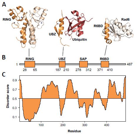
Genes 2020, 11, 138 2 of 15
Genes 2020, 11, 138 2 of 15
which is the E3 ubiquitin
ligase responsible for PCNAligase responsible for[8],
poly-ubiquitylation PCNA
play apoly-ubiquitylation [8], play a more
more direct role in orchestrating direct
the events
role in orchestrating the events leading to DNA damage
leading to DNA damage bypass than was originally thought. bypass than was originally thought.
In thisarticle,
In this article,wewe will
will discuss
discuss the regulation
the regulation of DNA
of these these damage
DNA damage bypass pathways
bypass pathways by
by ubiquitin
ubiquitin modifications of PCNA. We will focus on the structure, function, and interactions
modifications of PCNA. We will focus on the structure, function, and interactions of the E3 ubiquitin of the
E3 ubiquitin
ligases Rad18ligases
and Rad5Rad18 and
that areRad5 that arefor
responsible responsible
these PCNA for these PCNA modifications.
modifications. Then, we will Then, we
discuss
will discussideas
alternative alternative
aboutideas
how about how modifications
ubiquitin ubiquitin modifications regulate
regulate the the assembly
assembly of the multi-
of the multi-protein
protein complexes that carry out DNA
complexes that carry out DNA damage bypass. damage bypass.
2.
2. DNA
DNA Damage
Damage Bypass
Bypass and
and PCNA
PCNA Modifications
Modifications
The bypass
bypassofofdamaged
damaged DNA DNA is carried
is carried out byout
twobypathways:
two pathways: translesion
translesion synthesissynthesis and
and template
template
switchingswitching
(Figure 1).(Figure 1). In translesion
In translesion synthesis, thesynthesis,
stalled the stalled
classical DNAclassical DNA polymerase
polymerase is replaced byis
replaced by one or more specialized, non-canonical DNA polymerases [6,7,9–20].
one or more specialized, non-canonical DNA polymerases [6,7,9–20]. Each of these non-canonical Each of these non-
canonical DNA polymerases
DNA polymerases has evolvedhas evolved to
to efficiently efficientlynucleotides
incorporate incorporateopposite
nucleotides
one oropposite one or
more damaged
more
DNA damaged
templates.DNA templates.
For example, DNA Forpolymerase
example, DNA (pol)polymerase (pol) η (Rad30)
η (Rad30) efficiently efficiently
replicates throughreplicates
template
through template
thymine dimers thymine
and dimers [21,22].
8-oxoguanines and 8-oxoguanines
Similarly, Rev1 [21,22]. Similarly,
efficiently Rev1through
replicates efficiently replicates
minor-groove
through
adductedminor-groove adducted
guanines, exocyclic guanines,
guanine lesions,exocyclic
and abasic guanine lesions,
sites [23–25]. and
This abasic sites
pathway can be[23–25]. This
error-prone
pathway can be error-prone (i.e., mutagenic) or error-free (i.e., non-mutagenic) depending
(i.e., mutagenic) or error-free (i.e., non-mutagenic) depending on which non-canonical polymerase is on which
non-canonical polymerase
utilized to bypass the lesion.is utilized to bypass the lesion.
Figure 1. DNA damage bypass pathways and proliferating cell nuclear antigen (PCNA) modifications.
Figure 1. DNA damage bypass pathways and proliferating cell nuclear antigen (PCNA)
(A) The translesion synthesis pathway involves nucleotide incorporation using the damaged DNA as a
modifications. (A) The translesion synthesis pathway involves nucleotide incorporation using the
template. The template switching pathway involves the formation and extension of the chicken foot
damaged DNA as a template. The template switching pathway involves the formation and
intermediate. (B) Mono-ubiquitylation of PCNA by Rad6–Rad18 promotes translesion synthesis, and
extension of the chicken foot intermediate. (B) Mono-ubiquitylation of PCNA by Rad6–Rad18
poly-ubiquitylation of PCNA by Ubc13–Mms2–Rad5 promotes template switching. (C) Translesion
promotes translesion
synthesis involves synthesis, and
non-canonical poly-ubiquitylation
polymerases such as polofη,PCNA by Ubc13–Mms2–Rad5
pol ζ, and Rev1. promotes
template switching. (C) Translesion synthesis involves non-canonical polymerases such as pol η, pol
ζ,
Inand Rev1. switching, the stalled classical polymerase is replaced by a fork-remodeling helicase
template
[6,7,26–29]. In yeast, this fork-remodeling helicase is Rad5. Although the precise role of Rad5 in
In template
template switchingswitching, the stalled
remains unclear, thereclassical polymerase
is strong is replaced
in vitro evidence by that
showing a fork-remodeling
Rad5 uses the
helicase [6,7,26–29]. In yeast, this fork-remodeling helicase is Rad5. Although
energy derived from ATP hydrolysis to reverse the replication fork [29]. This leads the precise
to the role of
stalled
Rad5
strandinbase-pairing
template switching remains
with the newly unclear, there
synthesized is on
strand strong in vitro
the sister evidence
duplex showing
to form that Rad5
the “chicken foot”
uses the energy derived from ATP hydrolysis to reverse the replication fork [29]. This leads to the
stalled strand base-pairing with the newly synthesized strand on the sister duplex to form the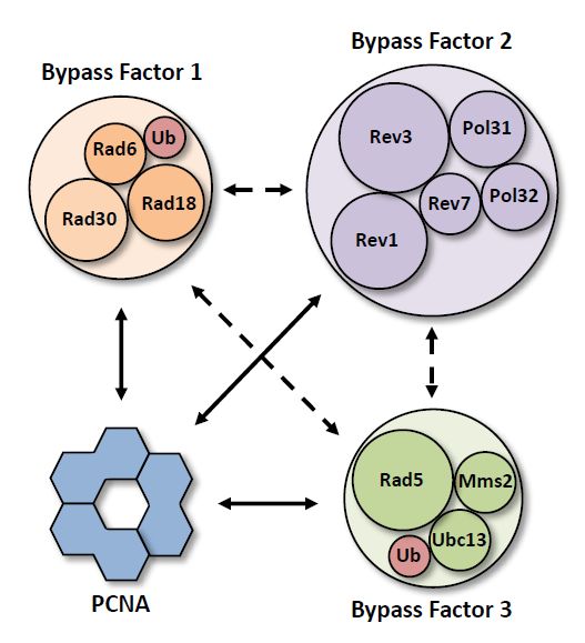
Genes 2020, 11, 138 3 of 15
Genes 2020, 11, 138 3 of 15
“chicken foot” intermediate (Figure 1) [29]. A DNA polymerase then extends the “middle toe” of
the chicken foot structure, and Rad5 finally remodels the chicken foot structure back to a replication
fork. This results
intermediate in the
(Figure 1) damage having
[29]. A DNA been bypassed,
polymerase and normal
then extends DNA replication
the “middle toe” of the can resume.
chicken It
foot
is important
structure, andtoRad5
note, however,
finally that the
remodels thechicken
fork reversal activityback
foot structure of Rad5 has not been
to a replication fork.confirmed in
This results
wild-type cells.
in the damage having been bypassed, and normal DNA replication can resume. It is important to note,
Thesethat
however, DNA damage
the fork bypass
reversal pathways
activity of Rad5 are
haslargely
not been regulated by in
confirmed thewild-type
ubiquitylation
cells. of PCNA
(Figure 1) DNA
These [8,30–40].
damage Thebypass
mono-ubiquitylation
pathways are largely of PCNA on lysine-164
regulated promotes translesion
by the ubiquitylation of PCNA
synthesis [8]. This is catalyzed by the E2 protein Rad6 and the E3 protein Rad18. This
(Figure 1) [8,30–40]. The mono-ubiquitylation of PCNA on lysine-164 promotes translesion synthesis [8]. is thought to
trigger the recruitment and activation of effector proteins such as the translesion
This is catalyzed by the E2 protein Rad6 and the E3 protein Rad18. This is thought to trigger the synthesis
polymerases
recruitment and η, pol ζ (Rev3–Rev7–Pol31–Pol32),
polactivation of effector proteins such asand the the Rev1 protein.
translesion synthesis polymerases pol η, pol
The K63-linked poly-ubiquitylation
ζ (Rev3–Rev7–Pol31–Pol32), and the Rev1 protein. of PCNA on lysine-164 promotes template switching [8].
This The
modification
K63-linked is catalyzed by the E2 protein
poly-ubiquitylation of PCNA Ubc13 with the E2-like
on lysine-164 promotesprotein Mms2
template and the [8].
switching E3
protein Rad5. Unlike mono-ubiquitylation, poly-ubiquitylation does not trigger
This modification is catalyzed by the E2 protein Ubc13 with the E2-like protein Mms2 and the E3 the recruitment of
an additional effector protein. This is because Rad5 is a dual-functional enzyme,
protein Rad5. Unlike mono-ubiquitylation, poly-ubiquitylation does not trigger the recruitment of being also the fork-
remodeling
an additional helicase
effectorthat forms the
protein. chicken
This foot intermediate
is because [29].
Rad5 is a dual-functional enzyme, being also the
fork-remodeling helicase that forms the chicken foot intermediate [29].
3. Rad6–Rad18
3. Rad6–Rad18
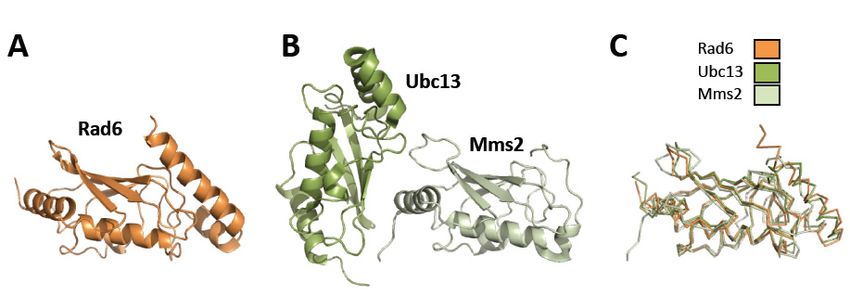
Rad6–Rad18 is the E2 ubiquitin-conjugating enzyme/E3 ubiquitin ligase that catalyzes the
mono-ubiquitylation
Rad6–Rad18 is theof PCNA [8]. Rad6 is a 20 kDa enzyme/E3
E2 ubiquitin-conjugating globular protein. The X-ray
ubiquitin ligasecrystal structuresthe
that catalyzes of
both yeast and human
mono-ubiquitylation of Rad6
PCNA have
[8]. been
Rad6 determined (Figure 2)
is a 20 kDa globular [41,42].The
protein. Both yeast
X-ray and human
crystal Rad6
structures of
proteins have
both yeast andahuman
nearly Rad6
identical
havefold to determined
been Ubc13, the E2 ubiquitin-conjugating
(Figure enzyme
2) [41,42]. Both yeast andthat catalyzes
human Rad6
the poly-ubiquitylation
proteins of PCNA,
have a nearly identical foldand to Mms2
to Ubc13, the E2[8].ubiquitin-conjugating
Rad18 is a 55 kDa enzymeprotein that
withcatalyzes
several
structured domains separated
the poly-ubiquitylation by intrinsically
of PCNA, and to Mms2disordered
[8]. Rad18 regions (Figure
is a 55 kDa 3). with several structured
protein
domains separated by intrinsically disordered regions (Figure 3).
2. Structures
Figure 2.
Figure StructuresofofRad6,
Rad6,Ubc13,
Ubc13,and
andMms2.
Mms2. (A)
(A)Ribbon
Ribbondiagram of Rad6
diagram (orange)
of Rad6 (1AYZ.pdb)
(orange) [41].
(1AYZ.pdb)
(B)
[41].Ribbon diagram
(B) Ribbon of Ubc13-Mms2
diagram (dark(dark
of Ubc13-Mms2 green and and
green lightlight
green, respectively)
green, (1JAT.pdb)
respectively) [43].[43].
(1JAT.pdb) (C).
Overlay of the structures of Rad6 (orange), Ubc13 (dark green), and Mms2 (light green).
(C). Overlay of the structures of Rad6 (orange), Ubc13 (dark green), and Mms2 (light green).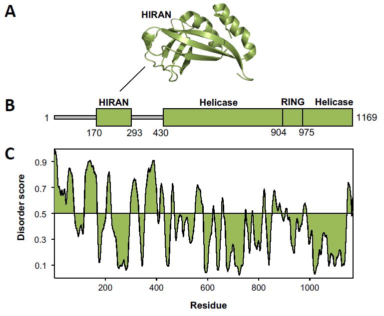
Genes 2020, 11, 138                                                                                                    4 of 15
Genes 2020, 11, 138                                                                                                    4 of 15

      Figure       Structure of
       Figure 3.3. Structure   of Rad18.   (A) Ribbon
                                  Rad18. (A)    Ribbon diagrams
                                                          diagrams ofof the
                                                                          the Rad18
                                                                              Rad18 Really
                                                                                       Really Interesting
                                                                                               Interesting New
                                                                                                             New Gene
                                                                                                                    Gene
      (RING)
       (RING) domain
               domain dimer
                          dimer (dark
                                 (dark orange
                                       orange and
                                                and light
                                                    light orange)
                                                           orange) (2Y43.pdb)
                                                                    (2Y43.pdb) [44],
                                                                                 [44], the
                                                                                        the Rad18
                                                                                            Rad18 Ubiquitin-Binding,
                                                                                                    Ubiquitin-Binding,
      Zinc-Binding
       Zinc-Binding (UBZ)
                      (UBZ) domain
                               domain (dark
                                       (dark orange)
                                              orange) bound
                                                       bound to to ubiquitin
                                                                   ubiquitin (red)
                                                                              (red) (2MRE.pdb)
                                                                                    (2MRE.pdb) [45],
                                                                                                   [45], and
                                                                                                         and the
                                                                                                               the Rad18
                                                                                                                   Rad18
      R6BD   domain    (dark  orange) bound   to Rad6  (light  orange)  (2YBF.pdb)   [42]. The
       R6BD domain (dark orange) bound to Rad6 (light orange) (2YBF.pdb) [42]. The zinc ions arezinc ions  are  shown as
                                                                                                                   shown
      spheres.   (B) A  linear diagram  of Rad18  shows    the structured   domains   as thick, orange
       as spheres. (B) A linear diagram of Rad18 shows the structured domains as thick, orange rectanglesrectangles  and
      the
       andintrinsically  disordered
            the intrinsically       regionsregions
                               disordered    as thin,as
                                                      grey  rectangles.
                                                        thin,            (C) A disorder
                                                               grey rectangles.  (C) A plot   of Rad18
                                                                                          disorder  plot was   generated
                                                                                                          of Rad18   was
      using  the  Protein  Disorder  Prediction  Server  [46].
       generated using the Protein Disorder Prediction Server [46].

    Unlike some E2:E3 pairs, which interact transiently and are often found not bound together,
     Unlike some E2:E3 pairs, which interact transiently and are often found not bound together,
Rad6–Rad18 form a tight complex comprised of two Rad18 molecules and one Rad6 molecule [47].
Rad6–Rad18 form a tight complex comprised of two Rad18 molecules and one Rad6 molecule [47].
While Rad6 is stable alone and is present in cells in high copy number, Rad18 is not found in the
While Rad6 is stable alone and is present in cells in high copy number, Rad18 is not found in the
absence of Rad6 and is approximately 10 times less abundant in cells than Rad6 [48,49].
absence of Rad6 and is approximately 10 times less abundant in cells than Rad6 [48,49].
3.1. Structure and Function of Rad18
3.1. Structure and Function of Rad18
      Structural studies of Rad18 have been limited to its structured domains and motifs. The structured
       Structural studies of Rad18 have been limited to its structured domains and motifs. The
domains include the Really Interesting New Gene (RING) domain, Ubiquitin-Binding, Zinc-Binding
structured domains include the Really Interesting New Gene (RING) domain, Ubiquitin-Binding,
(UBZ) domain, SAF-A/B, Acinus, Pias (SAP) domain, and Rad6-Binding Domain (R6BD) (Figure 3). The
Zinc-Binding (UBZ) domain, SAF-A/B, Acinus, Pias (SAP) domain, and Rad6-Binding Domain
regions of Rad18 between these structured domains are intrinsically disordered [46,50]. Intrinsically
(R6BD) (Figure 3). The regions of Rad18 between these structured domains are intrinsically
disordered regions are often sites for protein–protein interactions [51,52]. In Rad18, two such interaction
disordered [46,50]. Intrinsically disordered regions are often sites for protein–protein interactions
motifs have been identified: a SUMO-interaction motif (SIM) and a pol η-binding region.
[51,52]. In Rad18, two such interaction motifs have been identified: a SUMO-interaction motif (SIM)
and aThe
3.1.1.      η-binding
        pol RING       region.
                   Domain

3.1.1.The
       TheX-ray
            RINGcrystal
                    Domain structure of the human Rad18 RING domain shows that this domain forms a
homodimer (Figure 3) [44]. The RING domain (corresponding to residues 28 to 65 of yeast Rad18)
      The X-ray
is located   at thecrystal structure
                     N-terminus    of of the human
                                      Rad18.         Rad18
                                               This is       RINGzinc
                                                        a C3 HC   domain   shows
                                                                      finger      that this
                                                                             comprised    of domain  forms a
                                                                                              seven cysteine
                                                                4
homodimer      (Figure  3) [44]. The RING    domain   (corresponding to residues 28 to 65 of  yeast
residues (corresponding to residues 28, 31, 43, 48, 51, 62, and 65 of yeast Rad18) and one histidineRad18) is
located   at  the  N-terminus     of Rad18.   This is  a  C3HC4 zinc finger comprised of seven cysteine
residue (corresponding to residue 45 of yeast Rad18) that coordinate two zinc ions. Two α-helicases
residues (corresponding to residues 28, 31, 43, 48, 51, 62, and 65 of yeast Rad18) and one histidine
residue (corresponding to residue 45 of yeast Rad18) that coordinate two zinc ions. Two α-helicases
Genes 2020, 11, 138 5 of 15
(corresponding to residues 15 to 25 and residues 78 to 94 of yeast Rad18) flank the zinc finger. These
α-helicases form a four-helix bundle in the RING domain homodimer. This four-helix bundle and a
series of hydrogen bonds between residues of the zinc finger motifs stabilize the RING domain dimer.
While RING domain dimerization is required for the function of several other RING E3 ubiquitin
ligases, it is not known if this dimerization is required for the ligase function of Rad18.
The RING domain of Rad18 binds to Rad6 via a characteristic RING-E2 interface consisting of
both hydrophobic and electrostatic interactions [44]. The RING domain binds Rad6 with a dissociation
constant in the low micromolar range, and mutations in the RING domain that block this interaction
greatly reduce Rad18’s ubiquitin ligase activity [44].
3.1.2. The UBZ Domain
The NMR structure of the human Rad18 UBZ domain (corresponding to residues 187 to 210 of
yeast Rad18) shows that this domain has an overall β-strand/β-strand/α-helix fold (Figure 3) [45]. It is
a C2 HC zinc-binding domain comprised of three cysteine residues (corresponding to residues 190, 193,
and 210 of yeast Rad18) and one histidine residue (corresponding to residue 206 of yeast Rad18). NMR
titrations show that the Rad18 UBZ domain binds to ubiquitin with a dissociation constant in the low
micromolar range [45]. Moreover, the α-helix of the Rad18 UBZ (corresponding to residues 200 to 211
of yeast Rad18) and the β-strand 1 (corresponding to residues 187 to 190 of yeast Rad18) interact with
the canonical hydrophobic patch of ubiquitin comprised of leucine-8, isoleucine-44, and valine-70.
Interestingly, mutations in the UBZ domain do not block the ability of Rad18 to function as a
ubiquitin ligase during the mono-ubiquitylation of PCNA (47). This suggests that this domain may be
involved in a function of Rad18 other than translesion synthesis. In fact, the UBZ domain is essential
for Rad18 auto-ubiquitylation [53], which localizes Rad18 to the cytoplasm making it unavailable to
ubiquitylate nuclear proteins such as PCNA and Replication Factor C (RFC), the protein that loads
PCNA onto DNA.
3.1.3. The SAP Domain
This small domain (residues 278 to 312) is named after the three proteins in which it was first
identified: SAF-A/B, Acinus, and Pias [54]. The structure of the Rad18 SAP domain has not yet been
determined, but homology models predict that it has a helix–turn–helix fold similar to that of other
SAP domains. This domain binds both single- and double-stranded DNA with dissociation constants
in the low micromolar range [55]. It is not known, however, whether these DNA binding activities
are necessary for Rad18 ligase function. Inactivation of the SAP domain does, however, affect the
recruitment of non-canonical pol η to sites of DNA damage, indicating that this domain plays an
important but poorly understood role in the recruitment of non-canonical DNA polymerases [53].
3.1.4. The Rad6-Binding Domain
The R6BD is a small domain (residues 371 to 410) near the C-terminus of Rad18 that binds Rad6.
This domain has a helix–loop–helix fold (Figure 3) [42]. This domain binds to the opposite side of
Rad6 from the site of ubiquitin conjugation (cysteine-88) and interacts with β-strand 1, β-strand 2, and
β-strand 3 of Rad6 [42]. Both surface plasmon resonance and NMR titrations show that the dissociation
constant for R6BD and Rad6 is in the mid micromolar range [42]. Mutations in the R6BD in human
Rad18 decrease the binding affinity of Rad18 for Rad6 and substantially reduce Rad18’s ubiquitin ligase
activity. While the RING domain and the R6BD of Rad18 both bind Rad6, the functional significance of
this bidentate interaction is unclear. One possibility is that, like other bidentate E2–E3 interactions, the
RING domain can transiently dissociate from E2 facilitating the re-charging of E2 by E1 without the
complex dissociating [56]. This could allow for the processive ubiquitylation of the PCNA subunits
by Rad6–Rad18.Genes 2020, 11, 138 6 of 15
3.1.5. The SUMO-Interacting Motif
Yeast Rad18 contains a SIM (residues 139 to 142) in the predicted disordered region between
the RING domain and the UBZ domain [57]. Conserved hydrophobic residues in the yeast SIM
(leucine-139, isoleucine-141, and valine-142) are necessary for interaction with PCNA. Moreover, a
fraction of PCNA during normal DNA replication is sumoylated, and the sumoylation of PCNA
stimulates the ubiquitin ligase activity of Rad18. These findings support the notion that Rad18 is a
SUMO-directed ubiquitin ligase [57].
3.1.6. The Pol η-Binding Region
Pol η is a non-canonical DNA polymerase that carries out translesion synthesis through
thymine–thymine dimers and 8-oxoguanine lesions [21,22]. Human pol η binds to a region in
the disordered C-terminus of human Rad18 following the R6BD [55]. This interaction is dependent on
phosphorylation of serine-409 of human Rad18 [58]. The lack of phosphorylation at this site results in
an increased sensitivity to ultraviolet radiation and a decrease in nuclear foci containing pol η.
3.2. Other Interactions of Rad18
As described above, Rad18 interacts with Rad6, pol η, and PCNA. In addition to these proteins,
Rad18 interacts with several other proteins involved in DNA damage bypass including RPA (replication
protein A), Rev1, and Rad5.
3.2.1. Replication Protein A
Replication Protein A (RPA) is a single-stranded DNA-binding protein involved in DNA replication,
repair, and recombination [59]. It has three subunits: Rfa1, Rfa2, and Rfa3. The interaction between
yeast RPA and Rad18 involves the Rfa1 and Rfa2 subunits [60]. This interaction is disrupted by the
deletion of residues 112 to 192 of Rad18, which contain the SIM [60]. It is unclear, however, whether
this is due to the loss of a direct interaction with RPA or of an indirect interaction with RPA through
sumoylated PCNA or some other mediator protein.
3.2.2. Rev1
Rev1 has two functions in DNA damage bypass. First, it is a non-canonical DNA polymerase
that catalyzes translesion synthesis of minor-groove and exocyclic guanine adducts as well abasic
sites [23–25]. Second, it is a structural protein that can simultaneously bind PCNA and other
non-canonical DNA polymerases forming a Rev1 bridge [61,62]. In this way, it can help organize
the structure of the multi-protein complexes that carry out DNA damage bypass. Rad18 and Rev1
have been shown to physically interact [63]. Despite this, however, the structural basis and functional
implications of this interaction remain poorly understood.
3.2.3. Rad5
Rad5 is the E3 ubiquitin ligase that is necessary for the template-switching pathway of DNA
damage bypass (see below). Rad18 and Rad5 interact with each other [64]. This interaction has been
partially mapped to residues 83 to 246 of Rad18, which contain both the SIM and the UBZ domain [64].
More work is needed to understand the structural basis and functional implications of this interaction.
4. Ubc13–Mms2–Rad5
Ubc13–Mms2–Rad5 is the E2 ubiquitin-conjugating enzyme/E3 ubiquitin ligase that catalyzes the
K63-linked poly-ubiquitylation of PCNA [8]. Ubc13 is an 18 kDa globular protein and is the actual
E2 ubiquitin-conjugating enzyme. It forms a heterodimer with Mms2, a 16 kDa globular protein that
is a ubiquitin-conjugating enzyme variant. This heterodimer is required for DNA damage bypass
by template switching. The X-ray crystal structures of yeast and human Ubc13–Mms2 have beenGenes 2020, 11, 138 7 of 15
Genes 2020, 11, 138 7 of 15
Mms2 havewith
determined beenand determined with andcovalently
without ubiquitin without ubiquitin
attached to covalently attached
Ubc13 (Figure to Ubc13
2) [43,65]. The(Figure 2)
structure
[43,65].
of Ubc13The structure
is similar of Ubc13
to that is similar
of other to that
E2 proteins, of other
such E2 proteins,
as Rad6. Mms2 hassuch as Rad6.
a similar foldMms2 has a
with slight
similar fold with
modifications slight itmodifications
allowing allowing
to form a binding it tofor
surface form a binding surface for Ubc13.
Ubc13.
Yeast Rad5
Yeast Rad5 is aa 134
134kDakDaprotein
proteinthat
thathas
hastwotwofunctions
functions[66,67]. First,First,
[66,67]. it is an E3an
it is ubiquitin ligase
E3 ubiquitin
that binds
ligase Ubc13–Mms2
that binds Ubc13–Mms2and forms K63-linked
and forms poly-ubiquitin
K63-linked chainschains
poly-ubiquitin on mono-ubiquitylated PCNA
on mono-ubiquitylated
[8]. Second,
PCNA it is aitfork-remodeling
[8]. Second, helicase
is a fork-remodeling thatthat
helicase converts
convertsstalled replication
stalled replicationforks
forksto
to chicken foot
foot
intermediates [29].
intermediates [29]. Like
Like Rad18,
Rad18, Rad5
Rad5 has
has structured
structured domains
domains separated
separated by by intrinsically
intrinsically disordered
disordered
regions(Figure
regions (Figure4).4).Unlike
UnlikeRad18,
Rad18,however,
however,Rad5Rad5cancanbebeoverexpressed
overexpressedwithout
withoutUbc13–Mms2.
Ubc13–Mms2.
Figure 4. Structure of Rad5. (A) Ribbon diagram of the Rad5 HIP116 Rad5p N-terminal (HIRAN)
Figure 4. Structure of Rad5. (A) Ribbon diagram of the Rad5 HIP116 Rad5p N-terminal (HIRAN)
domain (green) (4XZG.pdb) [68]. (B) A linear diagram of Rad5 shows the structured domains as thick,
domain (green) (4XZG.pdb) [68]. (B) A linear diagram of Rad5 shows the structured domains as
green rectangles and the intrinsically disordered regions as thin, grey rectangles. (C) A disorder plot of
thick, green rectangles and the intrinsically disordered regions as thin, grey rectangles. (C) A
Rad5 was generated using the Protein Disorder Prediction Server [46].
disorder plot of Rad5 was generated using the Protein Disorder Prediction Server [46].
4.1. Structure and Function of Rad5
4.1. Structure and Function of Rad5
Currently, there is no high-resolution structural information about Rad5. Our limited structural
Currently,ofthere
understanding is no
Rad5 has beenhigh-resolution structuralmodeling
obtained from homology information
of its about Rad5.
structured Our limited
domains. These
structural understanding of Rad5 has been obtained from homology modeling
domains include the HIP116 Rad5p N-terminal (HIRAN) domain, the helicase domain, and the of its structured
domains.
RING These domains include the HIP116 Rad5p N-terminal (HIRAN) domain, the helicase
domain.
domain, and the RING domain.
4.1.1. HIRAN Domain
4.1.1. HIRAN Domain
The HIRAN domain of Rad5 (corresponding to residues 170 to 293 of yeast Rad5) is located
TheN-terminus
near the HIRAN domain of theofprotein
Rad5 (corresponding
and is flanked by to intrinsically
residues 170disordered
to 293 of yeast Rad5)
regions. is located
While there
near
are nothe N-terminus ofstructures
high-resolution the protein anddomain,
of this is flanked
theby intrinsically
HIRAN domaindisordered regions.
of Helicase-Like While there
Transcription
are no (HLTF),
Factor high-resolution
a human structures
homologofofthis domain,
Rad5, the HIRAN
has been domain
determined of Helicase-Like
(Figure 4) [68–70]. Transcription
The HIRAN
Factor (HLTF),
domain containsaanhuman homolog of Rad5, has been determined
oligosaccharide/oligonucleotide-binding (OB) fold(Figure
that binds the 30 ends
4) [68–70]. The of
HIRAN
DNA.
domain contains an oligosaccharide/oligonucleotide-binding (OB) fold that binds the 3′ ends of
DNA. Recognition of the 3′ OH is important for HIRAN binding, and the structure of the HIRANGenes 2020, 11, 138 8 of 15
Recognition of the 30 OH is important for HIRAN binding, and the structure of the HIRAN domain
prevents binding of double-stranded DNA. This suggests that the 30 end of a DNA strand must be
unwound before the HIRAN domain can bind it. The HIRAN domains of Rad5 and of HLTF are
important for fork-remodeling activity. It has been proposed that the HIRAN domain is important for
substrate recognition and positioning of the helicase domain on the DNA substrate [71].
The HIRAN domain of HLTF is required for unwinding forked DNA substrates containing
double-stranded DNA on both the leading and the lagging strand of the fork without single-stranded
gaps at the fork junction [71]. By contrast, this domain is not as important when unwinding forked
DNA substrates with single-stranded gaps on either the leading or the lagging strand. However,
unwinding of substrates with gaps on the lagging strand was more hindered by the deletion of the
HIRAN domain than by substrates with gaps on the leading strand. This suggests that a functional
HIRAN domain is more important when the 30 end of the leading strand is located immediately at the
fork junction [71].
4.1.2. Helicase Domain
Rad5 is a member of the Swi/Snf subfamily within the SF2 superfamily of helicases [72,73]. The
helicase domain of Rad5 (which begins around residue 430 of yeast Rad5 and ends at residue 1169)
spans the C-terminal half of the protein. Like other helicases in this superfamily, the Rad5 helicase
domain contains seven conserved motifs, including the Walker A and Walker B ATP-binding motifs.
In the structures of other SF2 helicases, these conserved motifs constitute an ATP-binding site between
the two RecA-like folds that make up the helicase domain [73]. Moreover, the helicase domain also
binds the DNA substrate.
The helicase domain is largely responsible for the ATPase and fork-remodeling activity of Rad5 [29].
The ATPase activity of Rad5 is stimulated by single-stranded DNA as well as double-stranded DNA
in the context of three-way junctions (forks) and four-way junctions (chicken foot structures and
Holiday junctions). In addition, Rad5 can unwind forked substrates with homologous arms (i.e., ones
in which the primer strands of the leading and lagging arms are complementary to one another) but
not heterologous arms [29]. Moreover, Rad5 is capable of regressing several hundred base pairs of
DNA. Whether this is the result of processive or distributive unwinding is unclear.
In the case of human HLTF, footprinting experiments suggest that translocation occurs in the
0 0
3 –5 direction, with the helicase domain positioned on the lagging strand template of the parental
duplex in front of the fork [71]. For Rad5, ATP binding to the helicase domain induces the unwinding
of the primer strand of the leading arm from the template strand, creating a free 30 end [74]. This free
30 end can then capture the primer strand of the lagging arm to form a four-way junction—a chicken
foot structure or a Holiday junction. Formation of this four-way junction has been proposed to be the
rate-limiting step of fork reversal [74].
4.1.3. RING Domain
Swi/Snf helicases often contain a small, folded domain between the two RecA-like folds of the
helicase domain [73]. In the case of Rad5, there is a RING domain (residues 904 to 975) inserted at this
position. Homology models based on X-ray crystal structures of other RING domains show that this is
a C3 HC4 zinc finger-type domain comprised of seven cysteine residues (residues 914, 917, 932, 937,
940, 957, and 960) and one histidine residue (residue 934) that coordinate zinc ions [75]. There is no
evidence of the RING domain of Rad5 forming dimers. Instead, the RING domain binds Ubc13 and is
important for Rad5’s E3 ubiquitin ligase activity [64].
4.2. Structural Models of Full-Length Rad5
Though Rad5 has several structured domains, approximately 30% of it is likely unstructured.
There are two large intrinsically disordered regions: one at the N-terminus (starting at residue 1 and
ending at residue 169 in yeast Rad5), and one between the HIRAN domain and the helicase domainGenes 2020, 11, 138 9 of 15
(starting at residue 294 and ending around residue 525 in yeast Rad5). The role of these unstructured
regions is likely to provide conformational flexibility to Rad5, allowing the HIRAN domain and the
helicase domain to reorient themselves with respect to one another. In addition, these regions likely
mediate protein–protein interactions.
A recent structural model of full-length Rad5 has been proposed on the basis of a combination of
molecular simulations and small-angle X-ray scattering (SAXS) data [75]. The molecular simulation
generated an ensemble of 5000 structures, which collectively agree well with the SAXS data. In this
structural model, the disordered regions are highly flexible and adopt many conformational states.
Interestingly, this structural model revealed the presence of an intra-molecular interaction between
the HIRAN and the helicase domains, which resulted in a more compact structure than what would
have been expected given the high degree of intrinsic disorder in the protein [75]. It should be noted,
however, that the SAXS data only examined the protein in the absence of ATP, DNA, and other binding
partners. Thus, it is unclear whether the putative HIRAN domain–helicase domain interaction persists
in the presence of these ligands.
4.3. Other Interactions of Rad5
As described above, Rad5 interacts with Ubc13 and Mms2. In addition to these proteins, Rad5
interacts with several other proteins including PCNA, Rev1, and Rad18.
4.3.1. PCNA
Rad5 binds PCNA through its N terminus (amino acids 1–430) [8]. The precise residues involved
in the interaction are unknown. This region contains a putative PCNA-interacting protein PIP-like
motif (residues 6 to 13), which binds Rev1 (see below) [31,76]. It is not known if this motif mediates
interactions with PCNA as well. Rad5 binds unmodified PCNA and mono-ubiquitylated PCNA with
similar affinities [77].
4.3.2. Rev1
Like Rad18, Rad5 interacts with Rev1, the non-canonical polymerase that helps to organize the
structure of the complex that carries out DNA damage bypass [76]. Based on this, it has been suggested
that Rad5 may regulate Rev1-mediated translesion synthesis. The structural basis of this interaction
is known. An X-ray crystal structure shows that the Rev1 C-terminal domain, which binds PIP-like
motifs, binds to a region of Rad5 containing a PIP-like motif (residues 6 to 13) [76]. As mentioned
above, this is possibly the same motif that Rad5 uses to bind PCNA, although this has not yet been
experimentally tested.
4.3.3. Rad18
As described above, Rad5 interacts with Rad18, the E3 ubiquitin ligase that is necessary for
translesion synthesis [64]. This interaction has been partially mapped to a region of Rad5 spanning
residues 1 to 556, which contains the N-terminal disordered regions and the HIRAN domain. The
structural basis and functional implications of this interaction are unclear. However, given that
Rad5 binds to unmodified PCNA and to mono-ubiquitylated PCNA with the same affinity [77],
the interaction between Rad5 and Rad18 may play a role in recruiting Rad5–Ubc13–Mms2 to
mono-ubiquitylated PCNA.
5. The Role of PCNA Modifications in DNA Damage Bypass
The coordination of these DNA damage bypass pathways is not well understood. A common
view of the role of Rad6–Rad18 and of Ubc13–Mms2–Rad5 in regulating DNA damage bypass is
as follows. When replication forks stall upon encountering DNA damage, Rad6–Rad18 catalyzes
the mono-ubiquitylation of PCNA. This signals for the recruitment of non-canonical polymerasesGenes 2020,
Genes 11, 138
2020, 11, 10
10 of
of 15
15
The protein–protein interactions discussed above and several observations in the literature cast
such as
doubt onpol Rev1, andmodel.
theη,traditional pol ζ. For
If translesion
example, polsynthesis fails,with
η interacts thenthe
Ubc13–Mms2–Rad5
Rad6–Rad18 complex, catalyzes the
and this
poly-ubiquitylation of PCNA. This signals for fork reversal and template switching.
interaction is essential for the recruitment of these proteins to stalled replication forks [63,78–80].
The protein–protein
Moreover, the presence of interactions discussed
pol η increases theabove and several
efficiency observations
with which Rad6–Rad18 in the literature
catalyzes cast
the
doubt on the traditional model. For example, pol η interacts with
mono-ubiquitylation of PCNA both in vitro and in vivo [80]. This strongly suggests that polthe Rad6–Rad18 complex, andη
this interaction
directly is essential
facilitates for the
the transfer recruitment
of the ubiquitinoffrom
thesetheproteins to stalled
Rad6 protein toreplication
PCNA. This forks [63,78–80].
likely occurs
Moreover,
because polthe presence of
η stabilizes thepol η increases
complex the efficiency
of Rad6–Rad18 andwithPCNA whichin aRad6–Rad18
more catalyticallycatalyzes the
active
mono-ubiquitylation of PCNA both in vitro and in vivo [80]. This strongly
conformation. Regardless of the specifics of the mechanism, this implies that pol η is already suggests that pol η directly
facilitates
present in the
thetransfer
complexofwiththe ubiquitin
Rad6–Rad18 fromand the with
Rad6 PCNA
proteinwhento PCNA. This likely
the ubiquitin occurs reaction
transfer because
pol η
occurs.stabilizes the complex of Rad6–Rad18 and PCNA in a more catalytically active conformation.
Regardless
These of the specifics
pieces of evidenceof thesuggest
mechanism, that this
the implies that pol η is already
mono-ubiquitylation present in the complex
and poly-ubiquitylation of
with Rad6–Rad18 and with PCNA when the ubiquitin transfer reaction
PCNA do not merely act as signals to recruit the non-canonical polymerases and fork-remodelingoccurs.
These
helicase. pieces they
Instead, of evidence
suggestsuggest
that thethat the mono-ubiquitylation
ubiquitylation machinery plays and apoly-ubiquitylation of PCNA
more direct role, recruiting
these other proteins. According to this alternative view, DNA damage bypass is carried outhelicase.
do not merely act as signals to recruit the non-canonical polymerases and fork-remodeling by the
Instead, they suggest that the ubiquitylation machinery plays a more direct role,
coordinated activity of three multi-protein factors (Figure 5). The first factor, which we dub Bypass recruiting these other
proteins. According to this alternative view, DNA damage bypass is carried
Factor 1, is comprised of Rad6–Rad18 and pol η. The second factor, which we refer to as Bypass out by the coordinated
activity2,ofisthree
Factor multi-protein
comprised of Rev1factors
and pol(Figure
ζ. The5). third
The first factor,
factor, whichwhich
we we calldub
BypassBypass Factor
Factor 1,
3, is
is comprised of Rad6–Rad18 and pol η. The second factor, which we
comprised of Ubc13–Mms2–Rad5. These factors may act sequentially or may assemble into a large refer to as Bypass Factor 2,
is comprised
complex of Rev1replication
at a stalled and pol ζ.fork, Theathird factor, whichcomprised
“bypassosome”, we call Bypass
of PCNA Factorand3,the is comprised
three bypassof
Ubc13–Mms2–Rad5. These factors may act sequentially or may assemble into a large complex at a
factors.
stalled replication fork, a “bypassosome”, comprised of PCNA and the three bypass factors.
Figure 5. Composition of the putative bypass factors. Diagrams showing the protein components of
Figure
putative5. Bypass
Composition
Factor of the putative
1 (orange) bypasspol
containing factors. Diagrams
η (Rad30), showing
Bypass Factor the proteincontaining
2 (purple) components polofζ
putative Bypass Factor 1 (orange)
(Rev3–Rev7–Pol31–Pol32), containing
and Bypass Factor 3pol η (Rad30),
(green) Bypass
containing Factor
Rad5. 2 (purple)
Ubiquitin containing
moieties pol
are shown
ζin(Rev3–Rev7–Pol31–Pol32), and Bypass Factor 3 (green) containing Rad5. Ubiquitin moieties
red. Solid arrows represent direct interactions with PCNA that allow for the formation of PCNA tool are
shown
belts. Dashed arrows represent interactions that allow for the formation of alternative architectures.of
in red. Solid arrows represent direct interactions with PCNA that allow for the formation
PCNA tool belts. Dashed arrows represent interactions that allow for the formation of alternative
architectures.
Whether or not there is a bypassosome, the evidence reviewed here provides a compelling case for
the presence of three bypass factors. According to this model, Bypass Factor 1 is directed to the stalled
Whether
replication or via
fork not its
there is a bypassosome,
Rad6–Rad18 the evidence
components. reviewed
In this way, here
its pol provides a rides
η component compelling case
piggyback
for the presence of three bypass factors. According to this model, Bypass Factor 1 is directed
on its Rad6–Rad18 components. If this model is correct, then pol η is already present at the stalled to the
stalled replication
replication fork whenforkthevia its Rad6–Rad18
ubiquitin moiety is components.
transferred to In this way, its pol η component rides
PCNA.
piggyback on its Rad6–Rad18 components. If this model is correct, then pol η is already present at
the stalled replication fork when the ubiquitin moiety is transferred to PCNA.Genes 2020, 11, 138 11 of 15
Next, Bypass Factor 2 is directed to the stalled fork via interactions of its Rev1 component with
ubiquitin-modified PCNA or with the pol η component of Bypass Factor 1. Bypass Factor 2 may also
be directed to stalled replication forks via interactions of its pol ζ with ubiquitin-modified PCNA.
Finally, Bypass Factor 3 is directed to the stalled replication fork via the E2 ubiquitin-conjugating
and E3 ubiquitin ligase activities of Ubc13–Mms2–Rad5. Because Rad5 also possesses a fork-remodeling
helicase activity, this activity rides piggyback on the ubiquitylation activity. This factor may also
be recruited via interactions of its Rad5 component with mono-ubiquitylated PCNA, with the
Rad18 component of Bypass Factor 1, or with the Rev1 component of Bypass Factor 2. Because
poly-ubiquitylation of PCNA likely requires the mono-ubiquitylation of PCNA, the recruitment of
Bypass Factor 3 likely occurs after the recruitment of Bypass Factor 1.
6. Conclusions
In this review, we have discussed the regulation of translesion synthesis and template switching
by post-translational modifications of PCNA. We have focused on the structures, functions, and
interactions of the E3 ubiquitin ligases Rad18 and Rad5, which facilitate the mono-ubiquitylation and
poly-ubiquitylation of PCNA. We have focused on these E3 ubiquitin ligases because they play an
important and arguably more direct role in orchestrating the steps of translesion synthesis and template
switching than was initially thought. Unfortunately, we still know very little about the structures,
functions, and interactions of these proteins.
One reason we know so little about these E3 ubiquitin ligases is that they contain extensive
regions of intrinsic disorder conferring to them a high degree of conformational flexibility. Another
reason is that they are components of large, dynamic, multi-protein complexes that likely change
their composition during the course of the DNA damage bypass process. Consequently, these are
extremely difficult proteins to study from a structural and biochemical perspective because traditional
experimental approaches, such as X-ray crystallography, NMR, and even cryo-electron microscopy, do
not have the capability to tackle such conformationally and compositionally flexible systems.
Fortunately, new approaches, both experimental and computational, are emerging that should
allow us to gain valuable new structural and biochemical information about such conformationally
flexible, multi-protein complexes. For example, hybrid approaches, such as full ensemble methods that
combine small-angle X-ray scattering data and molecular dynamics simulations, should be extremely
useful in studying such systems [81]. Similarly, single-molecule total internal reflection fluorescence
microscopy studies of the assembly and composition of multi-protein complexes should also prove
useful [82]. Ultimately, these emerging technologies should provide important and exciting insights
into these DNA damage bypass pathways and the maintenance of genome stability.
Author Contributions: Conceptualization, B.M.R., M.S.G., and M.T.W.; writing—original draft preparation,
B.M.R., M.S.G., and M.T.W.; writing—reviewing and editing B.M.R., M.S.G., and M.T.W.; project administration,
M.T.W.; funding acquisition, M.T.W. All authors have read and agreed to the published version of the manuscript.
Funding: This research was funded by the National Institute of General Medical Sciences, grant number GM081433.
The content is solely the responsibility of the authors and does not necessarily reflect the official views of the
National Institute of General Medical Sciences or the National Institutes of Health.
Acknowledgments: We thank Christine Kondratick, Justin Ling, Devin Reusch, Kyle Powers, and Elizabeth
Boehm for discussions.
Conflicts of Interest: The authors declare no conflicts of interest.
Abbreviations
HIRAN, HIP116 Rad5p N-terminal; HLTF, helicase-like transcription factor; PCNA, proliferating cell nuclear
antigen; PIP, PCNA-interacting protein; pol, polymerase; R6BD, Rad6-binding domain; RFC, replication factor C;
RING, really interesting new gene; RPA, replication protein A; SAP, SAF-A/B, Acinus, Pias; SAXS, small-angle
X-ray scattering; SIM, SUMO-interacting motif; UBZ, ubiquitin-binding, zinc-binding.Genes 2020, 11, 138 12 of 15
References
1. Friedberg, E.C.W.; Walker, G.C.; Siede, W.; Wood, R.D.; Schultz, R.A.; Ellenberger, T. DNA Repair and
Mutagenesis, 2nd ed.; American Society of Microbiology Press: Washington, DC, USA, 2006.
2. Hanahan, D.; Weinberg, R.A. Hallmarks of cancer: The next generation. Cell 2011, 144, 646–674. [CrossRef]
3. Kennedy, S.R.; Loeb, L.A.; Herr, A.J. Somatic mutations in aging, cancer and neurodegeneration. Mech.
Ageing Dev. 2012, 133, 118–126. [CrossRef] [PubMed]
4. Veltman, J.A.; Brunner, H.G. De novo mutations in human genetic disease. Nat Rev. Genet. 2012, 13, 565–575.
[CrossRef] [PubMed]
5. Acuna-Hidalgo, R.; Veltman, J.A.; Hoischen, A. New insights into the generation and role of de novo
mutations in health and disease. Genome Biol. 2016, 17, 241. [CrossRef] [PubMed]
6. Boiteux, S.; Jinks-Robertson, S. DNA repair mechanisms and the bypass of DNA damage in Saccharomyces
cerevisiae. Genetics 2013, 193, 1025–1064. [CrossRef]
7. Marians, K.J. Lesion Bypass and the Reactivation of Stalled Replication Forks. Ann. Rev. Biochem. 2018, 87,
217–238. [CrossRef]
8. Hoege, C.; Pfander, B.; Moldovan, G.L.; Pyrowolakis, G.; Jentsch, S. RAD6-dependent DNA repair is linked
to modification of PCNA by ubiquitin and SUMO. Nature 2002, 419, 135–141. [CrossRef]
9. Friedberg, E.C.; Wagner, R.; Radman, M. Specialized DNA polymerases, cellular survival, and the genesis of
mutations. Science 2002, 296, 1627–1630. [CrossRef]
10. Lawrence, C.W. Cellular roles of DNA polymerase zeta and Rev1 protein. DNA Repair 2002, 1, 425–435.
[CrossRef]
11. Prakash, S.; Prakash, L. Translesion DNA synthesis in eukaryotes: A one- or two-polymerase affair. Genes
Dev. 2002, 16, 1872–1883. [CrossRef]
12. Lehmann, A.R. Replication of damaged DNA. Cell Cycle 2003, 2, 300–302. [CrossRef] [PubMed]
13. Prakash, S.; Johnson, R.E.; Prakash, L. Eukaryotic translesion synthesis DNA polymerases: Specificity of
structure and function. Ann. Rev. Biochem. 2005, 74, 317–353. [CrossRef] [PubMed]
14. Lehmann, A.R. Replication of damaged DNA by translesion synthesis in human cells. FEBS Lett. 2005, 579,
873–876. [CrossRef] [PubMed]
15. Lehmann, A.R.; Niimi, A.; Ogi, T.; Brown, S.; Sabbioneda, S.; Wing, J.F.; Kannouche, P.L.; Green, C.M.
Translesion synthesis: Y-family polymerases and the polymerase switch. DNA Repair 2007, 6, 891–899.
[CrossRef] [PubMed]
16. Waters, L.S.; Minesinger, B.K.; Wiltrout, M.E.; D’Souza, S.; Woodruff, R.V.; Walker, G.C. Eukaryotic translesion
polymerases and their roles and regulation in DNA damage tolerance. Microbiol. Mol. Biol. Rev. 2009, 73,
134–154. [CrossRef] [PubMed]
17. Washington, M.T.; Carlson, K.D.; Freudenthal, B.D.; Pryor, J.M. Variations on a theme: Eukaryotic Y-family
DNA polymerases. Biochim. Biophys. Acta 2010, 1804, 1113–1123. [CrossRef]
18. Sale, J.E.; Lehmann, A.R.; Woodgate, R. Y-family DNA polymerases and their role in tolerance of cellular
DNA damage. Nat. Rev. Mol. Cell Biol. 2012, 13, 141–152. [CrossRef]
19. Pryor, J.M.; Dieckman, L.M.; Boehm, E.M.; Washington, M.T. Eukaryotic Y-Family Polymerases: A Biochemical
and Structural Perspective. Nucleic Acids Mol. Biol. 2014, 30, 85–108.
20. Powers, K.T.; Washington, M.T. Eukaryotic translesion synthesis: Choosing the right tool for the job. DNA
Repair 2018, 71, 127–134. [CrossRef]
21. Johnson, R.E.; Prakash, S.; Prakash, L. Efficient bypass of a thymine-thymine dimer by yeast DNA polymerase,
Pol eta. Science 1999, 283, 1001–1004. [CrossRef]
22. Haracska, L.; Yu, S.L.; Johnson, R.E.; Prakash, L.; Prakash, S. Efficient and accurate replication in the presence
of 7,8-dihydro-8-oxoguanine by DNA polymerase eta. Nat. Genet. 2000, 25, 458–461. [CrossRef] [PubMed]
23. Haracska, L.; Unk, I.; Johnson, R.E.; Johansson, E.; Burgers, P.M.; Prakash, S.; Prakash, L. Roles of yeast DNA
polymerases delta and zeta and of Rev1 in the bypass of abasic sites. Genes Dev. 2001, 15, 945–954. [CrossRef]
[PubMed]
24. Washington, M.T.; Minko, I.G.; Johnson, R.E.; Haracska, L.; Harris, T.M.; Lloyd, R.S.; Prakash, S.; Prakash, L.
Efficient and error-free replication past a minor-groove N2-guanine adduct by the sequential action of yeast
Rev1 and DNA polymerase zeta. Mol. Cell Biol. 2004, 24, 6900–6906. [CrossRef] [PubMed]Genes 2020, 11, 138 13 of 15
25. Pryor, J.M.; Washington, M.T. Pre-steady state kinetic studies show that an abasic site is a cognate lesion for
the yeast Rev1 protein. DNA Repair 2011, 10, 1138–1144. [CrossRef]
26. Unk, I.; Hajdu, I.; Blastyak, A.; Haracska, L. Role of yeast Rad5 and its human orthologs, HLTF and SHPRH
in DNA damage tolerance. DNA Repair 2010, 9, 257–267. [CrossRef]
27. Xu, X.; Blackwell, S.; Lin, A.; Li, F.; Qin, Z.; Xiao, W. Error-free DNA-damage tolerance in Saccharomyces
cerevisiae. Mutat. Res. Rev. Mutat. Res. 2015, 764, 43–50. [CrossRef]
28. Poole, L.A.; Cortez, D. Functions of SMARCAL1, ZRANB3, and HLTF in maintaining genome stability. Crit.
Rev. Biochem. Mol. Biol. 2017, 52, 696–714. [CrossRef]
29. Blastyak, A.; Pinter, L.; Unk, I.; Prakash, L.; Prakash, S.; Haracska, L. Yeast Rad5 protein required for
postreplication repair has a DNA helicase activity specific for replication fork regression. Mol. Cell 2007, 28,
167–175. [CrossRef]
30. Stelter, P.; Ulrich, H.D. Control of spontaneous and damage-induced mutagenesis by SUMO and ubiquitin
conjugation. Nature 2003, 425, 188–191. [CrossRef]
31. Moldovan, G.L.; Pfander, B.; Jentsch, S. PCNA, the maestro of the replication fork. Cell 2007, 129, 665–679.
[CrossRef]
32. Bergink, S.; Jentsch, S. Principles of ubiquitin and SUMO modifications in DNA repair. Nature 2009, 458,
461–467. [CrossRef] [PubMed]
33. Shaheen, M.; Shanmugam, I.; Hromas, R. The Role of PCNA Posttranslational Modifications in Translesion
Synthesis. J. Nucleic Acids 2010, 2010, 761217. [CrossRef] [PubMed]
34. Ulrich, H.D.; Walden, H. Ubiquitin signalling in DNA replication and repair. Nat. Rev. Mol. Cell Biol. 2010,
11, 479–489. [CrossRef] [PubMed]
35. Dieckman, L.M.; Freudenthal, B.D.; Washington, M.T. PCNA structure and function: Insights from structures
of PCNA complexes and post-translationally modified PCNA. Sub-Cell. Biochem. 2012, 62, 281–299.
36. Ulrich, H.D.; Takahashi, T. Readers of PCNA modifications. Chromosoma 2013, 122, 259–274. [CrossRef]
37. Boehm, E.M.; Gildenberg, M.S.; Washington, M.T. The Many Roles of PCNA in Eukaryotic DNA Replication.
Enzymes 2016, 39, 231–254.
38. Cipolla, L.; Maffia, A.; Bertoletti, F.; Sabbioneda, S. The Regulation of DNA Damage Tolerance by Ubiquitin
and Ubiquitin-Like Modifiers. Front. Genet. 2016, 7, 105. [CrossRef]
39. Choe, K.N.; Moldovan, G.L. Forging Ahead through Darkness: PCNA, Still the Principal Conductor at the
Replication Fork. Mol. Cell 2017, 65, 380–392. [CrossRef]
40. Kanao, R.; Masutani, C. Regulation of DNA damage tolerance in mammalian cells by post-translational
modifications of PCNA. Mutat. Res. 2017, 803-805, 82–88. [CrossRef]
41. Worthylake, D.K.; Prakash, S.; Prakash, L.; Hill, C.P. Crystal structure of the Saccharomyces cerevisiae
ubiquitin-conjugating enzyme Rad6 at 2.6 A resolution. J. Biol. Chem. 1998, 273, 6271–6276. [CrossRef]
42. Hibbert, R.G.; Huang, A.; Boelens, R.; Sixma, T.K. E3 ligase Rad18 promotes monoubiquitination rather than
ubiquitin chain formation by E2 enzyme Rad6. Proc. Natl. Acad. Sci. USA 2011, 108, 5590–5595. [CrossRef]
[PubMed]
43. VanDemark, A.P.; Hofmann, R.M.; Tsui, C.; Pickart, C.M.; Wolberger, C. Molecular insights into polyubiquitin
chain assembly: Crystal structure of the Mms2/Ubc13 heterodimer. Cell 2001, 105, 711–720. [CrossRef]
44. Huang, A.; Hibbert, R.G.; de Jong, R.N.; Das, D.; Sixma, T.K.; Boelens, R. Symmetry and asymmetry of the
RING-RING dimer of Rad18. J. Mol. Biol. 2011, 410, 424–435. [CrossRef] [PubMed]
45. Rizzo, A.A.; Salerno, P.E.; Bezsonova, I.; Korzhnev, D.M. NMR structure of the human Rad18 zinc finger in
complex with ubiquitin defines a class of UBZ domains in proteins linked to the DNA damage response.
Biochemistry 2014, 53, 5895–5906. [CrossRef]
46. Ishida, T.; Kinoshita, K. PrDOS: Prediction of disordered protein regions from amino acid sequence. Nucleic
Acids Res. 2007, 35, W460–W464. [CrossRef]
47. Masuda, Y.; Suzuki, M.; Kawai, H.; Suzuki, F.; Kamiya, K. Asymmetric nature of two subunits of RAD18, a
RING-type ubiquitin ligase E3, in the human RAD6A-RAD18 ternary complex. Nucleic Acids Res. 2012, 40,
1065–1076. [CrossRef]
48. Ghaemmaghami, S.; Huh, W.K.; Bower, K.; Howson, R.W.; Belle, A.; Dephoure, N.; O’Shea, E.K.; Weissman, J.S.
Global analysis of protein expression in yeast. Nature 2003, 425, 737–741. [CrossRef]
49. Siepmann, T.J.; Bohnsack, R.N.; Tokgoz, Z.; Baboshina, O.V.; Haas, A.L. Protein interactions within the N-end
rule ubiquitin ligation pathway. J. Biol. Chem. 2003, 278, 9448–9457. [CrossRef]Genes 2020, 11, 138 14 of 15
50. Ishida, T.; Kinoshita, K. Prediction of disordered regions in proteins based on the meta approach. Bioinformatics
2008, 24, 1344–1348. [CrossRef]
51. Dunker, A.K.; Brown, C.J.; Lawson, J.D.; Iakoucheva, L.M.; Obradovic, Z. Intrinsic disorder and protein
function. Biochemistry 2002, 41, 6573–6582. [CrossRef]
52. Oldfield, C.J.; Dunker, A.K. Intrinsically disordered proteins and intrinsically disordered protein regions.
Ann. Rev. Biochem. 2014, 83, 553–584. [CrossRef] [PubMed]
53. Nakajima, S.; Lan, L.; Kanno, S.; Usami, N.; Kobayashi, K.; Mori, M.; Shiomi, T.; Yasui, A.
Replication-dependent and -independent responses of RAD18 to DNA damage in human cells. J. Biol. Chem.
2006, 281, 34687–34695. [CrossRef] [PubMed]
54. Aravind, L.; Koonin, E.V. SAP—A putative DNA-binding motif involved in chromosomal organization.
Trends Biochem. Sci. 2000, 25, 112–114. [CrossRef]
55. Notenboom, V.; Hibbert, R.G.; van Rossum-Fikkert, S.E.; Olsen, J.V.; Mann, M.; Sixma, T.K. Functional
characterization of Rad18 domains for Rad6, ubiquitin, DNA binding and PCNA modification. Nucleic Acids
Res. 2007, 35, 5819–5830. [CrossRef] [PubMed]
56. Deshaies, R.J.; Joazeiro, C.A. RING domain E3 ubiquitin ligases. Ann. Rev. Biochem. 2009, 78, 399–434.
[CrossRef] [PubMed]
57. Parker, J.L.; Ulrich, H.D. A SUMO-interacting motif activates budding yeast ubiquitin ligase Rad18 towards
SUMO-modified PCNA. Nucleic Acids Res. 2012, 40, 11380–11388. [CrossRef]
58. Barkley, L.R.; Palle, K.; Durando, M.; Day, T.A.; Gurkar, A.; Kakusho, N.; Li, J.; Masai, H.; Vaziri, C. c-Jun
N-terminal kinase-mediated Rad18 phosphorylation facilitates Poleta recruitment to stalled replication forks.
Mol. Biol. Cell 2012, 23, 1943–1954. [CrossRef]
59. Wold, M.S. Replication protein A: A heterotrimeric, single-stranded DNA-binding protein required for
eukaryotic DNA metabolism. Ann. Rev. Biochem. 1997, 66, 61–92. [CrossRef]
60. Davies, A.A.; Huttner, D.; Daigaku, Y.; Chen, S.; Ulrich, H.D. Activation of ubiquitin-dependent DNA
damage bypass is mediated by replication protein a. Mol. Cell 2008, 29, 625–636. [CrossRef]
61. Nelson, J.R.; Gibbs, P.E.; Nowicka, A.M.; Hinkle, D.C.; Lawrence, C.W. Evidence for a second function for
Saccharomyces cerevisiae Rev1p. Mol. Microbiol. 2000, 37, 549–554. [CrossRef]
62. Boehm, E.M.; Spies, M.; Washington, M.T. PCNA tool belts and polymerase bridges form during translesion
synthesis. Nucleic Acids Res. 2016, 44, 8250–8260. [CrossRef] [PubMed]
63. Yuasa, M.S.; Masutani, C.; Hirano, A.; Cohn, M.A.; Yamaizumi, M.; Nakatani, Y.; Hanaoka, F. A human DNA
polymerase eta complex containing Rad18, Rad6 and Rev1; proteomic analysis and targeting of the complex
to the chromatin-bound fraction of cells undergoing replication fork arrest. Genes Cells 2006, 11, 731–744.
[CrossRef] [PubMed]
64. Ulrich, H.D.; Jentsch, S. Two RING finger proteins mediate cooperation between ubiquitin-conjugating
enzymes in DNA repair. EMBO J. 2000, 19, 3388–3397. [CrossRef] [PubMed]
65. Eddins, M.J.; Carlile, C.M.; Gomez, K.M.; Pickart, C.M.; Wolberger, C. Mms2-Ubc13 covalently bound to
ubiquitin reveals the structural basis of linkage-specific polyubiquitin chain formation. Nat. Struct. Mol. Biol.
2006, 13, 915–920. [CrossRef]
66. Gangavarapu, V.; Haracska, L.; Unk, I.; Johnson, R.E.; Prakash, S.; Prakash, L. Mms2-Ubc13-dependent
and -independent roles of Rad5 ubiquitin ligase in postreplication repair and translesion DNA synthesis in
Saccharomyces cerevisiae. Mol. Cell Biol. 2006, 26, 7783–7790. [CrossRef]
67. Chen, S.; Davies, A.A.; Sagan, D.; Ulrich, H.D. The RING finger ATPase Rad5p of Saccharomyces cerevisiae
contributes to DNA double-strand break repair in a ubiquitin-independent manner. Nucleic Acids Res. 2005,
33, 5878–5886. [CrossRef]
68. Hishiki, A.; Hara, K.; Ikegaya, Y.; Yokoyama, H.; Shimizu, T.; Sato, M.; Hashimoto, H. Structure of a Novel
DNA-binding Domain of Helicase-like Transcription Factor (HLTF) and Its Functional Implication in DNA
Damage Tolerance. J. Biol. Chem. 2015, 290, 13215–13223. [CrossRef]
69. Kile, A.C.; Chavez, D.A.; Bacal, J.; Eldirany, S.; Korzhnev, D.M.; Bezsonova, I.; Eichman, B.F.; Cimprich, K.A.
HLTF’s Ancient HIRAN Domain Binds 30 DNA Ends to Drive Replication Fork Reversal. Mol. Cell 2015, 58,
1090–1100. [CrossRef]
70. Korzhnev, D.M.; Neculai, D.; Dhe-Paganon, S.; Arrowsmith, C.H.; Bezsonova, I. Solution NMR structure of
the HLTF HIRAN domain: A conserved module in SWI2/SNF2 DNA damage tolerance proteins. J. Biomol.
NMR 2016, 66, 209–219. [CrossRef]You can also read