Enzymes in the Cholesterol Synthesis Pathway: Interactomics in the Cancer Context
←
→
Page content transcription
If your browser does not render page correctly, please read the page content below
biomedicines
Article
Enzymes in the Cholesterol Synthesis Pathway: Interactomics
in the Cancer Context
Pavel Ershov * , Leonid Kaluzhskiy                    , Yuri Mezentsev, Evgeniy Yablokov             , Oksana Gnedenko
and Alexis Ivanov

                                          Institute of Biomedical Chemistry, 10 Building 8, Pogodinskaya Street, 119121 Moscow, Russia;
                                          la-kaluzhskiy@yandex.ru (L.K.); yu.mezentsev@gmail.com (Y.M.); evgeyablokov1988@mail.ru (E.Y.);
                                          gnedenko.oksana@gmail.com (O.G.); professor-ivanov@yandex.ru (A.I.)
                                          * Correspondence: pavel79@inbox.ru; Tel.: +7-499-246-71-15

                                          Abstract: A global protein interactome ensures the maintenance of regulatory, signaling and struc-
                                          tural processes in cells, but at the same time, aberrations in the repertoire of protein–protein interac-
                                          tions usually cause a disease onset. Many metabolic enzymes catalyze multistage transformation of
                                          cholesterol precursors in the cholesterol biosynthesis pathway. Cancer-associated deregulation of
                                          these enzymes through various molecular mechanisms results in pathological cholesterol accumula-
                                          tion (its precursors) which can be disease risk factors. This work is aimed at systematization and
                                          bioinformatic analysis of the available interactomics data on seventeen enzymes in the cholesterol
                                          pathway, encoded by HMGCR, MVK, PMVK, MVD, FDPS, FDFT1, SQLE, LSS, DHCR24, CYP51A1,
                                          TM7SF2, MSMO1, NSDHL, HSD17B7, EBP, SC5D, DHCR7 genes. The spectrum of 165 unique and
                                          21 common protein partners that physically interact with target enzymes was selected from sev-
                                          eral interatomic resources. Among them there were 47 modifying proteins from different protein

                                   kinases/phosphatases and ubiquitin-protein ligases/deubiquitinases families. A literature search,
Citation: Ershov, P.; Kaluzhskiy, L.;
                                          enrichment and gene co-expression analysis showed that about a quarter of the identified protein
Mezentsev, Y.; Yablokov, E.;              partners was associated with cancer hallmarks and over-represented in cancer pathways. Our re-
Gnedenko, O.; Ivanov, A. Enzymes in       sults allow to update the current fundamental view on protein–protein interactions and regulatory
the Cholesterol Synthesis Pathway:        aspects of the cholesterol synthesis enzymes and annotate of their sub-interactomes in term of
Interactomics in the Cancer Context.      possible involvement in cancers that will contribute to prioritization of protein targets for future
Biomedicines 2021, 9, 895. https://       drug development.
doi.org/10.3390/biomedicines9080895

                                          Keywords: cholesterol; cholesterol precursors; pathway; enzymes; protein partners; interactome;
Academic Editor: Ciro Isidoro             cancer; tumor; TCGA; protein–protein interaction

Received: 1 July 2021
Accepted: 22 July 2021
Published: 26 July 2021
                                          1. Introduction
Publisher’s Note: MDPI stays neutral
                                                In normal cells, cholesterol is a crucial component of biomembranes. It plays a regu-
with regard to jurisdictional claims in
                                          latory and signaling role interacting with cholesterol-binding proteins. Cholesterol is the
published maps and institutional affil-   precursor of steroid hormones and bile acid biosynthesis pathways [1]. In the last ten years,
iations.                                  cholesterol emerged as a modulator of tumor progression [2–6]. Cholesterol accumula-
                                          tion is a characteristic feature for cancer cells [5,7]. Cholesterol-containing membrane raft
                                          domains regulate certain types of proteins, including various cell signaling ones that are
                                          critical to tumor cell survival and invasiveness. This suggests that membrane rafts have
Copyright: © 2021 by the authors.
                                          a regulatory role in tumor progression [8]. Exogenous cholesterol intake was positively
Licensee MDPI, Basel, Switzerland.
                                          associated with higher incidence of stomach, kidney, pancreas, lung, breast, bladder, colon,
This article is an open access article
                                          rectum cancers and lymphoma [7].
distributed under the terms and                 Enzymes of cholesterol synthesis pathway represent molecular targets for drugs,
conditions of the Creative Commons        mainly, statins. Although the number of registered clinical trials of statin therapy in cancer
Attribution (CC BY) license (https://     treatment has reached about two hundred (keywords: disease—cancer; other terms—
creativecommons.org/licenses/by/          statin; (https://clinicaltrials.gov/, accessed on 1 June 2021), statins efficacy remains quite
4.0/).                                    controversial due to their pleiotropic effects [9,10]. Statin application was associated with

Biomedicines 2021, 9, 895. https://doi.org/10.3390/biomedicines9080895                               https://www.mdpi.com/journal/biomedicines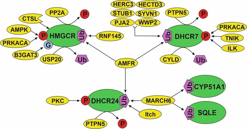
Biomedicines 2021, 9, 895                                                                                                              2 of 28

                                     reduction of cancer risk by 20% [11]. However, the addition of statins to standard anticancer
                                     therapy does not improve overall survival [12,13].
                                          Involvement of enzymes of cholesterol synthesis pathway (cholesterol synthesis en-
                                     zymes) in provoking and maintaining pathological cancer-dependent processes is closely
                                     linked with regulation on transcriptional and post-translational levels [1,14]. The last
                                     one can be realized through protein–protein interactions (PPIs) and post-translational
                                     modifications (PTMs) resulting in a change of protein stability and enzymatic activity.
                                          With the growing number of experimentally determined PPIs of cholesterol synthesis
                                     enzymes, it is necessary to assess and make comparative analysis of available information
                                     on both interactomes of individual enzymes (sub-interactomes) and the group interactome
                                     for several enzymes, functioning in a single metabolic pathway. The aim of the work was
                                     to systematize interactomics data on the cholesterol synthesis enzymes with respect to the
                                     cancer context. This is intended to update the current understanding of the functional and
                                     regulatory aspects of the cholesterol synthesis pathway in socially significant diseases.

                                     2. Materials and Methods
                                     2.1. TCGA Datasets Analysis
                                          The Cancer Genome Atlas (TCGA) program has generated, analyzed, and made avail-
                                     able data on the genomic sequence, expression, methylation, and copy number variation of
                                     over 11,000 individuals with over 30 different types of cancer [15,16]. The TCGA database
                                     was used to determine the expression patterns of genes, encoding cholesterol synthesis
                                     enzymes, as well as to find the associations with patient survival. The Gene Expression
                                     Profiling Interactive Analysis (GEPIA2) web server [17] (http://gepia2.cancer-pku.cn/,
                                     accessed on 23 April 2021) was used to obtain median values of gene expression in tumor
                                     and normal tissues from TCGA datasets and to determine the tumor/normal ratio or
                                     fold change (FC). The selection of differentially expressed genes (DEGs) was performed
                                     according to FC ≥ 3 and cut-off significance level 0.01. A heat map was plotted using web
                                     server NG-CHM GUI 2.19.1 [18]. Prognostic value of DEGs and Kaplan–Meier plotting
                                     were performed using GEPIA2 with the following settings: significance level 0.05; p-value
                                     adjustment by FDR; group cut-off-median. Principal component analysis (PCA) of data
                                     matrix with FC values for 17 genes, encoding cholesterol synthesis enzymes, and 30 differ-
                                     ent cancers was performed using ClustVis resource [19] (https://biit.cs.ut.ee/clustvis/,
                                     accessed on 26 April 2021). The assessment of mutational variability (somatic muta-
                                     tion frequency) of genes was performed using TCGA PanCancer Atlas Studies dataset
                                     (10953 patients/10967 samples) at the cBioPortal (https://www.cbioportal.org/, accessed
                                     on 2 May 2021).

                                     2.2. Interactomics Data Acquisition and Processing
                                          All possible spectrum of experimentally established (physical interactions) protein
                                     partners for each of 17 cholesterol synthesis enzymes (target enzymes) was retrieved with a
                                     set of interactome browsers [20–32] (Table 1). Common protein partners for target enzymes
                                     were obtained using the Venn diagram tool (http://bioinformatics.psb.ugent.be/webtools/
                                     Venn/, accessed on 26 March 2021).

                               Table 1. Interactome browsers for retrieving protein–protein interactions data.

  No.       Name and Ref.                                         Site                                                Notes
                                                                                                        Evidence type—protein interactions,
   1        Funcoup 5.0 [20]              http://FunCoup.sbc.su.se (accessed on 15 March 2021)            LLR Score > 2, confidence > 0.9,
                                                                                                           network: “PPI”, “Complex”
   2          Mentha [21]               https://mentha.uniroma2.it/ (accessed on 15 March 2021)         Evidence type: physical interactions
   3          CORUM [22]         http://mips.helmholtz-muenchen.de/corum/ (accessed on 15 March 2021)                    -
   4         ExoCarta [23]               http://www.exocarta.org/ (accessed on 15 March 2021)                            -
                                                                                                            Methods (binary, indirect),
   5           APID [24]                   http://apid.dep.usal.es (accessed on 15 March 2021)
                                                                                                                ≥1 publication
Biomedicines 2021, 9, 895                                                                                                           3 of 28

                                                              Table 1. Cont.

  No.       Name and Ref.                                       Site                                               Notes
                                                                                                       Interaction type: association,
   6           MINT [25]             https://mint.bio.uniroma2.it/ (accessed on 16 March 2021)
                                                                                                           physical association
   7        SIGNOR 2.0 [26]           https://signor.uniroma2.it/ (accessed on 16 March 2021)                         -
   8           HuRI [27]           http://www.interactome-atlas.org/ (accessed on 16 March 2021)                      -
   9            IID [28]              http://iid.ophid.utoronto.ca (accessed on 16 March 2021)         Interaction type: experimental
            Bioplex Explorer
   10                          https://bioplex.hms.harvard.edu/explorer/ (accessed on 16 March 2021)    Interaction probability > 0.9
                3.0 [29]
   11         Wiki-Pi [30]       https://hagrid.dbmi.pitt.edu/wiki-pi/ (accessed on 16 March 2021)                    -
                               http://cbdm-01.zdv.uni-mainz.de/~mschaefer/hippie/ (accessed on 16
   12       HIPPIE 2.0 [31]                                                                                     Score > 0.6
                                                          March 2021)
   13          HINT [32]                 http://hint.yulab.org/ (accessed on 16 March 2021)                  Binary interaction

                                  2.3. Annotation and Enrichment Analysis
                                       Functional annotation of the final list of target enzyme protein partners with Gene Ontol-
                                  ogy, KEGG, REACTOME, Panther (FDR ≤ 0.05) and Wiki Pathways terms (p-value ≤ 0.05)
                                  was performed with over-representation analysis (ORA) on the Web Gestalt server (WEB-
                                  based Gene SeT AnaLysis Toolkit) [33]. Annotation of proteins by subcellular localization
                                  was performed using NextProt database (https://www.nextprot.org/, accessed on 29
                                  March 2021). Gene annotation by “common essential genes” category was done using
                                  CRISPR-Cas9 whole-genome drop out screens to identify dependencies in cancer cells [34]
                                  on the website Cancer Dependency Map (https://score.depmap.sanger.ac.uk/, accessed
                                  on 2 April 2021) and DepMap portal (https://depmap.org/, accessed on 2 April 2021).
                                  Pubmed (https://pubmed.ncbi.nlm.nih.gov/, accessed on 10 June 2021) and Litsense [35]
                                  (https://www.ncbi.nlm.nih.gov/research/litsense, accessed on 10 June 2021) resources
                                  were used to search for available literature data. Annotation of cancer driver genes was car-
                                  ried out using Cancer Gene Census portal (https://cancer.sanger.ac.uk/census, accessed
                                  on 5 April)).
                                       Prediction of post-translational modification (PTM) sites of cholesterol synthesis
                                  enzymes was performed using the web-server PhosphoSitePlus (https://www.phosphosite.
                                  org/, accessed on 4 May 2021) containing experimental results of mass-spectrometry PTMs
                                  mapping.

                                  3. Results
                                  3.1. The Main Characteristics of Cholesterol Synthesis Enzymes
                                       Seventeen enzymes (target enzymes), functioning in a single metabolic cascade of
                                  multi-step transformations of cholesterol precursors (Figure 1), were selected from a path-
                                  way module M00100 KEGG (https://www.genome.jp/kegg-bin/show_pathway?map001
                                  00, accessed on 3 March 2021) and Hallmark Cholesterol Homeostasis (ID M5892, 74 genes,
                                  Molecular Signature Database (http://www.gsea-msigdb.org/, accessed on 6 March 2021)).
                                  The main characteristics of the cholesterol synthesis enzymes are also shown in Table S1
                                  (Supplementary File #1).
                                       Approximately 70% of cholesterol synthesis enzymes represent membrane proteins
                                  containing one or several transmembrane domains localized, mostly, in the endoplasmic
                                  reticulum membrane (ERM), Golgi apparatus membrane, cytoplasmic vesicles, plasma
                                  membrane and nucleus inner membrane (Table S1, Supplementary File #1). The remain-
                                  ing 30% of enzymes (MVK, PMVK, MVD, FDPS and LSS) are localized in the cytosol,
                                  peroxisomes and lipid droplets. Different subcellular localizations of enzymes may also
                                  suggest the additional functions besides transformation of cholesterol precursors. Multiple
                                  functions for this group of enzymes are poorly studied, possibly due to insufficient data
                                  on the functional significance of mapped PPIs. For example, the MoonProt database [36]
                                  (http://www.moonlightingproteins.org) does not contain information about multiple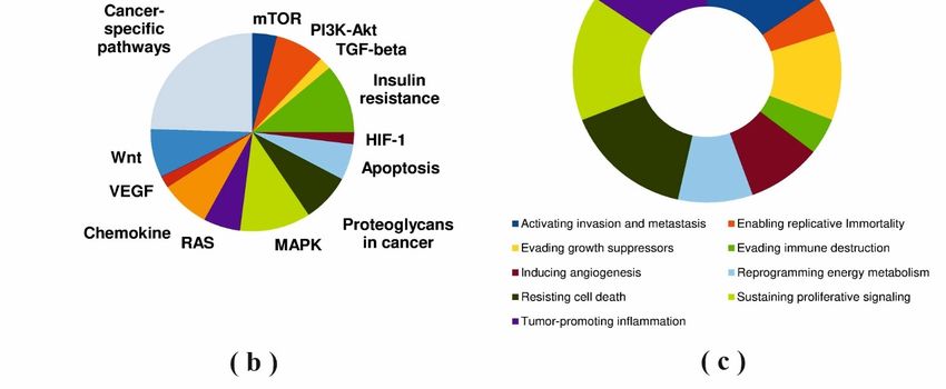
Biomedicines 2021, 9, 895 4 of 28
Biomedicines 2021, 9, x FOR PEER REVIEW 4 of 30
functions of the target enzymes, except the mevalonate kinase (MVK). It is known that
3.1. The Main
lanosterol Characteristics
synthase (LSS) canof Cholesterol Synthesis
regulate protein Enzymes by effectively reducing the
aggregation
number Seventeen
and/or enzymes (target enzymes), functioning
size of sequestosomes/aggresomes in a by
formed single metabolicproteins
endogenous cascadein of
multi‐step
the normal transformations
and cancer cellsof[37].
cholesterol precursors
In addition, (Figure
farnesyl 1), were selected
pyrophosphate from(FDPS)
synthase a path‐
way an
obtains module
additionalM00100 KEGG growth
function in fibroblast (https://www.genome.jp/kegg‐bin/show_path‐
factor (FGF) signaling pathway through
way?map00100,
binding accessed on 03
to the FGF-receptors March
[38]. 2021) and Hallmark
It is interesting Cholesterol
to note that Homeostasis
delta(24)-sterol (ID
reductase
(DHCR24)
M5892, 74 can protect
genes, neuronal
Molecular cells from
Signature ER stress-induced
Database apoptosis by attenuating
(http://www.gsea‐msigdb.org/, ER
accessed
stress
on 06 signaling, possibly
March 2021)). through
The main scavenging
characteristics ofintracellular reactive
the cholesterol oxygen
synthesis species
enzymes areand
also
elevating
shown incholesterol levels [39].
Table S1 (Supplementary File #1).
Figure 1.
Figure 1. AAlogical
logicalscheme
schemeof of
enzymes
enzymesandand
metabolites involved
metabolites in theincholesterol
involved synthesis
the cholesterol path‐
synthesis
way. The following metabolites are indicated by numbers: (1)—hydroxymethylglutaryl‐CoA;
pathway. The following metabolites are indicated by numbers: (1)—hydroxymethylglutaryl- (2)—
(R)‐mevalonate; (3)—(R)‐5‐phosphomevalonate; (4)—(R)‐5‐diphosphomevalonate; (5)—isopen‐
CoA; (2)—(R)-mevalonate; (3)—(R)-5-phosphomevalonate; (4)—(R)-5-diphosphomevalonate; (5)—
tenyl diphosphate; (6)—(2E,6E)‐farnesyl diphosphate; (7)—presqualene diphosphate; (8)—squa‐
isopentenyl diphosphate; (6)—(2E,6E)-farnesyl diphosphate; (7)—presqualene diphosphate; (8)—
lene; (9)—(S)‐squalene‐2,3‐epoxide; (10)—lanosterol; (11)—4,4‐dimethyl‐5‐alpha‐cholesta‐8,14,24‐
squalene; (9)—(S)-squalene-2,3-epoxide;
trien‐3‐beta‐ol (10)—lanosterol;
(FF‐MAS); (12)—14‐demethyllanosterol (11)—4,4-dimethyl-5-alpha-cholesta-
(T‐MAS); (13)—4‐alpha‐methyl zymosterol‐
8,14,24-trien-3-beta-ol (FF-MAS); (12)—14-demethyllanosterol
4‐carboxylate; (14)—3‐keto‐4‐methylzymosterol; (T-MAS);
(15)—4‐alpha‐methyl (13)—4-alpha-methyl
zymosterol; (16)—4‐alpha‐
zymosterol-4-carboxylate; (14)—3-keto-4-methylzymosterol;
carboxy‐5‐alpha‐cholesta‐8,24‐dien‐3‐beta‐ol; (15)—4-alpha-methyl
(17)—zymosterone; (18)—zymosterol;zymosterol;
(19)—zy‐(16)—
mostenol; (20)—lathosterol; (21)—7‐dehydrocholesterol;
4-alpha-carboxy-5-alpha-cholesta-8,24-dien-3-beta-ol; (22)—cholesterol;(18)—zymosterol;
(17)—zymosterone; (23)—5‐alpha‐cholesta‐
(19)—
7,24‐dien‐3‐beta‐ol;
zymostenol; (24)—7‐dehydrodesmosterol;
(20)—lathosterol; (25)—desmosterol.
(21)—7-dehydrocholesterol; (22)—cholesterol; (23)—5-alpha-cholesta-
7,24-dien-3-beta-ol; (24)—7-dehydrodesmosterol; (25)—desmosterol.
Approximately 70% of cholesterol synthesis enzymes represent membrane proteins
The oligomeric
containing state of
one or several target enzymes
transmembrane is an important
domains localized,interactomic
mostly, in thefactor. It was
endoplasmic
shown that a dimeric form is characteristic for cholestenol delta-isomerase
reticulum membrane (ERM), Golgi apparatus membrane, cytoplasmic vesicles, plasma (EBP) [40], sterol
14-alpha-demethylase
membrane and nucleus (CYP51A1) [41] and (Table
inner membrane MVK [42]. Squalene epoxidase
S1, Supplementary File (SQLE)
#1). Theexists
remain‐in
aing
monomeric form [43],
30% of enzymes whilePMVK,
(MVK, 3-hydroxy-3-methylglutaryl-CoA reductasein
MVD, FDPS and LSS) are localized (HMGCR)
the cytosol,forms
pe‐
tight tetramers
roxisomes and that
lipidimplies
droplets. theDifferent
influence of the enzyme’s
subcellular oligomeric
localizations state onmay
of enzymes thealso
activity
sug‐
and
gestmechanisms
the additionalfor allosteric
functionsmodulation by ligands [44].
besides transformation Since half ofprecursors.
of cholesterol these data concerns
Multiple
non-human
functions fororthologues,
this group of the conceptare
enzymes of the oligomeric
poorly studied,state of most
possibly duehuman cholesterol
to insufficient data
synthesis enzymes is not yet completely studied.
on the functional significance of mapped PPIs. For example, the MoonProt database [36]
Many cholesterol synthesis enzymes for
(http://www.moonlightingproteins.org) doescatalytic reactions
not contain require coenzymes
information NADH
about multiple
and
functions of the target enzymes, except the mevalonate kinase (MVK). It is knowncon-
NADPH, which intracellular levels can be correlated with tumor progression under that
dition of metabolism
lanosterol reprogramming
synthase (LSS) can regulate in cancers
protein[45]. At present,
aggregation byno coenzyme-dependent
effectively reducing the
oxidoreductases,
number and/or size which can bind with target enzymes,
of sequestosomes/aggresomes have
formed bybeen identified
endogenous [46], except
proteins in the
for CYP51A1 belonging to the multigene cytochrome P450 family.
normal and cancer cells [37]. In addition, farnesyl pyrophosphate synthase (FDPS) It strongly requires
obtains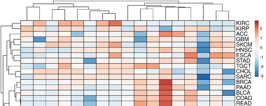
Biomedicines 2021, 9, 895                                                                                                        5 of 28

                                   electron transfer from its direct redox partner NADPH-dependent cytochrome P450 ox-
                                   idoreductase (POR) for catalysis [47]. It is interesting to note that SQLE contributes to
                                   cancers via its metabolites. An increase in SQLE expression promotes the cholesteryl ester
                                   production to induce hepatocellular carcinoma cell growth. Moreover, SQLE can raise the
                                   NADP+/NADPH ratio what triggers DNA methyltransferase 3A (DNMT3A) expression,
                                   DNMT3A-mediated epigenetic silencing of phosphatase tensin homolog (PTEN) and ac-
                                   tivation of pro-oncogenic mammalian target of rapamycin pathway [48]. Thus, levels of
                                   intracellular coenzymes and their ratio can be important factors in proper functioning of
                                   enzymes in the cholesterol pathway.
                                        Then, we searched for literature data on involvement of 17 target enzymes in carcino-
                                   genesis and important findings are shown in Table 2.

                            Table 2. Associations between cholesterol synthesis enzymes and carcinogenesis.

            Enzyme                                                     Thesis                                             Ref.
                                   Genetically proxied inhibition of HMGCR is significantly associated with lower
            HMGCR                                                                                                         [49]
                                       odds of epithelial ovarian cancer ([odds ratio 0.60 [95% CI, 0.43–0.83]).
                                    Inhibition of HMGCR by fluvastatin disrupts the non-small cell lung cancer
                                     (NSCLC) tumorigenesis. Knockdown of HMGCR in NSCLC cells induces                     [50]
                                                        apoptosis in vitro and in vivo models.
                                      Statin drugs, inhibiting HMGCR, increase the efficacy of some genotoxic
                                                                                                                         [51–54]
                                                                    anti-cancer drugs.
                                    HMGCR is expressed on carcinoma cells but not on normal epithelial cells in
                                  thymic tissue. Inhibition of HMGCR by fluvastatin suppresses cell proliferation         [55]
                                                         and induces the carcinoma cell death.
                                  PMVK can be considered as a novel prognostic biomarker for high-grade serous
                                 ovarian carcinoma (HGSOC). High expression of PMVK is significantly improves
             PMVK                                                                                                         [56]
                                    the survival of patients with HGSOC (adjusted hazard ratio, 0.430; [95% CI,
                                                                      0.228–0.809]).
                                 PMVK expression is positively correlated with drug response in estrogen receptor
                                                                                                                          [57]
                                           (ER) positive cells and negatively correlated in ER negative cells
                                 Increased FDPS expression is an independent risk factor of prostate cancer (PC) for
              FDPS                                                                                                        [58]
                                                              early biochemical recurrence.
                                   FDPS inhibitors, the carnosic acid derivatives, induces apoptosis in pancreatic
                                                                                                                          [59]
                                                                     cancer cell lines.
                                 Inhibition of the FDPS in the mevalonate pathway mediates the cytotoxic effects of
                                 a platinum (II) complex with zoledronic acid against human gastric cancer cell line      [60]
                                                                        SGC7901.
                                     FDPS expression significantly correlates with TNM stage and metastasis in
                                  non-small cell lung cancer (NSCLC). Inhibition or knockdown of FDPS disrupts            [61]
                                  the TGF-β1-induced cell invasion and epithelial-mesenchymal transition (EMT).
                                 FDPS inhibitors improve survival of multiple myeloma (MM) patients and results
                                                                                                                          [62]
                                        in down-regulation of ERK phosphorylation in human MM cell lines.
                                   FDFT1 is highly expressed in liver, lung, prostate, breast, ovary, small intestine,
             FDFT1                  bladder, cervix, thyroid, and esophageal cancers. FDFT1 regulates cell cycle          [63]
                                     progression and is directly or indirectly associated with apoptotic signals.
                                     High expression of FDFT1 is associated with poor prognosis and promotes
                                 metastasis of lung cancer. Loss of function of FDFT1 or its knockdown significantly      [64]
                                       inhibits invasion/migration and metastasis in cell and animal models.
                                         FDFT1 acts as a critical tumor suppressor in colorectal cancer (CRC).
                                   Down-regulation of FDFT1 is correlated with CRC malignant progression and              [65]
                                                                     poor prognosis.
                                 The knockdown of expression or activity inhibition of FDFT1 leads to a significant
                                                                                                                          [66]
                                                     decrease in prostate cancer cell proliferation.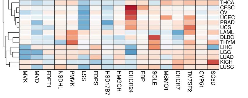
Biomedicines 2021, 9, 895                                                                                                    6 of 28

                                                         Table 2. Cont.

            Enzyme                                                Thesis                                            Ref.
                                  Overexpression of SQLE promotes lung squamous-cell carcinoma (SCC)
                             proliferation, migration and invasion, whereas knockdown of SQLE expression
              SQLE                                                                                                  [67]
                               shows the opposite effect. High expression of SQLE corresponds with poor
                                                            prognosis in lung SCC.
                              The expression of SQLE is upregulated in the hepatocellular carcinoma (HCC)
                                  tissues and its overexpression promotes cell proliferation and migration.
                                                                                                                    [68]
                              Downregulation of SQLE inhibits the tumorigenicity of HCC cells in vitro and
                                                                     in vivo.
                            SQLE overexpression is more prevalent in aggressive breast cancer (BC) and is an
                                                                                                                    [69]
                                           independent prognostic factor of unfavorable outcome.
                             SQLE epoxidase serves as a novel prognostic biomarker for patients with HCC.
                                Overexpression of SQLE in non-alcoholic fatty liver disease HCC tumors is          [48,70]
                              significantly associated with worse overall survival and disease-free survival.
                            SQLE reduction helps colorectal cancer cells to overcome constraints by inducing
                             the EMT required for generation cancer stem cells. SQLE depletion disrupts the         [71]
                                          GSK3B/p53 complex, resulting in a metastatic phenotype.
                              SQLE promotes nasopharyngeal carcinoma (NPC) proliferation by cholesteryl
                                                                                                                    [72]
                                                   ester accumulation instead of cholesterol.
                            Lanosterol synthase is a molecular target for menin inhibitor leading to the loss of
               LSS                                                                                                  [73]
                                               cholesterol homeostasis and cell death in glioma.
                            LSS activity increases in the daunorubicin-resistant leukemia cell line (CEM/R2).       [74]
                            CYP51A1 is significantly upregulated in the drug-tolerant (DT) human lung cancer
           CYP51A1           cell lines. The CYP51A1 inhibitor, ketoconazole, shows the synergy in apoptosis        [75]
                              induction with tyrosine kinase inhibitors of epidermal growth factor receptor.
                                CYP51 is present at a significantly higher level in primary colorectal cancer,
                            compared with normal colon. The strong CYP51 immunoreactivity is associated             [76]
                                                              with poor prognosis.
                                 The VFV, a potent non-azole inhibitor of human CYP51A1, decreases the
                            proliferation rates of lung cancer, hormone-responsive and -nonresponsive breast        [77]
                                         and skin cancer cells in a concentration-dependent manner.
                            TM7SF2 knockout (KO) mice show no alteration in cholesterol content. However,
            TM7SF2          delayed cell cycle progression to the G1/S phase was shown in TM7SF2 KO mice,           [78]
                                                       resulting in reduced cell division.
                            Loss of TM7SF2 increases incidence and multiplicity of skin papillomas. The null
                            genotype shows reduced expression of nur77, a gene associated with resistance to        [79]
                                                           neoplastic transformation.
                            NSDHL knockdown affects the cell cycle, survival, proliferation, and migration of
                               breast cancer cells, resulting in suppression of breast tumor progression and
            NSDHL                                                                                                   [80]
                            metastasis. High NSDHL expression is a potential predictor of poor prognosis in
                                                             breast cancer patients.
                            Inhibition NSDHL can be an effective strategy against carcinomas with activated
                                                                                                                    [81]
                                                            EGFR-KRAS signaling.
                            The inactivation of NSDHL or its partner SC4MOL sensitizes tumor cells to EGFR
                                                                                                                    [82]
                                                                   inhibitors.
                            NSDHL is significantly overexpressed in gastric cancer tissues that correlates with
                                                                                                                    [83]
                                       local tumor invasion, histological grade and TNM II-IV staging.
                             The NSDHL up-regulated in the metastasizing mammalian mouse cell line 4T1
                                                                                                                    [84]
                                                  compared to the non-metastasizing 67NR.
                            DHCR24 knockdown reduces whereas DHCR24 overexpression enhances breast
            DHCR24           cancer stem-like cell populations, mammosphere and aldehyde dehydrogenase              [85]
                                                                  positive cell.
                               High expression of DHCR24 in human HCC specimens correlates with poor
                                                                                                                    [86]
                            clinical outcome. Interfering DHCR24 alters growth and migration of HCC cells.
                               DHCR24 is up-regulated in bladder cancer (BC) cells compared with that in
                              normal tissues. DHCR24 might promote the proliferation of BC cells through            [87]
                                                      several cancer-associated processes.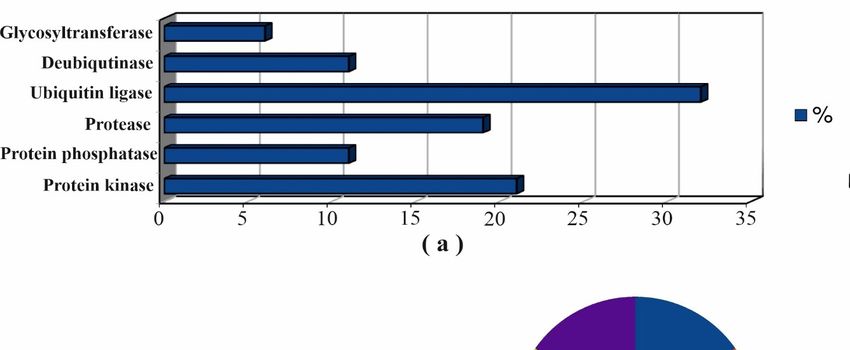
Biomedicines 2021, 9, 895                                                                                                7 of 28

                                                        Table 2. Cont.

            Enzyme                                               Thesis                                          Ref.
                            The EBP inhibitors show the good potency and efficacy in inhibiting proliferation
               EBP                                                                                                [88]
                                                 of human prostate cancer PC-3 cell line.
                             mRNA and protein accumulation are observed in anaplastic lymphoma kinase
                                                                                                                  [89]
                                                             (ALK+) tumors.
                              Inhibition of the EBP leads to cancer cell death via depletion of downstream
                                                                                                                  [90]
                                                                 sterols.
                             Decreased SC5D activity in cancer might increase prenylation of RAS, RAC or
              SC5D                                                                                                [91]
                                              RHOC thereby promoting cancer progression.

                                   Thus, the associations between functioning, at least, three quarters of the cholesterol
                              synthesis enzymes and solid cancers progression were found [48–91]. Not only overexpres-
                              sion and post-translational activation of enzymes correlated with pathogenic abnormalities,
                              but also a decrease in catalytic function due to down-regulation of expression or phar-
                              macological inhibition (SC5D, NSDHL and EBP). Another important implication is that
                              several target enzymes have differentiated contributions to cancers depending on the
                              tissue specificity.

                              3.2. Transcriptomic Landscape of Genes, Encoding Cholesterol Synthesis Enzymes
                                    The panoramic transcriptome profile of 17 target genes is shown in Figure S1
                              (Supplementary File #1). It can be conditionally distinguished two groups of tumors. The
                              first group includes COAD, DLBC, PAAD, READ and THYM tumors, while the second
                              one includes SKCM, LAML, CHOL and TGCT tumors with a preferential up-regulation
                              or down-regulation of DEGs, respectively. Principal component analysis (PCA) showed
                              that, in fact, only the CYP51A1 and NSDHL genes had the closest expression profiles in
                              almost all tumors (Figure S2, Supplementary File #1). A clusterogram, visualizing PCA
                              output, revealed, at least, four large tumor clusters with similar profiles of DEGs (Figure 2):
                              (1)—KIRC and KIRP; (2)—ACC, GBM, SKCM, HNSC, ESCA, etc.; (3)—BRCA, PAAD,
                              BLCA, COAD, READ, etc.; (4)—LAML, DLBC, THYM, LIHC, LGG, LUAD, KIRC and
                              LUSC. It is worth mentioning that alterations of expression profiles of target genes are
                              often due to binding of cancer-activated master regulators in their promoter regions. It can
                              be demonstrated, for example, by the network R-HSA-2426168 from Reactome database
                              (https://reactome.org/, accessed on 20 April 2021)). Master regulators such as SREBF1,
                              SREBF2, SP1 and NF-Y have broad promoter binding specificity and are subjected to
                              modulation through several oncogenic signaling pathways [92–96], but this “transcription
                              regulation issue” is out of scope of the present work.
                                    Five genes from these maps meet the strictest selection criteria: p < 0.001, hazard
                              ratio (HR) ≤ 0.5 or HR ≥ 2 as well as a number of cases ≥ 200. The maps of overall
                              sur-vival (OS) and disease-free survival (RFS) for 17 target genes are shown in Figure S3a,b,
                              respectively (Supplementary File #1). For all of them (OS: CYP51A1 in KIRC, FDFT1 in
                              KIRC, HMGCR in KIRC, SC5D in KIRC, TM7SF2 in LGG; RFS: FDFT1 in KIRC, SC5D
                              in PRAD) the Kaplan–Meier plots are shown in Supplementary File #4, Figure S9. It
                              should be noted that multigenic signatures have a higher priority in prognostic value
                              than single transcriptome markers [97]. First of all, survival analysis of a panel of 17
                              target genes versus each TCGA tumor was performed (Table S3, Supplementary File
                              #4). It is shown that the selection criteria were met in the case of KIRC only. More
                              negative prognosis for survival was associated with lower profile of cholesterol biosynthesis
                              genes expression (Figure S10, Supplementary File #4). It should be noted, that the severe
                              cholesterol metabolism disrupting was observed in the KIRC cells [98–100]. We also carried
                              out the survival analysis for each target enzyme versus all tumors (pan-cancer prognostic
                              significance). Results obtained are presented in Table S4 (Supplementary File #4), but no
                              hits were found. Finally, the gene signature 1 (HMGCR, DHCR7, SC5D, NSDHL, CYP51A1
                              and FDFT1) (Figure S4a, Supplementary File #1) and the gene signature 2 (SC5D, TM7SF2
                              and MVD) (Figure S4b, Supplementary File #1) were found to have prognostic value for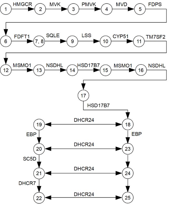
Biomedicines 2021, 9, 895                                                                                                                   8 of 28

Biomedicines 2021, 9, x FOR PEER REVIEW
                                   KIRC and LGG tumors, respectively.                                                  9 of 30
                                                                       Thus, high expression of these genes can be associated
                                  with the decrease of mortality by 2.5 times (HR value = 0.4).

       Figure2.2.The
      Figure     The clusterogram
                      clusterogram ofof expression
                                        expression profiles
                                                   profiles of
                                                            of genes,
                                                               genes, involved
                                                                      involved in cholesterol synthesis pathway,
                                                                                                        pathway, in
                                                                                                                 in different
                                                                                                                    different tumors.
                                                                                                                              tumors.
      The
       Thecolor
           colorscale
                  scaleshows
                        showsthe
                               thecluster
                                   clusterdistances.
                                           distances.

                                  3.3. Interactomics
                                        Five genes fromLandscape
                                                             theseofmaps
                                                                       Cholesterol
                                                                             meet theSynthesis
                                                                                        strictestEnzymes
                                                                                                    selection criteria: p
the coincidence of protein partners found by, at least, three different interactome browsers
and by the similar profiles of subcellular localization of a target enzyme and its interaction
partners (Figure 3). In addition, protein names and Uniprot IDs for each protein partner
are listed in Supplementary File #3. List A, containing 165 unique protein partners for all
target enzymes, was used for functional annotation. List B, containing in total 186 proteins
Biomedicines 2021, 9, 895 9 of 28
(165 unique and 21 common protein partners), was then used to visualize the PPI network
or, if it may say so, cluster interactome of cholesterol synthesis enzymes (Figure 4).
Biomedicines 2021, 9, x FOR PEER REVIEW 11 of 30
3. Selection
FigureFigure roundsrounds
3. Selection of found proteinprotein
of found partners.
partners.
Networkconstruction
Figure4.4.Network
Figure constructionofofPPIs
PPIsinvolving
involvingthe
thecholesterol
cholesterolsynthesis
synthesisenzymes.
enzymes.Legend:
Legend:target
targetenzymes,
enzymes,unique
uniqueand
and
commonprotein
common proteinpartners
partnersare
areshown
shownininred
reddiamonds,
diamonds,blue
blueellipses
ellipsesand
andblue
bluerectangles,
rectangles,respectively.
respectively.Edges,
Edges,connecting
connecting
enzymesthrough
enzymes throughcommon
commonprotein
proteinpartners,
partners,are
arehighlighted
highlighted in
in bold.
bold.
Somatic mutation frequency of genes, encoding cholesterol synthesis enzymes,
ranged from 0.4–0.7% and they were not annotated as cancer driver genes. Annotation of
genes, encoding 165 unique protein partners of enzymes, showed that following ones
were found as cancer drivers with corresponding somatic mutation frequency values:Biomedicines 2021, 9, 895 10 of 28
Somatic mutation frequency of genes, encoding cholesterol synthesis enzymes, ranged
from 0.4–0.7% and they were not annotated as cancer driver genes. Annotation of genes,
encoding 165 unique protein partners of enzymes, showed that following ones were
found as cancer drivers with corresponding somatic mutation frequency values: PRKACA
(0.7%); EWSR1 (1.2%); NDRG1 (0.8%); FGFR1 (1.4%); IDH1 (5.1%); PABPC1 (0.9%); LMNA
(0.8%); MYH9 (3.2%); SUZ12 (1.0%); TP53 (35.3%); HRAS (1.2%); IKBKB (1.4%); CYLD
(1.3%); ERBB2 (2.9%). Cancer-dependent PPIs can consist of a protein encoded by a driver
gene [101]. It was showed that a vast proportion of mutations in driver genes occurred in
the binding interfaces of two proteins, which means that “non-canonical” protein complexes
can emerge in tumor tissue, and vice versa, formation of canonical biologically significant
complexes in normal tissue can be suppressed during neoplastic transformation. Due to
some of the found protein partners, encoded by cancer driver genes, it was hypothesized
for protein complexes with their participation to be directly regulated by carcinogenesis.
Functional annotation of found protein partners with Gene Ontology (GO) terms is
shown in Figure S5, Supplementary File #1. Over-representation analysis (ORA) revealed
molecular functions enrichment (“transferase activity”, “chaperone binding”, “protease
binding”, “ubiquitin-like protein binding”, “ubiquitinyl hydrolase activity”) (Figure S6,
Supplementary File #1) and biological processes enrichment (“endoplasmic reticulum to
cytosol transport”, “nucleobase-containing small molecule interconversion”, “protein exit
from endoplasmic reticulum”, “ER-nucleus signaling pathway”, “response to topologically
incorrect protein”, “response to endoplasmic reticulum stress”) (Figure S7, Supplementary
File #1). ORA of protein partners in KEGG, REACTOME, Panther and WikiPathways
databases showed their involvement in nucleotides biosynthesis and metabolism, amino
acids pathways, synaptic signal transmission, protein processing in endoplasmic reticulum
(ER), ER quality control (ERQC) and cancer-related pathways (apoptotic pathway, MAPK
family signaling cascades, metabolic reprogramming and central carbon metabolism in can-
cer, Ras signaling, RAC1/PAK1/p38/MMP2 pathway and three cancer-specific pathways
in prostate, endometrial and bladder cancers) (Table S2, Supplementary File #1). Thus,
overall pool of protein partners, forming PPIs with cholesterol synthesis enzymes, can be
divided into two big groups involved in protein processing and cell signaling.
Taking into account the over-representation of protein partners in cancer-related
pathways, we tried to find the associations of each binary PPI with the cancer-specific
context. A way to hypothesize such associations is to correlate of expression profiles
of two genes encoded protein products, forming a PPI (gene co-expression analysis).
A positive or a negative correlation in different tumor tissues indirectly point to such
associations. Co-expression analysis was performed only for those tumor tissues, in which
DEGs (tumor/normal fold change ≥ 3), encoding the cholesterol synthesis enzymes, were
observed (Figure S1, Supplementary File #1). A Pearson correlation coefficient (R) equal
to 0.4 was used as a cut-off level. An average (|0.4 < R < 0.6|) and strong correlation
(|0.6 < R < 0.7|) of gene co-expression were found in five different tumor tissues for 42 and
7 binary PPIs, respectively (Table 3). R-values were mostly positive, however, a negative
correlation was observed for PPIs with participation of EBP in thymoma tissues (339 and
118 normal and tumor cases, respectively) (Table 3). The Human Proteome Atlas database
(https://www.proteinatlas.org) was then used for comparison of tissue-specific expression
profiles for each binary PPI with co-expressed genes. Concordance of the gene and protein
expression profiles can be seen only for HMGCR–VCP, DHCR24–CKAP5 and DHCR7–
FADS1 protein interactions in rectum adenocarcinoma datasets (318 and 92 normal and
tumor cases, respectively) as well as for DHCR7–FADS1 in pancreatic adenocarcinoma
(171 and 179 normal and tumor cases, respectively). However, it turned out that protein
expression data for several cholesterol synthesis enzymes and their protein partners were
absent in tumor tissues.Biomedicines 2021, 9, 895 11 of 28
Table 3. Gene co-expression analysis of protein–protein interactions.
Tumors Protein–Protein Interactions
MVK **—GSS (L—L) ***, MVK—ENO1 (L—M), MVD—EIF4EBP1 (L—M),
SQLE—FAF2 (M—L), SQLE—TMCO3 (M—n/d), LSS—MYO5C (M—n/d),
DLBC CYP51A1—ETS1 (n/d—H), MSMO1—C3AR1 (n/d—n/d), MSMO1—ELAVL1
(n/d—M, MSMO1—P2RX5 (n/d—M), EBP—VKORC1 (n/d—n/d),
EBP—ENO1 (n/d—M)
PAAD MSMO1—GPR35(n/d—n/d), EBP—MOV10 (L—n/d), DHCR7—FADS1 (H—M)
DHCR24—ERAP1(n/d—M), DHCR24—CKAP5 (n/d—M), DHCR24—PTPN1
PRAD
(n/d—n/d), DHCR24—REEP5 (n/d—M), DHCR24—UBL4A (n/d—M)
HMGCR—VCP (M—M), DHCR24—CKAP5 (M—M), HMGCR—INSIG1
READ
(M—n/d), DHCR7—FADS1 (H—M)
MVK—TACC3, FDPS—ATXN1, FDPS—PSME4, FDPS—TALDO1,
FDPS—SEC31A, SQLE—MARCH6, SQLE—CHRND, LSS—ALOX5, LSS—MYH9,
CYP51A1—SPINT2, MSMO1—ATXN1, MSMO1—ELAVL1, HSD17B7—GORAB,
THYM
SC5D—IPPK, EBP—HSP90B1 (neg *), EBP—ABCE1 (neg), EBP—KSR1 (neg),
EBP—TCTN2 (neg), EBP—BSCL2 (neg), EBP—MFSD8 (neg), FDPS—NME1,
SQLE—FAF2, SQLE—TMCO3, MSMO1—EIF3A, EBP—NCSTN (neg)
Abbreviations: DLBC—lymphoid neoplasm diffuse large B-cell lymphoma; PAAD—pancreatic adenocarcinoma;
PRAD—prostate adenocarcinoma; READ—rectum adenocarcinoma; THYM—thymoma. * Negative correlation.
** Cholesterol synthesis enzymes are underlined. *** Prevalent protein expression (according to Human Proteome
Atlas): H—high; M—medium; L—low; n/d—no data.
3.4. Post-Translational Modifications (PTM)
Further, we analyzed the known PTM spectrum of cholesterol synthesis enzymes as
well as the spectrum of modifying proteins among found protein partners. The amino acid
sequences of the target enzymes contain a large number of phosphorylation and ubiquityla-
tion sites (Figure S8, Supplementary File #1). Forty-seven (25%) protein partners which can
potentially function as modifying proteins were summarized in Table S3 (Supplementary
File #1). It should be noted that 15 proteins (AMPK, PP2A, CTSL, RNF145, gp78, USP20,
MEK5, GSK3β, MARCH6, IDOL, PKC, AMFR, Itch, PRKACA, SYVN1) were described as
modifying proteins [71,102–119] (Table S3). Results of annotation of all modifying proteins
with cancer hallmarks and cancer pathways terms are shown in Figure 5.
Figure 5A shows that ubiquitin-protein ligases and protein kinases were the main
functional classes of modifying protein partners which was in agreement with the known
Biomedicines 2021, 9, x FOR PEER REVIEW 13 of 30
PTMs sites on the cholesterol synthesis enzymes (Figure S8, Supplementary File #1). The
diagrams in Figure 5B,C and the data in Table S3 demonstrated that protein kinases (AMPK,
PRKACA, MEK5/ERK5, GSK3B, PKC and ILK), protein phosphatases (PP2A, PPP1CB,
described
DUSP6 and as PTPN5)
modifying proteins
and [71,102–119]
proteases (Table and
(CTSL, CASP7 S3). Results
CAPN1)ofare
annotation of all mod‐
highly enriched in
ifying proteins with cancer hallmarks and cancer
terms of cancer hallmark and cancer pathways terms. pathways terms are shown in Figure 5.
Figure 5. Cont.Biomedicines 2021, 9, 895 12 of 28
Figure 5.
Figure Annotation of
5. Annotation of modifying
modifying protein
protein partners
partnersof
oftarget
targetenzymes:
enzymes:distribution
distributionofoffunctional
functionalclasses (a);
classes involvement
(a); in
involvement
cancer pathways (b); enrichment with cancer hallmarks terms (c).
in cancer pathways (b); enrichment with cancer hallmarks terms (c).
4. Discussion
Figure 5A shows that ubiquitin‐protein ligases and protein kinases were the main
4.1. Mapping
functional of Protein–Protein
classes of modifyingInteraction
protein partners which was in agreement with the known
PTMsPPIs sitesrepresent
on the cholesterol
four majorsynthesis enzymes proteins
types: (1)—two (Figure S8, Supplementary
form File through
a direct complex #1). The
physical contacts;
diagrams in Figures(2)—proteins
5B and 5C and interact with
the data the same
in Table protein, without
S3 demonstrated thatforming a direct
protein kinases
complex with each other (indirect complex formation); (3)—both
(AMPK, PRKACA, MEK5/ERK5, GSK3B, PKC and ILK), protein phosphatases (PP2A, proteins are combined
in a single
PPP1CB, metabolic
DUSP6 and cascade,
PTPN5) andbut do not form
proteases eitherCASP7
(CTSL, a directand
or indirect
CAPN1)complex; (4)—a
are highly en‐
functionally
riched in terms significant
of cancermulti-protein
hallmark and complex, with each
cancer pathways component interacting physi-
terms.
cally with one or more adjacent components [120,121]. Studies of sub-interactome of an
individual
4. Discussion protein are aimed at mapping of a repertoire of PPIs for elucidating regulatory
4.1. Mapping of pathways
and signaling as well
Protein–Protein as annotating other unknown functions of a protein by
Interaction
known functions of its protein partners. Previously, we have studied sub-interactomes of
PPIs represent four major types: (1)—two proteins form a direct complex through
clinically significant enzymes thromboxane and prostacyclin synthases, which are associ-
physical contacts; (2)—proteins interact with the same protein, without forming a direct
ated with several human pathologies, including cancer. It was found that two enzymes
complex
with 20%with eachacid
amino other (indirectidentity
sequence complex formation);
had (3)—both
both common and proteins are combined
tissue-specific protein
in a single metabolic cascade, but do not form either a direct or indirect complex;
partners [122–124]. In general, the identification of PPIs of a target protein by affinity (4)—a
functionally significant multi‐protein complex, with each component interacting
chromatography and mass spectrometry showed that the number of its potential protein physi‐
cally withcan
partners one or more
reach adjacent
several dozen components
[122,125–127],[120,121]. Studies ofexperimental
and the complete sub‐interactome of an
validation
of all combinations of binary PPIs is an overly complex methodological problem.
Studies of sub-interactomes and the construction of global PPIs networks allow to
form comprehensive understanding of complexity of cellular processes and determine
a variety of interactomic profiles which can discriminate molecular events occurring in
normal and disease states [128–130]. Since cancer is one the most widespread disease in the
human population, many scientific reports focus on identifying not only DEGs or proteins
(DEPs), but also on establishing cancer-specific differential protein–protein interactions
(dPPIs) [131]. Thus, one can reasonably suggest that endogenous and exogenous factors
provoking carcinogenesis could alter the PPIs repertoire.
4.2. Protein–Protein Interactions of Cholesterol Synthesis Enzymes in the Cancer Context
4.2.1. SQLE
Cancer-associated small integral membrane open reading frame 1 protein (CASIMO1)
is overexpressed in breast tumors and interacts with SQLE. It is interesting that overexpres-
sion of CASIMO1 leads to SQLE protein accumulation, while knockdown of CASIMO1
decreased SQLE protein [132].Biomedicines 2021, 9, 895 13 of 28
The SQLE, like HMGCR, is believed to be a proto-oncogene and marker of aggressive
colorectal cancer (CRC) while interacting with GSK3B and p53. It was shown that SQLE
reduction caused by cholesterol accumulation aggravates CRC progression via the acti-
vation of the β-catenin oncogenic pathway and deactivation of the p53 tumor suppressor
pathway [71].
4.2.2. CYP51A1
PGRMC1 (progesterone receptor membrane component 1) is required for modula-
tion of microsomal P450 cytochromes in yeast and humans by binding with CYP51A1
and positively regulates it. Loss of PGRMC1 function reduces CYP51A1 activity and
increase production of toxic sterol intermediates [133]. In addition, PGRMC1 was shown to
interact with FDFT1, SCD1 (SCD5 homologue) and is overexpressed in hormone receptor-
positive breast cancer that correlated with enhanced cancer cell proliferation and lipid raft
formation [134].
4.2.3. TM7SF2
It is known that Beclin 1 is an essential autophagy protein and has been shown to play
a role in tumor suppression [135]. Interactions of Beclin 1 with TM7SF2 [136] may also
have a significance in cancer-dependent autophagy [137].
4.2.4. MVD
MVD enzyme was identified as a binding partner of the mortalin which belongs to
the HSP70 protein family and is involved in cell senescence and immortalization pathways.
There is an indication that MVD/mortalin interaction affects the activity of p21(Ras)
and its downstream modulators of normal and cancer cell proliferation in the Ras-Raf
pathway [138].
4.2.5. DHCR24
Hepatitis C virus induced overexpression of DHCR24 enzyme in human hepatocytes
was resulted in resistance to inhibition of the p53 stress response by stimulating the accumu-
lation of the MDM2—p53 complex in the cytoplasm and inhibition of the p53 acetylation in
the nucleus [139]. It follows that DHCR24 and SQLE enzymes in the cholesterol synthesis
pathway can act not only as the targets for modifying enzymes but also as a molecular
switches, triggering the alternative p53-dependent cancer cascades.
Thus, at least for five enzymes SQLE, FDFT1, MVD, CYP51A1 and TM7SF2, the
significance of PPIs with their participation in the cancer context was shown, so the
revealing of other cancer-specific PPIs through gene co-expression analysis, functional and
clinical annotation of common protein partners for target enzymes can help to expand
current understanding of cancer interactomics.
4.3. Common Protein Partners of Cholesterol Synthesis Enzymes
From Figure 4, it follows that there were several common proteins HSCB, CREB3,
MOV10 and VKORC1, which interacted with three or more target enzymes in the choles-
terol synthesis pathway. In fact, common proteins are of certain importance because they
act as “connectors” in the PPI network and could link different enzymes’ sub-interactomes.
Twice as many protein partners (ABCE1, UBL4A, PNKD, SYVN1, FAF2, SRPRB, PNKD,
REEP5 and KSR1) were found to interact with only two different enzymes (Figure 4).
Further, we discussed below the possible associations between PPIs with participation of
common protein partners and cancer-related events.
4.3.1. Molecular Chaperones
The accumulation of misfolded proteins can promote pathological processes due to
dysfunction of molecular chaperones. Heat shock cognate B (HSCB) is a co-chaperone; it
inserts Fe-S cluster into a number of proteins but does not have intrinsic chaperone activityBiomedicines 2021, 9, 895 14 of 28
and lacks a domain necessary for interaction with misfolded proteins [140]. CYP51A1,
NSDHL, PMVK, MSMO1 and EBP enzymes do not have Fe-S clusters but interact with
HSCB, so, it could be implied that HSCB may function as a scaffold to facilitate the
positioning of protein substrates on HSCA which possesses chaperone activity [141].
ATP-binding cassette transporter (ABCE1), being a chaperone, associates with chemother-
apy resistance in glioma via PI3K/Akt/NF-κB pathway [142,143] form PPIs with EBP and
FDPS enzymes.
4.3.2. Ubiquitin-Protein Ligases
HRD1 complex (ID 6859, Corum database, https://mips.helmholtz-muenchen.de/
corum/) mediates ubiquitin-dependent degradation of misfolded proteins. It consists of
13 proteins, including AUP1, ERLIN2, FAF2, HSP90B1 and SYVN1, which form PPIs with
several cholesterol synthesis enzymes (Figure 4). E3 ubiquitin-protein ligase synoviolin
(SYVN1), interacting with HMGCR and FDFT1, establishes the crosstalk between endo-
plasmic reticulum associated degradation (ERAD) and p53-mediated apoptosis under ER
stress in proliferative diseases [144,145].
Fas associated factor family member 2 (FAF2) interacts with HMGCR and SQLE
(Figure 4). FAF2 is considered to be a potential therapeutic target due to its regulatory
effect on the oncogenic Ras signaling via disrupting the stability of GTPase-activating
protein neurofibromin [146] and an essential determinant of metabolically stimulated
degradation of HMGCR [147]. It is assumed that, by analogy with HMGCR, a similar way
of post-translational regulation of protein stability can take place for SQLE.
4.3.3. Metabolic Enzymes
Alpha-enolase (ENO1) is a well-studied multifunctional enzyme with a large PPI
network consisting of 441 interactors (BioGRID database, https://thebiogrid.org/108338).
A pool of recent findings has confirmed a strong relationship between ENO1 overexpression
and cancer development [148,149]. ENO1 forms PPIs with EBP and MVK enzymes, with
the latter showing a positive correlation of gene expression profiles with ENO1 (Table 3).
PPIs with participation of ENO1 could be regarded as an important element in the initiation
of cancer-related events.
Vitamin K 2,3-epoxide reductase subunit 1 (VKORC1) is, presumably, capable to
PPIs with disulfide isomerase (PDI) [150] and regulated by pro-oncogenic mTOR sig-
naling [151]. Interesting, that VKORC1–EBP and VKORC1–HSD17B7 interactions were
described in [152], but associations with cancers are still unrevealed.
4.3.4. Signaling Proteins
Signal recognition particle receptor (SRPRB) plays a role in cell proliferation and
apoptosis, probably, via NF-κB pathway [153]. SRPRB belongs to the Ras family of small
GTPases and can take part in translocation of proteins in the ER membrane [154]. Due to
the lack of evidences on exact SRPRB function, the biological meaning of HMGCR–SRPRB
or FDFT1–SRPRB interactions remains unclear.
Paroxysmal non-kinesiogenic dyskinesia protein (PNKD) causes disordered cell dif-
ferentiation, initiates malignant transformation and accelerates metastasis [155,156]. It
forms different heterogeneous protein complexes [155], and, therefore, the interactions of
FDFT1–PNKD and LSS–PNKD can be considered from regulatory point of view.
Receptor expression-enhancing protein 5 (REEP5) interacts with DHCR24 and EBP
(Figure 4). REEP5, as well as REEP6, modulates oncogenic signaling through C-X-C
chemokine receptor type 1 (CXCR1): the depletion of REEP5 and REEP6 significantly
reduced cell growth and invasion via downregulating IL-8 mediated ERK phosphorylation,
actin polymerization and the expression of genes related to metastasis [157]. Co-expression
analysis showed a positive correlation of REEP5 and DHCR24 gene expression profiles in
PRAD and BRCA tumors (Table 3).Biomedicines 2021, 9, 895 15 of 28
4.3.5. Transport Proteins
Annotation of SC5D–BSCL2 and EBP–BSCL2 interactions is possible through the
known function of BSCL2 (seipin) in the lipid droplet (LD) biogenesis. BSCL2 supports the
formation of structurally uniform contacts between ER membrane and LDs [158]. Besides,
BSCL2 facilitates the delivery of molecular cargo from ER to LDs in human cells [159]. Thus,
the educated guess could be made about involvement of ER membrane bound proteins
SC5D and EBP into the ER-LD contacts.
4.3.6. Modifying Proteins
The dysregulation of protein tyrosine phosphatases is associated with several human
diseases, including cancers. PTPN1 directly dephosphorylates the terminal kinase c-Jun N-
terminal kinase (JNK) of the MAPK pathway [160]. Negative regulation of the JNK/MAPK
pathway by PTPN1 was found to reduce the tumor necrosis factor α (TNFα)-dependent
cell death response [161]. However, it is unknown if “non-signaling” PTPN1’s protein
substrates exist.
4.4. Regulation of Cholesterol Synthesis Enzymes through Post-Translational Modifications (PTM)
4.4.1. PTMs of HMGCR Are the Most Studied
Ubiquitin-dependent proteolytic degradation of HMGCR occurs under cholesterol-
replete conditions. This provokes insulin-induced gene 1 (INSIG1) to bind to the sterol-
sensing domain of HMGCR and to recruit E3 ligases with subsequent ubiquitination
of HMGCR [162,163]. Ubiquitination of HMGCR contributes to the recognition by the
ATPase VCP/p97 complex that mediates extraction and delivery of HMGCR from ER
membranes to cytosolic 26S proteasomes [164]. Theoretically, in this machinery, a cancer-
driven reduction of INSIG1 expression could dysregulate HMGCR proteolytic degra-
dation under cholesterol-replete conditions and promote excess cholesterol accumula-
tion. Analysis of TCGA datasets shows that, indeed, INSIG1 expression was reduced
in 6 from 19 tumors and was statistically significant only in CHOL and LUAD com-
pared to normal tissues. However, under these conditions, no gene co-expression be-
tween INSIG1 and HMGCR was observed. Additional analysis of CHOL expression
datasets (GSE132305, 182 tumor and 38 normal tissue samples) using NCBI-GEO deposi-
tory (https://www.ncbi.nlm.nih.gov/gds, accessed on 18 July 2021) showed that INSIG1
logFC value was 0.32 (adj.P.Val = 0.026, Bonferroni&Hochberg correction). As for LUAD,
we found logFC value 0.33 (adj.P.Val = 6.15 × 10⁻⁵ ) for INSIG1 expression (GSE63459 con-
sisting of 30 paired adjacent and tumor tissue samples). In another dataset GSE43458, we
compared 30 samples from non-smokers vs 40 tumor samples obtained from non-smokers
and from smokers. It was shown that INSIG1 expression was characterized logFC values
0.31 (adj.P.Val = 0.01) and 0.5 (8.41 × 10⁻⁵ ), respectively. It should still be said that there
was no obvious convergence between TCGA and NCBI-GEO datasets. Nevertheless, it
is interesting that a positive Pearson correlation (R > 0.6) between up-regulated INSIG1
and HMGCR expression in the TCGA-READ dataset (Table 3) may indicate the cancer-type
specific expression of INSIG1.
On the other hand, interaction of HMGCR with UbiA prenyltransferase domain-
containing protein-1 (UBIAD1) and heat shock protein 90 (HSP90) inhibits HMGCR pro-
teolysis. These PPIs can be referred cancer-related molecular events under conditions
of HSP90 and UBIAD1 overexpression which is associated with tumor progression, up-
regulation of the mevalonate pathway and cholesterol accumulation [165,166].
Proteolytic stability of HMGCR may also be negatively regulated by adenosine
monophosphate-activated protein kinase (AMPK) [102], with its activity being closely
correlated with tumor suppression when AMPK modulates mTOR and Akt pathways [104].
De-phosphorylation of HMGCR was mediated by PP2A and PP2C phosphatases resulted
in an increase of HMGCR activity [105]. PP2A was shown to suppress tumors but PP2A
is often down-regulated in tumors and its re-activation can induce apoptosis. However,
factors of PP2A inactivation are still unknown [106].Biomedicines 2021, 9, 895 16 of 28
4.4.2. A Model of PTM Regulation of Different Parts of Cholesterol Synthesis Pathway
As demonstrated above, the post-translational regulation (activity and stability)
of HMGCR realized through PPIs and protein modifications (i.e., ubiquitination and
phosphorylation/de-phosphorylation) have been studied most carefully. This cannot be
said about PTM patterns of other enzymes, involved in the cholesterol synthesis pathway.
Assuming the presence of similar PTM patterns for most of enzymes, “mechanics” of
HMGCR regulation should be extrapolated to those enzymes in the metabolic pathway,
which PPI spectrum was only fragmentarily characterized from the point of biological
meaning. That favors, firstly, by the presence of common modifying proteins for different
cholesterol synthesis enzymes (Table S3) and, secondly, by the coincidence of the annotated
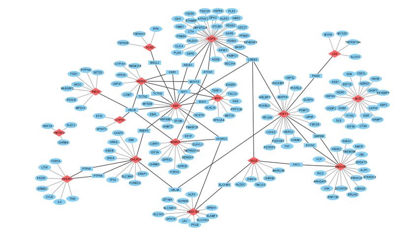
functions of protein partners that interact with them. In Figure 6, we visualize the PTM
spectrum and modifying proteins in the various “parts” of the cholesterol pathway: initial
part (HMGCR), middle part (SQLE and CYP51A1) and distant part (DHCR24 and DHCR7).
There are, at least, four common modifying proteins (AMFR, MARCH6, PRKACA and
PTPN5). Each of them can potentially affect several target enzymes from different parts
of the pathway. For example, MARCH6 interacts with SQLE, CYP51A1 and DHCR24
as well as AMFR interacts with HMGCR, DHCR24 and DHCR7. In view of the fact that
MARCH6 and AMFR are poly-specific E3 ubiquitin-protein ligases, it could be suggested
that there are conservative PPIs, participating in the proteolytic stability regulation of the
cholesterol synthesis enzymes. It is interesting to note that E3 ubiquitin-protein ligases
Biomedicines 2021, 9, x FOR PEER REVIEW
are applied in targeting low molecular weight degradators based on the Proteolysis 18 of 30
Tar-
geting Chimera (PROTAC technology) [167]. In brief, one “arm” of an PROTAC inhibitor
specifically interacts with a target protein while the other is covalently linked to the E3
ubiquitin-protein
linked ligase. The recruitment
to the E3 ubiquitin‐protein ligase. of
Thethe latter to a target
recruitment of theprotein
latter tomakes it possible
a target protein
makes it possible to trigger ubiquitin‐dependent proteasome degradation. Thus, onefor
to trigger ubiquitin-dependent proteasome degradation. Thus, one of the strategies of
therapeutic
the managing
strategies the cholesterol
for therapeutic managingpathway can be linked
the cholesterol with PROTAC
pathway technology
can be linked with
development.
PROTAC technology development.
Figure
Figure 6.
6. А
A model
model of
of the
the effects
effects of
of modifying
modifying proteins on some
proteins on some cholesterol
cholesterol synthesis
synthesis enzymes.
enzymes.
4.5. A Multiprotein Cholesterol “Metabolon”
Obviously, the question of whether the enzymes, functioning in the cholesterol syn‐
thesis pathway, physically interact with each other to form a multi‐protein cluster
(“metabolon”) in human cells, remains not entirely clear. Such spatial clustering of en‐
zymes would create conditions for more energetically favorable and effective transfor‐
mation of cholesterol precursors. In the FunCoup database (https://funcoup5.scilife‐
lab.se/search/), human protein interactions FDFT1‐MSMO1, LSS‐HSD17B1, MSMO1‐
SQLE and MSMO1‐CYP51A1 were predicted on interactions between orthologous yeastYou can also read