SURVEY AND SUMMARY A critical analysis of methods used to investigate the cellular uptake and subcellular localization of RNA therapeutics
←
→
Page content transcription
If your browser does not render page correctly, please read the page content below
Published online 9 July 2020                                                       Nucleic Acids Research, 2020, Vol. 48, No. 14 7623–7639
                                                                                                                   doi: 10.1093/nar/gkaa576

SURVEY AND SUMMARY
A critical analysis of methods used to investigate the
cellular uptake and subcellular localization of RNA
therapeutics
                                                                                                   *
Kirsten Deprey, Nefeli Batistatou and Joshua A. Kritzer

                                                                                                                                                                  Downloaded from https://academic.oup.com/nar/article/48/14/7623/5869349 by guest on 26 September 2020
Department of Chemistry, Tufts University, 62 Talbot Ave, Medford, MA 02155, USA

Received May 25, 2020; Revised June 17, 2020; Editorial Decision June 23, 2020; Accepted June 24, 2020

ABSTRACT                                                                          tamers. ASOs and siRNAs, first described in 1978 and 1998
                                                                                  (1,2), respectively, are currently the most widely applied in
RNA therapeutics are a promising strategy to treat                                drug development. Single-stranded ASOs modify protein
genetic diseases caused by the overexpression or                                  expression by binding to the target mRNA, and then, de-
aberrant splicing of a specific protein. The field has                            pending on chemical modifications and their targeted loca-
seen major strides in the clinical efficacy of this class                         tion on the mRNA, either cause RNase H-mediated degra-
of molecules, largely due to chemical modifications                               dation, correct aberrant splicing, or block ribosomal as-
and delivery strategies that improve nuclease resis-                              sembly (3,4). siRNAs are larger, double-stranded oligonu-
tance and enhance cell penetration. However, a ma-                                cleotides that cause mRNA degradation through an RNA-
jor obstacle in the development of RNA therapeutics                               induced silencing complex (RISC)-mediated pathway (5).
continues to be the imprecise, difficult, and often                               Because they could in principle target any mRNA, RNA
problematic nature of most methods used to mea-                                   therapeutics have a vast potential, especially to treat ge-
                                                                                  netic diseases that are currently untreatable by conventional
sure cell penetration. Here, we review these methods
                                                                                  medicine. Decades of work have made the design and syn-
and clearly distinguish between those that measure                                thesis of ASOs and siRNAs relatively straightforward (6,7).
total cellular uptake of RNA therapeutics, which in-                              Additionally, in contrast to small molecules and other drug
cludes both productive and non-productive uptake,                                 modalities, the pharmacokinetic properties of RNA thera-
and those that measure cytosolic/nuclear penetra-                                 peutics can, for the most part, be optimized independently
tion, which represents only productive uptake. We                                 from their target affinity, which is largely determined by
critically analyze the benefits and drawbacks of each                             their base sequence (8). Justifiably, drug development us-
method. Finally, we use key examples to illustrate                                ing RNA therapeutics has seen an exponential rise in in-
how, despite rigorous experimentation and proper                                  vestment during the last two decades (9).
controls, our understanding of the mechanism of                                      There are currently many RNA therapeutics in use and in
gymnotic uptake of RNA therapeutics remains lim-                                  clinical trials. As of May 2020, a total of seven ASO drugs
                                                                                  have been approved for use in humans: fomivirsen for cy-
ited by the methods commonly used to analyze RNA
                                                                                  tomegalovirus retinitis (10), mipomersen for familiar hyper-
delivery.                                                                         cholesterolemia (11), nusinersen for spinal muscular atro-
                                                                                  phy (12), eteplirsen and golodirsen for Duchenne muscu-
INTRODUCTION                                                                      lar dystrophy (13,14), inotersen for hereditary transthyretin
RNA therapeutics are an emerging drug class currently                             amyloidosis (15) and volanesorsen for familial chylomi-
being applied to the treatment of genetic diseases caused                         cronemia (16). Of these, four were approved by the United
by an overexpressed or aberrantly spliced protein. RNA                            States Food and Drug Administration (US FDA), and two
therapeutics are short, chemically modified nucleic acids                         were discontinued due to a decrease in the number of treat-
whose base sequences target disease-associated genetic ma-                        able patients (fomivirsen) or competing treatments (mipom-
terial in the cell with high selectivity. The term ‘RNA thera-                    ersen) (17). Two siRNA drugs have been approved by the
peutics’ includes antisense oligonucleotides (ASOs), small                        US FDA, patisiran for hereditary transthyretin amyloido-
interfering RNAs (siRNAs), microRNAs, and RNA ap-                                 sis (18) and givosiran for acute hepatic porphyria (19).

* To   whom correspondence should be addressed. Tel: +1 617 627 0451; Email: joshua.kritzer@tufts.edu

C The Author(s) 2020. Published by Oxford University Press on behalf of Nucleic Acids Research.
This is an Open Access article distributed under the terms of the Creative Commons Attribution Non-Commercial License
(http://creativecommons.org/licenses/by-nc/4.0/), which permits non-commercial re-use, distribution, and reproduction in any medium, provided the original work
is properly cited. For commercial re-use, please contact journals.permissions@oup.com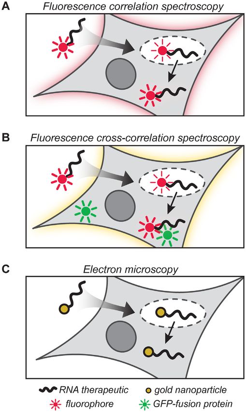
7624 Nucleic Acids Research, 2020, Vol. 48, No. 14

Many additional ASOs and siRNAs are currently undergo-             mers’ were introduced (29). Gapmers have a middle region
ing clinical trials (17,20). In a recent and well-publicized ‘N-   of PS-deoxynucleotides, allowing for RNase H1 recruit-
of-one’ trial, a child with a rare neurodegenerative genetic       ment, flanked by 2 -modified PS-nucleotides on either side.
disorder called Batten’s disease was treated with a person-        Additional 2 modifications including 2 -O-methoxyethyl
alized ASO drug called milasen. Milasen was designed with          (2 -MOE) are used to increase the hydrophobicity of the
the same chemical scaffold as nusinersen, but with a base se-      therapeutic, which enhances its binding to the membrane
quence that would correct the specific splicing error caused       receptors and facilitates cellular uptake (30). Conforma-
by the child’s unique genetic mutation (21). Within a year,        tional restriction, which can improve binding affinity and
milasen was produced, tested, approved, and administered           selectivity for the mRNA target, can be introduced by link-
to the young patient. These and other success stories pro-         ing the 2 -oxygen and 4 -carbon of the ribose, giving rise to
vide a glimpse into the vast potential of RNA therapeutics         a locked nucleic acid (LNA) (31). In a separate approach
to treat genetic disorders, and potentially other acute and        to backbone modification, cationic groups such as guani-
chronic diseases, which are difficult or impossible to treat       dinium can be added to the backbone. Guanidinium and

                                                                                                                                      Downloaded from https://academic.oup.com/nar/article/48/14/7623/5869349 by guest on 26 September 2020
with traditional small molecule therapies.                         other cationic groups are known to interact with the an-
   Despite their promise, development of RNA therapeu-             ionic cell surface and facilitate cellular uptake (32), and this
tics is fraught with many of the same difficulties as small        modification has shown some promise in promoting cellu-
molecule drug development, along with many difficulties            lar uptake for ASOs (33). For siRNAs, which are larger and
unique to this modality. RNA therapeutics have failed clin-        require separate sense and antisense strands, a large vari-
ical trials for a large variety of reasons, including toxicity,    ety of backbone and sugar modifications have been evalu-
off-target tissue sequestration, inactivity once delivered to      ated. Commonly, siRNAs incorporate specific patterns of
the target tissue, and even failure to reach the clinical end-     2 -OMe, 2 -fluoro (2 -F) and phosphorothioate modifica-
point despite causing alterations in protein expression (22–       tions at the 5 and 3 ends of both the sense and antisense
24). Some of these failures are due to incomplete under-           strands. While the impact of specific modification patterns
standing of the underlying biology and the poor predictive         on the activity of siRNAs is still being investigated, these
power of animal models, which are unfortunate features of          modifications together enhance target binding affinity and
all modern drug development. However, other clinical fail-         nuclease stability, while still allowing for association with
ures can be attributed to poor tissue targeting and cell pen-      the RISC complex (8).
etration, and these remain major obstacles in the field. Over          Incorporating the chemical modifications described
the last 15 years, a large effort has been undertaken in both      above can render ASOs and siRNAs highly resistant to
academic and industrial labs to gain a mechanistic under-          degradation by nucleases, and they can also improve ap-
standing of how RNA therapeutics are internalized by cells.        parent cell penetration. However, gymnosis (the process by
In this review, we discuss experimental methods that have          which RNA therapeutics are taken up by the cell, unfacili-
been used to measure cell penetration by RNA therapeu-             tated by other chemical or physical means of drug delivery)
tics, with the goal of distinguishing methods that measure         (34) remains highly inefficient. One way of bypassing inef-
total cellular uptake from methods that can more specif-           ficient gymnosis is to employ physical or chemical delivery
ically measure penetration to the cytosol or nucleus. This         strategies (35). Physical strategies for drug delivery include
distinction is important for a critical analysis of methods        microinjection, electroporation and compression (36–38).
used to monitor how RNA therapeutics enter cells, and for          They require the application of a physical needle or probe,
illustrating how those methods currently limit our under-          electric field, or high pressure in order to physically disrupt
standing of the underlying mechanisms.                             the plasma membrane. Though these techniques ensure ef-
                                                                   ficient delivery of the therapeutic to the cytosol with little
DISCUSSION                                                         loss to other compartments, they are painstaking and labo-
                                                                   rious, and can be damaging to the cell. Further, most phys-
Chemical modifications and delivery strategies for RNA ther-
                                                                   ical delivery strategies are not immediately applicable to in
apeutics
                                                                   vivo studies or clinical administration. Chemical strategies
Cellular delivery of RNA therapeutics faces two main is-           for delivery, by contrast, have been applied in animals and
sues: oligonucleotides are susceptible to degradation by           even in humans. The most commonly used chemical strat-
nucleases, and their cellular uptake can be inefficient (8).       egy in vitro is transfection using cationic lipids such as lipo-
For ASOs and siRNAs, the primary means of address-                 fectamine (39,40), but applications in vivo are limited due to
ing both of these issues has been through chemical mod-            high potential for toxicity (41). Formulation of RNA ther-
ifications. RNA therapeutics can employ a great diver-             apeutics into liposomes or lipid nanoparticles, containing
sity of chemical modifications on their phosphates, sug-           both cationic and neutral lipids to promote delivery while
ars and nucleobases––this area has been reviewed (25), so          avoiding toxicity, can promote delivery across the plasma
here we will summarize the most widely used modifica-              membrane (42). This strategy is used clinically– the siRNA
tions. Phosphorothioate (PS) backbones and 2 -O-methyl            drug patisiran is delivered using lipid nanoparticles (18).
(2 -OMe) ribose sugars are commonly used to increase nu-          Direct chemical conjugation of RNA therapeutics to lipids
clease resistance and promote cellular uptake (26–28). Im-         such as cholesterol or to cell-penetrating peptides also pro-
portantly, ASOs that are fully modified with PS- and 2 -          motes cell penetration (43–45). Conjugation of RNA ther-
O-methyl groups are not able to recruit RNase H1 and               apeutics to other chemical groups can further promote cell
cleave the target mRNA. To balance favorable pharmacoki-           entry as well as tissue targeting (46). For example, ASOs
netic properties with mRNA-degradation activity, ‘gap-             conjugated to N-acetylgalactosamine bind specifically to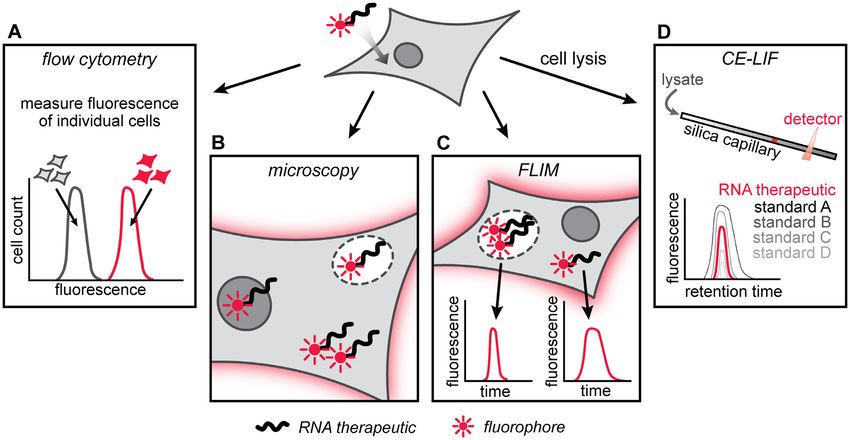
Nucleic Acids Research, 2020, Vol. 48, No. 14 7625

the asialoglycoprotein receptor, which is abundant in liver       activity, which indirectly suggested uptake of the exogenous
tissue, and are selectively taken up by hepatocytes in vitro      antisense oligonucleotide.
and in vivo (47). Chemical delivery strategies have great ad-        While the methodology for monitoring relevant pheno-
vantages, but each strategy has strengths and weaknesses.         types has advanced, the most common ways to measure
Overall, the main drawbacks are fast clearance, which re-         the productive uptake of RNA therapeutics still involve
quires administration of high doses, and accumulation of          readouts of functional activity. Today, reverse-transcriptase
toxic material, which can be dose-limiting.                       polymerase chain reaction (RT-PCR) is often used to quan-
   Investigation of the molecular mechanisms of gymnosis          titate target mRNA knockdown or splicing (Figure 2A). Af-
and various chemical delivery strategies has intensified in       ter treatment with the RNA therapeutic, cells or tissues are
recent years (see below). Often, RNA therapeutics are ob-         lysed and mRNA is extracted from the lysates. Primers cor-
served to be taken up by cells, but still do not exert a change   responding to the target mRNA transcript are added, and
in mRNA levels or protein expression. This phenomenon             RT-PCR is performed to quantitate the extent of mRNA
has been termed ‘non-productive uptake’, while uptake that        knockdown or splicing. RT-PCR is quantitative and high

                                                                                                                                   Downloaded from https://academic.oup.com/nar/article/48/14/7623/5869349 by guest on 26 September 2020
leads to a phenotypic effect has been termed ‘productive          throughput, and since it measures activity of RNA thera-
uptake’ (48). Mechanistically, gymnosis and most chemi-           peutics that have presumably penetrated to the cytosol or
cal delivery strategies rely on endocytosis for cellular up-      nucleus, endosomally trapped material does not contribute
take (Figure 1). Most of the material taken up by endocy-         to its signal. In parallel, knockdown of the target protein is
tosis remains trapped in endosomal vesicles. This trapped         typically monitored by Western blot (Figure 2B).
material is inactive since it cannot interact with its cellular      A hybridization-based approach can also be used to de-
target in the cytosol or nucleus, and either remains trapped      tect degradation of target mRNA. Fluorescence in situ hy-
in endosomes or gets delivered to the lysosome, where it is       bridization (FISH) uses a dye-labeled oligonucleotide probe
degraded. Collectively, this is the material that can be said     to detect changes in mRNA levels (Figure 2A) (53–56). The
to have undergone non-productive uptake, because it re-           dye-labeled probe is complementary to the target mRNA
mains associated with the cell but does not lead to any cel-      which allows it to be used to monitor knockdown. After
lular activity. In a poorly understood process, RNA ther-         treatment with RNA therapeutic, the cells are fixed and
apeutics can escape into the cytosol from late endosomes.         treated with the dye-labeled probe. Unhybridized probe is
This material can access the cellular target in the cytosol or    removed during wash steps, and the location and intensity
nucleus, and it can be said to have undergone productive          of hybridized probe is analyzed by fluorescence microscopy.
uptake.                                                           This allows direct and quantitative measurement of mRNA
   While assays that measure the activity of an RNA ther-         levels, allowing one to measure the activities of different
apeutic can imply the relative degree of productive up-           RNA therapeutics and thus indirectly to measure their de-
take, assays that measure cell penetration more directly          gree of cytosolic/nuclear penetration. Unlike RT-PCR, the
do not measure productive versus non-productive up-               FISH signal is directly proportional to the abundance of the
take. Rather, they measure either total cellular uptake or        target mRNA because the readout is non-amplified. How-
cytosolic/nuclear penetration. Total cellular uptake is the to-   ever, the lack of amplification means FISH is not as sensitive
tal amount of RNA therapeutic that remains associated             as RT-PCR. Additionally, wash steps must be extensive to
with the cell following treatment. Importantly, this includes     avoid high background from excess, unhybridized probe.
material bound at the cell surface and material trapped              Some investigations do not involve a specific endogenous
in endosomal compartments. Cytosolic/nuclear penetration          target. In these cases, an exogenously introduced reporter
refers to the fraction of RNA therapeutic that has success-       protein can be used to measure activity (Figure 2B). The
fully accessed the cytosolic/nuclear compartment. While           most common reporters are luciferase and green fluores-
RNA therapeutics can be active in the cytosol or in the           cent protein, whose expression can be measured through
nucleus (49), and some studies have shown selective local-        straightforward spectroscopic techniques (57–60). Such re-
ization in one compartment over the other (50–52), for the        porters can be used for RNA therapeutics that induce
sake of this discussion we will group these compartments to-      degradation of their target mRNA, but also for RNA ther-
gether as ‘cytosolic/nuclear’ to distinguish them from com-       apeutics that modulate mRNA splicing. For these ‘splice-
partments that prevent activity, such as endosomes and            switching’ assays, the RNA therapeutic is incubated with a
lysosomes. Ultimately, cytosolic/nuclear material is what         cell line that expresses either reporter protein, in which the
leads to the observation of productive uptake, while the dif-     mRNA transcript is interrupted by a large intron (61,62).
ference between total cellular uptake and cytosolic/nuclear       If the oligonucleotide can access the nucleus and reaches
penetration constitutes non-productive uptake (Figure 1).         the pre-mRNA transcript, it will redirect splicing and re-
                                                                  move the interruption, resulting in a functional full-length
                                                                  protein. For experiments using cultured cells, luciferase or
Methods to measure functional activity of RNA therapeutics
                                                                  GFP expression is commonly measured with a plate reader
In the studies that first described antisense technology, a 13-   or flow cytometer (61,62).
mer DNA oligonucleotide was applied to chick embryo fi-              While measuring mRNA and protein levels is more di-
broblasts infected with the Rous sarcoma virus to inhibit vi-     rect, activity can also be quantitated by measuring pheno-
ral replication (1). The incorporation of synthetic radioac-      typic changes (Figure 2C). In cell culture, these phenotypic
tive nucleotides was used as a measure of the virus’ reverse      assays may take the form of cell viability experiments af-
transcriptase activity (1). Cells treated with the antisense      ter treatment with an antisense oligonucleotide that knocks
oligonucleotide showed a decrease in reverse transcriptase        down a critical cellular protein. For example, PS- and 2 -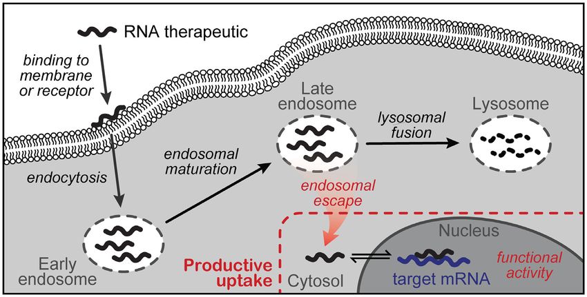
7626 Nucleic Acids Research, 2020, Vol. 48, No. 14
Downloaded from https://academic.oup.com/nar/article/48/14/7623/5869349 by guest on 26 September 2020
Figure 1. Productive uptake of RNA therapeutics. The RNA therapeutic binds the plasma membrane or a membrane receptor and is taken up by the
cell via endocytosis. It is initially trapped in early endosomes, which mature into late endosomes. The RNA therapeutic can be trafficked to the lysosome
to be degraded, and the total amount of material that is trapped in endosomes and degraded is referred to as ‘non-productive uptake.’ Alternatively, the
RNA therapeutic can escape from the endosome into the cytosol, from which it can access the nucleus and exert its therapeutic effect (functional activity).
‘Productive uptake’ therefore includes only material that accessed the cytosol and/or nucleus. Methods for measuring cell penetration of RNA therapeutics
can either measure total cellular uptake, which includes all material associated with the cell including material trapped in endosomes, or cytosolic/nuclear
penetration, which includes only material that contributes to functional activity.
Figure 2. Assays that measure functional activity of RNA therapeutics. After application of the RNA therapeutic, functional activity can be measured
in many different ways. Some common examples include (A) detection of mRNA knockdown by RT-PCR or fluorescence in situ hybridization (FISH),
(B) detection of altered protein expression through Western blot or splice correction of a reporter protein and (C) detection of phenotypic changes by cell
death or tumor shrinkage.
MOE-modified gapmer oligonucleotides targeted to Bcl-2 meabilization or changes in global gene expression (67).
and Bcl-xL mRNAs were incubated with human glioma In vivo, if the target is involved in cancer, the phenotypic
cells in culture, and one assay that was used to compare assay can involve reduction in tumor growth or inhibi-
the relative cytosolic/nuclear penetration of these oligonu- tion of tumor formation. For example, an LNA-containing
cleotides was a crystal violet assay for cell viability (63). oligonucleotide targeting TGF-2 was analyzed for effi-
However, viability assays can be problematic since RNA cacy through a reduction in tumor growth of lung metas-
therapeutics can be toxic at high concentrations (depend- tases (68). Overall, such phenotypic assays are indirect mea-
ing on chemical modifications and the delivery strategy, sures of cytosolic/nuclear penetration, because they are in-
0.2–10 M or higher) (64,65). Sequence-specific cytotox- fluenced by many other factors.
icity of modified oligonucleotides has also been observed Of course, functional activity assays are critical for drug
(66). Thus, it can be difficult to deconvolute on-target cell- development, as the goal for any RNA therapeutic is to have
killing activity from less specific toxicity due to cell per- a biological effect. Activity assays integrate all aspects of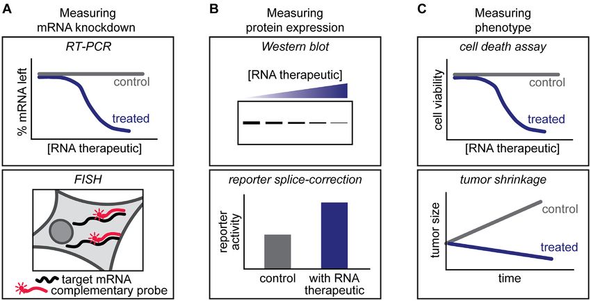
Nucleic Acids Research, 2020, Vol. 48, No. 14 7627

a molecule’s efficacy, from target access to target binding       tal radioactivity was measured after extensive washing
to subsequent translation, in a fairly straightforward and        (39,72,73). This method has also been applied in vivo to
high-throughput manner. Further, the readout from activ-          measure accumulation of a 3 H- or 35 S-labeled oligonu-
ity assays does not require a covalent label for detection, so    cleotide in specific tissues (74–76). Radioactive labeling and
these assays can compare productive uptake independent            detection methods are no longer widely used in early stage
from the impact of a chemical label.                              preclinical research due to high cost of materials and safety
   Although these studies are undeniably crucial in the           concerns. Radioactivity-based assays have been largely re-
pipeline of an RNA therapeutic, they also inherently miss         placed in favor of other detection methods, such as fluores-
a great deal of information on cellular uptake, subcellular       cence (77).
trafficking, and endosomal escape. For instance, the activ-          The most commonly used method for measuring total
ity assays discussed in this section are amplificative, so the    cellular uptake is tracking fluorescence of dye-labeled RNA
signal is not directly proportional to the extent of uptake.      therapeutics. For these experiments, dye-labeled oligonu-
Amplified assays provide for greater sensitivity, but do not      cleotides are incubated with cells in culture or administered

                                                                                                                                      Downloaded from https://academic.oup.com/nar/article/48/14/7623/5869349 by guest on 26 September 2020
allow for quantitative comparisons. Thus, they are not ideal      in vivo with subsequent tissue harvesting. Total cellular up-
for structure-activity relationship studies that seek to un-      take is most often measured by flow cytometry (Figure 3A)
derstand the effects of different chemical modifications. In      or fluorescence microscopy (Figure 3B) (44,78–82). Flow
addition, activity-based assays integrate a large number of       cytometry is quantitative and high-throughput, but the sig-
processes, any of which could impact the final readout. If        nal of internalized dye-labeled molecules is indistinguish-
an RNA therapeutic does not have the expected biologi-            able from that of dye-labeled molecules bound to the out-
cal effect, it could be due to lack of total uptake, lack of      side of the plasma membrane or trapped in endosomes.
endosomal escape, trafficking to the lysosome, sequestra-         Live-cell fluorescence microscopy is lower-throughput than
tion within subcellular structures, nonspecific interactions      flow cytometry, but microscopy is better able to distin-
with proteins or protein complexes, aggregation, degrada-         guish subcellular localization. However, it is difficult even
tion, low target affinity, high degree of secondary struc-        with confocal fluorescence microscopy to determine defini-
ture, failure to recruit ribonucleases or block the spliceo-      tively the extent to which a dye-labeled molecule is cytosolic
some, or many other factors. Ideally, one would subject the       or endosomal without advanced techniques. Further, each
RNA therapeutics of interest, plus controls, to many as-          of these methods requires labeling of the RNA therapeu-
says that each measure one of these diverse factors. How-         tic of interest with a bulky and hydrophobic dye. In some
ever, because cytosolic/nuclear penetration is such an im-        cases, addition of the dye can influence the extent of uptake
portant roadblock to activity for RNA therapeutics, failure       and subcellular trafficking by altering interactions with the
in an activity-based assay is often ascribed to poor produc-      plasma membrane and embedded proteins, and effects of
tive uptake (poor cytosolic/nuclear penetration, Figure 1)        dye labeling have been observed for cell-penetrant peptides
(69). One can control for some aspects of internalization us-     as well (83–85). Another liability of these commonly used
ing an experiment in which RNA therapeutics and controls          assays is that degradation of the molecule releases the free
are introduced into the cell via different delivery strategies.   dye, and thus could result in a false-positive signal (67,86).
However, this experiment only controls for a limited part         Despite these caveats, flow cytometry and fluorescence mi-
of the internalization process and does not directly address      croscopy are still two of the most widely used methods to
most of the other factors listed above. Ultimately, activity      measure total cellular uptake of RNA therapeutics.
assays are most informative in conjunction with assays that          Other, more advanced fluorescence-based techniques
measure total cellular uptake and assays that directly mea-       have been implemented to address some of the drawbacks
sure cytosolic/nuclear penetration.                               of flow cytometry and fluorescence microscopy (Figure 3C)
                                                                  (87,88). Fluorescence lifetime imaging microscopy (FLIM)
                                                                  can measure the total cellular uptake and stability of RNA
Methods to measure total cellular uptake
                                                                  therapeutics in cultured cells (52,89–93). The fluorescence
Understanding how to promote productive uptake of RNA             lifetime of a fluorophore can be altered when it is in close
therapeutics is critical to the development of effective ther-    proximity to another fluorophore or when it is in differ-
apeutics, yet the methods available to measure cell penetra-      ent chemical environments, such as packaged in a deliv-
tion have distinct weaknesses. The ideal method would be          ery vector, trapped in an endosome, or free in the cy-
quantitative, high-throughput, label-free, and able to distin-    tosol (91,94,95). FLIM has been used for the detection
guish unambiguously among subcellular compartments. In            of porphyrin-oligonucleotide conjugates, where the por-
this section, we discuss methods that measure total cellular      phyrin aided in both delivery and detection of the oligonu-
uptake; methods that measure cytosolic/nuclear penetra-           cleotide (90). In principle, FLIM can distinguish endoso-
tion are discussed in the next section. Methods commonly          mally trapped and cytosolic material, but in practice FLIM
applied to measure the total cellular uptake of peptide drugs     signal is altered in subtle ways by the type of dye used and
were recently reviewed (70,71). Here, we focus on an over-        the chemical environment of each subcellular structure. As
lapping set of methods commonly applied to RNA thera-             a result, it remains difficult to quantitate localization to sub-
peutics.                                                          cellular structures using FLIM, and thus FLIM is best char-
                                                                  acterized as a method for quantitating total cellular uptake.
Measuring total cellular uptake of labeled RNA therapeu-             Another alternative that uses dye-labeled oligonu-
tics. In early studies, oligonucleotides radiolabeled with        cleotides is capillary electrophoresis with laser-induced
35
   S phosphorothioates were incubated with cells, and to-         fluorescence (CE-LIF, Figure 3D), which has been used to
7628 Nucleic Acids Research, 2020, Vol. 48, No. 14
Downloaded from https://academic.oup.com/nar/article/48/14/7623/5869349 by guest on 26 September 2020
Figure 3. Assays that measure total cellular uptake of dye-labeled RNA therapeutics. Cells in culture can be treated with a dye-labeled RNA therapeutic
and the fluorescence within live cells can then be measured by (A) flow cytometry, (B) confocal fluorescence microscopy or (C) fluorescence lifetime imaging
microscopy (FLIM). (D) Fluorescence can also be measured in cell lysates by capillary electrophoresis with laser-induced fluorescence (CE-LIF).
quantitate the concentration of dye-labeled nucleic acids from the secondary antibody is detected by fluorescence mi-
in solution (96). In a related experiment, CE-LIF was croscopy, and fluorescence can be quantitated to measure
also used to measure concentration of endogenous target the relative amount of internalized oligonucleotide. Addi-
mRNA in plasma and human cells through the use of a tional co-localization studies with membrane proteins in-
complementary dye-labeled DNA probe (97–99). CE-LIF volved in endocytosis can aid in the study of subcellular
has also been applied to quantitation of RNA therapeutics localization (44,105,107). Immunofluorescence assays can
delivered by scrape-loading (100,101), liposome encap- be performed using unlabeled oligonucleotides, and the use
sulation (102,103) and peptide conjugation (104). In a of antibodies for detection renders this method highly spe-
typical CE-LIF experiment, the cells are lysed and the cific. However, immunofluorescence must be performed on
soluble fraction is injected onto a capillary column for fixed and permeabilized cells in order to deliver the anti-
separation by capillary electrophoresis. The dye-labeled bodies to the interior of the cell. Oligonucleotides and other
oligonucleotide is detected with a laser excitation beam biomolecules have been shown to redistribute throughout
at the appropriate wavelength, and the concentration of the cell as a result of fixation, leading to false-positive arti-
internalized molecule is calculated from a calibration curve facts (33,51,86,108,109). Finally, even with careful colocal-
of standards of known concentration spiked into untreated ization studies, it can be difficult to deconvolute cytosolic
cell lysate (100,101). This method is very sensitive and versus endosomal material in a definitive manner.
requires very small (nL-pL) sample volumes (102). CE-LIF Another label-free method for measuring total cellular
measures total cellular uptake because cell lysis results uptake of RNA therapeutics is in-cell nuclear magnetic res-
in the mixing and re-equilibration of membrane-bound, onance (NMR, Figure 4B). Unique NMR chemical shifts
endosomal, and cytosolic/nuclear material prior to analy- for artificial nucleic acids can be observed in cell lysates and
sis. It also carries the same caveats as flow cytometry and live cells. (110–113) The Trantirek group was the first to per-
fluorescence microscopy with respect to the dye potentially form in-cell NMR to detect exogenously applied oligonu-
altering penetration properties. cleotides, delivered by physical injection of live Xenopus lae-
vis oocytes (111). The oligonucleotides were doubly-labeled
Measuring total cellular uptake of label-free RNA therapeu- with 13 C and 15 N. The spectra obtained from live cells were
tics. A handful of assays that measure total cellular up- compared to those obtained from the lysates of treated cells
take eliminate the need for a label entirely. Immunofluo- and to those obtained in vitro. These NMR-based exper-
rescence, traditionally used to detect proteins, and also has iments have since been applied to live human cells (113),
recently been adapted for the detection of RNA therapeu- and have been adapted to quantify the uptake of RNA
tics in vitro using anti-oligonucleotide antibodies (Figure therapeutics. In this adaptation, the molecules were deliv-
4A) (44,105–107). Cells treated with PS-oligonucleotides ered to the cell via electroporation or transfection, and the
1
are washed, fixed, permeabilized, blocked, treated with an H spectra were recorded (114,115). To enhance sensitivity,
anti-oligonucleotide primary antibody, and finally treated the Petzold group performed 1 H,31 P cross-polarization dy-
with a dye-labeled secondary antibody. The fluorescence namic nuclear polarization NMR on frozen cells electropo-Nucleic Acids Research, 2020, Vol. 48, No. 14 7629
Downloaded from https://academic.oup.com/nar/article/48/14/7623/5869349 by guest on 26 September 2020
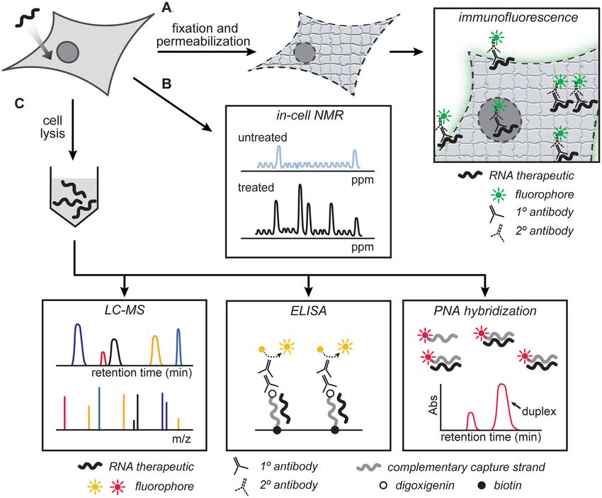
Figure 4. Assays that measure total cellular uptake of label-free RNA therapeutics. First, cells or tissues are treated with unlabeled RNA therapeutic.
(A) For immunofluorescence detection of unlabeled RNA therapeutics, cells or tissues are fixed, permeabilized, and incubated with dye-labeled antibodies
that selectively recognize the RNA therapeutic. (B) Using advanced NMR techniques, delivered RNA therapeutics can be measured in intact cells by
quantitating peak volumes from the NMR spectra of 31 P or other nuclei. (C) After homogenization and lysis of cells or tissues, the concentration of
unlabeled RNA therapeutic in the cell lysate can be measured by enzyme-linked immunosorbent assay (ELISA), a peptide nucleic acid (PNA) hybridization
assay, or by liquid chromatography–mass spectrometry (LC–MS).
rated with a PS-modified gapmer oligonucleotide (115). The chromatography and mass spectrometry (LC–MS, Figure
peak volumes of the 31 P NMR spectra were used to estimate 4C) (42,107,116–119). LC–MS can be made quantitative by
the concentration of the oligonucleotide. No detectable sig- calibrating with standards of known concentration spiked
nal was observed in cells that underwent free uptake of into cell lysate. With appropriate calibration, this method
the oligonucleotide (115). This may be due to the micro- can have both high sensitivity and excellent quantitation.
molar concentrations required for detection by NMR us- Other label-free assays take advantage of the selective
ing moderate acquisition times. Other limitations of NMR- nature of hybridization by using a labeled complementary
based quantitation include the requirement for careful con- strand for isolation and quantification of the RNA ther-
struction of calibration curves, and the interference in apeutic. One example is the adaptation of the enzyme-
NMR spectra from other molecules in cells and complex linked immunosorbent assay (ELISA) to measure total cel-
lysates. lular uptake and tissue distribution of antisense oligonu-
While NMR-based quantitation suffers from a lack of cleotides (Figure 4C) (50,120–127). In most of these exam-
sensitivity, mass spectrometry methods are notable for their ples, a cell lysate is incubated with a biotinylated oligonu-
excellent sensitivity. Mass spectrometry methods are be- cleotide complementary to the RNA therapeutic. Then the
coming more and more common for measuring the total cel- hybridized duplex is captured on avidin-coated magnetic
lular uptake of RNA therapeutics, both in cultured cells and beads. The biotinylated complementary oligonucleotide is
ex vivo (42,107,116–119). Cells or tissues are homogenized also labeled at the opposite end with digoxigenin, to allow
and lysed, and the cleared lysates are analyzed by liquid for detection of the immobilized duplexes with an alkaline7630 Nucleic Acids Research, 2020, Vol. 48, No. 14
phosphatase-conjugated anti-digoxigenin antibody. Alka- Methods to measure cytosolic/nuclear penetration
line phosphatase activity is used as a measure of the relative
Assays that measure material that has localized to the cy-
amount of original oligonucleotide that was pulled down.
tosol and/or nucleus, without interference from material
ELISA-based detection methods are very sensitive and can
stuck at the plasma membrane or in endosomal compart-
detect concentrations as low as picomolar in complex matri-
ments, are crucial for the development of RNA thera-
ces including tissue lysates (122,125,127). However, with the
peutics. In this section, we discuss methods that can se-
enhanced sensitivity comes the drawback that this assay is
lectively quantitate cytosolic and nuclear material. Many
amplificative, which complicates quantitative comparisons
of the assays described in the previous section on total
of total cellular uptake. ELISA, as with Western blotting,
cellular uptake can offer information about subcellular
involves many steps with a high number of manipulations,
localization through the use of subcellular fractionation
which reduces throughput and can lead to artifacts. Addi-
(50,72,75,107,136). Ultracentrifugation of cell lysates al-
tionally, antibodies must be carefully selected to ensure that
lows for separation of cellular structures, which in principle
they are specific and robust to manipulation.
Downloaded from https://academic.oup.com/nar/article/48/14/7623/5869349 by guest on 26 September 2020
allows for the separation of membrane-bound, endosomal,
Another assay that relies on hybridization is the peptide-
cytosolic and nuclear-localized material (137). However, in
nucleic acid (PNA) hybridization assay (Figure 4C). PNA
practice subcellular fractionation is technically challenging
hybridization assays are widely used to measure the total
and low-throughput, involving painstaking manipulations
cellular uptake of modified oligonucleotides and siRNAs in
of the lysate samples. Cross-contamination between sub-
cultured cells, as well as in plasma samples and in tissues ex
cellular compartments must be rigorously tested for and
vivo (81,128–135). For this assay, cells or tissues that have
controlled against. Even with careful controls, material can
been exposed to the RNA therapeutic are homogenized and
re-equilibrate during lysis prior to ultracentrifugation, fur-
lysed. The soluble lysates are incubated with a complemen-
ther reducing confidence that this method can faithfully dis-
tary, dye-labeled PNA, which hybridizes to the RNA ther-
tinguish material that resided in different compartments in
apeutic. HPLC retention time is used to distinguish duplex
the live, intact cell (137). Several alternatives can more reli-
PNA-RNA from unhybridized PNA, and peak volume can
ably eliminate endosomally trapped material from interfer-
be used to quantitate the concentration of duplexed PNA
ing with measurements of cytosolic/nuclear penetration of
using a calibration curve of PNA duplexes of known con-
RNA therapeutics.
centration spiked into the cell lysate (128). PNA hybridiza-
As described above, conventional fluorescence mi-
tion assays are highly specific, quantitative, and label-free
croscopy techniques such as immunofluorescence and con-
with respect to the RNA therapeutic. However, tissue ho-
focal fluorescence microscopy are largely qualitative and
mogenization and cell lysis allow the PNA probes to hy-
cannot easily distinguish between cytosolic and endosomal
bridize with RNA therapeutic that was once endosomally
material. Immunofluorescence and confocal microscopy
trapped or membrane-bound. Therefore, while this assay
work well in principle to distinguish endosomal material
has many advantages, it only measures total cellular uptake
from cytosolic/nuclear material, but in practice endosomal
and cannot distinguish between cytosolic and endosomal
and cytosolic/nuclear material can be confounding. These
compartments.
assays require extensive co-localization analysis to provide
All of the assays described in this section measure total
confidence in any conclusions about subcellular localiza-
cellular uptake, which is the total amount of material asso-
tion.
ciated with the cell or tissue (Figure 1). This includes mate-
rial that is trapped in endosomes or lysosomes, and material
that remains bound to the cell surface after wash steps. For Fluorescence correlation spectroscopy assays. More ad-
most methods, the need to fix or lyse cells results in mix- vanced fluorescence techniques are capable of better quan-
ing and equilibration of components from different com- titation of the amount of dye-labeled molecule within the
partments. Confocal microscopy is performed on live cells, cell, and they can also better distinguish subcellular com-
and therefore does not require cells to be fixed and perme- partments. A key example is fluorescence correlation spec-
abilized. With careful co-localization, confocal microscopy troscopy (FCS) (138), which has been applied to investi-
is sometimes able to distinguish endosomal and lysosomal gate the concentrations of RNA therapeutics in live cells
material from cytosolic material. However, this distinction (Figure 5A) (52,53,91,93). In FCS, the diffusion of a dye-
requires extensive and time-consuming analysis, and signal labeled molecule is tracked within a small (femtoliter) focal
from endolysosomal compartments cannot entirely be elim- volume by measuring fluctuations in fluorescence intensity
inated. of molecules entering and exiting the focal volume. The ab-
Overall, there is a great deal of evidence that RNA ther- solute number of molecules present within the defined focal
apeutics, especially when delivered via gynmosis, accumu- volume can be calculated using a Poisson distribution (139).
late in endosomal compartments with a relatively small Cytosolic material is distinguished from endosomal mate-
proportion escaping to the cytosol. Endosomally trapped rial through imaging analysis that identifies regions that
RNA therapeutics are unable to reach their intracellular do not include punctate signal, which represent endolyso-
target, leading to potential overestimation of productive somal vesicles (140). Focal volumes within these regions
uptake if conclusions are drawn solely from assays that are chosen manually. The Mundigl group at Roche recently
measure total cellular uptake. Such conclusions are to be used FCS to calculate the absolute number of microinjected
avoided, as this may be a primary reason for poor suc- LNA-gapmer oligonucleotides required for target gene sup-
cess when using in vitro experiments to predict in vivo pression (53). Scientists in the Brock group at Radboud
potency. University Medical Center also used FCS to measure theNucleic Acids Research, 2020, Vol. 48, No. 14 7631
Wagner and Lamb groups at the University of Munich used
FCCS to assess the cytosolic degradation of dual-labeled
oligonucleotides through simultaneous tracking of both flu-
orophores of a FRET pair (52).
FCS and FCCS allow calculation of absolute concen-
tration of a dye-labeled RNA therapeutic, as opposed to
the relative fluorescence intensities obtained from simpler
fluorescence microscopy techniques (143). Despite this and
other advantages, FCS and FCCS are low-throughput, re-
quire specialized instrumentation, are limited to applica-
tions in cell culture, and require a fluorescent dye to be con-
jugated to the RNA of interest. Further, the process requires
the user to manually select focal volumes to analyze within
Downloaded from https://academic.oup.com/nar/article/48/14/7623/5869349 by guest on 26 September 2020
the cell, which may introduce some degree of subjectivity.
These drawbacks may explain why, despite their sensitivity,
their absolute quantitation, and their ability to interrogate
exclusively cytosolic or nuclear material, FCS and FCCS
have yet to be adopted by a larger number of groups inter-
ested in RNA therapeutics.
Electron microscopy assays with siRNA-gold. Gilleron et
al used electron microscopy to measure the cellular amount
of siRNA-gold after in vitro and in vivo delivery using
lipid nanoparticles (144). Cells or tissues were administered
siRNA which was covalently labeled with gold nanoparti-
cles. At different timepoints, cells or tissues were washed,
fixed, stained, and subjected to electron microscopy to de-
tect the subcellular localization of the gold nanoparticles
(Figure 5C) (144). Electron microscopy allows one to mor-
phologically distinguish subcellular structures such as early
endosomes, late endosomes, and lysosomes. The high reso-
lution of this method enables clear measurement of the pro-
portion of siRNA-gold in endosomal, cytosolic and nuclear
compartments. However, this method is low throughput, re-
quires fixation, and necessitates labeling of the siRNA with
Figure 5. Assays that measure cytosolic/nuclear penetration of labeled gold nanoparticles, and has not been widely adopted.
RNA therapeutics. (A) Fluorescence correlation spectroscopy measures
the diffusion of a small number of dye-labeled molecules through a defined
The available methods for measuring functional activity
cytosolic focal volume. (B) Fluorescence cross-correlation spectroscopy and total cellular uptake are far more numerous and far
measures the diffusion of a small number of dye-labeled molecules through more accessible than the available methods for measuring
a defined cytosolic focal volume, tracked simultaneously with another flu- cytosolic/nuclear penetration. New, more easily adopted
orescent molecule or protein of interest. (C) Electron microscopy detects methods that measure cytosolic/nuclear penetration of
gold nanoparticle-labeled molecules, and can distinguish material in the
cytosol from material in various endosomal compartments. RNA therapeutics are thus critically needed. Existing meth-
ods that have been developed for peptide and protein deliv-
ery could readily be adapted for use with RNA therapeutics
nuclear concentration of a splice-switching oligonucleotide (70,71). For example, several assays measure a signal that
delivered by polyplex formation with cell-penetrant pep- depends on interaction between the exogenously applied
tides, correlating the concentration to the observed thera- molecule and a protein expressed in the cytosol/nucleus.
peutic effects in a cell culture model of myotonic dystrophy These assays include split-protein complementation assays
(93). (145–147), glucocorticoid receptor transcriptional reporter
FCS was further adapted into a dual-color technique assays (148,149), biotin ligase assays (150), and assays that
called fluorescence cross-correlation spectroscopy (FCCS) involve enzyme-specific fluorogenic probes (151). Each of
(Figure 5B) (141). FCCS involves the same principles as these assays comes with caveats of their own, which are dis-
FCS but tracks two fluorescent dyes simultaneously. If the cussed elsewhere (70,71), but they also offer valuable infor-
variations of intensity over time within a small focal volume mation about the cytosolic/nuclear penetration of exoge-
align well in the two different color channels, then that ob- nously applied molecules. With relatively straightforward
servation is indicative that the two fluorophores are in close adjustments, each of these assays could be directly applied
proximity to one another (141). The Schwille group used to measuring the cytosolic/nuclear penetration of RNA
FCCS to analyze the incorporation of microinjected dye- therapeutics.
labeled siRNAs into the RISC complex, visualized using Another promising assay that relies on interactions with
an expressed Argonaute2-EGFP fusion in both the cytosol expressed proteins is the chloroalkane penetration assay
and nucleus (142). In another example, researchers in the (CAPA) (152–154). CAPA uses a HeLa cell line that sta-7632 Nucleic Acids Research, 2020, Vol. 48, No. 14
bly expresses the engineered protein HaloTag to quanti- targeting genes associated with vesicle trafficking (105). A
tate the cytosolic penetration of molecules labeled with list of specific proteins identified as important for gymnosis
a small chloroalkane ligand (155). Cells are pulsed with is included in Table 1. Typically, once the protein involved
the chloroalkane-labeled molecule, which covalently reacts was implicated using knockdown, methods for quantitating
with HaloTag upon reaching the cytosol. This covalent re- total cellular uptake were used to more precisely define that
action blocks the active sites of the HaloTag protein. Cells protein’s role in gymnotic uptake. However, as mentioned
are then chased with a chloroalkane-labeled dye, which above, different assays have different limitations, which po-
reacts with open active sites. The total fluorescence from tentially limit the conclusions that can be drawn about the
the dye, measured by flow cytometry, is inversely propor- role of the implicated protein. Below, we highlight three il-
tional to the concentration of cytosolic RNA therapeu- lustrative examples of authors who rigorously implemented
tic. CAPA signal excludes material stuck in endosomes available assays and drew sound conclusions, but whose
or at the cell surface, and CAPA is non-amplified so the conclusions are inherently limited due to the nature of the
signal is a direct and quantitative measurement of the assays.
Downloaded from https://academic.oup.com/nar/article/48/14/7623/5869349 by guest on 26 September 2020
amount of material delivered. Our group originally devel-
oped CAPA to measure the cytosolic penetration of various Example 1. AP2M1 mediates productive uptake. In 2011,
cell-penetrating peptides, and others have adapted it for use Koller et al. determined that the adaptor protein AP2M1
with small molecules, peptidomimetics and cell-penetrant is crucial for the functional uptake of single-stranded RNA
proteins (156–161). We are currently applying CAPA to therapeutics into primary murine MHT cells. AP2M1 is an
diverse RNA therapeutics with different chemical modi- adaptor protein, involved in clathrin-mediated endocyto-
fications, in order to better understand factors that drive sis, that is recruited to the membrane in a cargo-dependent
cytosolic/nuclear penetration. manner. AP2M1 binds cytosolic-facing PI(4,5)-P2 mem-
brane lipids, clathrin, and additional accessory and adap-
tor proteins. It thus provides an indirect link between ex-
Methodology limits our understanding of the molecular
ogenous cargo interaction with the plasma membrane and
mechanisms of gymnosis
clathrin polymerization required for clathrin-mediated en-
RNA therapeutics can be delivered to the cell using a va- docytosis (171,172). Knockdown of AP2M1 decreased the
riety of strategies, all of which may have different mecha- total cellular uptake of a dye-labeled oligonucleotide by
nisms, rates of uptake and degrees of cytosolic/nuclear pen- nearly 50%, and decreased functional activity by a simi-
etration. Gymnosis refers to the cytosolic/nuclear penetra- lar degree. Uptake was measured by applying Cy3- and
tion of nucleic acids without facilitation by physical disrup- fluorescein-labeled oligonucleotides to MHT cells in cul-
tion or chemical agents (34). The mechanisms of gymno- ture and then analyzing using flow cytometry. Additionally,
sis are poorly understood, yet gymnotic uptake is central LC–MS was performed on the lysates of treated cells with
to the drug development strategy of many clinically impor- standards of known concentration spiked into the lysates
tant RNA therapeutics (34,162,163). It is critical to recog- for quantitation. Further, immunofluorescence studies were
nize that the methods used to analyze uptake directly affect performed on fixed cells using an anti-PS-oligonucleotide
the conclusions that can, and cannot, be drawn. Thus, in antibody. In parallel, RT-PCR and Western blot were used
this section, we will address what is known about the mech- to monitor the knockdown of target mRNA and protein, re-
anisms of gymnosis of RNA therapeutics, highlighting the spectively. Taken together, the data from these assays were
limitations of the methods used. used to conclude that AP2M1 is important for productive
It is generally accepted that RNA therapeutics are in- uptake.
ternalized by the cell through endocytosis after associat- While these data can firmly conclude that AP2M1 is in-
ing with proteins on the cell membrane (69,163–165). Af- volved in the earliest steps of gymnotic uptake (binding the
ter the initial uptake, the RNA therapeutic is trapped inside cell surface and endocytosis, Figure 1), they cannot con-
the early endosome. During endosome maturation, RNA clude anything about its role in later steps such as endo-
therapeutics may escape these vesicles and subsequently ex- somal escape. Surprisingly, while both total cellular up-
ert their therapeutic effect in the nucleus or cytosol (Figure take and functional activity were reduced upon siRNA-
1) (69,163,166,167). Endosomal escape is thought to be at- mediated knockdown of AP2M1, only total cellular uptake
tributed to some degree of membrane deformation of the was reduced upon siRNA-mediated knockdown of clathrin,
late endosome or a small degree of leakage from vesicle fu- while functional activity was unaffected. The reason for this
sion processes (69,80,144,163,166,167). discrepancy could not be addressed with the methods used.
The experiments required to understand the subcellular Specifically, flow cytometry and LC–MS can only measure
trafficking of RNA therapeutics are challenging, and their total material associated with the cell, and cannot distin-
difficulty has limited progress in understanding the exact guish among membrane-bound, cytosolic and endosomal
mechanisms of endosomal escape. Recently, some investi- material. The discrepancy between total cellular uptake and
gators have taken a genetics-based approach to implicate functional activity observed after AP2M1 and clathrin were
specific cellular proteins in gymnotic uptake and endoso- both knocked down could be explained by a role for AP2M1
mal escape. Most commonly, specific genes were individu- in endosomal escape, and potentially a degree of indepen-
ally knocked down to identify proteins involved in gymno- dence between rate of clathrin-mediated endosomal uptake
sis (166,168–170). Less commonly, specific genes involved and rate of endosomal escape. However, given the indirect
in uptake of RNA therapeutics were also identified using nature of functional activity assays, these hypotheses re-
siRNA knockdown screens with small libraries of siRNAs main untested.Nucleic Acids Research, 2020, Vol. 48, No. 14 7633
Table 1. Selected proteins shown to be important for gymnosis of RNA therapeutics, their known roles in gymnosis, and the methods used (other than
functional assays) to verify their importance for gymnosis. PS: phosphorothioate, MOE: 2 O-methoxyethyl, LNA: locked nucleic acid
Implicated step in gymnosis
Protein(s) (Figure 1) RNA therapeutic Methods used Reference
Stabilin-1 and Stabilin-2 Endocytosis Cy3- and 125 I-
labeled Fluorescence microscopy, (169)
PS-MOE gapmer ASOs immunohistochemistry,
radioactivity
Adaptor protein (AP2M1) Endocytosis Cy3- and Flow cytometry, (105)
fluorescein-labeled immunofluorescence, mass
PS-MOE gapmer ASOs spectrometry
Caprin-1 Not identified, Cy3-labeled peptide–PNA Fluorescence microscopy (173)
hypothesized role in conjugates
endocytosis
Systemic RNA interference Not identified, Alexa568-labeled Fluorescence microscopy (168)
Downloaded from https://academic.oup.com/nar/article/48/14/7623/5869349 by guest on 26 September 2020
deficient-1 transmembrane hypothesized role in PO-2’O-methyl ASOs,
family 2 (SIDT2) endocytosis dsRNA
Annexin A2 Endosomal maturation Cy3-labeled PS-MOE Subcelluluar fractionation, (166)
gapmer ASOs flow cytometry,
co-localization microscopy
after fixation
Epidermal growth factor Endocytosis, Endosomal Cy3-labeled gapmer ASOs Flow cytometry, (170)
receptor (EGFR) maturation with PS-MOE, PS-F, and co-localization microscopy
PS-cEt modifications after fixation
Protein Kinase C-alpha Endosomal maturation Cy5-labeled PS-LNA flow cytometry (174)
(PKC␣) gapmer ASOs
ESCRT-1 proteins: tumor Endocytosis Endosomal PS-DNA, cET and luciferase knockdown, (175)
susceptibility gene 101 maturation MOE-ASOs, and PS-F, viability
(TSG101) and VPS28 MOE-ASO
Coat protein complex II Endosomal escape Cy3-labeled PS-MOE Flow cytometry, (80)
(COPII) and associated gapmer ASOs, and co-localization microscopy
proteins: SEC31a, Sar1, Cy3-labeled 5-10-5 after fixation
STX5 PS-LNA gapmer ASOs
Mannose-6-phosphate Endosomal escape Cy3-labeled PS-MOE Flow cytometry, (176)
receptor (M6PR) and gapmer ASOs microscopy after fixation
associated tethering and co-localization
protein, GCC2
Rab5c Endosomal maturation PS-MOE gapmer ASO Radioactivity (177)
125 I-labeled PS-MOE
gapmer ASO
Early endosomal antigen 1 Endosomal maturation PS-MOE gapmer ASO Radioactivity (177)
(EEA1) 125 I-labeled PS-MOE
gapmer ASO
Rab7 Endosomal maturation, PS-MOE gapmer ASO Radioactivity (177)
Lysosome biogenesis and 125 I-labeled PS-MOE
fusion, Endosomal escape gapmer ASO
Lysobisphosphatidic acid Endosomal escape Cy3-labeled PS-MOE Flow cytometry, (167,177)
(LBPA) gapmer ASOs co-localization microscopy
after fixation
Alix Endosomal escape Cy3-labeled PS-MOE Flow cytometry, (167,177)
gapmer ASOs co-localization microscopy
after fixation
Example 2. Stabilin-1 and stabilin-2 promote productive up- the oligonucleotide to stabilins, followed by endocytic inter-
take. In 2016, the Harris and Seth groups found that, nalization of the receptor and the oligonucleotide.
in addition to previously reported scavenger receptors, Multiple assays were used in parallel to elucidate the
stabilin-1 and stabilin-2 are implicated in the internalization roles of stabilin proteins in the internalization of PS-
of chemically modified oligonucleotides (169). The stabilin– oligonucleotides. Uptake was measured by: fluorescence mi-
oligonucleotide binding event triggered uptake by clathrin- croscopy of a Cy3-labeled oligonucleotide in cell culture
mediated endocytosis, resulting in functional antisense ac- using co-localization with lysotracker, immunohistochem-
tivity in cells and tissues. HEK-293 cell lines stably express- istry using an anti-PS-oligonucleotide antibody in tissue
ing stabilin-1 or stabilin-2 had both a higher degree of in- samples comparing wild type and stabilin-2-knockout mice,
ternalization and increased antisense activity compared to and total radioactivity of 125 I-labeled oligonucleotide in cell
a HEK-293 cell line stably expressing a blank vector. To- culture. Additionally, expression of target mRNA in vitro
tal cellular uptake of oligonucleotide in stabilin-expressing and ex vivo was measured by RT-PCR to assess the ac-
cells was reduced in the presence of known stabilin ligands, tivity of the applied oligonucleotide. Cells and tissues ex-
which indicates a process that can be competitively satu- pressing stabilin-1 and stabilin-2 had reduced signals by mi-
rated. These findings were consistent with direct binding of croscopy, immunohistochemistry, and total radioactivity ofYou can also read


