Viral Proteins as Emerging Cancer Therapeutics - MDPI
←
→
Page content transcription
If your browser does not render page correctly, please read the page content below
cancers
Review
Viral Proteins as Emerging Cancer Therapeutics
Ekta Manocha, Arnaldo Caruso and Francesca Caccuri *

                                          Section of Microbiology, Department of Molecular and Translational Medicine, University of Brescia Medical
                                          School, 25123 Brescia, Italy; ekta.manocha@unibs.it (E.M.); arnaldo.caruso@unibs.it (A.C.)
                                          * Correspondence: francesca.caccuri@unibs.it; Tel.: +39-030-394491; Fax: +39-030-395258

                                          Simple Summary: This review is focused on enlisting viral proteins from different host sources,
                                          irrespective of their origin, that may act as future cancer curatives. Unlike the viral proteins that are
                                          responsible for tumor progression, these newly emerged viral proteins function as tumor suppressors.
                                          Their ability to regulate various cell signaling mechanisms specifically in cancer cells makes them
                                          interesting candidates to explore their use in cancer therapy. The discussion about such viral
                                          components may provide new insights into cancer treatment in the absence of any adverse effects to
                                          normal cells. The study also highlights avian viral proteins as a substitute to human oncolytic viruses
                                          for their ability to evade pre-existing immunity.

                                          Abstract: Viruses are obligatory intracellular parasites that originated millions of years ago. Viral
                                          elements cover almost half of the human genome sequence and have evolved as genetic blueprints in
                                          humans. They have existed as endosymbionts as they are largely dependent on host cell metabolism.
                                          Viral proteins are known to regulate different mechanisms in the host cells by hijacking cellular
                                          metabolism to benefit viral replication. Amicable viral proteins, on the other hand, from several
                                          viruses can participate in mediating growth retardation of cancer cells based on genetic abnormalities

                                          while sparing normal cells. These proteins exert discreet yet converging pathways to regulate events
                                   like cell cycle and apoptosis in human cancer cells. This property of viral proteins could be harnessed
Citation: Manocha, E.; Caruso, A.;        for their use in cancer therapy. In this review, we discuss viral proteins from different sources as
Caccuri, F. Viral Proteins as Emerging    potential anticancer therapeutics.
Cancer Therapeutics. Cancers 2021, 13,
2199. https://doi.org/10.3390/            Keywords: viral proteins; anticancer drugs; cell cycle; proliferation; apoptosis
cancers13092199

Academic Editor: Karl Munger
                                          1. Introduction
Received: 6 April 2021
                                                Viruses have proven to be drivers of evolution since more than 500 million years
Accepted: 30 April 2021
                                          ago. They have turned out to be obligatory intracellular parasites and shaped genomes
Published: 3 May 2021
                                          by supplying essential mechanisms. They have adapted not just to eukaryotic cells but
                                          also prokaryotes for maintenance of the lysogeny state of phages inside bacteria [1]. DNA
Publisher’s Note: MDPI stays neutral
                                          viruses have been evolving and diversifying for millions of years while RNA viruses
with regard to jurisdictional claims in
published maps and institutional affil-
                                          are probably having a more recent evolution and human adaptation for only thousands
iations.
                                          of years [2]. Both DNA and RNA viruses trigger metabolic reprograming and hijack
                                          mechanisms like cell cycle and cell signaling in the host cells to facilitate optimal virus
                                          production [3].
                                                Viral genomes consist of two transcriptional units encoding for non-structural and
                                          structural proteins. Structural proteins are components of virus particles and perform func-
Copyright: © 2021 by the authors.
                                          tions like cell recognition, fusion, entry, or replication [4]. On the other hand, non-structural
Licensee MDPI, Basel, Switzerland.
                                          (NS) proteins have been involved with cellular hijacking mechanisms like inclusions for-
This article is an open access article
                                          mation, cytoskeleton interaction, apoptosis, and autophagy [5]. Not all NS proteins are
distributed under the terms and
conditions of the Creative Commons
                                          known for their functions, but several of them are involved in the virus replication cycle
Attribution (CC BY) license (https://
                                          and latency. Many of NS proteins are conserved and share homology with other viruses,
creativecommons.org/licenses/by/          e.g., Rep proteins, adeno-associated virus (AAV) Rep78 and human herpesvirus type 6
4.0/).

Cancers 2021, 13, 2199. https://doi.org/10.3390/cancers13092199                                              https://www.mdpi.com/journal/cancers
Cancers 2021, 13, 2199                                                                                           2 of 20

                         (HHV-6) Rep6 share salient structural similarities for which they were also considered to
                         share functional similarities [6].
                               Different viral proteins exert discreet yet converging pathways to regulate events like
                         cell cycle and apoptosis selectively in human cancer cells based on present aberrations.
                         However, the functions and mechanisms behind cancer suppression activity of the viral
                         proteins have not been completely deciphered yet. Nonetheless, enlisted molecules in this
                         review are potential subjects to explore and may pave the way to be novel candidates in
                         anticancer therapy. In this review, we discuss some of the proteins from human viruses
                         widely known for their growth hampering roles in cancer cells followed by a few newly
                         examined proteins from avian and alphaviruses, their mechanisms of action, role in cell
                         cycle, and unknowns to be elucidated in future studies.

                         2. Human Viral Proteins as Anticancer Agents
                         2.1. Parvovirus NS1
                               Oncolytic viruses replicate selectively in and lyse tumor tissues while showing non-
                         productive infection of normal non-neoplastic cells [7]. Several species within the Parvovirus
                         genus, in particular the rat parvovirus H-1 (H-1PV) and its mouse relative, the minute
                         virus of mice (MVMp), have attracted high interest for their potential as anticancer agents.
                         Parvovirus H-1 is an autonomous, single-stranded non-enveloped DNA virus of rat origin,
                         capable of selectively killing a large panel of human cancer cells of different origins [8,9].
                         H-1PV and MVMp infection appear to be harmless in humans. The viral non-structural pro-
                         tein NS1, a 672-amino acid (aa) protein, is a key regulator of the parvoviral life-cycle. NS1
                         performs multiple roles like adenosine triphosphate binding and hydrolysis, site-specific
                         DNA binding, DNA nicking, helicase, and promoter transregulation [10–12]. These prop-
                         erties enable NS1 to control a variety of processes that are necessary for progeny particle
                         production including viral DNA amplification and gene expression [12]. The intracellular
                         accumulation of NS1 protein owing to its bipartite nuclear localization sequence (NLS)
                         between aa residues 194 and 216, is a major effector of the virus-induced cytotoxicity of the
                         neoplastic cells. Furthermore, modification of specific residues (Thr-435 and Ser-473) of
                         NS1 is important for cancer cell toxicity which is exerted, at least in part, by dysregulation
                         of intracellular signaling pathways [13]. Cell death caused by NS1 was shown to be majorly
                         induced by apoptosis and dependent on caspase-9-driven caspase-3 activation [14].

                         Mechanism of Action
                               NS1 is able to specifically target cancer cells since cellular factors mediating post-
                         translational modifications are upregulated in transformed cells as compared to their
                         normal counterparts. For instance, protein kinase C (PKC) isoforms causing phospho-
                         rylation of certain NS1 residues are elevated in cancer cells which results in stimulation
                         of the cell-killing activity by the viral protein. NS1 from MVMp induces DNA damage
                         response (DDR) by recruiting checkpoint kinase 2 (Chk2) for Ataxia telangiectasia mutated
                         (ATM) phosphorylation which results in proteasomal degradation of cell division cycle
                         (cdc) 25A, cyclin B1, p53 upregulation, and finally cell cycle arrest [10] (Figure 1). At the
                         same time, p21 levels are maintained low by the viral protein during the early stages
                         of infection to redirect the cellular machinery towards a more efficient replication. So
                         far, reactive oxygen species (ROS) are considered to be a source of DNA damage by NS1
                         from H-1PV that contributes, at least partially, to both virus-induced DDR and cell cycle
                         arrest [10]. The cytostatic potential of NS1 is mediated by an accumulation of cells in the
                         G2 phase by upregulation of p21 and a block in cellular DNA replication [14]. The other
                         mechanisms behind cytotoxic activities of the protein are still under investigation. Never-
                         theless, other studies have suggested the protein to form a complex with protein kinase II
                         (CKII) which leads to the phosphorylation of components of the cytoskeleton. This, in turn,
                         activates actin-binding protein, gelsolin, and suppresses signal-transduction by the Neural
                         Wiskott–Aldrich syndrome protein (N-WASP) thereby causing cytoskeleton disruption [15].
                         H-1PV-induced cell death is facilitated by NS1-mediated p53 dependent or independent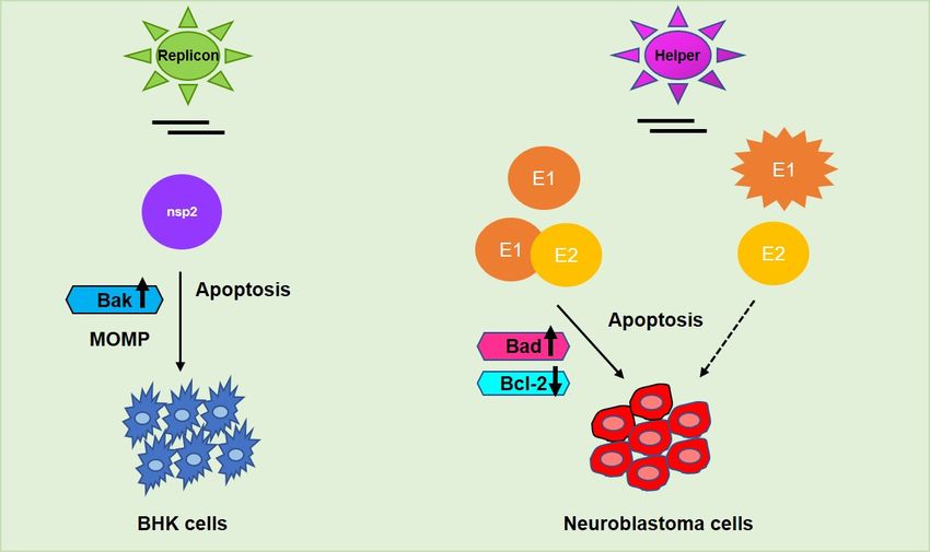
Cancers 2021, 13, 2199 3 of 20
Cancers 2021, 13, 2199 3 of 20
actin-binding protein, gelsolin, and suppresses signal-transduction by the Neural Wis-
kott–Aldrich syndrome protein (N-WASP) thereby causing cytoskeleton disruption [15].
H-1PV-induced cell death is facilitated by NS1-mediated p53 dependent or independent
mechanisms
mechanisms through
through the
the accumulation
accumulation of
of reactive
reactive oxygen
oxygen species,
species, mitochondrial
mitochondrial outer
outer
membrane permeabilization
membrane permeabilization (MOMP),
(MOMP), DNA
DNA damage,
damage, cell
cellcycle
cyclearrest,
arrest,and
andfinally,
finally, caspase
caspase
activation [14]. H-1PV
activation H-1PV has
hasalready
alreadybeen
beenrecruited inin
recruited phase
phase II clinical trials
II clinical for for
trials the the
treatment
treat-
of pancreatic
ment ductalductal
of pancreatic adenocarcinoma and presently
adenocarcinoma is in its
and presently is evaluation stage stage
in its evaluation [16]. [16].
Figure 1. Schematic representation of different cell death pathways mediated by viral proteins. Levels of kinases are up-
Figure 1. Schematic representation of different cell death pathways mediated by viral proteins. Levels of kinases are
regulated in cancer cells due to which phosphorylation and activation of viral protein residues (NS1 and p 10.8) lead to
upregulated in cancer cells due to which phosphorylation and activation of viral protein residues (NS1 and p 10.8) lead to
ER stress and DNA damage response (DDR) causing mitochondrial outer membrane permeabilization (MOMP), apopto-
ER stress
sis, and
and cell DNACell
death. damage
cycleresponse
arrest is (DDR) causing
mediated mitochondrial
by activation of DDRouter membrane
(NS1) permeabilization
and downstream kinases (MOMP), apoptosis,
like Ataxia telangi-
and cell death. Cell cycle arrest is mediated by activation of DDR (NS1) and downstream kinases like Ataxia
ectasia mutated (ATM) and checkpoint kinases (Chk1/2). Cell cycle progression is inhibited at the G1/S or G2/M phase of telangiectasia
mutated
the (ATM)
cell cycle and checkpoint
as respective cyclins kinases
and CDKs(Chk1/2). Cell cycle
are inactivated uponprogression
expressionisofinhibited
the viral at the G1/S
proteins or G2/M
mediated phase
caspase of
acti-
vation
the cell(Rep78).
cycle asAtrespective
the same cyclins
time, E2Fandinhibition
CDKs are is maintained
inactivated by dephosphorylation
upon expression of the of viral
retinoblastoma protein (pRb)
proteins mediated by
caspase
Chk1/2,
activation finally inhibiting
(Rep78). At thethe transcription
same time, E2F of proto-oncogenes.
inhibition Upward
is maintained arrow↑—upregulation;
by dephosphorylation downward arrow↓—
of retinoblastoma protein
downregulation;
(pRb) by Chk1/2, circled P inhibiting
finally in blue—phosphorylation.
the transcription of proto-oncogenes. Upward arrow↑—upregulation; downward
arrow↓—downregulation; circled P in blue—phosphorylation.
2.2. Adeno-Associated Viruses (AAV) Rep78
2.2. Adeno-Associated
AAVs, other membersVirusesof(AAV) Rep78
the parvovirus family, are a group of non-enveloped, small,
single-stranded
AAVs, other DNA viruses,
members thatparvovirus
of the rely on helper viruses
family, like adenoviruses
are a group or herpesvi-
of non-enveloped, small,
ruses for their efficient
single-stranded replication
DNA viruses, [17].on
that rely The autonomous
helper and
viruses like helper-dependent
adenoviruses parvovi-
or herpesviruses
ruses have
for their uniquereplication
efficient biological [17].
properties in common.and
The autonomous Members of both groups
helper-dependent efficiently
parvoviruses
suppress tumor
have unique growthproperties
biological in animalsinthrough
common. different
Members proteins,
of bothirrespective of the suppress
groups efficiently mode of
tumor
tumorinduction
growth in[18]. The rep
animals proteins,
through a family
different of multifunctional
proteins, irrespective NS AAV
of the proteins,
mode are
of tumor
inductionfor
required [18]. Thereplication
virus rep proteins,anda family of multifunctional
gene regulation. AAV Rep78 NS AAVwasproteins,
proposed aretorequired
impair
for virus
the replication
utilization and pathway
of cAMP gene regulation.
by helperAAV Rep78inwas
viruses proposed
HeLa to impair
cells and thereby the utilization
inhibit pro-
of cAMPreplication
ductive pathway byofhelper viruses
the helper in HeLa
virus [19].cells andearly
In the thereby inhibit
1990s, productive
AAVs replication
were reported to
of the helper virus [19]. In the early 1990s, AAVs were reported to
inhibit carcinogen-induced simian virus 40 (SV40) DNA amplification [20] and carcino-inhibit carcinogen-
induced simian virus 40 (SV40) DNA amplification [20] and carcinogen-induced resistance
against methotrexate associated with amplification of the dihydrofolate reductase (DHFR)
gene. Rep78 was found to interfere with both SV40 DNA amplification and herpesvirus
replication [18]. Interestingly, Rep78 shares several properties with parvovirus NS1 like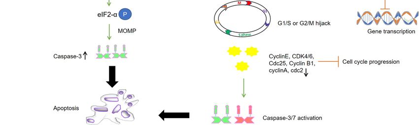
Cancers 2021, 13, 2199 4 of 20
specific DNA binding, site-specific endonuclease, helicase, and ATPase activities, along
with a cytostatic effect. The interaction of AAV Rep78 with p53 was suggested to be
responsible for the observed protection of p53 in adenovirus-infected cells which is usually
found to be degraded by the interaction of adenoviral E1B (early gene) with p53. By
protecting p53 from ubiquitin-mediated degradation by adenovirus, the function of p53 as
a cell cycle blocking agent is restored in the presence of Rep78 [21].
Mechanism of Action
It was earlier shown that Rep78-expressing cells display accumulation of the hy-
pophosphorylated form of retinoblastoma protein (pRb) that leads to downregulation of E2
transcription factor (E2F) target genes, namely cyclin A, cdc2, and cyclin B [22]. Moreover,
Rep78 inhibits the kinase activity of PRKX, a homolog of cAMP-dependent protein kinase
A (PKA), and PKA itself, which results in the blockage of cAMP response element-binding
protein (CREB)-dependent transcription in cervical cancer cells [19]. It was also found
to be associated with the oncogenic transcription factor c-Jun and alter c-Jun-dependent
transcription by inhibiting its binding to needed transcriptional partners/cofactors such as
c-Fos likely through mechanisms of steric hindrance [23]. Furthermore, binding of Rep78 to
the cell cycle regulatory phosphatase cdc25A prevents the latter access to substrates cyclin
dependent kinase 1 (CDK1) and CDK2 thus resulting in the inactivation of CDKs that are
required for continued DNA replication. However, the nicking activity of Rep78 together
with the inactivation of Cdc25A is required to attain a strong, if not total, pRb inactiva-
tion [22]. It is possible that Rep78 induces DDR that causes pRb hypophosphorylation by
Chk1/2 and forms a complex with E2F itself, eventually causing transcriptional inhibition
of proto-oncogenes in cancer cells (Figure 1). Collectively, Rep78 has been observed to
exert antiproliferative effects by blocking cell cycle in all of the phases and by inducing
apoptosis independently of p53 via the caspase-3 dependent pathway [24]. In light of these
findings, exploring the role of Rep78 in other cancer types and replicating the same in vivo
may mark another milestone in virus-based anticancer therapy.
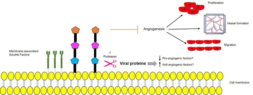
2.3. Human Herpesvirus Type 6 (HHV-6) Rep6/U94
HHV-6 is a double-stranded DNA lymphotropic β-herpesvirus existing as two closely
related strains, namely HHV-6A and HHV-6B. The HHV-6/U94 gene, also known as
Rep6, is highly conserved in both HHV-6A and B. It is a single-stranded DNA binding,
exonuclease, helicase-ATPase protein which might be involved in DNA replication. It
expresses at low levels during the early phases of viral replication [25]. U94 is known as a
negative regulator of viral replication as it does not support productive viral replication
in T-cell lines stably expressing U94. It accumulates in the treated cells and inhibits HHV-
6A/B, HHV-7, and cytomegalovirus replication by blocking the virus cycle before genome
replication [26]. U94 possesses a highly structural and functional similarity with AAV-
2 Rep68/78 for which the viral protein has been studied for its involvement in cancer
regulation [27]. Since U94 mRNA was detected in the peripheral blood mononuclear cells
(PBMC) from latently infected healthy individuals, U94 was considered as a molecular
marker of viral latency [28].
2.3.1. Mechanism of Action
The viral protein was initially known for its ability to suppress Harvey (H)-ras-induced
transformation in stably U94-expressing NIH 3T3 cell line [29]. Later, Ifon et al. [27] demon-
strated the anticancer activity of U94 on human prostate cancer in vivo, as the development
of human prostate cancer (PC3) cell line-derived tumor in nude mice was inhibited by treat-
ment with a recombinant U94 protein. The anticancer activity of the U94 protein was possi-
bly ascribed to Fibronectin 1 (FN1) upregulation and a concomitant Angiopoietin-like 4
(ANGPTL4) downregulation [27]. Indeed, increased levels of FN-1 are known to accelerate
FN1-FN1 polymerization and FN1 binding to the PC3 cell surface which could be, at least in
part, responsible for decreased clonogenicity in vitro and tumorigenesis in vivo. Moreover,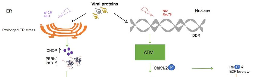
Cancers 2021, 13, 2199                                                                                           5 of 20

                         the expression of SPUVE 23, a serine protease associated with increased malignant potential,
                         was also observed to be downregulated in the recombinant U94-treated PC3 cell line [27].
                         Subsequently, U94 was also identified to impair tumor growth and invasion in glioma cells
                         by promoting AKT/GSK3β signaling [30] and migration of oligodendrocyte progenitor
                         cells (hOPC), thus highlighting its role in metastasis prevention [31,32]. Later, our group
                         reported U94 ability to impair triple-negative human breast cancer cell (MDA-MB 231)
                         migration, motility, invasion, and proliferation both in vitro and in vivo. The viral protein
                         operates in MDA-MB 231 cells by downmodulating the activation of proto-oncogene Src
                         and the downstream signaling pathways β-catenin/STAT3/cortactin/ARP2-3/Akt [33]. In
                         a 3D fluid-dynamic environment, U94-positive cultures displayed β-catenin localization
                         at the cell membrane, contrary to cytoplasmic localization of the same in transformed
                         cells, which indicates a U94-triggered mesenchymal to epithelial transition (MET). At
                         the same time vimentin, an epithelial to mesenchymal transition (EMT) marker highly
                         expressed in MDA-MB-231 cells, was strongly down-modulated in cells treated with U94.
                         Similarly, the expression of other EMT markers like TWIST, N-cadherin, Snail1, and matrix
                         metalloprotease 2 (MMP2) was strongly inhibited, thereby supporting a role of U94 in me-
                         diating a MET of MDA-MB-231 cells. This was further confirmed in vivo as U94 inhibited
                         tumor development of MDA-MB 231 xenografts in mouse models. Interestingly, similar
                         to MDA-MB-231 cells, U94 also inhibited HeLa cells migration, proliferation, and colony
                         formation both in vitro and in vivo through Src down-modulation [33]. U94 was found to
                         reversibly arrest cell cycle in S-phase when transiently expressed in MDA-MB-231 cells [33].
                         Of late, our group demonstrated U94 to be a DDR inhibitor as it displays anticancer activity
                         in MDA-MB-231 cells by downregulating DDR genes, cholesterol biosynthesis, and cell
                         cycle, out of which cyclin-dependent kinase inhibitor 3 (CDKN3), Non-SMC Condensin
                         II Complex Subunit G2 (NCAPG2), Ndc80 kinetochore complex component (NUF2), and
                         High Mobility Group Box 1 (HMGB1), being the major ones. U94-mediated DDR inhibition
                         likely occurs through downregulation of Bcl-2 and upregulation of Bax-, Bad-levels, Poly
                         (ADP-ribose) polymerase (PARP) cleavage, and caspase-9-exerted apoptotic cell death via
                         intrinsic apoptotic pathway activation [34]. The anticancer function of the viral protein was
                         also investigated and confirmed in other triple-negative human breast cancer cell lines like
                         MDA-MB 468 and BT-549 cells. In particular, U94 worked in synergy with DNA damaging
                         drugs such as cisplatin and doxorubicin to attack tumor cells [34].

                         2.3.2. Role of U94 in Blocking Angiogenesis
                               Previous work by our group showed that HHV-6 infection of endothelial cells (EC)
                         resulted in a strong inhibition of angiogenesis in vitro and ex vivo. In the latter condition,
                         treatment of rat aortic rings with U94 rendered them insensitive to vascular endothe-
                         lial growth factor (VEGF)-induced vasculogenic activity [26]. Later, we identified that
                         U94-expressing tumor xenografts of mouse origin displays impaired vasculogenesis [33].
                         Surprisingly, human umbilical vein endothelial cells (HUVEC) co-cultured with U94-
                         expressing MDA-MB-231 cells lost their ability to perform angiogenesis or migrate in vitro.
                         Consequently, the secretome of U94-expressing MDA-MB 231 cells was used to test its
                         activity on HUVECs angiogenic activity. As expected, the secretome derived from U94-
                         expressing breast cancer cells was found to completely inhibit angiogenesis of HUVECs
                         in vitro [33]. This finding was strongly suggesting on the involvement of a U94-induced
                         soluble factor in inhibiting EC angiogenesis. Recently, HHV-6A has been found to induce
                         the expression of the non-classical class I Human leukocyte antigen-G (HLA-G) molecule in
                         primary human mesothelial cells as a mechanism for viral immune-escape [35]. It is worth
                         noting that HHV-6 was found to promote HLA-G expression by activating the activating
                         transcription factor 3 (ATF3), a member of the basic leucine zipper domain (bZIP)/CREB
                         proteins, which can interact directly with the HLA-G promoter thereby stimulating the
                         HLA-G production [36]. For this reason, the involvement of HLA-G in sustaining the anti-
                         angiogenic activity of U94 on ECs was investigated and recently confirmed [36]. Altogether,
                         these results highlight the capability of U94 to mediate the impairment of cancer progres-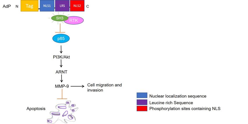
HHV-6 was found to promote HLA-G expression by activating the activating transcrip-
tion factor 3 (ATF3), a member of the basic leucine zipper domain (bZIP)/CREB proteins,
which can interact directly with the HLA-G promoter thereby stimulating the HLA-G pro-
Cancers 2021, 13, 2199 6 of 20
duction [36]. For this reason, the involvement of HLA-G in sustaining the anti-angiogenic
activity of U94 on ECs was investigated and recently confirmed [36]. Altogether, these
results highlight the capability of U94 to mediate the impairment of cancer progression
through a “two
sion through compartments”
a “two activity,
compartments” thusthus
activity, providing
providinganticancer
anticancertherapeutic
therapeuticbenefits
benefits
not
notonly
onlyininterms
termsofof antiproliferative
antiproliferative and lytic effects
effects on
ondifferent
differenttumor
tumorcells
cellsbut
butalso
alsoin-
hibiting the
inhibiting theneovascularization
neovascularizationprocess
processneeded
neededforforcancer
cancercell
cellgrowth
growthand metastatization
and metastatiza-
(Figure
tion 2). 2).
(Figure
Figure
Figure2.2.U94
U94regulates different
regulates mechanisms
different mechanisms to exert its activities
to exert mainly
its activities by downregulation
mainly of proto-oncogene
by downregulation Src, re-
of proto-oncogene Src,
sulting in inhibition of cancer cell proliferation both in vitro and in vivo. Secondly, U94 was found to suppress angiogen-
resulting in inhibition of cancer cell proliferation both in vitro and in vivo. Secondly, U94 was found to suppress angiogenesis
esis ex vivo by suppressing the vessel formation. The viral protein upregulates pro-apoptotic genes like Bax and Bad levels
ex vivo by suppressing the vessel formation. The viral protein upregulates pro-apoptotic genes like Bax and Bad levels and
and downregulates anti-apoptotic Bcl-2 levels culminating in Caspase-9 activation. Finally, the cleavage of Poly (ADP-
downregulates anti-apoptotic Bcl-2 levels culminating in Caspase-9 activation. Finally, the cleavage of Poly (ADP-ribose)
ribose) polymerase (PARP) results in inhibition of DNA repair and mediation of intrinsic apoptosis in cancer cells. Upward
polymerase (PARP) results
arrow↑—upregulation; in inhibition
downward of DNA repair and mediation of intrinsic apoptosis in cancer cells. Upward
arrow↓—downregulation.
arrow↑—upregulation; downward arrow↓—downregulation.
3. Why Avian Viral Proteins as Anticancer Therapeutics?
3. Why Avian Viral Proteins as Anticancer Therapeutics?
Avian viruses are known to cause morbidity and mortality including diseases like
Avian viruses are known to cause morbidity and mortality including diseases like
viral arthritis, hepatitis, respiratory syndromes, immune suppression in species like
viral arthritis, hepatitis, respiratory syndromes, immune suppression in species like goose,
goose, ducks, turkeys, pigeons, raptors, and quails, but not in humans. The economy and
ducks, turkeys, pigeons, raptors, and quails, but not in humans. The economy and poultry
poultry industry have been affected in several countries due to these viruses. However,
industry have been affected in several countries due to these viruses. However, viruses
viruses derived from such sources that do not circulate extensively in the human popula-
derived from such sources that do not circulate extensively in the human population
tion represent a potential source of viral proteins able to bypass any pre-existing immun-
represent a potential source of viral proteins able to bypass any pre-existing immunity [37].
ity [37]. Due to which they might prove to be safer and effective candidates to be utilized
Due to which they might prove to be safer and effective candidates to be utilized in cancer
in cancer therapy with lesser side effects as compared to conventional viral vectors with
therapy with lesser side effects as compared to conventional viral vectors with efficacy and
efficacy and risks.
processing processing risks. Interestingly,
Interestingly, mostviral
most of the avian of the avian viral
proteins proteins
possess nuclearpossess nu-
localization
clear
property that makes them effective in targeting cellular mechanisms. In the next section, In
localization property that makes them effective in targeting cellular mechanisms. we
the next
have section, wesome
summarized haveofsummarized some of
the newly emerged the viral
avian newly emerged
proteins avian
which haveviral proteins
recently been
which have recently
investigated for theirbeen investigated
potential forand
functions their
maypotential
prove tofunctions and may
be promising prove to be
anti-carcinogenic
promising anti-carcinogenic
agents in the future. agents in the future.
3.1. Chicken Anemia Virus (CAV) Apoptin
CAV, a member of the genus Gyrovirus, is an etiological agent of chicken infectious
anemia known to cause immunosuppression in young chickens and compromise immune
response in older birds. CAV mainly infects hematopoietic cells including bone marrow-
derived cells [38,39], myeloid progenitor cells, and T-lymphocyte precursor cells [40]. VP3,
a 121 amino acid-long structural protein from CAV is known for its property to induce
apoptosis and viral cytotoxicity in host cells, hence the name apoptin [41]. The C-terminalCancers 2021, 13, 2199 7 of 20
domain of apoptin contains a bipartite nuclear localization sequence (NLS, aa 82–88 and
111–121) and a secondary nuclear export sequence (NES, aa 97–105) and together these mo-
tifs confer to the protein a nucleocytoplasmic shuttling activity [42,43]. Apoptin is known
for its multimerization and nucleus retention activity in human transformed or tumor
cells from tissues of endodermal, ectodermal, and mesodermal origin. It interacts with
cellular proteins like anaphase promoting complex (APC/C) and transports the latter from
cytoplasm to nucleus to be deposited in promyelocytic leukemia (PML)-nuclear bodies,
whereas the same remains cytoplasmic in normal human cells [42]. Apoptin is commonly
phosphorylated at Thr-108 in osteosarcoma (U2OS, Saos-2), lung carcinoma (H1299), colon
carcinoma (HT29), cervical carcinoma (HeLa), hepatocellular carcinoma, and other trans-
formed cells [42,44–46]. Although phosphorylation status is not necessary for nuclear
localization, it does play a role in determining the toxicity of the viral protein. Instead,
mutations in leucine-rich sequence (LRS, aa 33–46) cause reduced nuclear accumulation of
apoptin in cancer cells [42]. In normal cells, apoptin has been shown to be localized in the
cytoplasm, aggregated, and eventually degraded [45]. Therefore, apoptin can selectively
kill various human tumor or transformed cells with little cytotoxic effect in normal cells.
3.1.1. Mechanism of Action
Apoptin triggers caspase-dependent cell death via the intrinsic apoptotic path-
way [47–49] independently of p53, but requires pro-apoptotic transcriptionally active p73
isoforms from p53 family [50,51]. Like parvovirus NS1, DDR signaling plays a key role
in nuclear localization and apoptosis induction by apoptin [52]. In a study by Kucharski
et al. [53], the authors show that the inhibition of kinases Chk1 and Chk2 in non-small cell
lung adenocarcinoma (NSCLC) results in cytoplasmic re-localization of apoptin. Therefore,
the phosphorylation of residues T56 and T61 is relevant in regulating the localization and
apoptotic activity of the protein. Other studies have found apoptin to trigger the nuclear
accumulation of the related kinase Akt in prostate cancer [54] and PKCβ1 in colon cancer
cells via interaction with the N-terminal region of the viral protein [55]. Recently, PKCβ1
was shown to phosphorylate apoptin in multiple myeloma cell lines [55], thereby indicating
PKCβ1 to be a tumor-specific target responsible for sensitizing cells to apoptin [56–58].
Apoptin also drives translocation of the transcription factor nuclear hormone receptor
(Nur) 77 from the nucleus into the cytoplasm, where it causes mitochondrial outer mem-
brane permeabilization and induces cytochrome C (cyt c) release mediating intrinsic cell
death pathway [47,48] (Figure 3). Other studies have shown that apoptin interacts with
and inhibits Abl/BCR-Abl1 kinase and downstream targets, like STAT5, CrkL, and c-myc
in chronic myeloid leukemia (CML) [59]. Jangamreddy et al. [60] designed an apoptin-
derived decapeptide (AdP, aa 81–90) as an alternative therapeutic agent, which acts as a
negative downregulator of BCR-Abl1 and is capable of downstream targeting c-myc with
comparable efficacy to full-length apoptin.
3.1.2. AdP
AdP is a 5.2 kDa hydrophilic, highly soluble peptide, consisting of four parts: a pen-
etrating peptide Tat (for entering facilitation), the core NLS1, the NLS2 sequence, and a
flexible connection (LRS) between NLS1 and NLS2 [61]. The peptide was tailored to facili-
tate targeting to cancer cells and nuclear accumulation. Owing to its small size, the peptide
was designed to exhibit reduced immunogenicity and strong antitumor activity against
glioma cells. The molecule led to reduced heat shock protein 70 (HSP70) mRNA and protein
levels in tumor cells and proved to be more effective in mediating glioma cell apoptosis
than apoptin itself both in vitro and in vivo [61]. Song et al. [62], postulated the potential
mechanisms for glioma inhibition to be linked with the interaction of AdP with heat shock
element-SRC homology 3 (HSE-SH3) domain, inactivation of RTK/PI3K/Akt pathway, and
MMP-9 suppression (Figure 3). However, compared to apoptin, AdP increases apoptosis
in human astrocytes, but to a lower extent than in glioma cells [61]. Interestingly, down the
line, Hou et al. [63] demonstrated that recombinant apoptin (GST tagged apoptin, chemi-Cancers 2021, 13, 2199 8 of 20
cally modified by folic acid for entering into the cells) inhibits the growth of breast cancer
cells likely by triggering apoptosis. They demonstrated that recombinant apoptin inhibits
proliferation and induces apoptosis in vitro following similar molecular mechanisms as
apoptin by facilitating the expression levels of Bax, Cyt c, p-Akt, and p-Nur77. Another
study by Zhou et al. [64], showed that AdP inhibited cell viability in cisplatin-resistant
gastric cancer cells, without affecting normal cells by PI3K/Akt/ARNT signaling pathway.
Overall, AdP causes an increase in the G2/M phase population leading to apoptosis and
priming the cells sensitive to chemotherapy likely due to decreased expression of AKT, p85,
and their phosphorylated forms in both therapy sensitive and resistant cells. Collectively,
these findings suggest that the apoptin derived peptide could be used in combination
Cancers 2021, 13, 2199 with other drugs and targeted for different kinds of cancer therapy, provided the safety of 8 of 20
normal cells has been assured.
FigureFigure 3. Schematic
3. Schematic representation
representation of apoptin
of apoptin derived
derived peptide
peptide (AdP) (AdP) targeted
targeted signaling
signaling pathways.
pathways. AdP binds
AdP binds to SRC-
to SRC-
homology 3 (SH3) domain of tyrosine kinases and deactivates PI3K/Akt pathway followed by Aryl hydrocarbon nuclear
homology 3 (SH3) domain of tyrosine kinases and deactivates PI3K/Akt pathway followed by Aryl hydrocarbon nuclear
translocator (ARNT) signaling and MMP-9 suppression leading to inhibition of cell migration along mediating apoptosis.
translocator (ARNT) signaling and MMP-9 suppression leading to inhibition of cell migration along mediating apoptosis.
3.1.2.Reovirus
3.2. Avian AdP (ARV) p17
ARV AdP is a to
belongs 5.2the
kDagenus
hydrophilic, highly in
Orthoreovirus soluble peptide, consisting
the Reoviridae family. It of hasfour parts: a pen-
a double-
etrating
stranded peptide Tat
10-segmented (for genome
RNA enteringencoding
facilitation), theleast
for at coreeight
NLS1, the NLS2
structural andsequence,
four NSand a
flexible connection (LRS) between NLS1 and NLS2 [61]. The
proteins. ARV p17 (p17) is a 17-kDa non-structural protein encoded by the S1 gene and peptide was tailored to facil-
contains 146 aa [65]. The S1 genome segment of ARV contains three open reading frames pep-
itate targeting to cancer cells and nuclear accumulation. Owing to its small size, the
tide wasinto
that translate designed
p10, p17, toand
exhibit reducedrespectively.
σC proteins, immunogenicity and strong
P10 displays membraneantitumor activity
destabi-
against glioma cells. The molecule led to reduced heat shock protein
lization activity [65–67], whereas σC is known to be a cell attachment protein [68] capable 70 (HSP70) mRNA
and protein
of inducing levels[69–71]
apoptosis in tumorandcells
p17and
as aproved to be more effective
nucleocytoplasmic shuttling in mediating
protein with glioma
an cell
apoptosis
unknown thanPrevious
function. apoptinstudies
itself both
haveinshown
vitro andthatin vivo
p17 has[61]. Song
a basic et al.spanning
region, [62], postulated
from the
aa 119potential mechanisms for glioma
to 128 (IAAKRGRQLD), which isinhibition
similar totothe befunctional
linked with the interaction
monopartite NLSofofAdPthe with
heat shock element-SRC homology 3 (HSE-SH3) domain, inactivation
c-Myc protein and is highly conserved in different ARV isolates [72]. A monopartite-type of RTK/PI3K/Akt
pathway,
functional NLS and
nearMMP-9 suppression
the C terminus of p17(Figure 3). However,
is necessary compared
for nuclear import.to NLSapoptin, AdP in-
interacts
creases
with the apoptosis
nucleoporin in human astrocytes,
translocated promoter region but to(Tpr)
a lower extentwithin
localized than inthe glioma
nuclearcells [61]. In-
pore
terestingly,
complex and causesdown the line, Hou
suppression of Tpret al. [63] demonstrated
thereby activating cellthat cycle recombinant
regulators like apoptin
p53, (GST
tagged and
Phosphatase apoptin,
tensinchemically modified
homolog (PTEN), andbyp21.
folicThe
acid for entering
activation of CDK intoinhibitors
the cells)causes
inhibits the
growth of breast
downregulation of bothcancer cells likely by
PI3K/Akt/mTOR andtriggering apoptosis.
ERK signaling They [73,74].
pathways demonstrated
Previous that re-
combinant apoptin inhibits proliferation and induces apoptosis
studies have shown that p17 causes retardation of cell growth by deactivation of mTORC1 in vitro following similar
molecular mechanisms as apoptin by facilitating the expression levels of Bax, Cyt c, p-Akt,
and p-Nur77. Another study by Zhou et al. [64], showed that AdP inhibited cell viability
in cisplatin-resistant gastric cancer cells, without affecting normal cells by
PI3K/Akt/ARNT signaling pathway. Overall, AdP causes an increase in the G2/M phase
population leading to apoptosis and priming the cells sensitive to chemotherapy likelyCancers 2021, 13, 2199 9 of 20
and downstream protein synthesis through activation of the p53 pathway [75]. In a recent
report, Chiu et al. [74] showed that p17 contains a NES spanning from aa 19 to 26 (LSLRE-
LAI), which is required for interaction with the heterogeneous nuclear ribonucleoprotein
(hnRNP) A1 and serves as a carrier in mediating nucleocytoplasmic shuttling of the viral
protein. While lamin A/C mediates p17 nuclear import, p17-hnRNP A1 carrier-cargo
complex causes downregulation of Tpr by direct interaction with lamin A/C and Tpr.
Altogether, p17 has been observed to induce cell growth retardation, cell cycle arrest,
and host cellular translation shutoff by suppression of CDK1- and polo-like kinase (PLK1)-
like signaling pathways and regulation of the p53/PTEN/mTORC1-like pathway [76].
Since p17 is also known to induce autophagy and trigger protein kinase RNA (PKR)-
activated signaling, it activates the innate immune system and can mount the immune
response against tumors. In summary, p17 appears to divert the cellular machinery required
for normal cell-cycling processes, including ATM/Chk1/2 signaling pathway [77] to allow
virus replication via induction of cell cycle arrest and cellular translation shutoff [75,76,78].
Secondly, p17 positively regulates PTEN, AMP-activated protein kinase (AMPK), and
PKR/eIF2 signaling pathways accompanied by downregulation of Akt and mTORC1,
thereby triggering autophagy [78]. Autophagy-induced activity of p17 has been observed
to be mediated by p17 nuclear localization, as the former is delayed and viral replication is
affected when the protein could not enter into the nucleus [79].
3.2.1. Role in Cell Cycle
P17 exhibits cell growth inhibition and cell cycle retardation in multiple cell lines
like African green monkey kidney epithelial cells (VERO), chicken fibroblasts (DF-1),
human adenocarcinoma (SW620), HeLa, and human lung cancer (A549), along displaying
reduced tumor size in vivo [76]. P17 acts on inhibiting CDK1 in two different manners:
first, by suppressing the phosphorylation of CDK1 via suppression of kinases like PLK1
and CDC25c; and secondly, by competing with cyclin B1 to bind CDK1 leading to CDK1
retention in the cytoplasm. Prevention of the cyclin B1/CDK1 complex formation in
the nucleus leads to G2/M cell cycle retardation [76]. Moreover, p17 expression is also
responsible for p53 and PTEN phosphorylation by impairing the targeting ability of the
corresponding E3 ubiquitin ligase, namely mouse double minute 2 homolog (MDM2) [80].
Enhancement of p53 interaction with cyclin H mediates suppression of cyclin-associated
kinase (CAK) activity by p17 and dissociates the CDK7/Cyclin H complex. The same
study reported a particular motif in the p17 protein spanning from aa 140 to 143 (WXFD)
and conserved residues at positions D113 and K122, as critical for CDK2 and CDK6
binding [74]. The p17 binding to cyclins via conserved motifs is a peculiar property of most
of the renowned tumor suppressor proteins. To conclude, p17 suppresses the formation of
CDK1/cyclin A2, CDK2/cyclin E1, and CDK6/cyclin D1 complexes by directly binding
to CDK, cyclin, or CDK/cyclin complexes, benefitting viral replication [74]. The two
prime pathways behind this activity are PI3K/AKT- and Tpr/p53/PTEN/ERK-dependent
inhibition of the mTORC1 pathway.
3.2.2. Role in Angiogenesis
We recently showed that p17 is also able to inhibit motility, migration, and angio-
genesis in human macrovascular (HUVEC) and microvascular ECs (HMVEC). Treatment
of ECs with recombinant GST-p17, or over-expression of p17 at the intracellular level by
nucleofection of a p17-expressing plasmid, led to the suppression of tube-like formation on
Matrigel, cell migration, and sprouts generation in a 3D spheroid assay. Interestingly, p17
was found to downregulate EC angiogenic activity in the presence of different mitogenic
stimuli like VEGF-A and basic fibroblast growth factor (FGF-2), thereby confirming its wide
anti-angiogenic spectrum of activity. The anti-angiogenic activity mediated by p17 was
also demonstrated ex vivo and in vivo by aortic ring assay and chick chorioallantoic mem-
brane (CAM) assay, respectively, where the viral protein proved to suppress the number of
neovessels formation while remaining non-toxic to the normal tissues [81]. Furthermore,cleofection of a p17-expressing plasmid, led to the suppression of tube-like formation on
Matrigel, cell migration, and sprouts generation in a 3D spheroid assay. Interestingly, p17
was found to downregulate EC angiogenic activity in the presence of different mitogenic
stimuli like VEGF-A and basic fibroblast growth factor (FGF-2), thereby confirming its
Cancers 2021, 13, 2199
wide anti-angiogenic spectrum of activity. The anti-angiogenic activity mediated 10 byofp17
20
was also demonstrated ex vivo and in vivo by aortic ring assay and chick chorioallantoic
membrane (CAM) assay, respectively, where the viral protein proved to suppress the
number of neovessels formation while remaining non-toxic to the normal tissues [81]. Fur-
we observed
thermore, wethe secretion
observed ofsecretion
the a well-known tumor suppressor
of a well-known molecule, namely
tumor suppressor dipeptidyl
molecule, namely
protease
dipeptidyl4 (DPP4), in the
protease supernatants
4 (DPP4), of p17
in the expressing of
supernatants HUVECs and HMVECs
p17 expressing HUVECs(Figureand
4).
Further
HMVECs studies may4).confirm
(Figure Furtherthe pathways
studies mayresponsible
confirm thefor upregulation
pathways of soluble-DPP4
responsible for upregu-in
the presence of p17. Whether pro-angiogenic factors are downregulated due
lation of soluble-DPP4 in the presence of p17. Whether pro-angiogenic factors are down-to the secretion
of anti-angiogenic
regulated due to thefactors by ARV
secretion p17 or vice versa,
of anti-angiogenic remains
factors to bep17
by ARV solved. Up-regulation
or vice versa, remains of
protease levelsUp-regulation
to be solved. in the presenceofofprotease
p17 responsible
levels infor thepresence
the secretionofofp17
membrane-associated
responsible for the
factors as DPP4
secretion is another field yet factors
of membrane-associated to be explored.
as DPP4 is another field yet to be explored.
Figure 4. Schematic representation of a possible protease-mediated mechanism, triggered by ARV p17, leading to secretion
Figure 4. Schematic representation of a possible protease-mediated mechanism, triggered by ARV p17, leading to secretion
of membrane-bound anti-angiogenic factors that prevent endothelial cells (EC) migration, proliferation, vessel formation,
of membrane-bound
and anti-angiogenic
angiogenesis. Upward factors that prevent
arrow↑—upregulation; endothelial
downward cells (EC) migration, proliferation, vessel formation,
arrow↓—downregulation.
and angiogenesis. Upward arrow↑—upregulation; downward arrow↓—downregulation.
3.3. Muscovy Duck Reovirus (MDRV) p10.8
3.3. Muscovy Duck Reovirus (MDRV) p10.8
Other viral proteins from avian species and homologous to ARV p17 serve important
Other viral proteins from avian species and homologous to ARV p17 serve impor-
cell growth regulatory functions and might exert anticancer activities in tumor cells.
tant cell growth regulatory functions and might exert anticancer activities in tumor cells.
MDRV is another member of the genus Orthoreovirus, an important poultry pathogen that
MDRV is another member of the genus Orthoreovirus, an important poultry pathogen that
is involved in several diseases including viral arthritis, pericarditis, hepatitis, respiratory
is involved in several diseases including viral arthritis, pericarditis, hepatitis, respiratory
syndromes, and sudden death. Ducklings infected with this reovirus were first reported
syndromes, and sudden death. Ducklings infected with this reovirus were first reported in
in 1950 and MDRV was first isolated in 1972 [82]. Its genome consists of 10 double-
1950 and MDRV was first isolated in 1972 [82]. Its genome consists of 10 double-stranded
stranded RNA segments which can be separated into three size classes: L (large), M (me-
RNA segments which can be separated into three size classes: L (large), M (medium), and
dium), and S (small). Like other avian reoviruses, MDRV appears to evolve mechanisms
S (small). Like other avian reoviruses, MDRV appears to evolve mechanisms that alter
that alter the physiology of host cells during infection to increase its replication. MDRV
the physiology of host cells during infection to increase its replication. MDRV was first
was first appeared to induce autophagy in chicken fibroblasts via suppression of mTOR
appeared to induce autophagy in chicken fibroblasts via suppression of mTOR phosphory-
lation and marked increased levels of Microtubule-associated protein 1A/1B-light chain 3
(LC3-II) induced by a NS protein named σNS [83]. Another MDRV protein, named p10.8,
is coded by the S4 gene sequence and is found highly conserved suggesting that p10.8
plays an important role in virus-host interaction [82]. This polypeptide has no significant
sequence similarity to other known proteins, so its amino acid sequence offers no clues
about its function [84]. Similar to ARV p17, p10.8 can localize to the nucleus independent of
the host cell type based on a signal mediated import. MDRV p10.8 has an aromatic amino
acid-rich NLS which enables it to pass through the nuclear pore complex and a leucine-rich
NES [84]. Recently, MDRV p10.8 has garnered attention due to its apoptosis-inducing
ability in DF-1 and VERO cells [84].
Cell Cycle Arrest
Like ARV σC, p10.8 induced apoptosis is associated with ER stress through unfolded
protein response-mediated BIP/IRE1/XBP1 pathway [85–87]. The viral protein is known
to dissociate the BIP/IRE1 complex and increase the phosphorylated form of inositol-Cancers 2021, 13, 2199 11 of 20
requiring enzyme 1 (IRE1) to activate X-box binding protein 1 (XBP1) as indicated by the
increased mRNA levels of binding immunoglobulin protein (BIP), XBP1, C/EBP homol-
ogous protein (CHOP), and caspase-3 [87]. Another study revealed that p10.8 induced
cell cycle arrest at the G0/G1 phase in DF-1 cells by dissociating BIP from protein kinase
R-like ER kinase (PERK) and increasing phosphorylated PERK and eukaryotic initiation
factor 2 subunit 1 (eIF2α) levels [82]. Taken together, the viral protein increases the protein
expression of BIP, p-PERK, p-eIF2α, CHOP, cleaved-Caspase 12, and cleaved-Caspase 3,
thus indicating that the p10.8 protein induces ER stress-mediated apoptosis (Figure 1). This
finding implies a cell cycle-regulated role of p10.8 in inducing apoptosis-related cell death.
Furthermore, high levels of kinases in tumor cells may promote high specificity of action for
p10.8. Recently, it has been demonstrated that both MDRV p10.8 and ARV σC can mediate
CDK4 ubiquitination by stabilizing Cdc20 with the aid of molecular chaperones, chaper-
onin containing TCP1 subunit 2 (CCT2), and 5 (CCT5) [88]. On the other hand, the fact that
both p10.8 of MDRV and σ1s of mammalian reovirus can localize to the nucleus and cause
apoptosis of infected cells suggests that σ1s and p10.8 may be functionally related [89].
Nuclear import of p10.8 mediates activation of p53 in VERO cells possibly via suppression
of nucleoporin Tpr as with ARV p17. This, in turn, leads to the activation of extrinsic
cell death via activation of the Fas/caspase 8/caspase 3 pathway [85]. This property of
the protein to modulate different apoptotic and cell cycle control pathways may make it
a suitable candidate for targeting tumor cells mostly because of its similarity with ARV
p17 in PKR activation and nuclear localization. However, based on these primary studies,
further characterization of the protein is required in cancer cells along with addressing its
cytotoxic activity on normal cells.
3.4. Newcastle Disease Virus (NDV) F Protein
Newcastle disease virus (NDV) is an avian paramyxovirus, a member of the Avulavirus
genus with a negative single-strand RNA genome. It is one of the most well-researched
oncolytic viruses for its activity against all kinds of cancer cell lines from ecto-, endo-, and
mesodermal origin, but not normal cells [90]. It has displayed an impressive safety profile
in phase I and II human clinical trials. NDV binds to the sialic acid (Sia) receptor on host
cells and, therefore, can infect a broad range of cell types. Different receptor isoform expres-
sion patterns between cell types contribute to the selection of cancer cells like HeLa by NDV
over normal cells like BHK fibroblasts [91,92]. NDV can achieve oncolysis via activation of
the extrinsic or intrinsic apoptosis, activation of PERK kinase followed by caspase-12, and
secretion of cytokines like tumor necrosis factor–α (TNF-α) amongst various others, from
the infected tumor cells [93]. Engineered NDV vectors expressing apoptin [94], immune
checkpoint blockades like anti-CTLA-4, anti-PD-1, anti-PDL, and cytokines like IL-2 [95]
and influenza virus NS1 [96] trigger oncolytic cell death in tumor cell lines from lung and
liver, tumor-bearing mice [94], and apoptosis-resistant cells, respectively [96].
The F protein from NDV is a class I viral membrane fusion protein present as a
trimer in the virion where the cleavage site of the F protein is known to be responsible
for virulence and the formation of syncytia [92]. Both Fusion (F) and Hemaglutinin-
Neuraminidase (HN) proteins expressed on the surface have been studied to interact and
fuse with host cellular membranes. Upon adsorption of HN to its cellular receptors, F
protein undergoes a conformational change which triggers the release of fusion peptides to
fuse the viral and cellular membranes [97]. Intracellular insertion of viral HN and F surface
antigens were reported to induce a strong inflammatory response with the secretion of
cytokines, chemokines, and type I interferons (IFN). This, in turn, modulates tumor cell
surface markers and induces downstream apoptosis [93]. However, Liu et al. [92] lately
reported that that the F protein plays a major role in NDV-induced oncolytic effect on
xenograftic mice from H22 and 4T1 cell lines, possibly via mtorc1 inhibition but it remains
to be confirmed in further studies. Like ARV p17, NDV F protein is also postulated to
induce autophagy by upregulating autophagy related 5 (ATG5), beclin-1, and microtubule-
associated proteins 1A/1B light chain 3B (MAP1LC3B) like markers.Cancers 2021, 13, 2199 12 of 20
4. Old World (OW) Alphaviruses
Alphaviruses belong to the family of Togaviridae. They are non-segmented, positive-
stranded RNA, enveloped viruses with an icosahedral structure [98]. OW alphaviruses
include sindbis virus (SINV), semliki forest virus (SFV), and chikungunya virus (CHIKV),
which share many common characteristics. Nearly all the alphaviruses are arthropod-
borne and are transmitted to their vertebrate hosts by mosquitoes [99]. The naturally
occurring OW alphaviruses are relatively milder in humans and severely infect species like
cattle, birds, and horses. The oncolytic SINV, SFV, and other alphavirus vectors have been
reviewed in [100]. Here, we focus on SINV as a representative member of the alphavirus
family and involvement of its structural proteins in mediating cytotoxicity in cancer cells.
4.1. Mechanism of Action
SINV has a genomic RNA of 11.7 kb and is primarily known to target lymph nodes. It
is transmitted to birds and mammals by mosquito bites [101] and subsequently spreads
throughout the body via the bloodstream [102]. In humans, SINV infection is considered to
induce no symptoms or only mild symptoms (fever, rash, and arthralgia) [98] suggesting
low infectivity and viral replication in normal tissues. SINV was shown to induce cyto-
pathic effects in ovarian and cervical cancer cells without affecting normal keratinocytes.
It was found to be stable in the bloodstream and effective in targeting remote tumors as
observed by regression of cervical tumor xenografts in SINV infected mouse models [103].
The virus induces cell death in human squamous carcinoma (HSC-3) cells through apopto-
sis related to caspase-3, 9, cytochrome c, Nuclear factor kappa B (NF-kB), NF-kB inhibitor
(IkBa), and IkB kinase (Ikk) modulation [104]. The extensive virus-induced activity was
also testified by neuroblastoma regression in nude mice upon intratumoral and intravenous
administration of SINV AR339 strain [105]. Additionally, SINV infection regulates the
cell cycle progression in HeLa cells by accumulating cells in the S phase and relatively
shortening the G1 phase by exerting upregulation of cyclin E, Cdc25A, and CDK4/6 levels.
The virus causes downregulation of p21 during the early phases of infection and facilitates
quick entry into the S phase. However, the S phase was found to be arrested during the
later stages of infection when SINV starts to downregulate cyclin A levels [102].
4.2. SINV E1 and E2
The alphavirus genome encodes six structural proteins (capsid, 6K, and three surface
glycoproteins, E1, E2, and E3) and four non-structural proteins, nsP1-4, which are compo-
nents of the viral replicase and transcriptase [106–108]. nsP2 is known for its cytotoxicity
since it is able to shut down cellular transcription, translation, and induce apoptosis in
BHK cells [109]. However, there has not been shown any strict correlation between nuclear
localization of nsP2 and cytotoxicity since the nsP2-mutant replicons retained nuclear local-
ization while remaining non-cytotoxic. The SINV envelope protein E2 is responsible for
cellular-receptor binding and E1 is required to promote the fusion between viral particles
and cell membrane [107]. It was earlier shown that alphaviral structural proteins contribute
to cell death by apoptosis as virus replicon particles (VRP) lacking E1 and E2, showed a de-
lay in caspase activation thereby concluding that structural proteins contribute to apoptotic
activity in cancer cells [109]. Both of the viral structural proteins, in addition to nsP2, have
been shown to play a role in alphavirus-induced apoptosis [110]. SINV induced apoptosis
is caspase-8 dependent and mediated by Bad [108]. The expression of SINV structural
envelope proteins, either E1 or E2, led to apoptosis in rat prostatic adenocarcinoma (AT-3)
cells [109]. However, a single amino acid change in the SINV E2 from Q55 to H55 conferred
both neurovirulence in mouse neuroblastoma (N18) cells and the ability to kill AT-3 cells
expressing bcl-2, likely due to the alteration of the interaction between E2 and bcl-2 [111].
Later, Hurtado et al. [112] investigated the functional amino acids of the E2 envelope
protein in SINV and identified that the change of amino acid E70 to K70 suppressed the
metastasis-targeting ability of the protein, possibly due to inhibited interaction between E2
and E1 in the vector spike confirmation. In a recent study, Saito et al. [98] demonstratedcarcinoma (AT-3) cells [109]. However, a single amino acid change in the SINV E2 from
Q55 to H55 conferred both neurovirulence in mouse neuroblastoma (N18) cells and the
ability to kill AT-3 cells expressing bcl-2, likely due to the alteration of the interaction be-
tween E2 and bcl-2 [111]. Later, Hurtado et al. [112] investigated the functional amino
Cancers 2021, 13, 2199 acids of the E2 envelope protein in SINV and identified that the change of amino acid E70
13 of 20
to K70 suppressed the metastasis-targeting ability of the protein, possibly due to inhibited
interaction between E2 and E1 in the vector spike confirmation. In a recent study, Saito et
al. [98] demonstrated that E1 induced higher cytotoxicity than E2 in human neuroblas-
that E1
toma induced
cell higherNGP,
lines (NB69, cytotoxicity than E2 in
and RT-BM-1). human neuroblastoma
Moreover, cell lines (NB69,
E1 and E2 heterodimers NGP,
or E1—but
and RT-BM-1). Moreover, E1 and E2 heterodimers or E1—but not E2 alone—was
not E2 alone—was able to exhibit cytotoxicity in neuroblastoma cells (Figure 5). Further- able to
exhibit cytotoxicity in neuroblastoma cells (Figure 5). Furthermore, in the presence of E1,
more, in the presence of E1, the UV-inactivated SINV induced cytotoxicity specifically in
the UV-inactivated SINV induced cytotoxicity specifically in human neuroblastoma cells
human neuroblastoma cells but not in normal human fibroblasts, affirming E1 as a potent
but not in normal human fibroblasts, affirming E1 as a potent therapeutic agent.
therapeutic agent.
Figure 5. Alphaviral induced apoptosis facilitated by its respective non-structural protein nsp2 (encoded by replicon vec-
Figure 5. Alphaviral induced apoptosis facilitated by its respective non-structural protein nsp2 (encoded by replicon vector)
tor) in baby hamster kidney (BHK) fibroblasts and structural proteins E1/E2 (encoded by helper vector) in neuroblastoma
in baby hamster kidney (BHK) fibroblasts and structural proteins E1/E2 (encoded by helper vector) in neuroblastoma and
and other cancer cells. E2 alone—could not mediate the same effect as E1 alone—or E1-E2 heterodimer in metastasis reg-
other cancer
ulation cells.
whereas E2 alone—could
UV-inactivated notmay
SINV mediate the same
commence effect asinE1the
oncolysis alone—or
presenceE1-E2
of E1,heterodimer
specifically in
in metastasis regulation
neuroblastoma cells.
whereas UV-inactivated
Upward SINV may
arrow↑—upregulation; commence
downward oncolysis in the presence of E1, specifically in neuroblastoma cells. Upward
arrow↓—downregulation.
arrow↑—upregulation; downward arrow↓—downregulation.
5. Administration Tools
Conventional delivery methods for transient gene expression like transfection, lipofec-
tion, nucleofection, or electroporation along with viral delivery methods like lentiviruses,
adenoviruses, baculoviruses are commonly used for intracellular administration of almost
all the listed viral proteins, particularly apoptin. For instance, integration-deficient lentivi-
ral vectors (IDLV) were implicated for delivery of Rep78 in HEK 293 cells [113] and flippase
(Flp)-derived recombination system to attain the stable expression of NS1 in HeLa cells [14].
Viral vectors co-expressing enhanced green fluorescence protein (EGFP) like Retroviral-
vector expressing Rep78 [22] and HSV-1 amplicon vector expressing U94 [33] were used to
ameliorate the need of a drug resistance gene and aid cellular tracking. Furthermore, Sind-
bis viral vectors have been produced by co-electroporation of in vitro-transcribed RNAs
from replicon (replicase and viral subgenomic promoter sequences) and helper plasmids
(viral subgenomic promoter, capsid, and envelope protein sequences) to administer nsp2
and E1/E2 proteins, respectively (Figure 5) to minimize collateral effects in vitro whereas
intraperitoneal injection of SCID mice with Sindbis vectors (~106 TU) resulted to be a
successful therapy in vivo [112]. Similarly, NDV chimeric rClone30 vectors were designed
by recombination of Clone30 lentogenic strain with F and HN genes from Anhinga meso-
genic strain to achieve desired oncolytic activity in the absence of hazardous consequences
associated with virulent velogenic strains [92].
However, to overcome targeting and internalization limitations associated with the
use of viral or non-viral vectors, cell-penetrating peptides (CPP) proved as an effective
approach towards accelerating the functional activity of anticancer molecules. Small CPPs,You can also read